Embed Size (px)
Citation preview
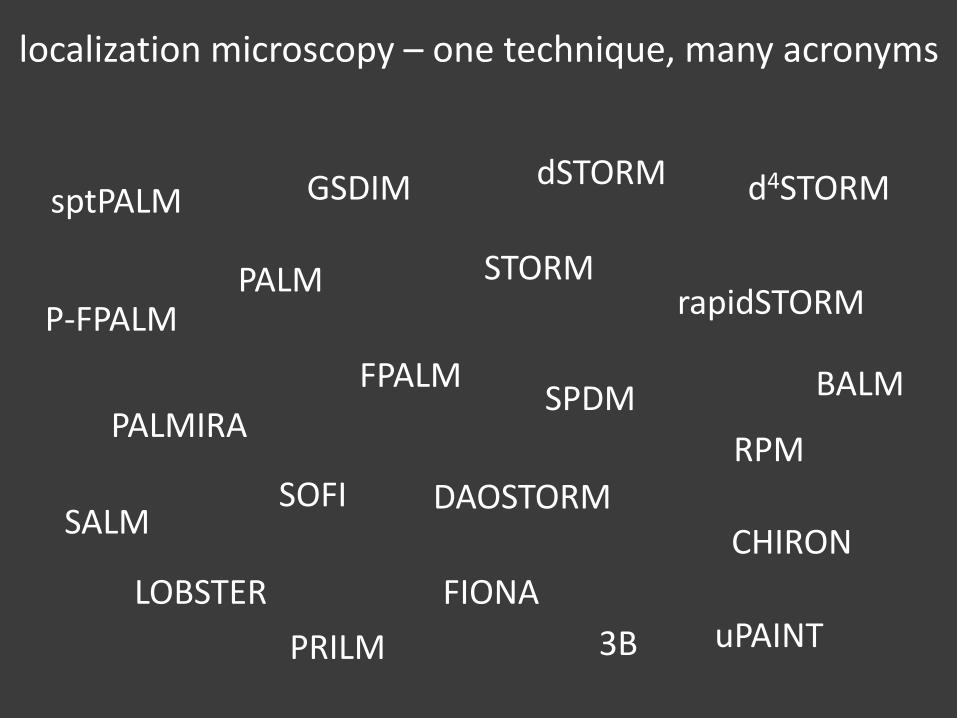
localization microscopy – one technique, many acronyms
PALM STORM
FPALM
PALMIRA
dSTORM
SPDM
GSDIM
RPM
d4STORMsptPALM
rapidSTORM
DAOSTORMSOFI
FIONA
3B
SALM
LOBSTER
CHIRON
uPAINTPRILM
P-FPALM
BALM
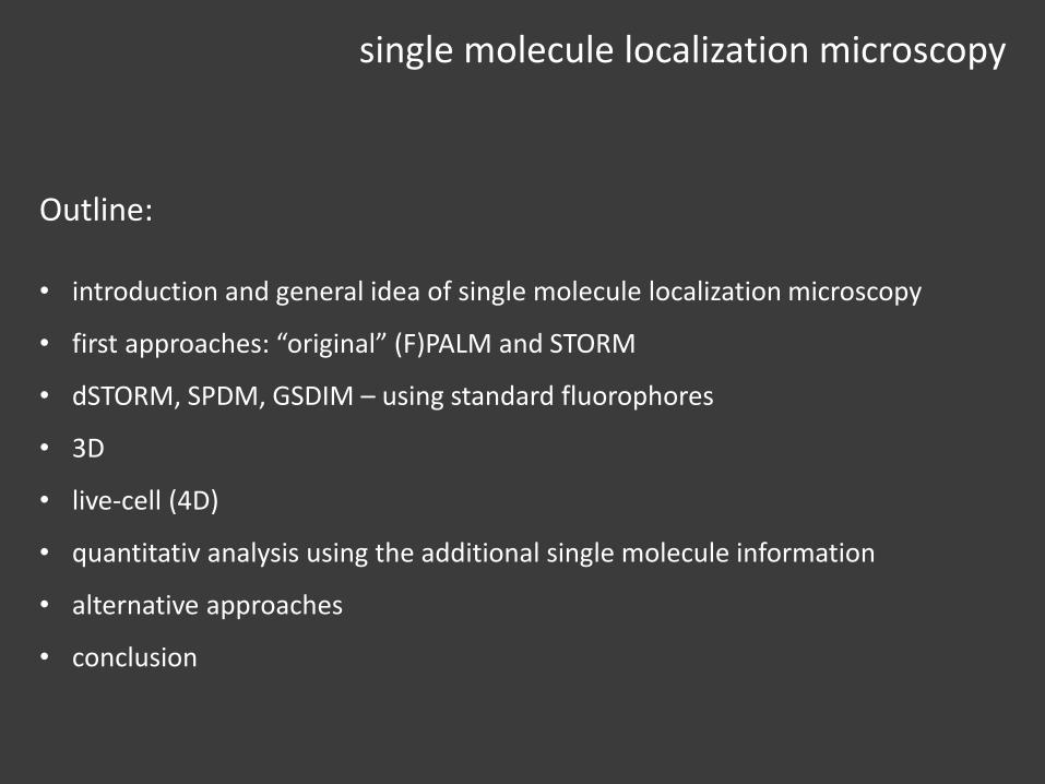
Outline:
• introduction and general idea of single molecule localization microscopy
• first approaches: “original” (F)PALM and STORM
• dSTORM, SPDM, GSDIM – using standard fluorophores
• 3D
• live-cell (4D)
• quantitativ analysis using the additional single molecule information
• alternative approaches
• conclusion
single molecule localization microscopy
introduction to localization microscopy
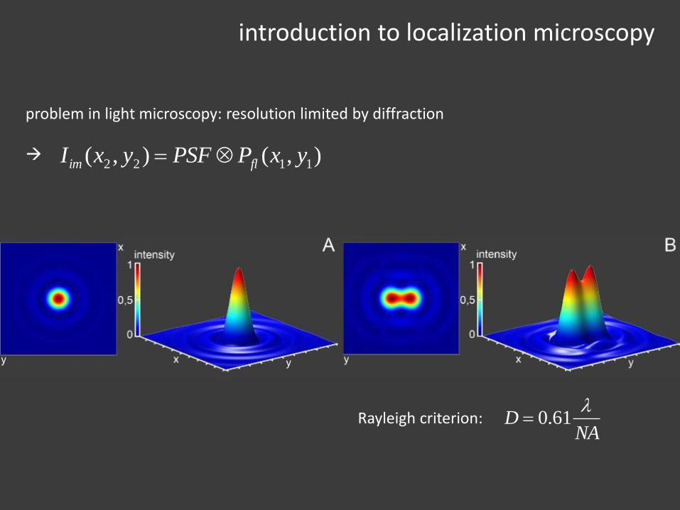
problem in light microscopy: resolution limited by diffraction
),(),( 1122 yxPPSFyxI flim
Rayleigh criterion:NA
D
61.0
introduction to localization microscopy
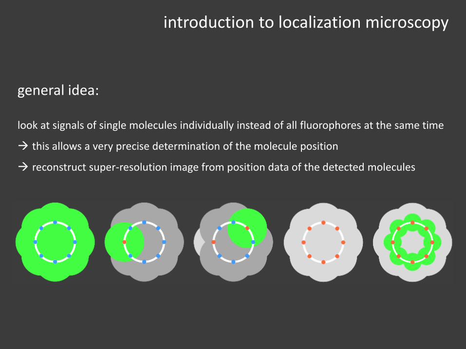
general idea:
look at signals of single molecules individually instead of all fluorophores at the same time
this allows a very precise determination of the molecule position
reconstruct super-resolution image from position data of the detected molecules
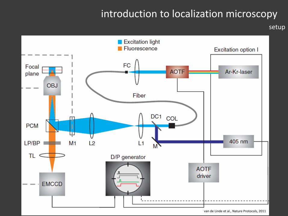
introduction to localization microscopysetup
van de Linde et al., Nature Protocols, 2011
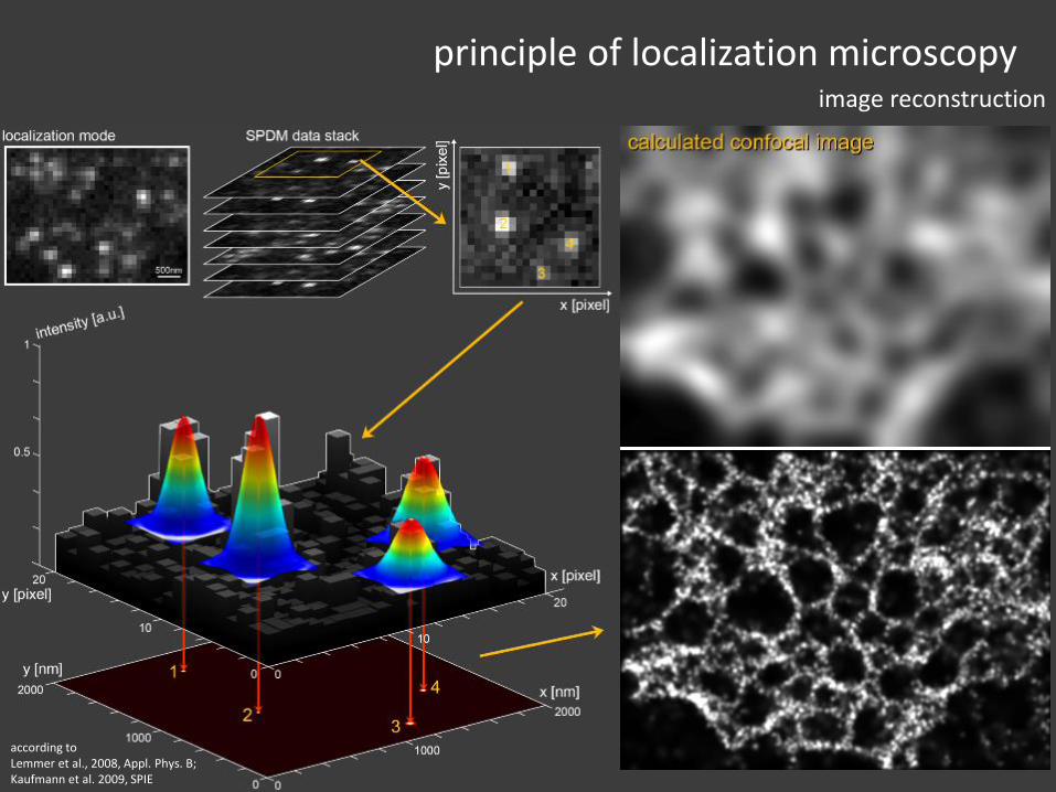
principle of localization microscopyimage reconstruction
according toLemmer et al., 2008, Appl. Phys. B;Kaufmann et al. 2009, SPIE

principle of localization microscopyposition determination
localisation accuracy 𝜎 of a single molecule is depended on
• width of the PSF s• number of detected photons N• background intensity b• size of the pixels on the camera a
𝜎2 =𝑠2 + 𝑎2/12
𝑁+
8𝜋𝑠4𝑏2
𝑎2𝑁2
𝐼(𝑥, 𝑦) = 𝐼0exp −(𝑥 − 𝑥0 )2+(𝑦 − 𝑦0 )2
2𝑠2+ 𝑏
typical model function: 2D Gaussian + linear background
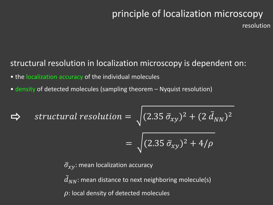
𝜎𝑥𝑦: mean localization accuracy
𝑑𝑁𝑁: mean distance to next neighboring molecule(s)
𝜌: local density of detected molecules
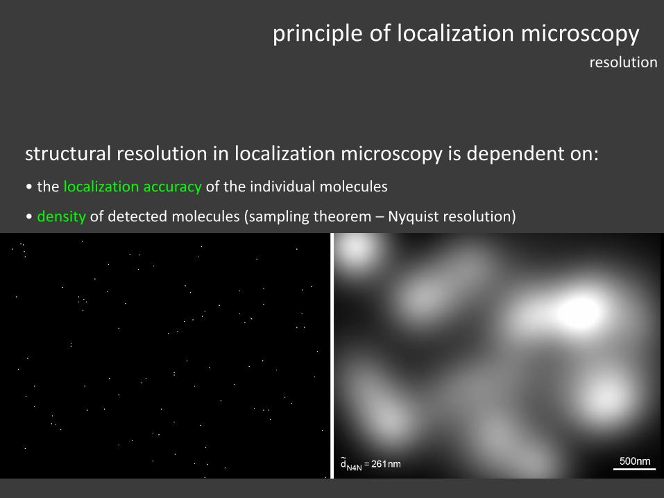
structural resolution in localization microscopy is dependent on:
• the localization accuracy of the individual molecules
• density of detected molecules (sampling theorem – Nyquist resolution)
𝑠𝑡𝑟𝑢𝑐𝑡𝑢𝑟𝑎𝑙 𝑟𝑒𝑠𝑜𝑙𝑢𝑡𝑖𝑜𝑛 = (2.35 𝜎𝑥𝑦)2 + (2 𝑑𝑁𝑁)
2
= (2.35 𝜎𝑥𝑦)2 + 4/𝜌
principle of localization microscopyresolution
𝜎𝑥𝑦: mean localization accuracy
𝑑𝑁𝑁: mean distance to next neighboring molecule(s)
𝜌: local density of detected molecules
structural resolution in localization microscopy is dependent on:
• the localization accuracy of the individual molecules
• density of detected molecules (sampling theorem – Nyquist resolution)
𝑠𝑡𝑟𝑢𝑐𝑡𝑢𝑟𝑎𝑙 𝑟𝑒𝑠𝑜𝑙𝑢𝑡𝑖𝑜𝑛 = (2.35 𝜎𝑥𝑦)2 + (2 𝑑𝑁𝑁)
2
= (2.35 𝜎𝑥𝑦)2 + 4/𝜌
principle of localization microscopyresolution
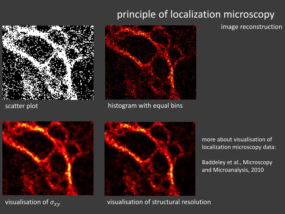
scatter plot histogram with equal bins
visualisation of 𝜎𝑥𝑦
principle of localization microscopyimage reconstruction
visualisation of structural resolution
more about visualisation of localization microscopy data:
Baddeley et al., Microscopy and Microanalysis, 2010
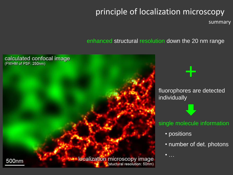
enhanced structural resolution down the 20 nm range
fluorophores are detected
individually
single molecule information
• positions
• number of det. photons
• …
+
principle of localization microscopysummary

(F)PALM and STORM

(some) history of localization microscopy
localisation of single molecules / point-like objects
Burns et al., 1985 theoretical paper about super-resolution distance measurements using spectral characteristics
Betzig, 1995 first measurements with SNOM under cryo conditions
Bornfleth et al., 1998 CLSM measurements of 3D distances < 60 nm using fluorescent markers of different wavelengths (@ RT)
Heilemann et al., 2002 using single molecule live time instead of colours to measure distances of 40 nm
localisation of many molecules to reconstruct structural information
2006: (PALM, FPALM, STORM) – photo-switchable / photo-activatable dyes
2008: (dSTORM, SPDM, GSDIM) – using standard fluorophores
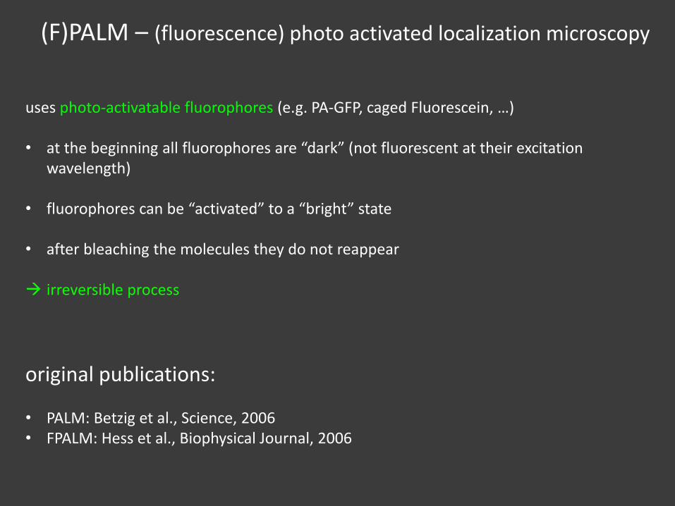
(F)PALM – (fluorescence) photo activated localization microscopy
uses photo-activatable fluorophores (e.g. PA-GFP, caged Fluorescein, …)
• at the beginning all fluorophores are “dark” (not fluorescent at their excitation wavelength)
• fluorophores can be “activated” to a “bright” state
• after bleaching the molecules they do not reappear
irreversible process
original publications:
• PALM: Betzig et al., Science, 2006• FPALM: Hess et al., Biophysical Journal, 2006
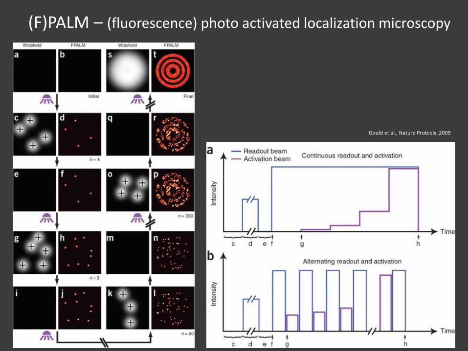
(F)PALM – (fluorescence) photo activated localization microscopy
Gould et al., Nature Protcols ,2009
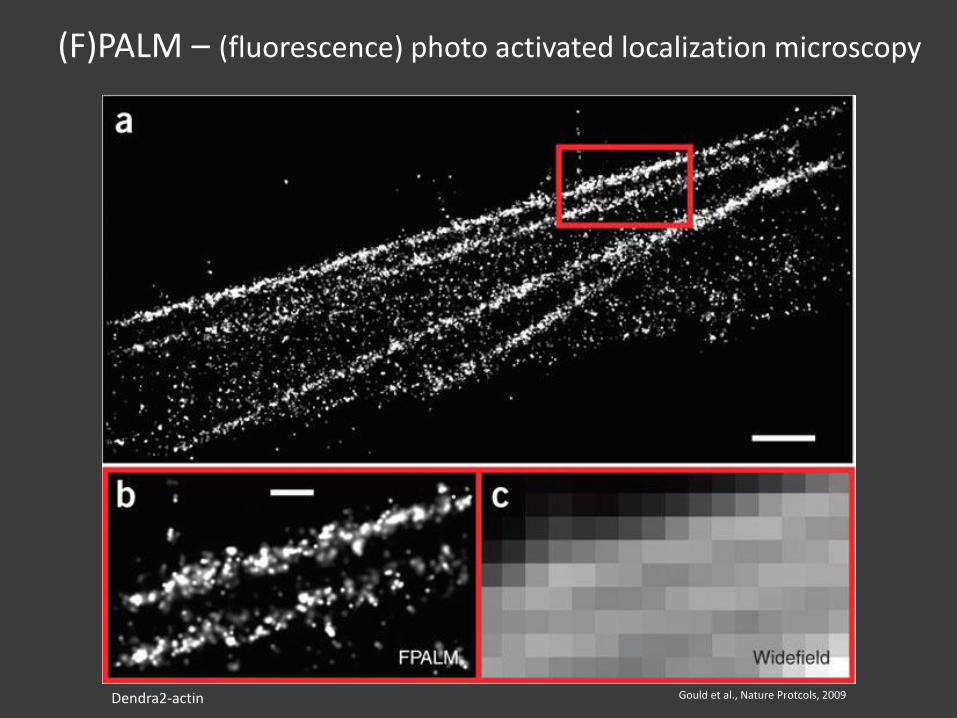
(F)PALM – (fluorescence) photo activated localization microscopy
Gould et al., Nature Protcols, 2009Dendra2-actin
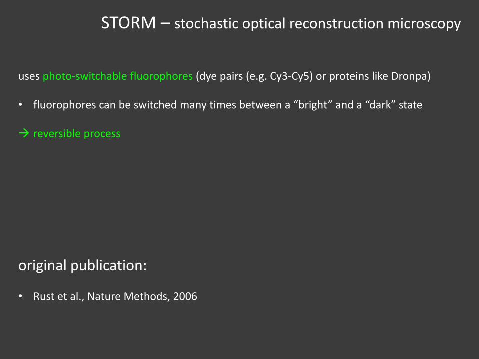
STORM – stochastic optical reconstruction microscopy
uses photo-switchable fluorophores (dye pairs (e.g. Cy3-Cy5) or proteins like Dronpa)
• fluorophores can be switched many times between a “bright” and a “dark” state
reversible process
original publication:
• Rust et al., Nature Methods, 2006
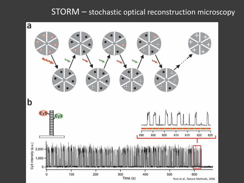
STORM – stochastic optical reconstruction microscopy
Rust et al., Nature Methods, 2006
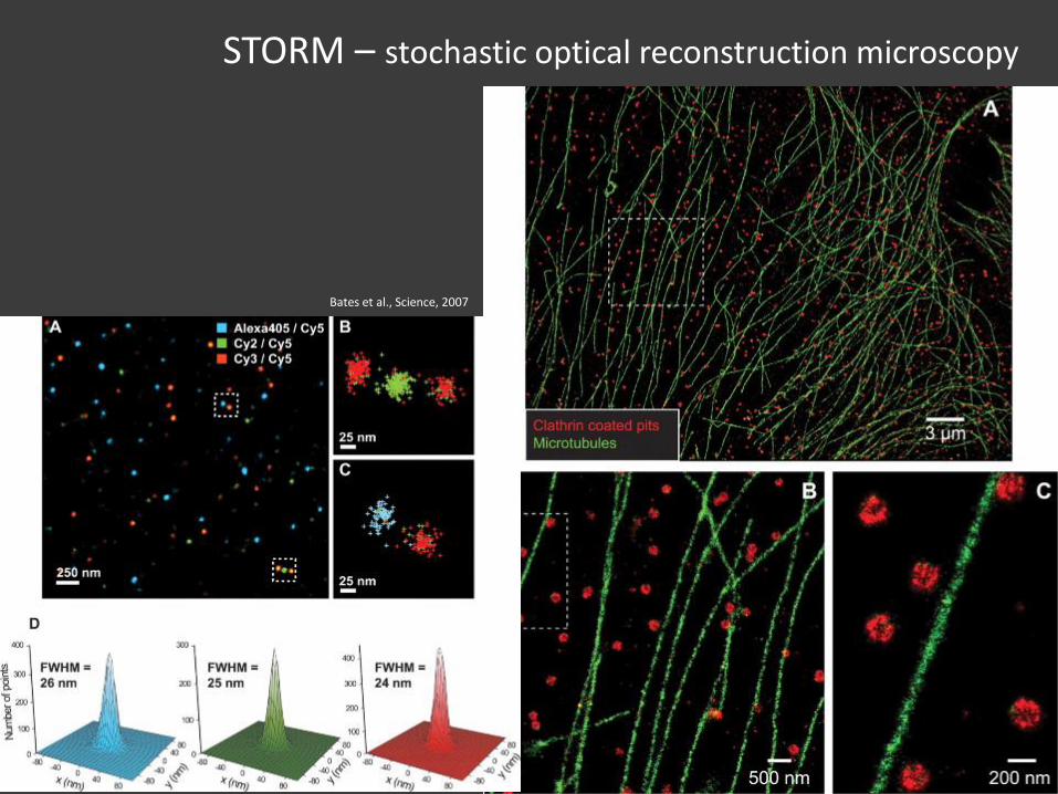
STORM – stochastic optical reconstruction microscopy
Bates et al., Science, 2007
dSTORM, SPDM, GSDIM, …
dSTORM, SPDM, GSDIM, …direct STROM
spectral position determination microscopy
ground state depletion microscopy followed by individual molecule return
uses standard fluorophores (e.g. Alexa and Atto dyes, GFP, YFP, RFP, …)
• switching mechanism based on a light induced long-lived “dark” state
• stochastic recovery to “bright” (fluorescent) state is used for optical isolation of the single molecule signals
original publication:
• dSTORM: Heilemann et. al., Angewandte Chemie International Edition, 2008• SPDM: Lemmer et al., Applied Physics B, 2008• GSDIM: Fölling et al., Nature Methods, 2008
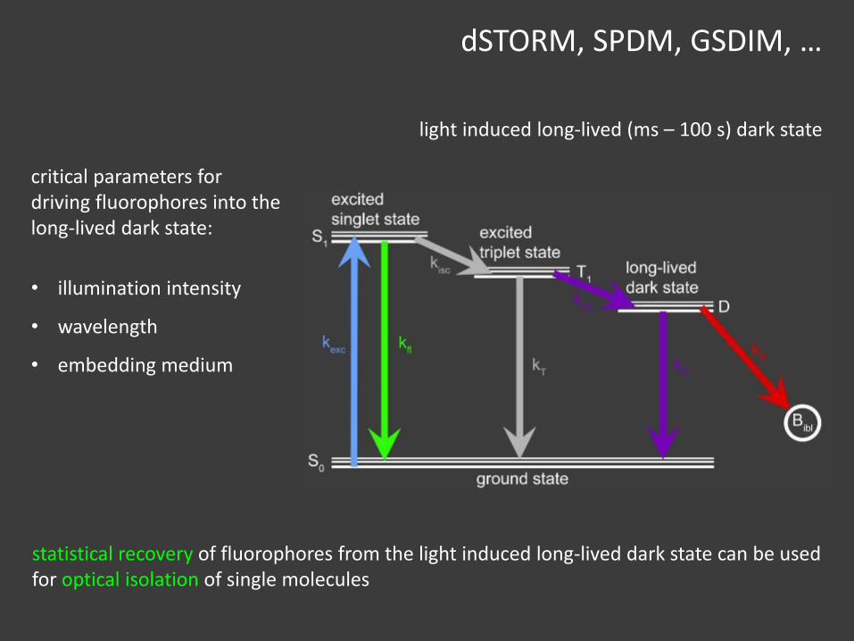
light induced long-lived (ms – 100 s) dark state
statistical recovery of fluorophores from the light induced long-lived dark state can be used for optical isolation of single molecules
dSTORM, SPDM, GSDIM, …
critical parameters for driving fluorophores into the long-lived dark state:
• illumination intensity
• wavelength
• embedding medium
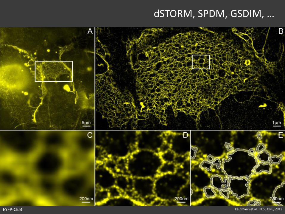
dSTORM, SPDM, GSDIM, …
Kaufmann et al., PLoS ONE, 2012EYFP-Cld3
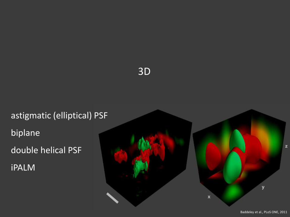
3D
Baddeley et al., PLoS ONE, 2011
astigmatic (elliptical) PSF
biplane
double helical PSF
iPALM
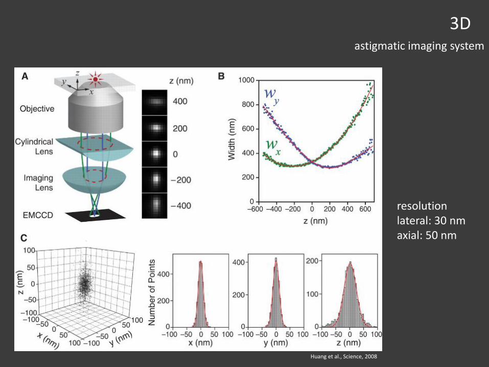
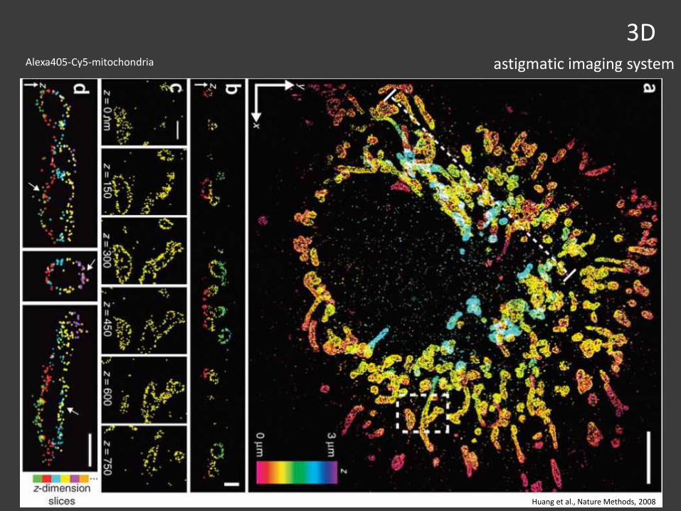
3Dastigmatic imaging system
Huang et al., Science, 2008
resolutionlateral: 30 nmaxial: 50 nm
3Dastigmatic imaging system
Huang et al., Nature Methods, 2008
Alexa405-Cy5-mitochondria
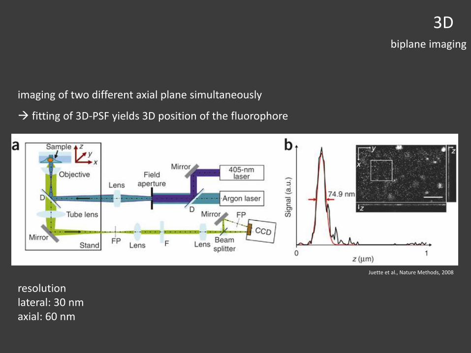
3Dbiplane imaging
imaging of two different axial plane simultaneously
fitting of 3D-PSF yields 3D position of the fluorophore
Juette et al., Nature Methods, 2008
resolutionlateral: 30 nmaxial: 60 nm
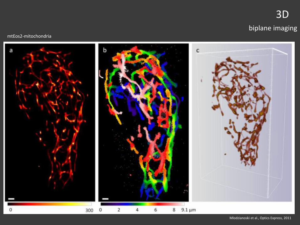
Mlodzianoski et al., Optics Express, 2011
3Dbiplane imaging
mtEos2-mitochondria
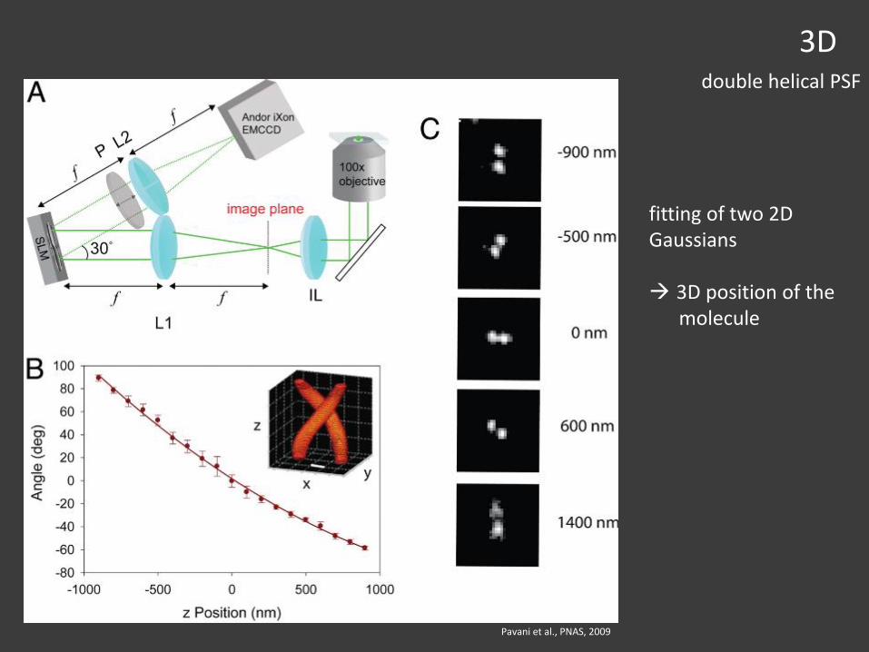
3Ddouble helical PSF
Pavani et al., PNAS, 2009
fitting of two 2D Gaussians
3D position of the molecule
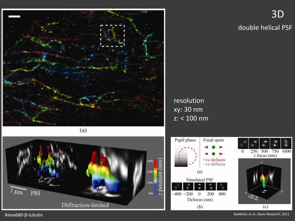
3Ddouble helical PSF
resolutionxy: 30 nmz: < 100 nm
Baddeley et al., Nano Research, 2011Alexa680-β-tubulin
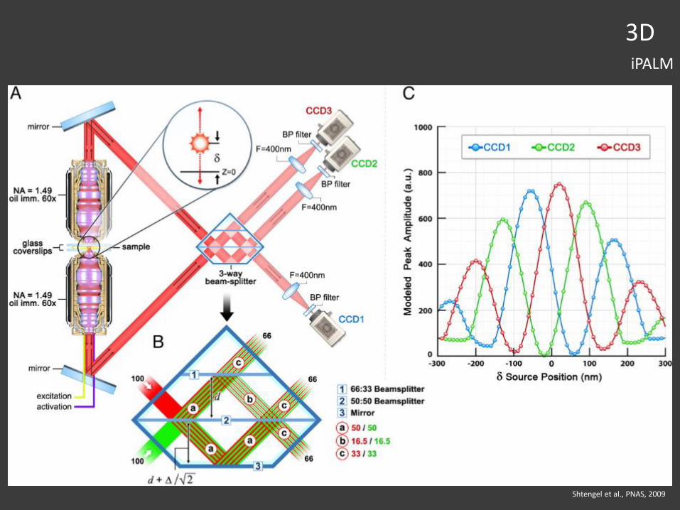
3DiPALM
Shtengel et al., PNAS, 2009
3DiPALM
KIT
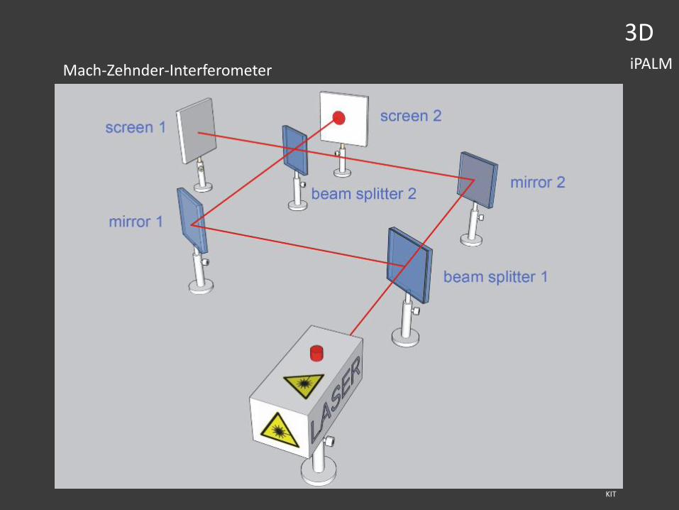
Mach-Zehnder-Interferometer

3DiPALM
Shtengel et al., PNAS, 2009
3DiPALM
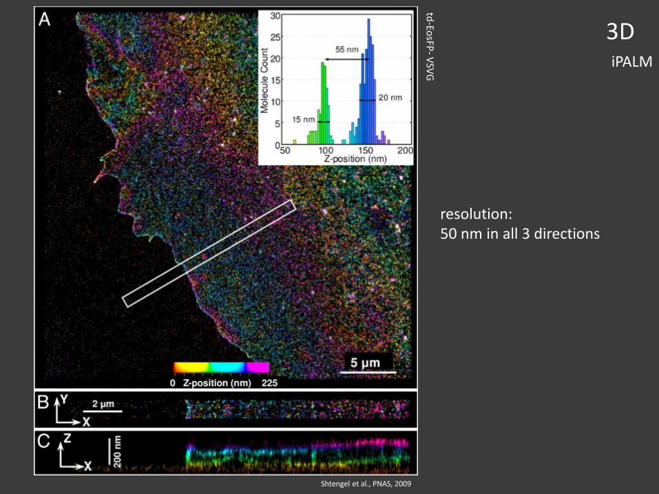
Shtengel et al., PNAS, 2009
resolution:50 nm in all 3 directions
td-Eo
sFP-
VSV
G
two examples for “live-cell” applications
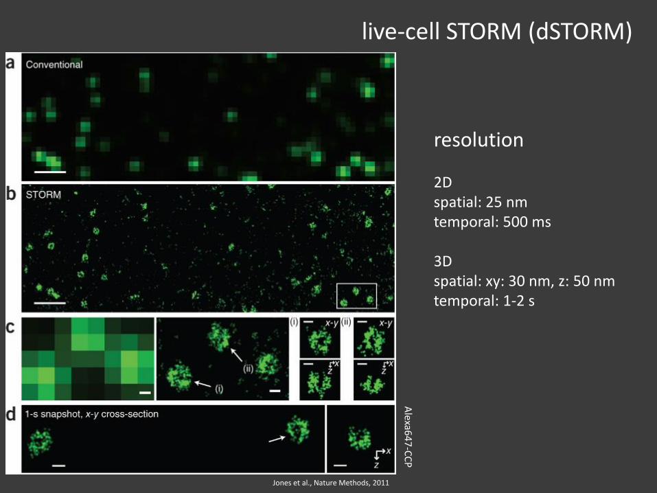
live-cell STORM (dSTORM)
Jones et al., Nature Methods, 2011
resolution
2Dspatial: 25 nmtemporal: 500 ms
3Dspatial: xy: 30 nm, z: 50 nmtemporal: 1-2 s
Alexa6
47
-CC
P
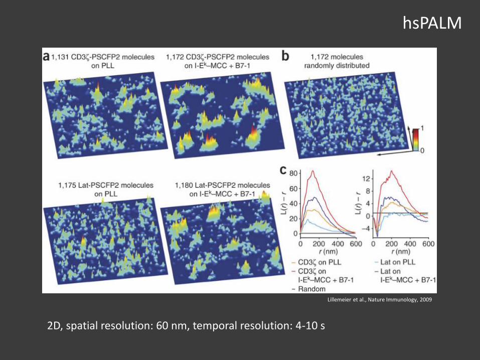
hsPALM
2D, spatial resolution: 60 nm, temporal resolution: 4-10 s
Lillemeier et al., Nature Immunology, 2009

how to get a lot more information from the data
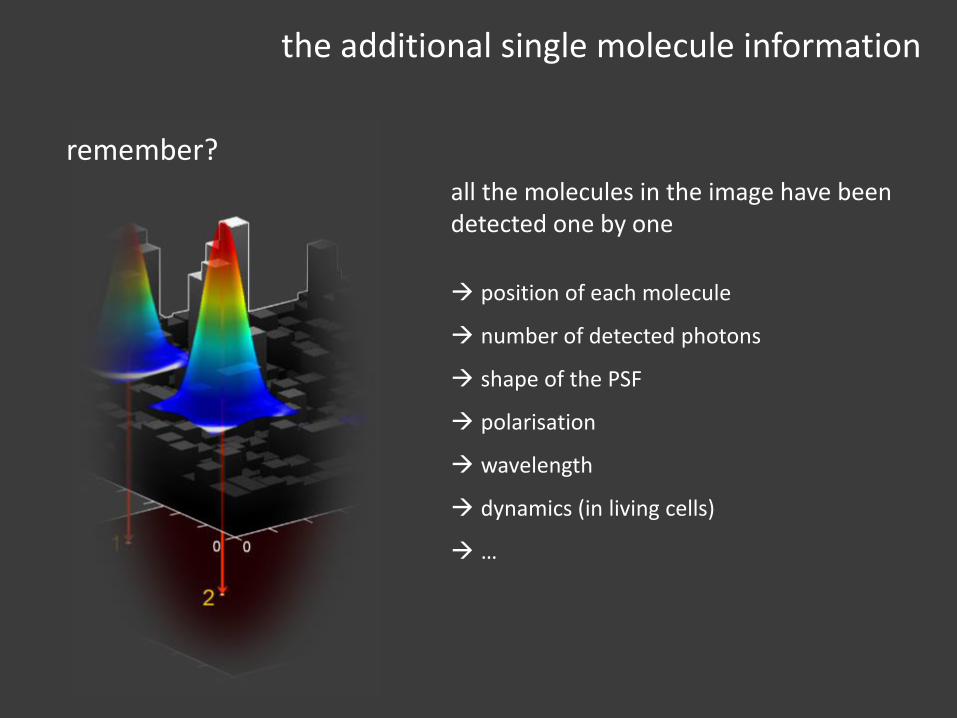
the additional single molecule information
all the molecules in the image have been detected one by one
position of each molecule
number of detected photons
shape of the PSF
polarisation
wavelength
dynamics (in living cells)
…
remember?
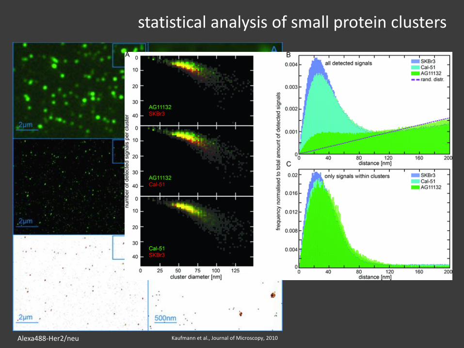
statistical analysis of small protein clusters
Kaufmann et al., Journal of Microscopy, 2010Alexa488-Her2/neu
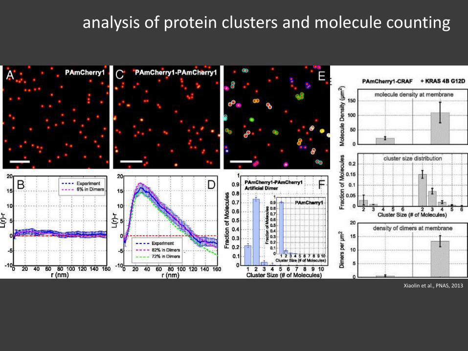
analysis of protein clusters and molecule counting
Xiaolin et al., PNAS, 2013
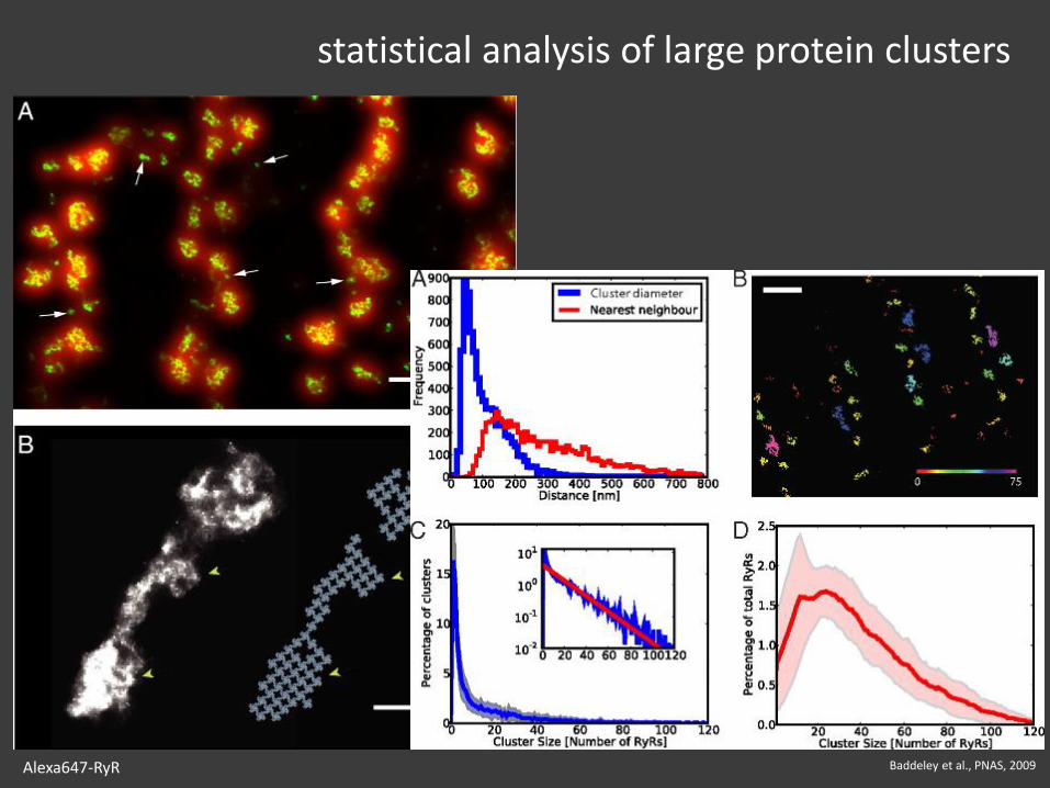
statistical analysis of large protein clusters
Baddeley et al., PNAS, 2009Alexa647-RyR

protein densities
Kaufmann et al., PLoS ONE, 2012
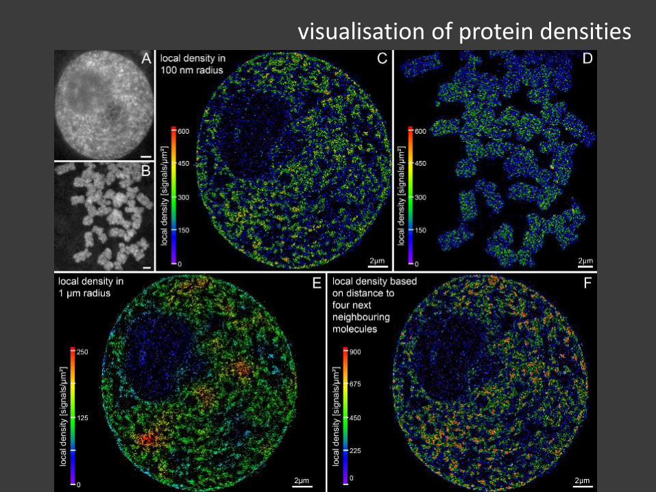
visualisation of protein densities
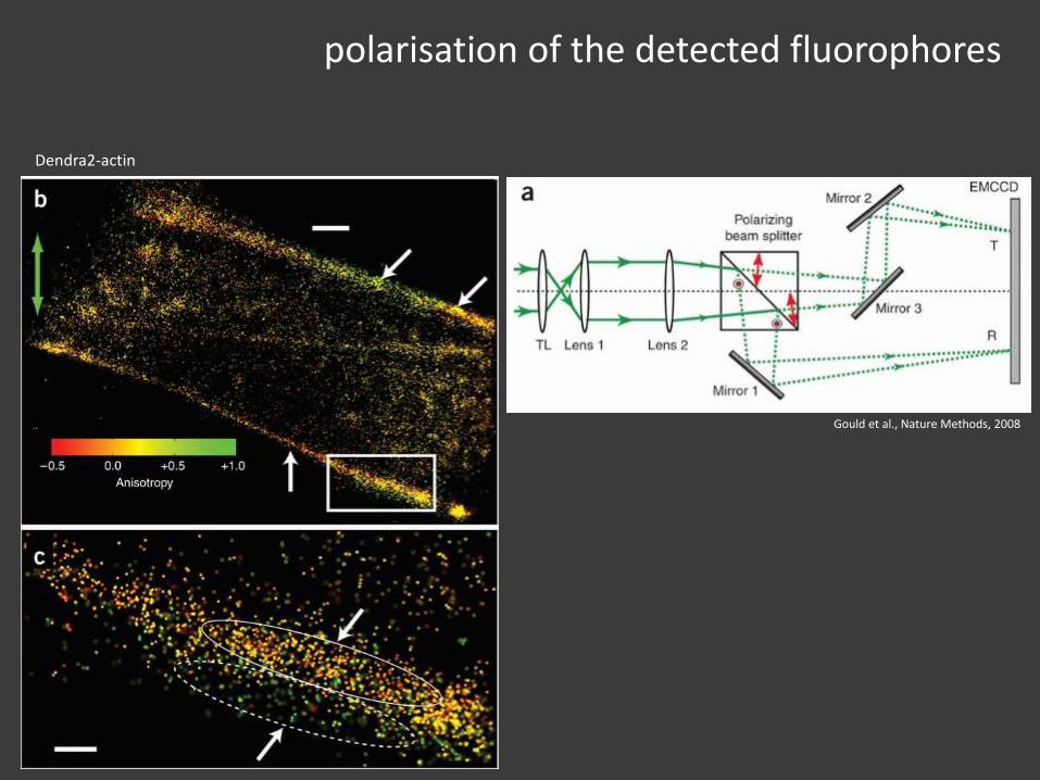
polarisation of the detected fluorophores
Gould et al., Nature Methods, 2008
Dendra2-actin
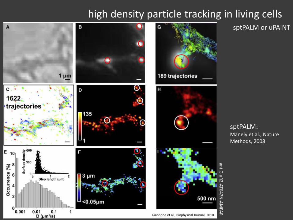
high density particle tracking in living cellssptPALM or uPAINT
Giannone et al., Biophysical Journal, 2010
sptPALM:Manely et al., Nature Methods, 2008
antiG
luR
2-AT6
47
N-A
MPA
R

alternative approaches
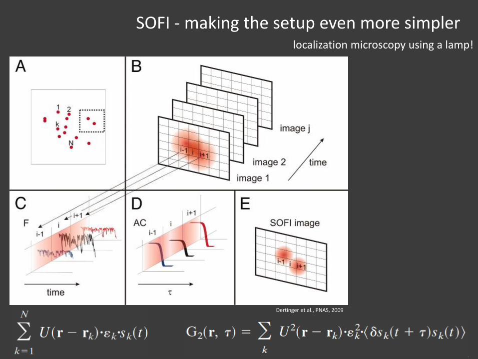
SOFI - making the setup even more simpler
Dertinger et al., PNAS, 2009
localization microscopy using a lamp!
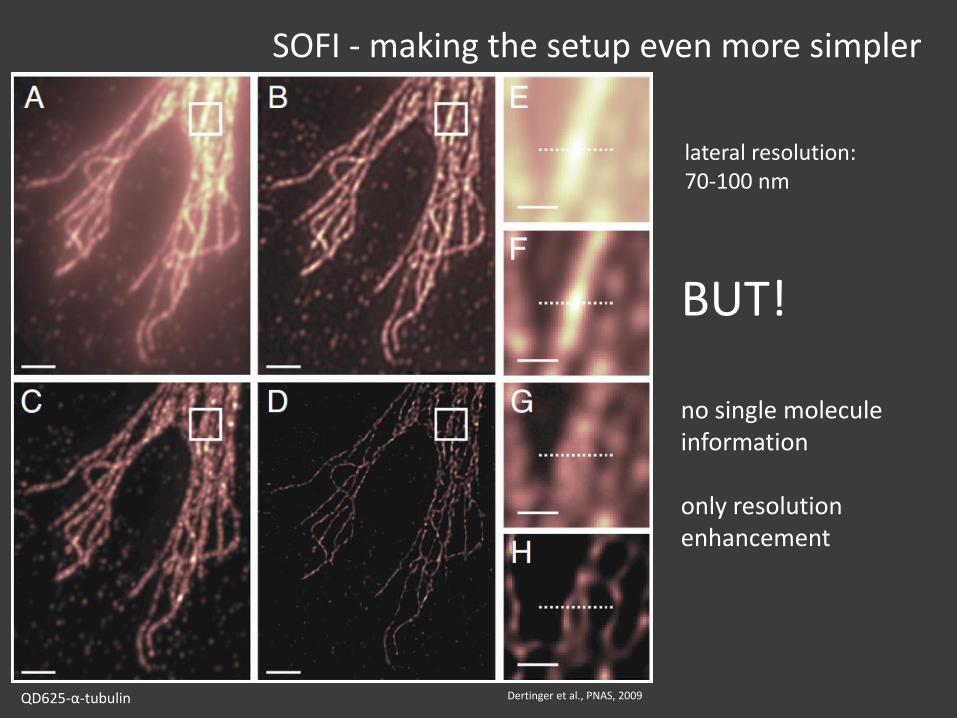
SOFI - making the setup even more simpler
Dertinger et al., PNAS, 2009
BUT!
no single molecule information
only resolution enhancement
lateral resolution:70-100 nm
QD625-α-tubulin
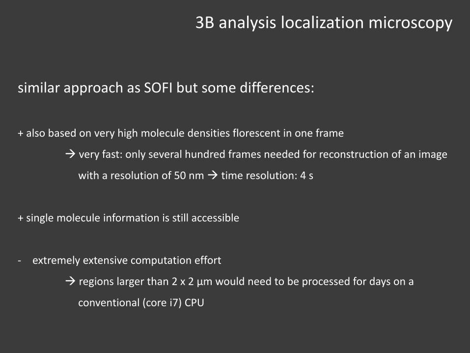
3B analysis localization microscopy
similar approach as SOFI but some differences:
+ also based on very high molecule densities florescent in one frame
very fast: only several hundred frames needed for reconstruction of an image
with a resolution of 50 nm time resolution: 4 s
+ single molecule information is still accessible
- extremely extensive computation effort
regions larger than 2 x 2 μm would need to be processed for days on a
conventional (core i7) CPU
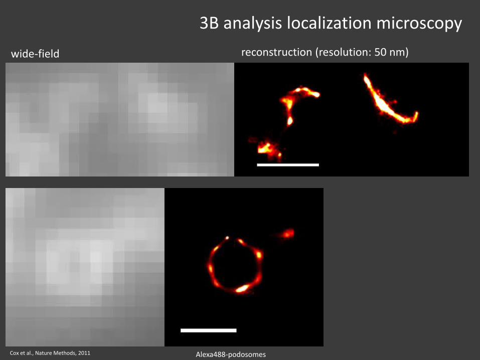
3B analysis localization microscopy
Cox et al., Nature Methods, 2011
wide-field reconstruction (resolution: 50 nm)
Alexa488-podosomes

conclusion
conclusion
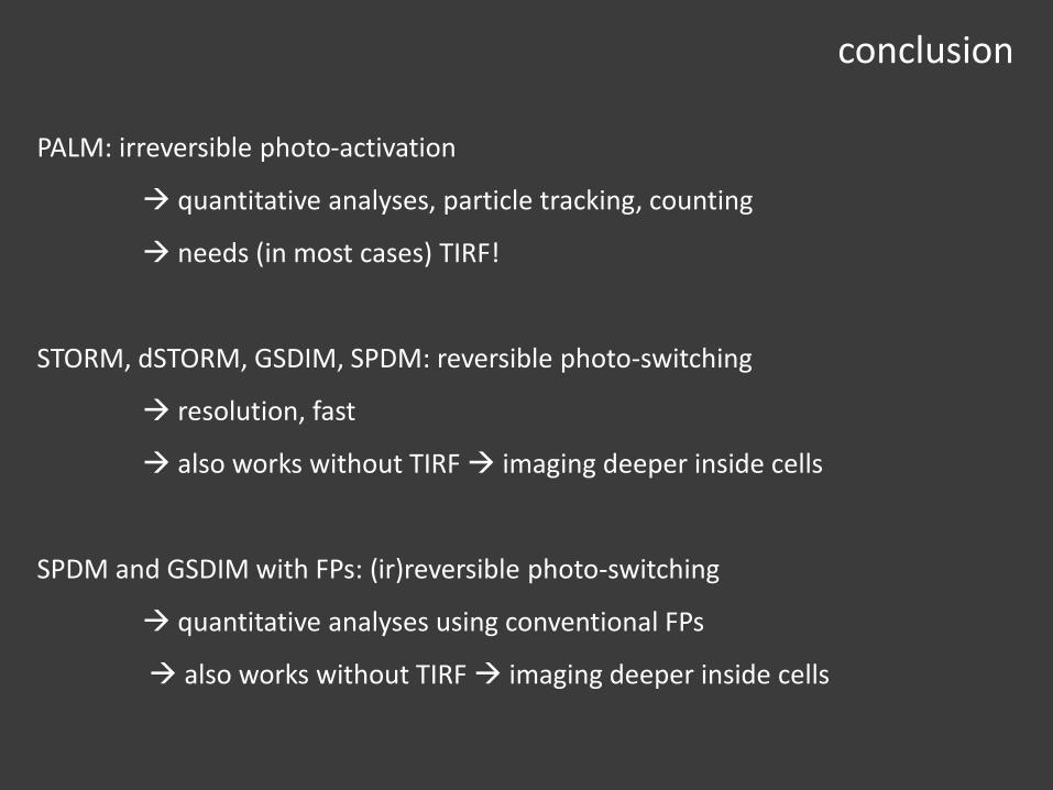
PALM: irreversible photo-activation
quantitative analyses, particle tracking, counting
needs (in most cases) TIRF!
STORM, dSTORM, GSDIM, SPDM: reversible photo-switching
resolution, fast
also works without TIRF imaging deeper inside cells
SPDM and GSDIM with FPs: (ir)reversible photo-switching
quantitative analyses using conventional FPs
also works without TIRF imaging deeper inside cells
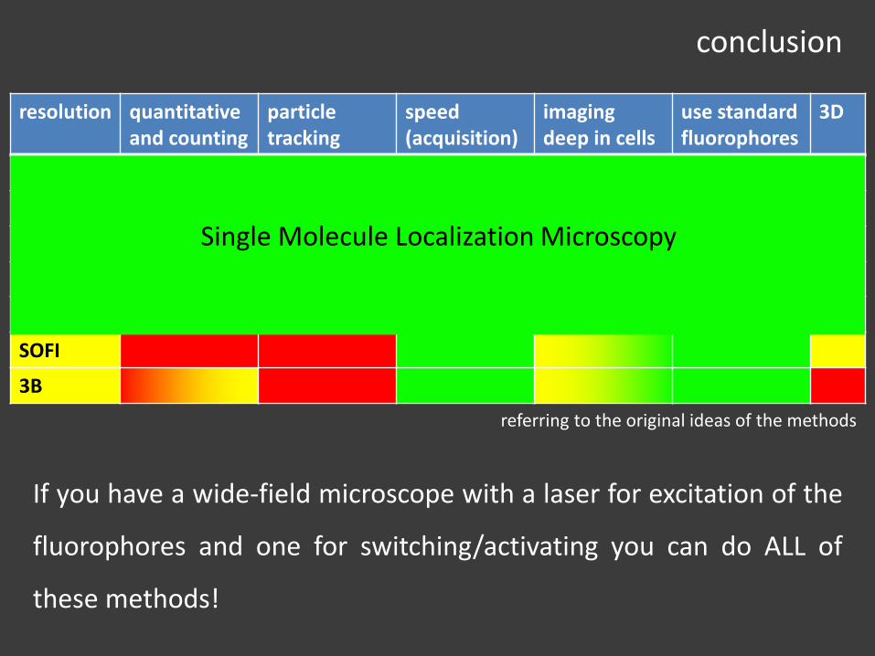
conclusion
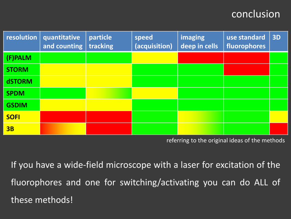
resolution quantitativeand counting
particletracking
speed (acquisition)
imaging deep in cells
use standard fluorophores
3D
(F)PALM
STORM
dSTORM
SPDM
GSDIM
SOFI
3B
If you have a wide-field microscope with a laser for excitation of the
fluorophores and one for switching/activating you can do ALL of
these methods!
referring to the original ideas of the methods
conclusion
resolution quantitativeand counting
particletracking
speed (acquisition)
imaging deep in cells
use standard fluorophores
3D
(F)PALM
STORM
dSTORM
SPDM
GSDIM
SOFI
3B
If you have a wide-field microscope with a laser for excitation of the
fluorophores and one for switching/activating you can do ALL of
these methods!
referring to the original ideas of the methods
Single Molecule Localization Microscopy
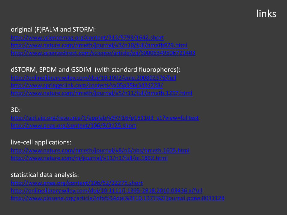
links
original (F)PALM and STORM:http://www.sciencemag.org/content/313/5793/1642.shorthttp://www.nature.com/nmeth/journal/v3/n10/full/nmeth929.htmlhttp://www.sciencedirect.com/science/article/pii/S0006349506721403
dSTORM, SPDM and GSDIM (with standard fluorophores):http://onlinelibrary.wiley.com/doi/10.1002/anie.200802376/fullhttp://www.springerlink.com/content/vx05p35kr3424228/http://www.nature.com/nmeth/journal/v5/n11/full/nmeth.1257.html
3D:http://apl.aip.org/resource/1/applab/v97/i16/p161103_s1?view=fulltexthttp://www.pnas.org/content/106/9/3125.short
live-cell applications:http://www.nature.com/nmeth/journal/v8/n6/abs/nmeth.1605.htmlhttp://www.nature.com/ni/journal/v11/n1/full/ni.1832.html
statistical data analysis:http://www.pnas.org/content/106/52/22275.shorthttp://onlinelibrary.wiley.com/doi/10.1111/j.1365-2818.2010.03436.x/fullhttp://www.plosone.org/article/info%3Adoi%2F10.1371%2Fjournal.pone.0031128

links
high density particle tracking:http://www.nature.com/nmeth/journal/v5/n2/full/nmeth.1176.htmlhttp://www.sciencedirect.com/science/article/pii/S0006349510007137
Nat. Protoc.:http://www.nature.com/nprot/journal/v4/n3/abs/nprot.2008.246.htmlhttp://www.nature.com/nprot/journal/v6/n7/abs/nprot.2011.336.html
commercial systems:http://zeiss-campus.magnet.fsu.edu/articles/superresolution/palm/introduction.htmlhttp://www.nikoninstruments.com/en_GB/Products/Microscope-Systems/Inverted-Microscopes/Biological/N-STORM-Super-Resolutionhttp://www.leica-microsystems.com/products/light-microscopes/life-science-research/fluorescence-microscopes/details/product/leica-sr-gsd/
algorithms:http://www.super-resolution.biozentrum.uni-wuerzburg.de/home/rapidstorm/http://code.google.com/p/quickpalm/
summary and links:http://www2.bioch.ox.ac.uk/microngroup/research/localization-microscopy.shtml