Embed Size (px)
Citation preview
Case ReportJ Gastric Cancer 2016;16(3):200-206 http://dx.doi.org/10.5230/jgc.2016.16.3.200
Copyrights © 2016 by The Korean Gastric Cancer Association www.jgc-online.org
This is an open-access article distributed under the terms of the Creative Commons Attribution Non-Commercial License (http://creativecommons.org/licenses/by-nc/4.0) which permits unrestricted noncommercial use, distribution, and reproduction in any medium, provided the original work is properly cited.
Introduction
In Korea, proximal gastrectomy (PG) has recently attracted
attention as a better choice of function-preserving surgery for
proximal early gastric cancer (EGC) than total gastrectomy (TG).
Although the remnant stomach after PG may be a source for
cancer recurrence, several data have advocated for the oncologic
safety of this procedure.1-3 With regard to conventional PG, the
more troublesome issue has been reflux symptoms induced by
esophagogastric anastomosis where the esophageal mucosa is
exposed to gastric juice. Therefore, various strategies have been
designed to reduce reflux from remnant stomach.2,4-32 Of these
procedures, jejunal interposition (JI) and double tract reconstruc-
tion (DTR) modify the structure of esophagojejunostomy (EJ),
and therefore the jejunum acts as a buffer between esophagus
and remnant stomach. However, JI is associated with abdominal
discomfort after meals, continuous gastric fullness, and hiccups
between meals. These symptoms may result from the interposed
segment, which may disturb the passage of food.25,33 Additional-
ly, it is difficult to reproduce the complicated structure of JI dur-
ing a laparoscopic procedure. Conversely, DTR not only reduces
the incidence of anastomosis-related complications, but is also
sufficiently reproducible as a laparoscopic procedure.34 For these
reasons, in Korea, DTR is recently supported by most surgeons
who prefer laparoscopic PG for the treatment of proximal EGC.
DTR was adopted in an ongoing multi-center prospective study,
KLASS05, in which clinical outcomes will be compared between
laparoscopic PG and TG for proximal EGC.
Nevertheless, single-port laparoscopic procedures have not
been easy to apply in PG with DTR. Even in conventional
laparoscopic PG with DTR, EJ embeds a high possibility of
anastomotic failure. Therefore, it has been considered impellent
to achieve EJ with bearing the poor ergonomics of single-port
laparoscopic surgery. Moreover, since DTR results in more com-
pISSN : 2093-582X, eISSN : 2093-5641
Correspondence to: Seong-Heum Park
Department of Surgery, Korea University College of Medicine, 73 Inchon-ro, Seongbuk-gu, Seoul 02841, Korea Tel: +82-31-412-4936, Fax: +82-31-413-4829 E-mail: [email protected] June 7, 2016Revised July 6, 2016Accepted July 6, 2016
Single-Port Laparoscopic Proximal Gastrectomy with Double Tract Reconstruction for Early Gastric Cancer:
Report of a Case
Chang Min Lee, Da Won Park, Do Hyun Jung, You Jin Jang, Jong-Han Kim, Sungsoo Park, and Seong-Heum Park
Department of Surgery, Korea University Medical Center, Korea University College of Medicine, Seoul, Korea
In Korea, proximal gastrectomy has recently attracted attention as a better choice of function-preserving surgery for proximal early gastric cancer than total gastrectomy. Of the various strategies to overcome reflux symptoms from remnant stomach, double tract reconstruction not only reduces the incidence of anastomosis-related complications, but is also sufficiently reproducible as a laparoscopic procedure. Catching up with the recent rise of single-port laparoscopic surgeries, we performed a pure single-port laparoscopic proximal gastrec-tomy with DTR. This procedure was designed by merging the function-preserving concept of proximal gastrectomy with single-port lapa-roscopic total gastrectomy.
Key Words: Stomach neoplasms; Single port; Laparoscopy; Gastrectomy
Single Port Laparoscopic PG with DTR for EGC
201
plex structures than conventional esophagogastrostomy, some
surgeons are concerned about the short-term safety of laparo-
scopic PG with DTR. Therefore, few surgeons have planned PG
with DTR in the setting of single-port laparoscopic surgery.
Recently, for a cT1 case, we performed a single-port laparo-
scopic proximal gastrectomy (SPPG) with DTR. Our center has
previously performed single-port laparoscopic distal gastrectomy
(SPDG) for EGC; therefore, the procedures presented in this
case report were planned based on our expertise. To the best of
our knowledge, this is the first report of SPPG with DTR.
Case Report
A 40-year-old woman with a poorly differentiated gastric
adenocarcinoma diagnosed via gastroscopic biopsy was referred
to our center. The tumor was located on the posterior wall of the
gastric high body. Computed tomography showed that there was
no regional lymph node (LN) involvement or distant metastasis
(cT1N0M0). Her body mass index (BMI) was 22.5 kg/m2 and
she did not have any comorbid conditions or history of previous
abdominal surgery.
We decided to perform a PG with D1+ lymph node dissec-
tion (LND) since the patient presented with early-stage disease
in the proximal stomach.35 DTR was adopted to avoid the pre-
viously described complications associated with conventional
esophagogastrostomy.34 In addition, considering her low BMI,
single-port laparoscopic surgery was planned. The details of our
procedure were as follows:
1. Preparative procedures
In the operating room, the patient was placed on the bed with
both legs abducted under general anesthesia. The bed was ad-
justed to create a reverse Trendelenburg position for the patient.
The operator stood between the patient’s legs. The scopist was
positioned on the left side of the patient.
A commercial 4-lumen single-port trocar (Gloveport®; Nelis,
Bucheon, Korea) was inserted through a transumbilical incision
using Hasson’s method.36 The Gloveport trocar system consists
of a self retractor that covers a 25 mm incision and four channels
(one 12 mm channel and three 5 mm channels). After a pneu-
moperitoneum was formed with carbon dioxide at a pressure of
15 mmHg, a flexible scope was inserted through a channel of
the umbilical port. The falciform ligament and the left lobe of
the liver were raised in the cephald direction by combined suture
retraction.37
2. Tumor localization
One surgeon performed intraoperative endoscopy for tumor
A
Left abdominal wall
Stomach
Transverse colon
B
Liver
Lymph nodeNo. 8a
RGA
CHA
C D E
Liver
Liver
Linear stapler
Distal stomach
Pancreas
Proximalstomach
Liver
Linearstapler
Esophagus
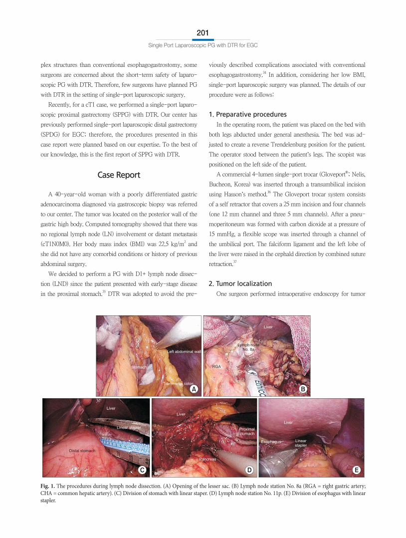
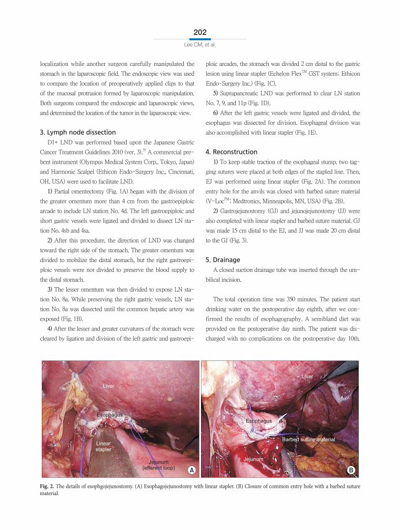
Fig. 1. The procedures during lymph node dissection. (A) Opening of the lesser sac. (B) Lymph node station No. 8a (RGA = right gastric artery; CHA = common hepatic artery). (C) Division of stomach with linear staper. (D) Lymph node station No. 11p. (E) Division of esophagus with linear stapler.
Lee CM, et al.
202
localization while another surgeon carefully manipulated the
stomach in the laparoscopic field. The endoscopic view was used
to compare the location of preoperatively applied clips to that
of the mucosal protrusion formed by laparoscopic manipulation.
Both surgeons compared the endoscopic and laparoscopic views,
and determined the location of the tumor in the laparoscopic view.
3. Lymph node dissection
D1+ LND was performed based upon the Japanese Gastric
Cancer Treatment Guidelines 2010 (ver. 3).35 A commercial pre-
bent instrument (Olympus Medical System Corp., Tokyo, Japan)
and Harmonic Scalpel (Ethicon Endo-Surgery Inc., Cincinnati,
OH, USA) were used to facilitate LND.
1) Partial omentectomy (Fig. 1A) began with the division of
the greater omentum more than 4 cm from the gastroepiploic
arcade to include LN station No. 4d. The left gastroepiploic and
short gastric vessels were ligated and divided to dissect LN sta-
tion No. 4sb and 4sa.
2) After this procedure, the direction of LND was changed
toward the right side of the stomach. The greater omentum was
divided to mobilize the distal stomach, but the right gastroepi-
ploic vessels were not divided to preserve the blood supply to
the distal stomach.
3) The lesser omentum was then divided to expose LN sta-
tion No. 8a. While preserving the right gastric vessels, LN sta-
tion No. 8a was dissected until the common hepatic artery was
exposed (Fig. 1B).
4) After the lesser and greater curvatures of the stomach were
cleared by ligation and division of the left gastric and gastroepi-
ploic arcades, the stomach was divided 2 cm distal to the gastric
lesion using linear stapler (Echelon FlexTM GST system; Ethicon
Endo-Surgery Inc.) (Fig. 1C).
5) Suprapancreatic LND was performed to clear LN station
No. 7, 9, and 11p (Fig. 1D).
6) After the left gastric vessels were ligated and divided, the
esophagus was dissected for division. Esophageal division was
also accomplished with linear stapler (Fig. 1E).
4. Reconstruction
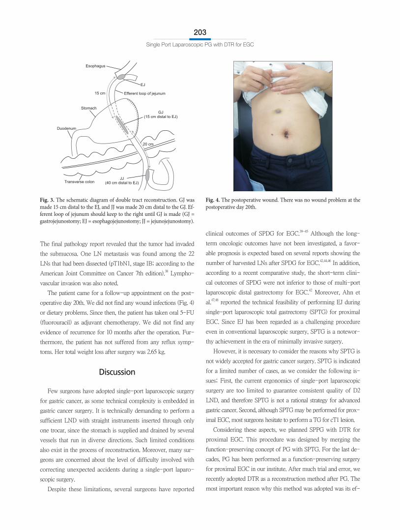
1) To keep stable traction of the esophageal stump, two tag-
ging sutures were placed at both edges of the stapled line. Then,
EJ was performed using linear stapler (Fig. 2A). The common
entry hole for the anvils was closed with barbed suture material
(V-LocTM; Medtronics, Minneapolis, MN, USA) (Fig. 2B).
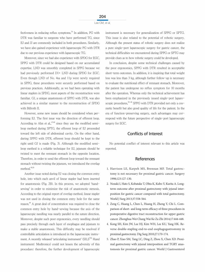
2) Gastrojejunostomy (GJ) and jejunojejunostomy (JJ) were
also completed with linear stapler and barbed suture material. GJ
was made 15 cm distal to the EJ, and JJ was made 20 cm distal
to the GJ (Fig. 3).
5. Drainage
A closed suction drainage tube was inserted through the um-
bilical incision.
The total operation time was 350 minutes. The patient start
drinking water on the postoperative day eighth, after we con-
firmed the results of esophagography. A semibland diet was
provided on the postoperative day ninth. The patient was dis-
charged with no complications on the postoperative day 10th.
A B
Liver
Esophagus
Linearstapler
Jejunum(efferent loop)
Esophagus
Liver
Jejunum
Barbed suture material
Fig. 2. The details of esophgojejunostomy. (A) Esophagojejunostomy with linear stapler. (B) Closure of common entry hole with a barbed suture material.
Single Port Laparoscopic PG with DTR for EGC
203
The final pathology report revealed that the tumor had invaded
the submucosa. One LN metastasis was found among the 22
LNs that had been dissected (pT1bN1, stage IB; according to the
American Joint Committee on Cancer 7th edition).38 Lympho-
vascular invasion was also noted.
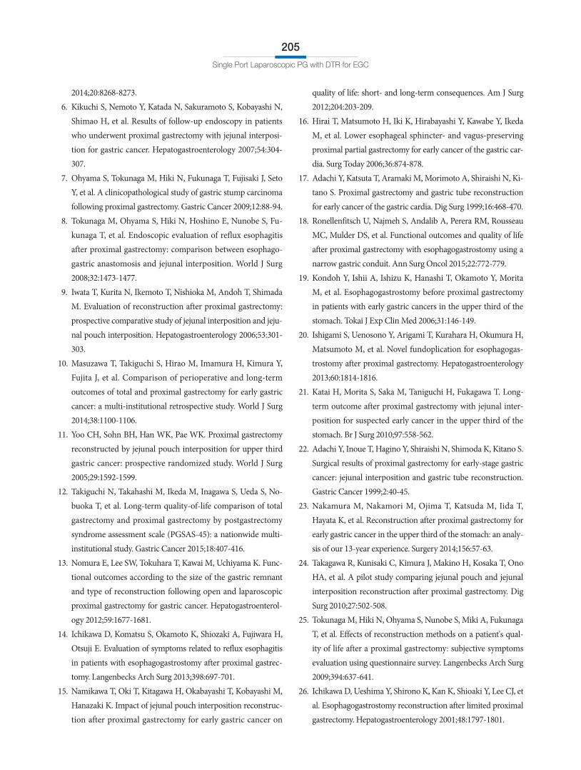
The patient came for a follow-up appointment on the post-
operative day 20th. We did not find any wound infections (Fig. 4)
or dietary problems. Since then, the patient has taken oral 5-FU
(fluorouracil) as adjuvant chemotherapy. We did not find any
evidence of recurrence for 10 months after the operation. Fur-
thermore, the patient has not suffered from any reflux symp-
toms. Her total weight loss after surgery was 2.65 kg.
Discussion
Few surgeons have adopted single-port laparoscopic surgery
for gastric cancer, as some technical complexity is embedded in
gastric cancer surgery. It is technically demanding to perform a
sufficient LND with straight instruments inserted through only
one trocar, since the stomach is supplied and drained by several
vessels that run in diverse directions. Such limited conditions
also exist in the process of reconstruction. Moreover, many sur-
geons are concerned about the level of difficulty involved with
correcting unexpected accidents during a single-port laparo-
scopic surgery.
Despite these limitations, several surgeons have reported
clinical outcomes of SPDG for EGC.39-45 Although the long-
term oncologic outcomes have not been investigated, a favor-
able prognosis is expected based on several reports showing the
number of harvested LNs after SPDG for EGC.42,44,46 In addition,
according to a recent comparative study, the short-term clini-
cal outcomes of SPDG were not inferior to those of multi-port
laparoscopic distal gastrectomy for EGC.42 Moreover, Ahn et
al.47,48 reported the technical feasibility of performing EJ during
single-port laparoscopic total gastrectomy (SPTG) for proximal
EGC. Since EJ has been regarded as a challenging procedure
even in conventional laparoscopic surgery, SPTG is a notewor-
thy achievement in the era of minimally invasive surgery.
However, it is necessary to consider the reasons why SPTG is
not widely accepted for gastric cancer surgery. SPTG is indicated
for a limited number of cases, as we consider the following is-
sues; First, the current ergonomics of single-port laparoscopic
surgery are too limited to guarantee consistent quality of D2
LND, and therefore SPTG is not a rational strategy for advanced
gastric cancer. Second, although SPTG may be performed for prox-
imal EGC, most surgeons hesitate to perform a TG for cT1 lesion.
Considering these aspects, we planned SPPG with DTR for
proximal EGC. This procedure was designed by merging the
function-preserving concept of PG with SPTG. For the last de-
cades, PG has been performed as a function-preserving surgery
for proximal EGC in our institute. After much trial and error, we
recently adopted DTR as a reconstruction method after PG. The
most important reason why this method was adopted was its ef-
Esophagus
EJ
Efferent loop of jejunum15 cm
Stomach
Duodenum
GJ(15 cm distal to EJ)
Transverse colonJJ
(40 cm distal to EJ)
20 cm
Fig. 3. The schematic diagram of double tract reconstruction. GJ was made 15 cm distal to the EJ, and JJ was made 20 cm distal to the GJ. Ef-ferent loop of jejunum should keep to the right until GJ is made (GJ = gastrojejunostomy; EJ = esophagojejunostomy; JJ = jejunojejunostomy).
Fig. 4. The postoperative wound. There was no wound problem at the postoperative day 20th.
Lee CM, et al.
204
fectiveness in reducing reflux symptoms.34 In addition, PG with
DTR was familiar to surgeons who have performed TG, since
EJ and JJ are commonly included in both procedures. Similarly,
we have also gained experience with laparoscopic PG with DTR
due to our previous experience with laparoscopic TG.
Moreover, since we had also experience with SPDG for EGC,
SPPG with DTR could be designed based on our accumulated
expertise. LND was smoothly completed in SPPG because we
had previously performed D1+ LND during SPDG for EGC.
Even though LND of No. 4sa and 11p were newly required
in SPPG, these procedures were securely performed based on
previous practices. Additionally, as we had been operating with
linear staplers in SPDG, most aspects of the reconstruction were
familiar. GJ, a unique anastomosis of SPPG with DTR, was also
achieved in a similar manner to the reconstruction of SPDG
with Billroth II.
However, some new issues should be considered when per-
forming EJ. The first issue was the direction of efferent loop.
According to Ahn et al.,47,48 since they use the modified semi-
loop method during SPTG, the efferent loop of EJ proceeded
toward the left side of abdominal cavity. On the other hand,
during SPPG with DTR, efferent loop should be kept to the
right until GJ is made (Fig. 3). Although the modified semi-
loop method is a reliable technique for EJ, jejunum should be
twisted to meet the remnant stomach in the opposite location.
Therefore, in order to send the efferent loop toward the remnant
stomach without twisting the jejunum, we introduced the overlap
method.49,50
Another issue noted during EJ was closing the common entry
hole, into which each anvil of linear stapler had been inserted
for anastomosis (Fig. 2B). In this process, we adopted ‘hand-sewing’ in order to minimize the risk of anastomotic stenosis.
According to the original report of overlap method, linear stapler
was not used in closing the common entry hole for the same
reason.49 A great deal of concentration was required to close the
common entry hole by hand-sewing because the axis of the
laparoscopic needling was nearly parallel to the suture direction.
Moreover, despite such poor ergonomics, every needling should
pass precisely through each layer of esophagus and jejunum to
make a stable anastomosis. This difficulty may be resolved if
controllable articulation is introduced in the laparoscopic instru-
ment. A recently released ‘articulating instrument’ (SILSTM Hand
instrument; Medtronics) could not lessen the adversity of this
procedure; therefore, the further development of laparoscopic
instrument is necessary for generalization of SPPG or SPTG.
This issue is also related to the potential of robotic surgery.
Although the present status of robotic surgery does not realize
a pure single-port laparoscopic surgery for gastric cancer, the
technical difficulties we encountered during SPPG or SPTG may
provide clues as to how robotic surgery could be developed.
In conclusion, despite some technical challenges caused by
the poor ergonomics, SPPG with DTR resulted in acceptable
short-term outcomes. In addition, it is inspiring that total weight
loss was less than 3 kg, although further follow-up is necessary
to evaluate the nutritional effect of remnant stomach. Moreover,
the patient has undergone no reflux symptom for 10 months
after the operation. Whereas only the technical achievement has
been emphasized in the previously issued single-port laparo-
scopic procedures,39-48 SPPG with DTR provided not only a cos-
metic benefit but also good quality of life for the patient. In the
era of function-preserving surgery, such advantages may cor-
respond with the future perspective of single-port laparoscopic
surgery for EGC.
Conflicts of Interest
No potential conflict of interest relevant to this article was
reported.
References
1. Harrison LE, Karpeh MS, Brennan MF. Total gastrec-tomy is not necessary for proximal gastric cancer. Surgery 1998;123:127-130.
2. Nozaki I, Hato S, Kobatake T, Ohta K, Kubo Y, Kurita A. Long-term outcome after proximal gastrectomy with jejunal inter-position for gastric cancer compared with total gastrectomy. World J Surg 2013;37:558-564.
3. Zeng C, Huang L, Chen L, Huang H, Zheng Y, Chi L. Com-parison of short- and long-term efficacy of three procedures in postoperative digestive tract reconstruction for upper gastric cancer. Zhonghua Wei Chang Wai Ke Za Zhi 2014;17:444-448.
4. Kong SH, Kim JW, Lee HJ, Kim WH, Lee KU, Yang HK. Re-verse double-stapling end-to-end esophagogastrostomy in proximal gastrectomy. Dig Surg 2010;27:170-174.
5. Zhao P, Xiao SM, Tang LC, Ding Z, Zhou X, Chen XD. Proxi-mal gastrectomy with jejunal interposition and TGRY anas-tomosis for proximal gastric cancer. World J Gastroenterol
Single Port Laparoscopic PG with DTR for EGC
205
2014;20:8268-8273. 6. Kikuchi S, Nemoto Y, Katada N, Sakuramoto S, Kobayashi N,
Shimao H, et al. Results of follow-up endoscopy in patients who underwent proximal gastrectomy with jejunal interposi-tion for gastric cancer. Hepatogastroenterology 2007;54:304-307.
7. Ohyama S, Tokunaga M, Hiki N, Fukunaga T, Fujisaki J, Seto Y, et al. A clinicopathological study of gastric stump carcinoma following proximal gastrectomy. Gastric Cancer 2009;12:88-94.
8. Tokunaga M, Ohyama S, Hiki N, Hoshino E, Nunobe S, Fu-kunaga T, et al. Endoscopic evaluation of reflux esophagitis after proximal gastrectomy: comparison between esophago-gastric anastomosis and jejunal interposition. World J Surg 2008;32:1473-1477.
9. Iwata T, Kurita N, Ikemoto T, Nishioka M, Andoh T, Shimada M. Evaluation of reconstruction after proximal gastrectomy: prospective comparative study of jejunal interposition and jeju-nal pouch interposition. Hepatogastroenterology 2006;53:301-303.
10. Masuzawa T, Takiguchi S, Hirao M, Imamura H, Kimura Y, Fujita J, et al. Comparison of perioperative and long-term outcomes of total and proximal gastrectomy for early gastric cancer: a multi-institutional retrospective study. World J Surg 2014;38:1100-1106.
11. Yoo CH, Sohn BH, Han WK, Pae WK. Proximal gastrectomy reconstructed by jejunal pouch interposition for upper third gastric cancer: prospective randomized study. World J Surg 2005;29:1592-1599.
12. Takiguchi N, Takahashi M, Ikeda M, Inagawa S, Ueda S, No-buoka T, et al. Long-term quality-of-life comparison of total gastrectomy and proximal gastrectomy by postgastrectomy syndrome assessment scale (PGSAS-45): a nationwide multi-institutional study. Gastric Cancer 2015;18:407-416.
13. Nomura E, Lee SW, Tokuhara T, Kawai M, Uchiyama K. Func-tional outcomes according to the size of the gastric remnant and type of reconstruction following open and laparoscopic proximal gastrectomy for gastric cancer. Hepatogastroenterol-ogy 2012;59:1677-1681.
14. Ichikawa D, Komatsu S, Okamoto K, Shiozaki A, Fujiwara H, Otsuji E. Evaluation of symptoms related to reflux esophagitis in patients with esophagogastrostomy after proximal gastrec-tomy. Langenbecks Arch Surg 2013;398:697-701.
15. Namikawa T, Oki T, Kitagawa H, Okabayashi T, Kobayashi M, Hanazaki K. Impact of jejunal pouch interposition reconstruc-tion after proximal gastrectomy for early gastric cancer on
quality of life: short- and long-term consequences. Am J Surg 2012;204:203-209.
16. Hirai T, Matsumoto H, Iki K, Hirabayashi Y, Kawabe Y, Ikeda M, et al. Lower esophageal sphincter- and vagus-preserving proximal partial gastrectomy for early cancer of the gastric car-dia. Surg Today 2006;36:874-878.
17. Adachi Y, Katsuta T, Aramaki M, Morimoto A, Shiraishi N, Ki-tano S. Proximal gastrectomy and gastric tube reconstruction for early cancer of the gastric cardia. Dig Surg 1999;16:468-470.
18. Ronellenfitsch U, Najmeh S, Andalib A, Perera RM, Rousseau MC, Mulder DS, et al. Functional outcomes and quality of life after proximal gastrectomy with esophagogastrostomy using a narrow gastric conduit. Ann Surg Oncol 2015;22:772-779.
19. Kondoh Y, Ishii A, Ishizu K, Hanashi T, Okamoto Y, Morita M, et al. Esophagogastrostomy before proximal gastrectomy in patients with early gastric cancers in the upper third of the stomach. Tokai J Exp Clin Med 2006;31:146-149.
20. Ishigami S, Uenosono Y, Arigami T, Kurahara H, Okumura H, Matsumoto M, et al. Novel fundoplication for esophagogas-trostomy after proximal gastrectomy. Hepatogastroenterology 2013;60:1814-1816.
21. Katai H, Morita S, Saka M, Taniguchi H, Fukagawa T. Long-term outcome after proximal gastrectomy with jejunal inter-position for suspected early cancer in the upper third of the stomach. Br J Surg 2010;97:558-562.
22. Adachi Y, Inoue T, Hagino Y, Shiraishi N, Shimoda K, Kitano S. Surgical results of proximal gastrectomy for early-stage gastric cancer: jejunal interposition and gastric tube reconstruction. Gastric Cancer 1999;2:40-45.
23. Nakamura M, Nakamori M, Ojima T, Katsuda M, Iida T, Hayata K, et al. Reconstruction after proximal gastrectomy for early gastric cancer in the upper third of the stomach: an analy-sis of our 13-year experience. Surgery 2014;156:57-63.
24. Takagawa R, Kunisaki C, Kimura J, Makino H, Kosaka T, Ono HA, et al. A pilot study comparing jejunal pouch and jejunal interposition reconstruction after proximal gastrectomy. Dig Surg 2010;27:502-508.
25. Tokunaga M, Hiki N, Ohyama S, Nunobe S, Miki A, Fukunaga T, et al. Effects of reconstruction methods on a patient's qual-ity of life after a proximal gastrectomy: subjective symptoms evaluation using questionnaire survey. Langenbecks Arch Surg 2009;394:637-641.
26. Ichikawa D, Ueshima Y, Shirono K, Kan K, Shioaki Y, Lee CJ, et al. Esophagogastrostomy reconstruction after limited proximal gastrectomy. Hepatogastroenterology 2001;48:1797-1801.
Lee CM, et al.
206
27. Shiraishi N, Adachi Y, Kitano S, Kakisako K, Inomata M, Ya-suda K. Clinical outcome of proximal versus total gastrectomy for proximal gastric cancer. World J Surg 2002;26:1150-1154.
28. Tomita R, Fujisaki S, Tanjoh K, Fukuzawa M. A novel opera-tive technique on proximal gastrectomy reconstructed by interposition of a jejunal J pouch with preservation of the vagal nerve and lower esophageal sphincter. Hepatogastroenterology 2001;48:1186-1191.
29. Yabusaki H, Nashimoto A, Matsuki A, Aizawa M. Evaluation of jejunal pouch interposition after proximal gastrectomy for early gastric cancer in the upper third of the stomach. Hepato-gastroenterology 2012;59:2032-2036.
30. Kobayashi M, Araki K, Okamoto K, Okabayashi T, Akimori T, Sugimoto T. Anti-reflux pouch-esophagostomy after proximal gastrectomy with jejunal pouch interposition reconstruction. Hepatogastroenterology 2007;54:116-118.
31. Hoshikawa T, Denno R, Ura H, Yamaguchi K, Hirata K. Proxi-mal gastrectomy and jejunal pouch interposition: evaluation of postoperative symptoms and gastrointestinal hormone secre-tion. Oncol Rep 2001;8:1293-1299.
32. Zhao Q, Li Y, Guo W, Zhang Z, Ma Z, Jiao Z. Clinical applica-tion of modified double tracks anastomosis in proximal gas-trectomy. Am Surg 2011;77:1593-1599.
33. Hiki N, Nunobe S, Kubota T, Jiang X. Function-preserving gastrectomy for early gastric cancer. Ann Surg Oncol 2013;20:2683-2692.
34. Ahn SH, Jung do H, Son SY, Lee CM, Park do J, Kim HH. Laparoscopic double-tract proximal gastrectomy for proximal early gastric cancer. Gastric Cancer 2014;17:562-570.
35. Japanese Gastric Cancer Association. Japanese gastric cancer treatment guidelines 2010 (ver. 3). Gastric Cancer 2011;14:113-123.
36. Hasson HM. A modified instrument and method for laparos-copy. Am J Obstet Gynecol 1971;110:886-887.
37. Shabbir A, Lee JH, Lee MS, Park DJ, Kim HH. Combined suture retraction of the falciform ligament and the left lobe of the liver during laparoscopic total gastrectomy. Surg Endosc 2010;24:3237-3240.
38. Washington K. 7th edition of the AJCC cancer staging manual: stomach. Ann Surg Oncol 2010;17:3077-3079.
39. Omori T, Tanaka K, Tori M, Ueshima S, Akamatsu H, Nishida T. Intracorporeal circular-stapled Billroth I anastomosis in single-incision laparoscopic distal gastrectomy. Surg Endosc 2012;26:1490-1494.
40. Park do J, Lee JH, Ahn SH, Eng AK, Kim HH. Single-port lap-
aroscopic distal gastrectomy with D1+β lymph node dissection for gastric cancers: report of 2 cases. Surg Laparosc Endosc Percutan Tech 2012;22:e214-e216.
41. Ahn SH, Son SY, Lee CM, Jung do H, Park do J, Kim HH. In-tracorporeal uncut Roux-en-Y gastrojejunostomy reconstruc-tion in pure single-incision laparoscopic distal gastrectomy for early gastric cancer: unaided stapling closure. J Am Coll Surg 2014;218:e17-e21.
42. Ahn SH, Son SY, Jung do H, Park do J, Kim HH. Pure single-port laparoscopic distal gastrectomy for early gastric cancer: comparative study with multi-port laparoscopic distal gastrec-tomy. J Am Coll Surg 2014;219:933-943.
43. Ahn SH, Jung do H, Son SY, Park do J, Kim HH. Pure single-incision laparoscopic D2 lymphadenectomy for gastric cancer: a novel approach to 11p lymph node dissection (midpancreas mobilization). Ann Surg Treat Res 2014;87:279-283.
44. Kim SM, Ha MH, Seo JE, Kim JE, Choi MG, Sohn TS, et al. Comparison of single-port and reduced-port totally laparo-scopic distal gastrectomy for patients with early gastric cancer. Surg Endosc 2015. doi: 10.1007/s00464-015-4706-8 [In print].
45. Kim SM, Lee SH, Ha MH, Seo JE, Kim JE, Choi MG, et al. Techniques of the single-port totally laparoscopic distal gas-trectomy. Ann Surg Oncol 2015;22 Suppl 3:S341.
46. Suh YS, Park JH, Kim TH, Huh YJ, Son YG, Yang JY, et al. Unaided stapling technique for pure single-incision distal gas-trectomy in early gastric cancer: unaided delta-shaped anas-tomosis and uncut Roux-en-Y anastomosis. J Gastric Cancer 2015;15:105-112.
47. Ahn SH, Son SY, Jung do H, Park YS, Shin DJ, Park do J, et al. Solo intracorporeal esophagojejunostomy reconstruction using a laparoscopic scope holder in single-port laparoscopic total gastrectomy for early gastric cancer. J Gastric Cancer 2015;15:132-138.
48. Ahn SH, Park do J, Son SY, Lee CM, Kim HH. Single-incision laparoscopic total gastrectomy with D1+beta lymph node dissection for proximal early gastric cancer. Gastric Cancer 2014;17:392-396.
49. Inaba K, Satoh S, Ishida Y, Taniguchi K, Isogaki J, Kanaya S, et al. Overlap method: novel intracorporeal esophagojeju-nostomy after laparoscopic total gastrectomy. J Am Coll Surg 2010;211:e25-e29.
50. Tsujimoto H, Uyama I, Yaguchi Y, Kumano I, Takahata R, Mat-sumoto Y, et al. Outcome of overlap anastomosis using a linear stapler after laparoscopic total and proximal gastrectomy. Lan-genbecks Arch Surg 2012;397:833-840.