Embed Size (px)
Citation preview

AWARD NUMBER: W81XWH-10-1-0226 TITLE: Function of ZFAND3 in the DNA Damage Response PRINCIPAL INVESTIGATOR: Bianca M. Sirbu
CONTRACTING ORGANIZATION: Vanderbilt University Nashville, TN 37232
REPORT DATE: June 2011 TYPE OF REPORT: Annual Summary PREPARED FOR: U.S. Army Medical Research and Materiel Command Fort Detrick, Maryland 21702-5012 DISTRIBUTION STATEMENT: Approved for Public Release; Distribution Unlimited The views, opinions and/or findings contained in this report are those of the author(s) and should not be construed as an official Department of the Army position, policy or decision unless so designated by other documentation.
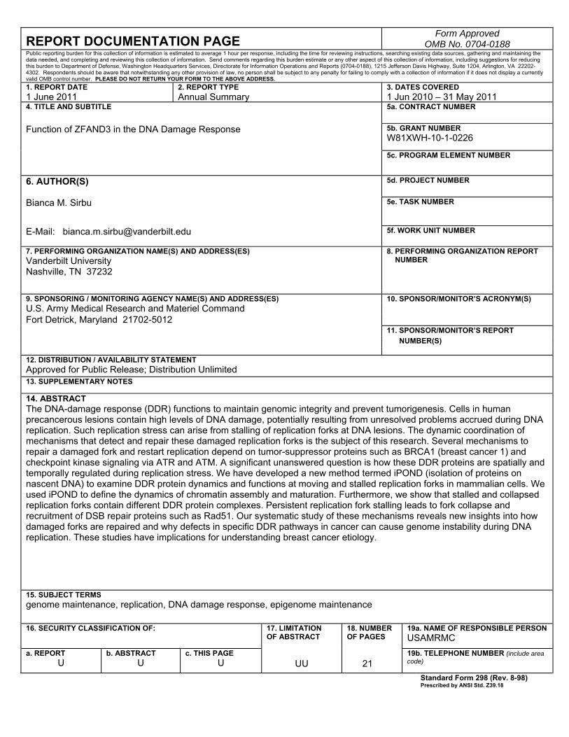
REPORT DOCUMENTATION PAGE Form Approved
OMB No. 0704-0188 Public reporting burden for this collection of information is estimated to average 1 hour per response, including the time for reviewing instructions, searching existing data sources, gathering and maintaining the data needed, and completing and reviewing this collection of information. Send comments regarding this burden estimate or any other aspect of this collection of information, including suggestions for reducing this burden to Department of Defense, Washington Headquarters Services, Directorate for Information Operations and Reports (0704-0188), 1215 Jefferson Davis Highway, Suite 1204, Arlington, VA 22202-4302. Respondents should be aware that notwithstanding any other provision of law, no person shall be subject to any penalty for failing to comply with a collection of information if it does not display a currently valid OMB control number. PLEASE DO NOT RETURN YOUR FORM TO THE ABOVE ADDRESS. 1. REPORT DATE 1 June 2011
2. REPORT TYPEAnnual Summary
3. DATES COVERED 1 Jun 2010 – 31 May 2011
4. TITLE AND SUBTITLE
5a. CONTRACT NUMBER
Function of ZFAND3 in the DNA Damage Response 5b. GRANT NUMBER W81XWH-10-1-0226
5c. PROGRAM ELEMENT NUMBER
6. AUTHOR(S)
5d. PROJECT NUMBER
Bianca M. Sirbu 5e. TASK NUMBER
E-Mail: [email protected] 5f. WORK UNIT NUMBER
7. PERFORMING ORGANIZATION NAME(S) AND ADDRESS(ES) Vanderbilt University Nashville, TN 37232
8. PERFORMING ORGANIZATION REPORT NUMBER
9. SPONSORING / MONITORING AGENCY NAME(S) AND ADDRESS(ES) 10. SPONSOR/MONITOR’S ACRONYM(S)U.S. Army Medical Research and Materiel Command Fort Detrick, Maryland 21702-5012 11. SPONSOR/MONITOR’S REPORT NUMBER(S)
12. DISTRIBUTION / AVAILABILITY STATEMENT Approved for Public Release; Distribution Unlimited 13. SUPPLEMENTARY NOTES
14. ABSTRACT The DNA-damage response (DDR) functions to maintain genomic integrity and prevent tumorigenesis. Cells in human precancerous lesions contain high levels of DNA damage, potentially resulting from unresolved problems accrued during DNA replication. Such replication stress can arise from stalling of replication forks at DNA lesions. The dynamic coordination of mechanisms that detect and repair these damaged replication forks is the subject of this research. Several mechanisms to repair a damaged fork and restart replication depend on tumor-suppressor proteins such as BRCA1 (breast cancer 1) and checkpoint kinase signaling via ATR and ATM. A significant unanswered question is how these DDR proteins are spatially and temporally regulated during replication stress. We have developed a new method termed iPOND (isolation of proteins on nascent DNA) to examine DDR protein dynamics and functions at moving and stalled replication forks in mammalian cells. We used iPOND to define the dynamics of chromatin assembly and maturation. Furthermore, we show that stalled and collapsed replication forks contain different DDR protein complexes. Persistent replication fork stalling leads to fork collapse and recruitment of DSB repair proteins such as Rad51. Our systematic study of these mechanisms reveals new insights into how damaged forks are repaired and why defects in specific DDR pathways in cancer can cause genome instability during DNA replication. These studies have implications for understanding breast cancer etiology.
15. SUBJECT TERMS genome maintenance, replication, DNA damage response, epigenome maintenance
16. SECURITY CLASSIFICATION OF:
17. LIMITATION OF ABSTRACT
18. NUMBER OF PAGES
19a. NAME OF RESPONSIBLE PERSONUSAMRMC
a. REPORT U
b. ABSTRACT U
c. THIS PAGEU UU 21
19b. TELEPHONE NUMBER (include area code)
Standard Form 298 (Rev. 8-98)Prescribed by ANSI Std. Z39.18
Table of Contents
Page Introduction…………………………………………………………….………..….. 4 BODY………………………………………………………………………………….. 5 Key Research Accomplishments………………………………………….…….. 8 Reportable Outcomes……………………………………………………………… 8 Conclusion…………………………………………………………………………… 9 References……………………………………………………………………………. 10 Appendices…………………………………………………………………………… 13
4
Introduction
Maintaining genome integrity is essential to prevent carcinogenesis (1,2). During DNA
replication, genome maintenance requires that several dynamic processes be coordinated at the
replication fork (3,4). Damage encountered during DNA replication, termed replication stress, stalls the
replication fork and challenges the accurate completion of genome and epigenome inheritance. The
DNA damage response (DDR) becomes activated in response to replication stress to protect, repair,
and promote successful completion of chromosome replication (3,5). Importantly, pre-cancerous
lesions, such as those of the breast, exhibit high levels of replication stress that activate the DDR (6,7).
However, the underlying mechanisms that result in genome instability and uncontrolled cell cycle in
these lesions remain unclear and are significant to understanding breast cancer etiology (7).
Upon replication stress, the activated DDR signals to recruit protein complexes to stalled
replication forks to maintain genome and epigenome integrity. In addition to localization, DDR proteins
can be post-translationally modified at stalled forks to modulate protein dynamics (3). For example,
recruitment of the ATR (ATM-and Rad3 related) kinase to stalled replication forks initiates DDR
signaling and regulation through a cascade of phosphorylation events (3). One target of ATR after
replication stress is the breast cancer susceptibility gene BRCA1 (8).
Our understanding of DDR signaling has benefited from genomic screens that have identified
potential regulators of genome integrity, such as ZFAND3 (zinc finger AN1-type domain containing
protein 3) (9). Furthermore, studying genome maintenance proteins has relied heavily on detection of
protein localization to sites of fork stalling. Unfortunately, in human cells, current methods for studying
protein dynamics at replication forks are limited in sensitivity and spatiotemporal resolution (10). To
overcome this challenge and analyze protein dynamics at active, stalled and collapsed replication forks,
we have developed a new methodology termed iPOND (isolation of proteins on nascent DNA) (11).
We used iPOND to define the dynamics of proteins and post-translational modification in the replisome
(the proteins necessary to complete DNA replication), particularly after replication stress. Replication
stress compromises not only genome, but also epigenome integrity (12). Chromatin alterations have
been hypothesized to present a source of genomic instability in cancer (13). Therefore, our iPOND
analyses included studies of changes that accompany chromatin deposition and maturation during DNA

5
replication. Overall, we demonstrate the power of iPOND for studies of replisome and chromatin
dynamics during normal and replication stress conditions.
Body
The initial goal of the proposed research project was to elucidate the function of the novel DNA
damage response gene ZFAND3. Several lines of evidence placed ZFAND3 in the DDR pathway.
ZFAND3 accumulates after DNA damage and functions to sustain the G2/M checkpoint after ionizing
radiation (IR). Furthermore, ZFAND3 was identified to interact with the breast cancer gene TOPBP1
(topoisomerase binding protein 1) in a yeast two-hybrid screen. Lastly, ZFAND3 depletion sensitized to
PARP inhibition. Synthetic lethality with PARP is significant since therapeutic gain has been reported in
patients lacking the breast cancer susceptibility gene BRCA1 (14).
As indicated in the statement of work, the first aim was to characterize the function of ZFAND3
in the DDR. In accordance with this, we found that depletion of ZFAND3 caused minimal reduction in
checkpoint signaling through Chk1 phosphorylation after IR, but not UV or HU treatments. To address
ZFAND3 knockdown efficiency in the loss of function assays, we raised an antibody to ZFAND3.
However, the antibody recognized only exogenously expressed tagged protein, which forced temporary
suspension of task 1.
The subsequent task was to identify ZFAND3 interacting proteins. In agreement with the
proposed outline, we sought to confirm the yeast two-hybrid interaction between TOPBP1 and ZFAND3
using pulldown methodologies and co-immunoprecipitation (co-IP). Recombinant GST-tagged
TOPBP1 fragments and full-length protein were purified and incubated with cell lysates expressing
tagged ZFAND3. No interaction between TOPBP1 and ZFAND3 was detectable before or after IR
damage. Co-IPs corroborated the pulldown results under all lysis conditions tested. Therefore, the
interaction identified in the yeast two-hybrid screen could not be confirmed using the outlined methods.
The unavailability of a ZFAND3 antibody made further protein-protein interaction studies difficult.
The final task of the proposal was to examine the mechanism of ZFAND3 accumulation after
genotoxic stress. To this end, the stability of HA-tagged protein was determined using pulse-chase
6
methods with cycloheximide. The ZFAND3-HA half-life did not change following various treatment
times and doses of IR. This suggested that accumulation of tagged ZFAND3 was not due to protein
stabilization after damage, as is the case for other DDR proteins such as p53 (15). Furthermore,
aggregation of ZFAND3-HA was found to be clonal and cell-type dependent. Therefore, the
subsequent experiments aimed to understand checkpoint regulation of ZFAND3 levels were
suspended.
At this point, we completed the list of tasks in the outlined experimental design that could be
performed without the essential tool—a good ZFAND3 antibody. With insufficient tools and evidence to
pursue ZFAND3 function as a TOPBP1 interacting protein that accumulates after DNA damage, we
redirected our studies to encompass a broader understanding of genome maintenance activities that
prevent breast cancer.
To maintain genome integrity, numerous dynamic processes must be coordinated and executed
with speed and accuracy at the replication fork (3). Exposure of cells to damaging agents during DNA
replication can stall replication forks and recruit DDR protein complexes to maintain genome integrity at
stalled forks (3,5). Understanding such dynamic activities requires identification of proteins located at
the replication fork. In mammalian cells, it has been difficult to stall replication forks at predictable
genomic loci, making chromatin immunoprecipitation (ChIP)-based methods inapplicable for isolation of
stalled replisomes. ChIP of specific endonuclease-induced DSBs has greatly facilitated understanding
of the DDR after DSBs (16,17,18). To provide a tool to study the DDR at moving and stalled replication
forks, we have developed a novel method termed iPOND (isolation of proteins on nascent DNA) (11).
The iPOND methodology identifies proteins at replication forks via a single-step streptavidin
purification of biotin-tagged nascent DNA and the associated proteins. We validated that iPOND
specifically isolates replisome components, even those that are present at one or two copies per
replication fork, such as polymerase epsilon. Therefore, iPOND provides improved sensitivity of
detection for replisome components compared to the commonly utilized method of
immunofluorescence. Of note, iPOND can purify replication proteins that are associated either directly
or indirectly with nascent DNA (11).
In addition to improved sensitivity, combining iPOND with pulse-chase methodologies provides
high spatial and temporal examination of the changing replisome (11). For example, the replication
7
protein PCNA (proliferating cell nuclear antigen) is recycled from completed Okazaki fragments on the
lagging strand; however, the timing of this event has remained undetermined in mammalian cells (19).
Using iPOND pulse-chase, we defined this timing of PCNA recycling, as well as the timing of chromatin
maturation. Furthermore, iPOND can monitor the in vivo assembly of chromatin and confirmed
proposed models of a step-wise deposition of histones onto nascent DNA (11,20).
To understand the DDR to replication stress, we next analyzed the dynamics of proteins
recruited and modified at stalled and collapsed replication forks. We showed that shortly upon
exposure to the replication stress agent hydroxyurea (HU), the histone variant H2AX is phosphorylated
(termed γH2AX) at stalled replication forks. Phosphorylated H2AX is typically considered a marker of
DSBs (21). However, we detected γH2AX early in the response to HU, prior to evidence of DSB
formation at a stalled fork (22). Early γH2AX corresponded to phosphorylation of RPA on Ser33, an
ATR kinase target site (23). The subsequent RPA phosphorylation on Ser4/8, which are DNA-PK sites,
occurred after prolonged exposure to replication stress (23). This suggests that ATR phosphorylates
RPA immediately after fork stalling, while DNA-PK phosphorylates RPA at persistently stalled forks.
Prolonged exposure to replication stress exhibited enrichment of DSB repair proteins Mre11,
KU70/80 and the recombinase Rad51 at stalled forks. Our data suggest that persistently stalled forks
collapse into DSBs within four hours of fork stalling. Furthermore, accumulation of Rad51 to
persistently, but not transiently stalled forks depends on the nuclease activity of Mre11, similarly to the
mechanism that recruits Rad51 to DSBs (24).
At DSBs, γH2AX spreads from the DSB to facilitate DDR signaling (25,26). Several
observations of γH2AX patterns at stalled forks led us to test whether γH2AX also spreads from a
stalled replication fork. Indeed, using various iPOND pulse-chase methods, we showed that γH2AX
spreads from stalled replication forks to include a large chromatin domain. Of note, the propagation of
γH2AX occurs shortly after exposure to replication stress. To examine the checkpoint kinases
responsible for initiating and spreading γH2AX, we tested the necessity of ATR, ATM and DNA-PK in
time course experiments. Our results are consistent with a model wherein ATR phosphorylates H2AX
at stalled forks and promotes initial γH2AX spreading. Longer exposures to replication stress require
the combined activities of ATR, ATM, and DNA-PK to initiate, spread and maintain γH2AX at the stalled
fork and in regions proximal to the fork.
8
The iPOND methodology and findings described above have been incorporated into a
manuscript that has been published in Genes and Development (see appendix).
Key Research Accomplishments
• Completion of proposed plan for characterization of ZFAND3 in the DDR
• Development of a novel methodology iPOND (isolation of proteins on nascent DNA) to study the
DDR at stalled replication forks in mammalian cells at high spatial and temporal resolution
• Validation of iPOND to show replisome dynamics and the timing of chromatin assembly and
maturation
• Characterization of dynamic replisome composition at stalled and collapsed replication forks
• DSB repair proteins are recruited to persistently stalled replication forks
• γH2AX spreads from stalled replication forks in an ATR, ATM, and DNA-PK dependent manner
• Rad51 protein accumulation at persistently stalled replication forks depends on Mre11 nuclease
activity
Reportable outcomes
1. Publication of iPOND as novel method to study genome maintenance activities at replication forks in mammalian cells
a. Title: Analysis of protein dynamics at active, stalled and collapsed replication forks b. Authors: Sirbu BM, Couch FB, Feigerle JT, Bhaskara S, Hiebert SW, Cortez D c. Published: June 2011 Genes and Development. 25(12):1320-7
2. Oral presentation of iPOND methodology
a. Title: Analysis of protein dynamics at active, stalled and collapsed replication forks b. Speaker: Sirbu BM c. Meeting: Gordon Research Conference, Cell Growth and Proliferation, June 29, 2011,
Biddeford, ME.
3. Poster presentation of iPOND methodology a. Title: Analysis of protein dynamics at active, stalled and collapsed replication forks b. Authors: Sirbu BM, Couch FB, Feigerle JT, Bhaskara S, Hiebert SW, Cortez D c. Meeting: Gordon Research Seminar, Cell Growth and Proliferation, June 26, 2011,
Biddeford, ME.
4. Poster presentation describing the iPOND methodology a. Title: Analysis of protein dynamics at active, stalled and collapsed replication forks b. Authors: Sirbu BM, Couch FB, Feigerle JT, Bhaskara S, Hiebert SW, Cortez D c. Retreat: Vanderbilt Ingram Cancer Center Retreat, Nashville, TN, May 2011
9
Conclusions
Genome integrity must be maintained at the replication fork to prevent cancer (3). Accurate
completion of chromosome replication involves the spatial and temporal coordination of genome
maintenance protein dynamics at replication forks. Previous studies of the replisome during normal
and stress responses at stalled forks have relied largely on immunofluorescent imaging to track protein
localization. While useful, immunofluorescence (IF) has the significant disadvantages of low resolution
and low sensitivity. For example, proteins that exist at only single copy levels at replication forks
cannot be tracked with IF. To overcome this difficulty, we have developed a new methodology termed
iPOND to isolate proteins on nascent DNA. iPOND has dramatically improved sensitivity compared to
IF and allows the interrogation of dynamic events that occur at the normal and stalled fork with high
spatial and temporal resolution.
We used this method to define the timing of histone deposition and chromatin maturation. We
found that replication fork stalling causes changes in the recruitment and phosphorylation of proteins at
the damaged fork. H2AX phosphorylation, typically considered a DSB marker, occurs at stalled
replication forks and spreads to include chromatin domains away from the replication fork. This occurs
in a checkpoint dependent manner prior to evidence of DSB formation at a stalled fork. Finally, we
demonstrated a switch in the DDR at persistently stalled forks that includes Mre11-dependent Rad51
assembly.
Overall the data we have obtained with iPOND provides the first high-resolution, time-
dependent analyses of protein dynamics at active, stalled, and collapsed replication forks in
mammalian cells. Furthermore, our results validate iPOND as a powerful method to study DNA damage
responses, chromatin deposition, and chromatin maturation during DNA replication. These dynamic
processes must be well defined to comprehend how their coordination during replication stress
prevents the genomic and epigenomic instability that is a hallmark of cancerous lesions, such as those
found in breast cancer.

10
References
1. Hoeijmakers, J.H. (2001). Genome maintenance mechanisms for preventing cancer. Nature
411:366-74.
2. Schar, P. (2001). Spontaneous DNA damage, genome instability, and cancer—when DNA
replication escapes control. Cell 104:329-32.
3. Cimprich, K.A., and Cortez, D. (2008). ATR: an essential regulator of genome integrity. Nat Rev
Mol Cell Biol 9, 616-627.
4. Probst, A.V., Dunleavy, E., and Almouzni, G. (2009). Epigenetic inheritance during the cell
cycle. Nat Rev Mol Cell Biol 10, 192-206.
5. Harper, J.W., and Elledge, S.J. (2007). The DNA damage response: ten years after. Mol Cell
28, 739-745.
6. Bartkova J, Horejsí Z, Koed K, Krämer A, Tort F, Zieger K, Guldberg P, Sehested M, Nesland
JM, Lukas C, Ørntoft T, Lukas J, Bartek J. (2005). DNA damage resonse as a candidate anti-
cancer barrier in early human tumorigenesis. Nature 434:864-70.
7. Gorgoulis VG, Vassiliou LV, Karakaidos P, Zacharatos P, Kotsinas A, Liloglou T, Venere M,
Ditullio RA Jr, Kastrinakis NG, Levy B, Kletsas D, Yoneta A, Herlyn M, Kittas C, Halazonetis TD.
(2005). Activation of the DNA damage checkpoint and genomic instability in human
precancerous lesions. Nature 434:907-13.
8. Tibbetts RS, Cortez D, Brumbaugh KM, Scully R, Livingston D, Elledge SJ, Abraham RT.
(2000). Functional interaction between BRCA1 and the checkpoint kinase ATR during
genotoxic stress. Genes Dev 14(23):2989-3002.
9. Polo S.E., Jackson S.P. (2011). Dynamics of DNA damage response proteins on DNA breaks: a
focus on protein modifications. Genes Dev 25(5):409-33.
10. Sirbu BM, Couch FB, Feigerle JT, Bhaskara S, Hiebert SW, Cortez D. (2011). Analysis of
protein dynamics at active, stalled and collapsed replication forks. Genes Dev 25(12):1320-7.
11. Lovejoy CA, Xu X, Bansbach CE, Glick GG, Zhao R, Ye F, Sirbu BM, Titus LC, Shyr Y, Cortez
D. (2009). Functional genomic screens identify CINP as a genome maintenance protein. Proc
Natl Acad Sci U S A 106(46):19304-9.
11
12. Jasencakova Z, Scharf AN, Ask K, Corpet A, Imhof A, Almouzni G, Groth A. (2010). Replication
stress interferes with histone recycling and predeposition marking of new histones. Mol Cell
37(5):736-43.
13. Jasencakova Z, Groth A. (2010). Replication stress, a source of genetic aberrations in cancer.
Bioessays 32(10):847-55.
14. Ashworth, A. (2008). A synthetic lethal therapeutic approach: Poly(ADP) Ribose Polymerase
inhibitors for the treatment of cancers deficient in DNA double-strand break repair. J Clin Oncol
26(22):3785-90.
15. Kubbutat MH, Jones SN, Vousden KH. (1997). Regulation of p53 stability by Mdm2. Nature
387(6630):299-303.
16. Rodrigue, A., Lafrance, M., Gauthier, M.C., McDonald, D., Hendzel, M., West, S.C., Jasin, M.,
and Masson, J.Y. (2006). Interplay between human DNA repair proteins at a unique double-
strand break in vivo. EMBO J 25, 222-231.
17. Rudin, N., and Haber, J.E. (1988). Efficient repair of HO-induced chromosomal breaks in
Saccharomyces cerevisiae by recombination between flanking homologous sequences. Mol
Cell Biol 8, 3918-3928.
18. Soutoglou, E., Dorn, J.F., Sengupta, K., Jasin, M., Nussenzweig, A., Ried, T., Danuser, G., and
Misteli, T. (2007). Positional stability of single double-strand breaks in mammalian cells. Nat Cell
Biol 9, 675-682.
19. Shibahara K, Stillman B. (1999). Replication dependent marking of DNA by PCNA facilitates
CAF-1-coupled inheritance of chromatin. Cell 96(4):575-85.
20. Worcel, A., Han, S., and Wong, M.L. (1978). Assembly of newly replicated chromatin. Cell 15,
969-977.
21. Dickey, J.S., Redon, C.E., Nakamura, A.J., Baird, B.J., Sedelnikova, O.A., and Bonner, W.M.
(2009). H2AX: functional roles and potential applications. Chromosoma 118, 683-692.
22. Petermann, E., and Helleday, T. (2010). Pathways of mammalian replication fork restart. Nat
Rev Mol Cell Biol 11, 683-687.

12
23. Anantha, R.W., Vassin, V.M., and Borowiec, J.A. (2007). Sequential and synergistic
modification of human RPA stimulates chromosomal DNA repair. J Biol Chem 282, 35910-
35923.
24. Mimitou, E.P., and Symington, L.S. (2009). DNA end resection: many nucleases make light
work. DNA Repair (Amst) 8, 983-995.
25. Berkovich, E., Monnat, R.J., Jr., and Kastan, M.B. (2008). Assessment of protein dynamics and
DNA repair following generation of DNA double-strand breaks at defined genomic sites. Nat
Protoc 3, 915-922.
26. Savic, V., Yin, B., Maas, N.L., Bredemeyer, A.L., Carpenter, A.C., Helmink, B.A., Yang-Iott,
K.S., Sleckman, B.P., and Bassing, C.H. (2009). Formation of dynamic gamma-H2AX domains
along broken DNA strands is distinctly regulated by ATM and MDC1 and dependent upon H2AX
densities in chromatin. Mol Cell 34, 298-310.
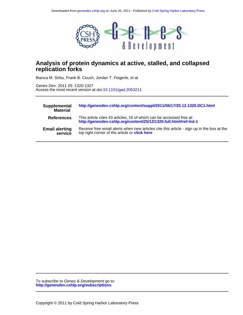
10.1101/gad.2053211Access the most recent version at doi: 2011 25: 1320-1327Genes Dev.
Bianca M. Sirbu, Frank B. Couch, Jordan T. Feigerle, et al. replication forksAnalysis of protein dynamics at active, stalled, and collapsed
MaterialSupplemental http://genesdev.cshlp.org/content/suppl/2011/06/17/25.12.1320.DC1.html
References http://genesdev.cshlp.org/content/25/12/1320.full.html#ref-list-1
This article cites 43 articles, 16 of which can be accessed free at:
serviceEmail alerting
click heretop right corner of the article orReceive free email alerts when new articles cite this article - sign up in the box at the
http://genesdev.cshlp.org/subscriptions go to: Genes & DevelopmentTo subscribe to
Copyright © 2011 by Cold Spring Harbor Laboratory Press
Cold Spring Harbor Laboratory Press on June 20, 2011 - Published by genesdev.cshlp.orgDownloaded from

Analysis of protein dynamics at active,stalled, and collapsed replication forks
Bianca M. Sirbu, Frank B. Couch, Jordan T. Feigerle, Srividya Bhaskara, Scott W. Hiebert,and David Cortez1
Department of Biochemistry, Vanderbilt University School of Medicine, Nashville, Tennessee 37232, USA
Successful DNA replication and packaging of newly synthesized DNA into chromatin are essential to maintaingenome integrity. Defects in the DNA template challenge genetic and epigenetic inheritance. Unfortunately,tracking DNA damage responses (DDRs), histone deposition, and chromatin maturation at replication forks isdifficult in mammalian cells. Here we describe a technology called iPOND (isolation of proteins on nascent DNA)to analyze proteins at active and damaged replication forks at high resolution. Using this methodology, we definethe timing of histone deposition and chromatin maturation. Class 1 histone deacetylases are enriched atreplisomes and remove predeposition marks on histone H4. Chromatin maturation continues even whendecoupled from replisome movement. Furthermore, fork stalling causes changes in the recruitment andphosphorylation of proteins at the damaged fork. Checkpoint kinases catalyze H2AX phosphorylation, whichspreads from the stalled fork to include a large chromatin domain even prior to fork collapse and double-strandbreak formation. Finally, we demonstrate a switch in the DDR at persistently stalled forks that includesMRE11-dependent RAD51 assembly. These data reveal a dynamic recruitment of proteins and post-translationalmodifications at damaged forks and surrounding chromatin. Furthermore, our studies establish iPOND as a usefulmethodology to study DNA replication and chromatin maturation.
[Keywords: DNA replication; chromatin; DNA damage response; H2AX; histone acetylation; EdU; click chemistry]
Supplemental material is available for this article.
Received March 22, 2011; revised version accepted May 16, 2011.
In human cells, more than 6 billion base pairs of DNAneed to be replicated and packaged into chromatin everycell division cycle. Failures lead to mutation, epigeneticchanges, and other chromosomal aberrations that ulti-mately cause diseases such as cancer. DNA replication iscoordinated with chromatin assembly (Probst et al. 2009).The replisome, containing the proteins necessary to com-plete replication, is a dynamic machine that must workwith speed and precision. DNA lesions, insufficient nucle-otides, and other types of replication stress cause forkstalling. In these circumstances, theDNAdamage response(DDR) mobilizes repair activities to stabilize the fork,resolve the problem, and complete DNA synthesis (Harperand Elledge 2007; Cimprich and Cortez 2008).The DDR to replication stress is poorly understood in
comparison with the response to double-strand breaks(DSBs). For example, there are extensive modifications tothe chromatin surrounding a DSB, including destabiliza-tion of nucleosomes, chromatin remodeling, and histonepost-translational modifications (Morrison and Shen 2009;van Attikum and Gasser 2009; Rossetto et al. 2010;
Venkitaraman 2010). These changes increase access to therepair machinery and recruit proteins involved in repairand DDR signaling. The extent to which chromatinchanges at a stalled forkmimic those at a DSB is unknown.Replication provides a unique landscape and set of
challenges compared with a DSB. The immediate vicinityof the replisome lacks nucleosomes. Also, half of thehistones on the nascent DNA are newly synthesized andrequire changes in post-translational modifications torestore the proper chromatin structure. Finally, severalmechanisms exist to recover stalled replication forks,which necessitate the recruitment of multiple enzymaticactivities and, perhaps, different chromatin changes.The difference in our knowledge of the responses at
stalled forks compared with DSBs is due primarily to theincreased technical challenges of studying replicationstress. For example, several investigators have used site-specific DSBs combined with chromatin immunoprecip-itation (ChIP) to examine proteins at breaks with highresolution (Rudin and Haber 1988; Rodrigue et al. 2006;Soutoglou et al. 2007; Berkovich et al. 2008). Thus far,site-specific analysis of active and stalled replisomes inmammalian cells has not been achieved. We addressedthis technical limitation by developing the iPOND (iso-lation of proteins on nascent DNA) methodology. iPOND
1Corresponding author.E-mail [email protected] is online at http://www.genesdev.org/cgi/doi/10.1101/gad.2053211.
1320 GENES & DEVELOPMENT 25:1320–1327 ! 2011 by Cold Spring Harbor Laboratory Press ISSN 0890-9369/11; www.genesdev.org
Cold Spring Harbor Laboratory Press on June 20, 2011 - Published by genesdev.cshlp.orgDownloaded from

permits the isolation and analysis of proteins at active,stalled, and collapsed replication forks. It can also probethe changes that accompany chromatin deposition andmaturation following DNA synthesis. We demonstratethe power of iPOND by defining the dynamics of proteinsand post-translational modifications in the replisome andon the newly deposited chromatin.
Results
Development of iPOND
Tracking the location of any single replisome in a mam-malian cell is not possible, limiting the utility of ChIP-based technologies. To overcome this technical limitation,we used the thymidine analog 5-ethynyl-29-deoxyuridine(EdU) (Salic and Mitchison 2008), which contains analkyne functional group. Covalent linkage to a biotin-azideusing click chemistry (Moses and Moorhouse 2007) facil-itates single-step purification of the EdU-labeled nascentDNA and associated proteins at replication forks (Fig. 1A).To validate this methodology we first asked whether
we could detect replisome proteins. We labeled cells withEdU for 10 min then performed iPOND. We detectedproliferating cell nuclear antigen (PCNA), chromatinassembly factor 1 (CAF-1), replication protein A (RPA),and two subunits of polymerase e (Fig. 1B). These resultsindicate that iPOND can purify replisome proteins, in-cluding those indirectly bound to DNA such as CAF-1(Shibahara and Stillman 1999). Furthermore, they indi-cate that iPOND is a highly sensitive methodology. Weare able to detect proteins such as POLE2 and POLE3,which are expected to be at a density of only one or twomolecules per fork (Fig. 1B). Thus, unlike immunofluores-cence, iPOND does not require high concentrations ofproteins within a small nuclear region to track proteinlocalization. Of note, proteins not present at replicationforks, such as GAPDH, are not detectable in iPOND cap-tures (data not shown).
In time-course experiments, we detected PCNA andCAF-1 after a 2.5-min pulse of EdU, histones H2B and H3after 5 min, and the linker histone H1 at 20 min after EdUaddition (Fig. 1C). Thus, with short labeling times, weselectively purify proteins at the replication fork, andlonger labeling times permit analysis of chromatin assem-bly. The order of histone deposition supports previousfractionation data indicating that H1 is added 10–20 minafter DNA replication to create higher-order chromatinstructures (Worcel et al. 1978).The resolution of this technique depends on the length
of the EdU pulse, the rate of DNA synthesis, and the size ofthe DNA fragments generated after cell lysis. In practice,the first two parameters are the most important, since weconsistently obtainDNA fragments of;150 base pairs (bp)(Supplemental Fig. 1). In mammalian cells, the rate ofDNA synthesis varies between 0.75 and 2.5 kb/min(Herrick and Bensimon 2008). Thus, a 2.5-min EdU pulselabels ;2–6 kb, although this is likely a significant over-estimation, since EdU must enter the cell and be phos-phorylated before incorporation into DNA. Thus, iPONDresolution is on the order of a few thousand base pairs.Importantly, iPOND can be combined with pulse-chase
methods to track how proteins assemble and disassemblefrom a nascent DNA segment with high spatial andtemporal resolution. Increasing chase times monitorDNA-associated proteins at greater and greater distancesfrom the moving fork. In these experiments, histonelevels remain constant, indicating that the procedureeffectively captures a maturing chromatin segment ofconstant length (Fig. 1D). However, PCNA and CAF-1levels purified with the EdU-labeled segment declinewith a half-life of considerably <10 min of chase time(Fig. 1D). These data indicate that iPOND isolates chro-matin-associated proteins specifically located at the rep-lication fork, and are consistent with rapid unloading ofPCNA andCAF-1 once Okazaki fragment DNA synthesisis complete.
Figure 1. Development of the iPONDtechnology. (A) iPOND begins by addingEdU to cultured cells. The cells are thentreated with formaldehyde to cross-linkprotein–DNA complexes, washed, and per-meabilized with detergent. Copper cata-lyzes the cycloaddition of biotin-azide tothe EdU-labeled DNA. The cells are thenlysed in denaturing conditions with sonica-tion. The biotin-labeled DNA–protein com-plexes are purified using streptavidin-coatedbeads, cross-links are reversed, and theeluted proteins are analyzed by immuno-blotting or other methods like mass spec-trometry. (B) Cells were incubated withEdU for 10 min prior to performing iPOND.Cells expressing POLE2-HA or POLE3-HAwere used to detect these proteins with the
HA antibody. (C) Cells were incubated in EdU-containing medium for increasing times prior to performing the iPOND protocol. (D)Cells were incubated with EdU for 10 min. The EdU-containing medium was removed and cells were washed once before incubating forincreasing times in medium containing 10 mM thymidine prior to performing iPOND. In all experiments, the No Clk control is theinput sample in the first lane processed with no biotin-azide.
Analysis of DNA replication using iPOND
GENES & DEVELOPMENT 1321
Cold Spring Harbor Laboratory Press on June 20, 2011 - Published by genesdev.cshlp.orgDownloaded from

Analysis of chromatin maturation using iPOND
Maturation of the new chromatin requires addition andremoval of histone post-translationalmodifications. Newlysynthesized histone H4 is acetylated on two lysines (5 and12), and these evolutionarily conserved marks are removedafter deposition (Sobel et al. 1995; Taddei et al. 1999). Ourtime course experiments indicate that acetylated H4K5(H4K5ac) is removed rapidly and H4K12ac deacetylationis slightly delayed (Fig. 2A,B). The delay in K12 deacety-lation could be due to the activity of chromatin-associatedhistone acetyltransferases (HATs) that promote the acety-lation of this site in some chromatin domains. Indeed, in thepresence of the nonselective HAT inhibitor anacardic acid,the rate of H4K12 deacetylation becomes identical to H4K5,with a half-life of <20 min (Fig. 2C,D).In principle, chromatin maturation—as measured by
H4K5,K12 deacetylation—could be coupled to fork progres-sion. To test this possibility, we used high concentrationsof hydroxyurea (HU) to stall active replisomes and stopDNA synthesis. HU addition stalls the fork effectively inthese cells, since the amount of histone capture does notincrease appreciably during the HU treatment (Fig. 2E).Deacetylation of newly deposited H4 proceeds at the samerate regardless of whetherDNA synthesis is inhibited. Thus,chromatin maturation can be uncoupled from replisomemovement.The histone deacetylase (HDAC) in human cells that
catalyzes the deacetylation of H4K5 and K12 is unknown.HDAC1 and HDAC2 associate with CAF-1 (Ahmad et al.1999), and HDAC3 is required—perhaps in late S phase orG2—to remove H4K5ac (Bhaskara et al. 2010). Indeed, inpulse-chase experiments, we found an enrichment of
HDAC1, HDAC2, and HDAC3 near the fork (Fig. 2A), andthe selective class I HDAC inhibitor FK228 (Furumai et al.2002) prevented deacetylation ofH4 (Fig. 2F), suggesting thatall three of these HDACs are involved.
DDR response at stalled replication forks
HU treatment causes DDR activation to stabilize the stalledfork and induce a cell cycle checkpoint. Previous studiessuggest that HU-stalled forks remain stable and competentto resume DNA synthesis for several hours; however,eventually, the stalled fork collapses and DSBs are formed(Petermann et al. 2010). To further examine this process,we monitored recruitment and modification of proteins atstalled forks. The amounts of PCNA and CAF-1 that arecaptured at the stalled fork decrease initially after addingHU to the medium, and then reach a steady state level ofbetween 20% and 30% of that found at an elongating fork(Fig. 3A). This PCNA pattern is likely due to unloading ofPCNA from the completed Okazaki fragments.We detectedRPA associated with the fork both before and after HUaddition (Fig. 3A). The amount of RPA detected remainedconstant even though RPA accumulates at stalled forks(Cimprich and Cortez 2008). This discrepancy is explainedbecause RPA binds only to the single-stranded, templatestrand of DNA, which lacks incorporated EdU. Therefore,iPOND detects only the RPA immediately adjacent to thenewly synthesized dsDNA (Supplemental Fig. 2).In these experiments, we noticed that at 120 and 240
min after addition of HU, the electrophoretic mobility ofRPA decreased, consistent with phosphorylation (Fig. 3A).RPA S33 phosphorylation could be detected within 10minof HU addition, and S4/S8 phosphorylation appeared at 2 h
Figure 2. HDACs are enriched at replication forksand deacetylate newly deposited histone H4 regardlessof fork movement. (A–E). Cells were labeled with EdUfor 10 min followed, by a chase into thymidine-con-taining medium for the indicated times prior to per-forming iPOND. (B) Quantitation of H4 acetylationlevels compared with total H4 in the click reactionsamples from three independent experiments. Errorbars in all figures are standard deviations. (C,D) Ana-cardic acid (30 mM) was added to the indicated samples.(E) HU (3 mM) was added to the indicated samples. (F)Cells labeled with EdU were chased into 3 mM HUmedium with or without 100 nM FK228 prior toperforming iPOND.
Sirbu et al.
1322 GENES & DEVELOPMENT
Cold Spring Harbor Laboratory Press on June 20, 2011 - Published by genesdev.cshlp.orgDownloaded from
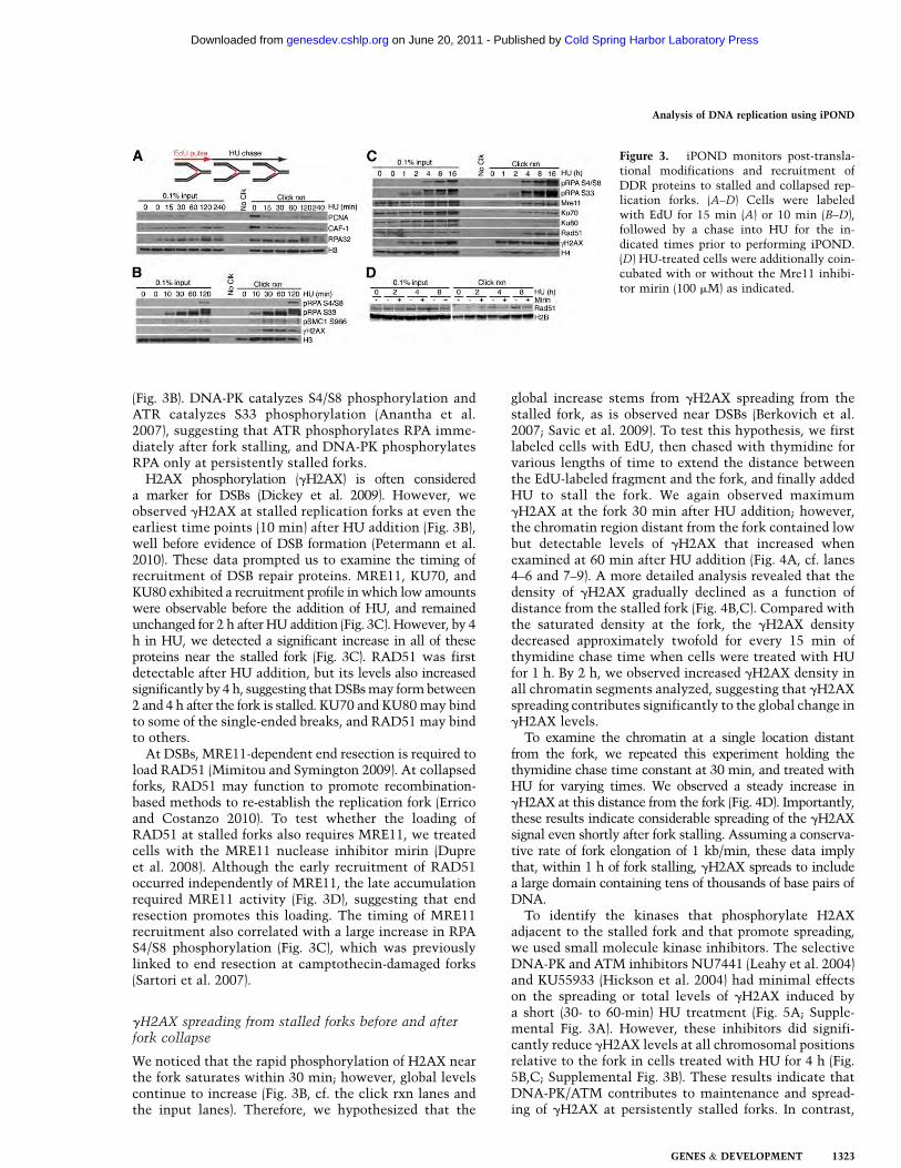
(Fig. 3B). DNA-PK catalyzes S4/S8 phosphorylation andATR catalyzes S33 phosphorylation (Anantha et al.2007), suggesting that ATR phosphorylates RPA imme-diately after fork stalling, and DNA-PK phosphorylatesRPA only at persistently stalled forks.H2AX phosphorylation (gH2AX) is often considered
a marker for DSBs (Dickey et al. 2009). However, weobserved gH2AX at stalled replication forks at even theearliest time points (10 min) after HU addition (Fig. 3B),well before evidence of DSB formation (Petermann et al.2010). These data prompted us to examine the timing ofrecruitment of DSB repair proteins. MRE11, KU70, andKU80 exhibited a recruitment profile in which low amountswere observable before the addition of HU, and remainedunchanged for 2 h after HU addition (Fig. 3C). However, by 4h in HU, we detected a significant increase in all of theseproteins near the stalled fork (Fig. 3C). RAD51 was firstdetectable after HU addition, but its levels also increasedsignificantly by 4 h, suggesting thatDSBsmay formbetween2 and 4 h after the fork is stalled. KU70 and KU80may bindto some of the single-ended breaks, and RAD51 may bindto others.At DSBs, MRE11-dependent end resection is required to
load RAD51 (Mimitou and Symington 2009). At collapsedforks, RAD51 may function to promote recombination-based methods to re-establish the replication fork (Erricoand Costanzo 2010). To test whether the loading ofRAD51 at stalled forks also requires MRE11, we treatedcells with the MRE11 nuclease inhibitor mirin (Dupreet al. 2008). Although the early recruitment of RAD51occurred independently of MRE11, the late accumulationrequired MRE11 activity (Fig. 3D), suggesting that endresection promotes this loading. The timing of MRE11recruitment also correlated with a large increase in RPAS4/S8 phosphorylation (Fig. 3C), which was previouslylinked to end resection at camptothecin-damaged forks(Sartori et al. 2007).
gH2AX spreading from stalled forks before and afterfork collapse
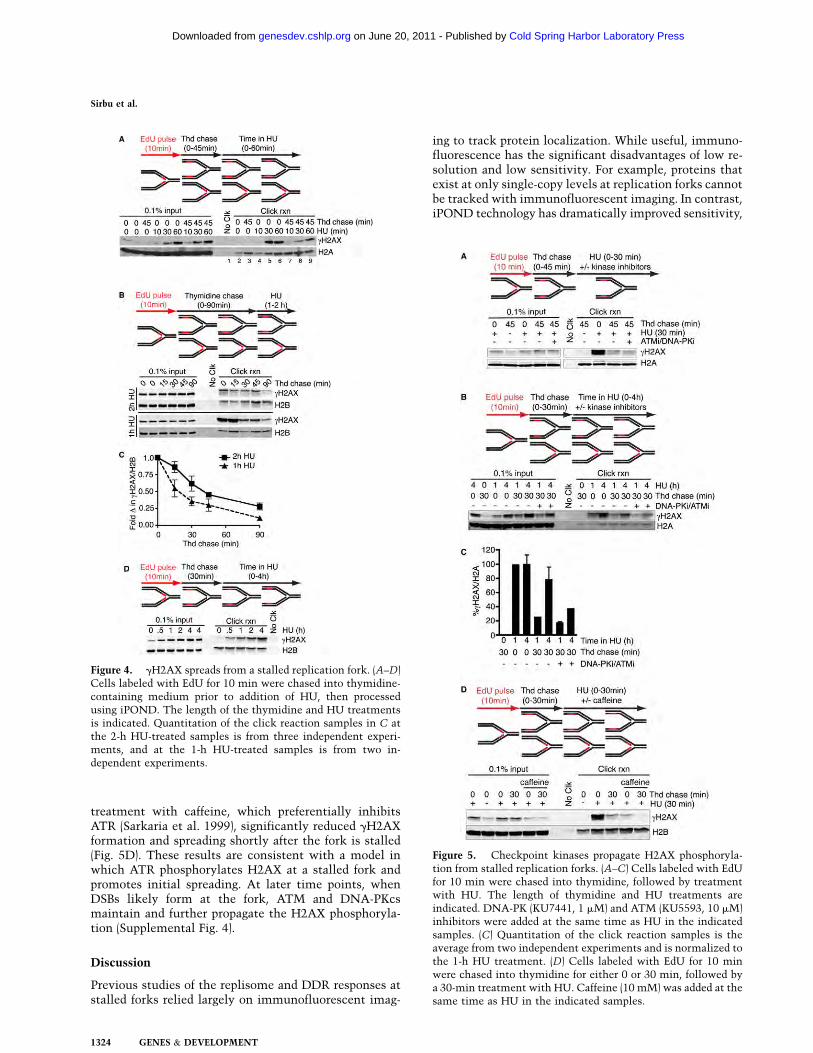
We noticed that the rapid phosphorylation of H2AX nearthe fork saturates within 30 min; however, global levelscontinue to increase (Fig. 3B, cf. the click rxn lanes andthe input lanes). Therefore, we hypothesized that the
global increase stems from gH2AX spreading from thestalled fork, as is observed near DSBs (Berkovich et al.2007; Savic et al. 2009). To test this hypothesis, we firstlabeled cells with EdU, then chased with thymidine forvarious lengths of time to extend the distance betweenthe EdU-labeled fragment and the fork, and finally addedHU to stall the fork. We again observed maximumgH2AX at the fork 30 min after HU addition; however,the chromatin region distant from the fork contained lowbut detectable levels of gH2AX that increased whenexamined at 60 min after HU addition (Fig. 4A, cf. lanes4–6 and 7–9). A more detailed analysis revealed that thedensity of gH2AX gradually declined as a function ofdistance from the stalled fork (Fig. 4B,C). Compared withthe saturated density at the fork, the gH2AX densitydecreased approximately twofold for every 15 min ofthymidine chase time when cells were treated with HUfor 1 h. By 2 h, we observed increased gH2AX density inall chromatin segments analyzed, suggesting that gH2AXspreading contributes significantly to the global change ingH2AX levels.To examine the chromatin at a single location distant
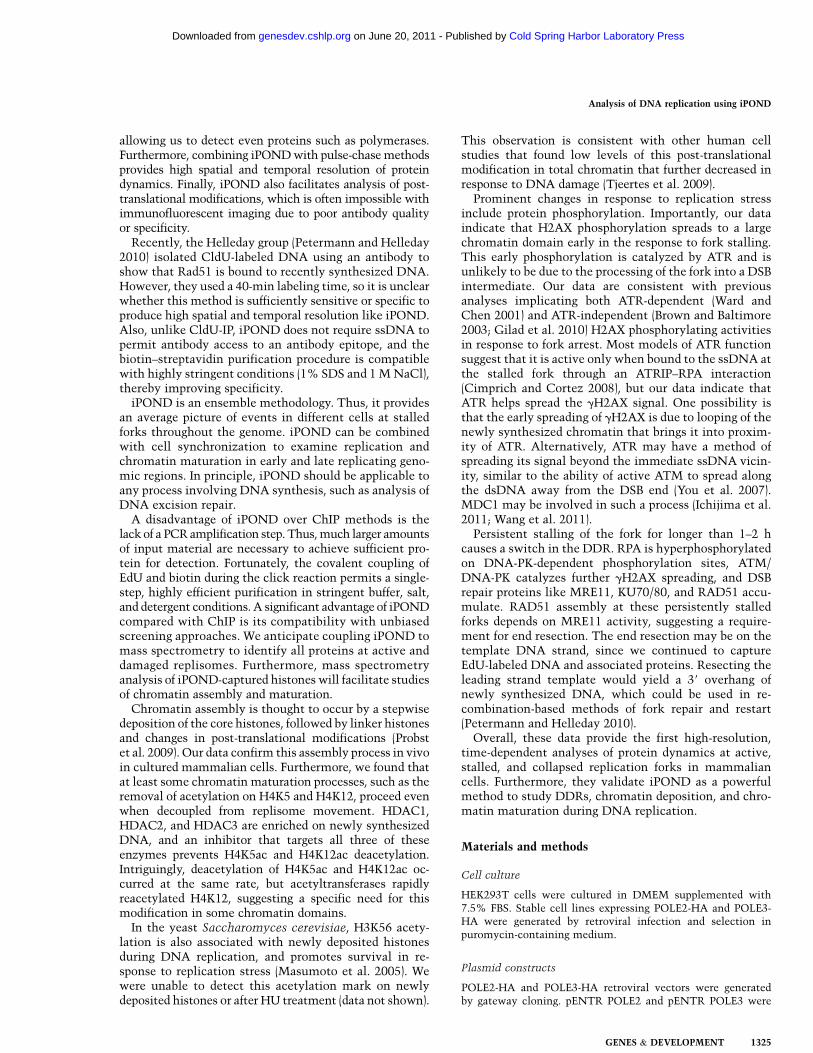
from the fork, we repeated this experiment holding thethymidine chase time constant at 30 min, and treated withHU for varying times. We observed a steady increase ingH2AX at this distance from the fork (Fig. 4D). Importantly,these results indicate considerable spreading of the gH2AXsignal even shortly after fork stalling. Assuming a conserva-tive rate of fork elongation of 1 kb/min, these data implythat, within 1 h of fork stalling, gH2AX spreads to includea large domain containing tens of thousands of base pairs ofDNA.To identify the kinases that phosphorylate H2AX
adjacent to the stalled fork and that promote spreading,we used small molecule kinase inhibitors. The selectiveDNA-PK and ATM inhibitors NU7441 (Leahy et al. 2004)and KU55933 (Hickson et al. 2004) had minimal effectson the spreading or total levels of gH2AX induced bya short (30- to 60-min) HU treatment (Fig. 5A; Supple-mental Fig. 3A). However, these inhibitors did signifi-cantly reduce gH2AX levels at all chromosomal positionsrelative to the fork in cells treated with HU for 4 h (Fig.5B,C; Supplemental Fig. 3B). These results indicate thatDNA-PK/ATM contributes to maintenance and spread-ing of gH2AX at persistently stalled forks. In contrast,
Figure 3. iPOND monitors post-transla-tional modifications and recruitment ofDDR proteins to stalled and collapsed rep-lication forks. (A–D) Cells were labeledwith EdU for 15 min (A) or 10 min (B–D),followed by a chase into HU for the in-dicated times prior to performing iPOND.(D) HU-treated cells were additionally coin-cubated with or without the Mre11 inhibi-tor mirin (100 mM) as indicated.
Analysis of DNA replication using iPOND
GENES & DEVELOPMENT 1323
Cold Spring Harbor Laboratory Press on June 20, 2011 - Published by genesdev.cshlp.orgDownloaded from
treatment with caffeine, which preferentially inhibitsATR (Sarkaria et al. 1999), significantly reduced gH2AXformation and spreading shortly after the fork is stalled(Fig. 5D). These results are consistent with a model inwhich ATR phosphorylates H2AX at a stalled fork andpromotes initial spreading. At later time points, whenDSBs likely form at the fork, ATM and DNA-PKcsmaintain and further propagate the H2AX phosphoryla-tion (Supplemental Fig. 4).
Discussion
Previous studies of the replisome and DDR responses atstalled forks relied largely on immunofluorescent imag-
ing to track protein localization. While useful, immuno-fluorescence has the significant disadvantages of low re-solution and low sensitivity. For example, proteins thatexist at only single-copy levels at replication forks cannotbe tracked with immunofluorescent imaging. In contrast,iPOND technology has dramatically improved sensitivity,
Figure 4. gH2AX spreads from a stalled replication fork. (A–D)Cells labeled with EdU for 10 min were chased into thymidine-containing medium prior to addition of HU, then processedusing iPOND. The length of the thymidine and HU treatmentsis indicated. Quantitation of the click reaction samples in C atthe 2-h HU-treated samples is from three independent experi-ments, and at the 1-h HU-treated samples is from two in-dependent experiments.
Figure 5. Checkpoint kinases propagate H2AX phosphoryla-tion from stalled replication forks. (A–C) Cells labeled with EdUfor 10 min were chased into thymidine, followed by treatmentwith HU. The length of thymidine and HU treatments areindicated. DNA-PK (KU7441, 1 mM) and ATM (KU5593, 10 mM)inhibitors were added at the same time as HU in the indicatedsamples. (C) Quantitation of the click reaction samples is theaverage from two independent experiments and is normalized tothe 1-h HU treatment. (D) Cells labeled with EdU for 10 minwere chased into thymidine for either 0 or 30 min, followed bya 30-min treatment with HU. Caffeine (10 mM) was added at thesame time as HU in the indicated samples.
Sirbu et al.
1324 GENES & DEVELOPMENT
Cold Spring Harbor Laboratory Press on June 20, 2011 - Published by genesdev.cshlp.orgDownloaded from
allowing us to detect even proteins such as polymerases.Furthermore, combining iPONDwith pulse-chasemethodsprovides high spatial and temporal resolution of proteindynamics. Finally, iPOND also facilitates analysis of post-translational modifications, which is often impossible withimmunofluorescent imaging due to poor antibody qualityor specificity.Recently, the Helleday group (Petermann and Helleday
2010) isolated CldU-labeled DNA using an antibody toshow that Rad51 is bound to recently synthesized DNA.However, they used a 40-min labeling time, so it is unclearwhether this method is sufficiently sensitive or specific toproduce high spatial and temporal resolution like iPOND.Also, unlike CldU-IP, iPOND does not require ssDNA topermit antibody access to an antibody epitope, and thebiotin–streptavidin purification procedure is compatiblewith highly stringent conditions (1% SDS and 1MNaCl),thereby improving specificity.iPOND is an ensemble methodology. Thus, it provides
an average picture of events in different cells at stalledforks throughout the genome. iPOND can be combinedwith cell synchronization to examine replication andchromatin maturation in early and late replicating geno-mic regions. In principle, iPOND should be applicable toany process involving DNA synthesis, such as analysis ofDNA excision repair.A disadvantage of iPOND over ChIP methods is the
lack of a PCRamplification step. Thus,much larger amountsof input material are necessary to achieve sufficient pro-tein for detection. Fortunately, the covalent coupling ofEdU and biotin during the click reaction permits a single-step, highly efficient purification in stringent buffer, salt,and detergent conditions. A significant advantage of iPONDcompared with ChIP is its compatibility with unbiasedscreening approaches. We anticipate coupling iPOND tomass spectrometry to identify all proteins at active anddamaged replisomes. Furthermore, mass spectrometryanalysis of iPOND-captured histones will facilitate studiesof chromatin assembly and maturation.Chromatin assembly is thought to occur by a stepwise
deposition of the core histones, followed by linker histonesand changes in post-translational modifications (Probstet al. 2009). Our data confirm this assembly process in vivoin cultured mammalian cells. Furthermore, we found thatat least some chromatin maturation processes, such as theremoval of acetylation on H4K5 and H4K12, proceed evenwhen decoupled from replisome movement. HDAC1,HDAC2, and HDAC3 are enriched on newly synthesizedDNA, and an inhibitor that targets all three of theseenzymes prevents H4K5ac and H4K12ac deacetylation.Intriguingly, deacetylation of H4K5ac and H4K12ac oc-curred at the same rate, but acetyltransferases rapidlyreacetylated H4K12, suggesting a specific need for thismodification in some chromatin domains.In the yeast Saccharomyces cerevisiae, H3K56 acety-
lation is also associated with newly deposited histonesduring DNA replication, and promotes survival in re-sponse to replication stress (Masumoto et al. 2005). Wewere unable to detect this acetylation mark on newlydeposited histones or after HU treatment (data not shown).
This observation is consistent with other human cellstudies that found low levels of this post-translationalmodification in total chromatin that further decreased inresponse to DNA damage (Tjeertes et al. 2009).Prominent changes in response to replication stress
include protein phosphorylation. Importantly, our dataindicate that H2AX phosphorylation spreads to a largechromatin domain early in the response to fork stalling.This early phosphorylation is catalyzed by ATR and isunlikely to be due to the processing of the fork into a DSBintermediate. Our data are consistent with previousanalyses implicating both ATR-dependent (Ward andChen 2001) and ATR-independent (Brown and Baltimore2003; Gilad et al. 2010) H2AX phosphorylating activitiesin response to fork arrest. Most models of ATR functionsuggest that it is active only when bound to the ssDNA atthe stalled fork through an ATRIP–RPA interaction(Cimprich and Cortez 2008), but our data indicate thatATR helps spread the gH2AX signal. One possibility isthat the early spreading of gH2AX is due to looping of thenewly synthesized chromatin that brings it into proxim-ity of ATR. Alternatively, ATR may have a method ofspreading its signal beyond the immediate ssDNA vicin-ity, similar to the ability of active ATM to spread alongthe dsDNA away from the DSB end (You et al. 2007).MDC1 may be involved in such a process (Ichijima et al.2011; Wang et al. 2011).Persistent stalling of the fork for longer than 1–2 h
causes a switch in the DDR. RPA is hyperphosphorylatedon DNA-PK-dependent phosphorylation sites, ATM/DNA-PK catalyzes further gH2AX spreading, and DSBrepair proteins like MRE11, KU70/80, and RAD51 accu-mulate. RAD51 assembly at these persistently stalledforks depends on MRE11 activity, suggesting a require-ment for end resection. The end resection may be on thetemplate DNA strand, since we continued to captureEdU-labeled DNA and associated proteins. Resecting theleading strand template would yield a 39 overhang ofnewly synthesized DNA, which could be used in re-combination-based methods of fork repair and restart(Petermann and Helleday 2010).Overall, these data provide the first high-resolution,
time-dependent analyses of protein dynamics at active,stalled, and collapsed replication forks in mammaliancells. Furthermore, they validate iPOND as a powerfulmethod to study DDRs, chromatin deposition, and chro-matin maturation during DNA replication.
Materials and methods
Cell culture
HEK293T cells were cultured in DMEM supplemented with7.5% FBS. Stable cell lines expressing POLE2-HA and POLE3-HA were generated by retroviral infection and selection inpuromycin-containing medium.
Plasmid constructs
POLE2-HA and POLE3-HA retroviral vectors were generatedby gateway cloning. pENTR POLE2 and pENTR POLE3 were
Analysis of DNA replication using iPOND
GENES & DEVELOPMENT 1325
Cold Spring Harbor Laboratory Press on June 20, 2011 - Published by genesdev.cshlp.orgDownloaded from
recombined with pLPCX-GW-HA3X (pDC1127) to generatea C-terminal HA-tagged POLE2 and POLE3 retroviral vectors.pDC1127was created by subcloning a 3XHA epitope into pLPCXbetween the Not1 and Cla1 restriction sites, then subcloning thegateway cassette containing attR1, ccdB gene, and attR2 as anEcoRV fragment between EcoR1 and Not1 sites.
iPOND
EdU-labeled sample preparation HEK 293T cells (;1.5 3 108
cells per sample) were incubated with 10–12 mMEdU (VanderbiltSynthesis Core). For pulse-chase experiments with thymidine(Sigma), EdU-labeled cells were washed once with temperature-and pH-equilibrated medium containing 10 mM thymidine toremove the EdU, then chased into 10 mM thymidine. Otherchemicals were added to the cell cultures at the followingconcentrations: HU (3 mM; Sigma), HAT inhibitor anacardicacid (30 mM; Enzo), HDAC inhibitor FK228 (100 nM; kindlyprovided by Dineo Khabele), Mre11 inhibitor Mirin (100 mM;Sigma), ATM inhibitor (KU55933, 10 mM; AstraZeneca), DNA-PK inhibitor (KU57788, 1 mM; AstraZeneca), and caffeine (10mM; ICN Biomedicals). DMSO was used as a vehicle controlwhere appropriate.
After labeling, cells were cross-linked in 1% formaldehyde/PBS for 20 min at room temperature, quenched using 0.125 Mglycine, and washed three times in PBS. Collected cell pelletswere frozen at !80°C, then resuspended in 0.25% Triton-X/PBSto permeabilize. Pellets were washed once with 0.5% BSA/PBSand once with PBS prior to the click reaction.
Click reaction Cells were incubated in click reaction buffer for1–2 h at a concentration of 2 3 107 cells per milliliter of clickreaction buffer. The click reaction buffer contains Invitrogen’sClick-iT cell reaction buffer and cell buffer additive (C10269), 2mM copper (II) sulfate (CuSO4), and 1 mM photocleavable biotin-azide (Kim et al. 2009) (kindly provided by Ned Porter). DMSOwas added instead of biotin-azide to the negative control samples(no clk in all figures). Cell pellets were washed once with 0.5%BSA/PBS and once with PBS.
Cell lysis Cells were then resuspended in lysis buffer contain-ing 1% SDS, 50 mM Tris (pH 8.0), 1 mg/mL leupeptin, and 1 mg/mL aprotinin. Samples were sonicated (Micro-tip, Misonix 4000or Fisher Scientific Sonic Dismembrator model 500) using thefollowing settings: 13–16W, 20-sec constant pulse, and 40- to 59-sec pause for a total of 4–5 min. Samples were centrifuged at13,200 rpm for 10min, filtered through a 90-mmnylonmesh, anddiluted 1:1 (v/v) with PBS containing 1 mg/mL leupeptin and 1mg/mL aprotinin prior to purification.
Purification Streptavidin–agarose beads (Novagen) were washed1:1 (v/v) twice in lysis buffer and once in PBS. Washed beads wereincubated with the samples for 14–20 h at 4°C in the dark. Thebeads were washed once with lysis buffer, once with 1 M NaCl,and then twice with lysis buffer. Captured proteins were elutedand cross-links were reversed in SDS sample buffer by incubatingfor 25 min at 95°C. Proteins were resolved on SDS-PAGE anddetected by immunoblotting. In most cases, quantitative immu-noblotting was performed using the Odyssey infrared imagingsystem.
Antibodies
Antibodies used were as follows: PCNA (Santa Cruz Biotechnol-ogy); CAF-1 p60, RPA32, pRPA32 S4/S8, pRPA32 S33, andpSMC1 S966 (Bethyl Laboratories); FK2 (Calbiochem); RAD51,
H2B, H2A, H3, H4,H4K5Ac, KU70, KU80,HDAC1,HDAC2, andHDAC3 (Abcam); gH2AX, H1 (Millipore); MRE11 (Genetex);H4K12Ac and H4K20me1 (ActiveMotif); and anti-HA (Covance).
Determination of DNA fragment size
To determine DNA fragment size, 5 mL of pre- and post-sonication samples were incubated at 65°C to reverse theDNA–protein cross-links, then incubated with RNaseA andproteinase K. DNA samples were resolved on a 1.5% agarosegel, stained with ethidium bromide, and visualized under UVlight. DNA fragment sizes ranged between 100–300 bp. It shouldbe noted that we determined that the CuSO4 in the clickreaction catalyzes cleavage of the phosphodiester bond andassists in generating the small fragment size.
Acknowledgments
We thank Drs. Ned Porter, Keri Tallman, Dineo Khabele, JanelMcLean, Kathy Gould, Mahesh Chandrasekharan, SimonaCodreanu, Daniel Liebler, and Larry Marnett for supplyingreagents and advice. This work was supported by NIH grantR01CA136933 to D.C. Additional support was provided by theVanderbilt-Ingram Cancer Center (CA06485) and the IngramCharitable Fund. B.M.S. is supported by a Department of DefenseBreast Cancer Research Program predoctoral fellowship(W81XWH-10-1-0226). F.B.C is supported by the MolecularToxicology training grant (T32 CA09582).
References
Ahmad A, Takami Y, Nakayama T. 1999. WD repeats of the p48subunit of chicken chromatin assembly factor-1 required forin vitro interaction with chicken histone deacetylase-2.J Biol Chem 274: 16646–16653.
Anantha RW, Vassin VM, Borowiec JA. 2007. Sequential andsynergistic modification of human RPA stimulates chromo-somal DNA repair. J Biol Chem 282: 35910–35923.
Berkovich E, Monnat RJ Jr, Kastan MB. 2007. Roles of ATM andNBS1 in chromatin structure modulation and DNA double-strand break repair. Nat Cell Biol 9: 683–690.
Berkovich E, Monnat RJ Jr, Kastan MB. 2008. Assessment ofprotein dynamics and DNA repair following generation ofDNA double-strand breaks at defined genomic sites. NatProtoc 3: 915–922.
Bhaskara S, Knutson SK, Jiang G, Chandrasekharan MB, WilsonAJ, Zheng S, Yenamandra A, Locke K, Yuan JL, Bonine-Summers AR, et al. 2010. Hdac3 is essential for the main-tenance of chromatin structure and genome stability. CancerCell 18: 436–447.
Brown EJ, Baltimore D. 2003. Essential and dispensable roles ofATR in cell cycle arrest and genome maintenance. GenesDev 17: 615–628.
Cimprich KA, Cortez D. 2008. ATR: an essential regulator ofgenome integrity. Nat Rev Mol Cell Biol 9: 616–627.
Dickey JS, Redon CE, Nakamura AJ, Baird BJ, Sedelnikova OA,Bonner WM. 2009. H2AX: functional roles and potentialapplications. Chromosoma 118: 683–692.
Dupre A, Boyer-Chatenet L, Sattler RM, Modi AP, Lee JH,Nicolette ML, Kopelovich L, Jasin M, Baer R, Paull TT,et al. 2008. A forward chemical genetic screen reveals aninhibitor of the Mre11-Rad50-Nbs1 complex. Nat Chem Biol4: 119–125.
Errico A, Costanzo V. 2010. Differences in the DNA replicationof unicellular eukaryotes and metazoans: known unknowns.EMBO Rep 11: 270–278.
Sirbu et al.
1326 GENES & DEVELOPMENT
Cold Spring Harbor Laboratory Press on June 20, 2011 - Published by genesdev.cshlp.orgDownloaded from

Furumai R, Matsuyama A, Kobashi N, Lee KH, Nishiyama M,Nakajima H, Tanaka A, Komatsu Y, Nishino N, Yoshida M,et al. 2002. FK228 (depsipeptide) as a natural prodrug thatinhibits class I histone deacetylases. Cancer Res 62: 4916–4921.
Gilad O, Nabet BY, Ragland RL, Schoppy DW, Smith KD,Durham AC, Brown EJ. 2010. Combining ATR suppressionwith oncogenic Ras synergistically increases genomic in-stability, causing synthetic lethality or tumorigenesis ina dosage-dependent manner. Cancer Res 70: 9693–9702.
Harper JW, Elledge SJ. 2007. The DNA damage response: tenyears after. Mol Cell 28: 739–745.
Herrick J, Bensimon A. 2008. Global regulation of genome dupli-cation in eukaryotes: an overview from the epifluorescencemicroscope. Chromosoma 117: 243–260.
Hickson I, Zhao Y, Richardson CJ, Green SJ, Martin NM, Orr AI,Reaper PM, Jackson SP, Curtin NJ, Smith GC. 2004. Identi-fication and characterization of a novel and specific inhibitorof the ataxia-telangiectasia mutated kinase ATM. CancerRes 64: 9152–9159.
Ichijima Y, Ichijima M, Lou Z, Nussenzweig A, Camerini-OteroRD, Chen J, Andreassen PR, Namekawa SH. 2011. MDC1directs chromosome-wide silencing of the sex chromosomesin male germ cells. Genes Dev 25: 959–971.
Kim HY, Tallman KA, Liebler DC, Porter NA. 2009. An azido-biotin reagent for use in the isolation of protein adducts oflipid-derived electrophiles by streptavidin catch and photo-release. Mol Cell Proteomics 8: 2080–2089.
Leahy JJ, Golding BT, Griffin RJ, Hardcastle IR, Richardson C,Rigoreau L, Smith GC. 2004. Identification of a highly potentand selective DNA-dependent protein kinase (DNA-PK) in-hibitor (NU7441) by screening of chromenone libraries.Bioorg Med Chem Lett 14: 6083–6087.
Masumoto H, Hawke D, Kobayashi R, Verreault A. 2005. A rolefor cell-cycle-regulated histone H3 lysine 56 acetylation inthe DNA damage response. Nature 436: 294–298.
Mimitou EP, Symington LS. 2009. DNA end resection: manynucleases make light work. DNA Repair (Amst) 8: 983–995.
Morrison AJ, Shen X. 2009. Chromatin remodelling beyondtranscription: the INO80 and SWR1 complexes. Nat RevMol Cell Biol 10: 373–384.
Moses JE, Moorhouse AD. 2007. The growing applications ofclick chemistry. Chem Soc Rev 36: 1249–1262.
Petermann E, Helleday T. 2010. Pathways of mammalianreplication fork restart. Nat Rev Mol Cell Biol 11: 683–687.
Petermann E, Orta ML, Issaeva N, Schultz N, Helleday T. 2010.Hydroxyurea-stalled replication forks become progressivelyinactivated and require two different RAD51-mediated path-ways for restart and repair. Mol Cell 37: 492–502.
Probst AV, Dunleavy E, Almouzni G. 2009. Epigenetic inheri-tance during the cell cycle. Nat Rev Mol Cell Biol 10: 192–206.
Rodrigue A, Lafrance M, Gauthier MC, McDonald D, HendzelM, West SC, Jasin M, Masson JY. 2006. Interplay betweenhuman DNA repair proteins at a unique double-strand breakin vivo. EMBO J 25: 222–231.
Rossetto D, Truman AW, Kron SJ, Cote J. 2010. Epigeneticmodifications in double-strand break DNA damage signalingand repair. Clin Cancer Res 16: 4543–4552.
Rudin N, Haber JE. 1988. Efficient repair of HO-inducedchromosomal breaks in Saccharomyces cerevisiae by re-combination between flanking homologous sequences. MolCell Biol 8: 3918–3928.
Salic A, Mitchison TJ. 2008. A chemical method for fast andsensitive detection of DNA synthesis in vivo. Proc NatlAcad Sci 105: 2415–2420.
Sarkaria JN, Busby EC, Tibbetts RS, Roos P, Taya Y, Karnitz LM,Abraham RT. 1999. Inhibition of ATM and ATR kinaseactivities by the radiosensitizing agent, caffeine. CancerRes 59: 4375–4382.
Sartori AA, Lukas C, Coates J, Mistrik M, Fu S, Bartek J, Baer R,Lukas J, Jackson SP. 2007. Human CtIP promotes DNA endresection. Nature 450: 509–514.
Savic V, Yin B, Maas NL, Bredemeyer AL, Carpenter AC,Helmink BA, Yang-Iott KS, Sleckman BP, Bassing CH.2009. Formation of dynamic g-H2AX domains along brokenDNA strands is distinctly regulated by ATM and MDC1 anddependent upon H2AX densities in chromatin. Mol Cell 34:298–310.
Shibahara K, Stillman B. 1999. Replication-dependent markingof DNA by PCNA facilitates CAF-1-coupled inheritance ofchromatin. Cell 96: 575–585.
Sobel RE, Cook RG, Perry CA, Annunziato AT, Allis CD. 1995.Conservation of deposition-related acetylation sites in newlysynthesized histones H3 and H4. Proc Natl Acad Sci 92:1237–1241.
Soutoglou E, Dorn JF, Sengupta K, Jasin M, Nussenzweig A, RiedT, Danuser G, Misteli T. 2007. Positional stability of singledouble-strand breaks in mammalian cells. Nat Cell Biol 9:675–682.
Taddei A, Roche D, Sibarita JB, Turner BM, Almouzni G. 1999.Duplication and maintenance of heterochromatin domains.J Cell Biol 147: 1153–1166.
Tjeertes JV, Miller KM, Jackson SP. 2009. Screen for DNA-damage-responsive histone modifications identifies H3K9Acand H3K56Ac in human cells. EMBO J 28: 1878–1889.
van Attikum H, Gasser SM. 2009. Crosstalk between histonemodifications during the DNA damage response. Trends CellBiol 19: 207–217.
Venkitaraman AR. 2010. Modifying chromatin architectureduring the response to DNA breakage. Crit Rev BiochemMol Biol 45: 2–13.
Wang J, Gong Z, Chen J. 2011. MDC1 collaborates with TopBP1in DNA replication checkpoint control. J Cell Biol 193: 267–273.
Ward IM, Chen J. 2001. Histone H2AX is phosphorylated in anATR-dependent manner in response to replicational stress.J Biol Chem 276: 47759–47762.
Worcel A, Han S, Wong ML. 1978. Assembly of newly replicatedchromatin. Cell 15: 969–977.
You Z, Bailis JM, Johnson SA, Dilworth SM, Hunter T. 2007.Rapid activation of ATM on DNA flanking double-strandbreaks. Nat Cell Biol 9: 1311–1318.
Analysis of DNA replication using iPOND
GENES & DEVELOPMENT 1327
Cold Spring Harbor Laboratory Press on June 20, 2011 - Published by genesdev.cshlp.orgDownloaded from