Embed Size (px)
Citation preview
REVIEW
Sirtuin 1 in immune regulation and autoimmunity
Sinyi Kong1, Michael W McBurney2 and Deyu Fang1
The NAD-dependent histone deacetylase sirtuin (Sirt)1 is implicated in a wide variety of physiological processes, ranging from
tumorigenesis to mitochondrial biogenesis to neuronal development. Recent studies indicate that Sirt1 is a critical regulator of
both the innate and adaptive immune response in mice and its altered functions are likely involved in autoimmune diseases.
Small molecules that modulate Sirt1 functions are potential therapeutic reagents for autoimmune inflammatory diseases. In this
review, we highlight the functions of Sirt1 in the immune system focusing on the underlying molecular mechanisms, and the
potential of Sirt1 as a therapeutic target for autoimmune diseases.
Immunology and Cell Biology (2012) 90, 6–13; doi:10.1038/icb.2011.102; published online 22 November 2011
Keywords: sirt1; T-cell activation; tolerance; autoimmunity
The Sirtuin family of proteins was initially identified as orthologs ofthe yeast Sir2 (silent information regulatory 2) protein. Recent studieshave demonstrated that sirtuins have diverse functions in a broadrange of physiological settings. In Saccharomyces cervisae, the Sir2 genewas found to prolong the replicative lifespan of yeast mother cellsbefore undergoing senescence.1 Adding a copy of Sir2 prolonged thereplicative lifespan by suppressing the recombination of rDNA, abyproduct of replication, which induces senescence when accumulatedto a critical amount.1 This initial discovery naturally sparked aninterest in Sir2 and its orthologs in higher organisms; the longevity-promoting phenotype has since then been extended to Caenohabditiselegans2 and Drosophila melanogaster,3 although the mechanismsresponsible for lifespan extension in these multicellular organismsare varied and not yet clear.
There are seven mammalian orthologs of yeast sir2, named Sirt1–Sirt7. Of the seven members, Sirt1 bears closest homology to theprotein encoded by the yeast gene.4 Like sir2, most Sirtuins possessNAD+-dependent deacetylase activity, except for Sirt4 and Sirt7,which still do not have any identified substrates, and have notdemonstrated deacetylase activity in vitro.5 Although Sir2 was initiallyidentified as a histone deacetylase (HDAC), Sirtuins, especially Sirt1,have both histone and nonhistone substrates. Endogenous substratesfor Sirt1 are particularly abundant and include p53, NBS1, p65, c-Jun,c-Myc, and the list goes on.6–10
The mechanism of deacetylation by Sirtuins is distinct from theclass I and class II HDAC families. Sirtuins hydrolyze one NAD+
molecule per deacetylation reaction, and each reaction results in theformation of nicotinamide (NAM), the deacetylated substrate, and anO-acetyl-ADP ribose.11 All Sirtuins share an B30-kDa core deacety-lase domain. Their N- and C-termini vary as does their subcellularlocalization and protein substrates.5 In addition to the core deacetylasedomain, Sirt1 has two nuclear localization signals at the N-terminus
and a coiled-coil domain C-terminus of the core domain.5 Most of theSirt1 activity on target proteins is dependent on its core deacetylasedomain, but other regions could serve to enhance substrate binding aswell. For example, a point mutation in the core deacetylase domain onHistidine 363 to a Tyrosine abrogates Sirt1 deacetylase activity and theability to suppress p65, p53 and many more target proteins.8,12,13 Theinteraction with c-Jun, however, seems to also rely on the C-terminusof Sirt1, as truncated mutants of Sirt1 lacking the C-terminus failed tointeract with c-Jun.14
The study of Sirtuins is a relatively new field, and although Sirt1 isthe best defined member of the Sirtuin family, the discovery of its rolein the immune system has only emerged in recent years. In this review,we aim to provide a comprehensive summary of the major pathwaysregulated by Sirt1, which has essential roles in the immune response,as well as physiological evidence for the role of Sirt1 in the immuneresponse based on knockout mouse models and pharmacologicalmanipulation of Sirt1.
REGULATION OF SIRT1
The activity and expression of Sirt1 are tightly regulated at manylevels; from broader, general mechanisms, such as substrate availabil-ity, tissue and subcellular localization, to gene-specific regulatorymechanisms, such as activation of transcription factors, posttransla-tional modification, protein–protein interaction and regulation bymicroRNA. As one NAD+ molecule is needed per deacetylase reaction,the activity of Sirt1, as well as that of all other Sirtuins, is closelyregulated by the availability of NAD+.15 In addition, NAM, which is aproduct of the transferase reaction, is an endogenous inhibitor ofSirtuins.16 This leads to the speculation that enzymes involved in therecycling and biosynthesis of NAD+ could have an important role inthe regulation of Sirt1 activity. Indeed, enzymes such as CD38, anNAD+ glycohydrolase and cyclase expressed in many hematopoietic
Received 8 September 2011; revised 26 September 2011; accepted 5 October 2011; published online 22 November 2011
1Department of Pathology, Northwestern University Feinberg School of Medicine, Chicago, IL, USA and 2Center for Cancer Therapeutics, Ottawa Hospital Research Institute, andDepartment of Medicine, University of Ottawa, Ottawa, Ontario, CanadaCorrespondence: Dr D Fang, Department of Pathology, Northwestern University Feinberg School of Medicine, 303 E Chicago Ave, Chicago, IL 60612, USA.E-mail: [email protected]
Immunology and Cell Biology (2012) 90, 6–13& 2012 Australasian Society for Immunology Inc. All rights reserved 0818-9641/12
www.nature.com/icb
cell lines are linked with Sirt1 regulation. Knocking out CD38 in amouse model led to an increased steady-state levels of NAD+, andCD38 KO mice were more resistant to age-dependent onset of diabetesand obesity, presumably due to increased Sirtuin activity.17 Similarly,Nampt, a mammalian enzyme, which catalyzes the recycling of NAMback to NAD+, is transcriptionally regulated by CLOCK–BMAL in acomplex with Sirt1, and can regulate the activity of Sirt1 in a 12-hcycle.18 Many exogenous small molecule activators and inhibitors ofSirt1, such as Sirtinol and splitomycin, are analogs of NAD+ andNAM, and modulate Sirt1 activity by mimicking NAD+ and NAMbinding with Sirt1.19
Another axis of regulation reported recently is the positive feedbackmechanism between Sirt1 and AMP-activated protein kinase (AMPK).AMPK senses AMP/ATP ratio in the cell and upon a reduction inenergy stores, activated AMPK can upregulate a host of genes toincrease ATP generation.20,21 One of the products of AMPK activationis also the increase of the NAD+/NADH ratio in the cell, which servesto activate Sirtuins.22 Furthermore, Sirt1 can regulate AMPK activityby deacetylating LKB1, an activator of AMPK. Deacetylation of LKB1by Sirt1 results in translocation of LKB1 from the nucleus to thecytoplasm, where it can subsequently undergo auto-activation andactivate AMPK.23 This positive feedback mechanism further corrobo-rates the close link between Sirt1 and metabolic regulation, givingparticular promise to the study of Sirt1 in calorie restriction andmetabolic disease.
Sirtuins are also regulated at the level of tissue and subcellularlocalization. Sirt1 is fairly ubiquitous in the body, and is expressed inneurons, heart, liver, kidney, blood and spleen. Sirt1 possesses twonuclear localization signals at the N-terminus, and along with Sirt6and Sirt7, is found primarily in the nucleus.24 On the other hand,Sirt3, 4 and 5 are localized in the mitochondria,25,26 and Sirt2 isprimarily cytosolic.24 The localization of Sirtuins at different cellularcompartments could therefore be a key regulatory factor in determin-ing Sirtuin activity on various nuclear and protein target proteins.
In terms of Sirt1 expression, multiple transcription factors areknown to regulate Sirt1 mRNA levels, including p53, FOXO3a,HIC1:CtBP complex, E2F1 and c-Myc, in response to various stimulisuch as growth factors, cell cycle conditions and induction ofapoptosis.10,27–29 Interestingly, most of these transcription factors,such as p53, Foxo3 and c-Myc, are also regulated by Sirt1 via proteindeacetylation, providing a feedback mechanism in the regulation ofSirt1 expression.10,30,31 In addition, Sirt1 expression can be regulatedposttranscriptionally by microRNA regulation. MiR-34a and miR-199have been recently reported to suppress the level of Sirt1 in prostatecancer cell line PC3 and cardiomyocytes, respectively.32,33
Lastly, at the posttranslational level, Sirt1 is regulated by posttran-slational modification and protein–protein interaction. Sirt1 can bephosphorylated by both c-Jun N-terminal kinase 1 (JNK1) and 2(JNK2); phosphorylation of Sirt1 at Ser27, Ser47 and Thr530 byJNK1-enhanced nuclear translocation of Sirt1 and Sirt1 activity.34,35
Interestingly, the phosphorylation of Sirt1 led to decreased acetylationof Histone 3 residues, but not p53.34 JNK2 on the other hand onlyphosphorylates Ser27 but not Ser47, and the phosphorylation on Sirt1Ser27 by JNK2 led to increased Sirt1 protein stability.35 Several proteinregulators of Sirt1 have also only recently been identified. Activeregulator of Sirt136 and the neuronal protein necdin physically interactwith Sirt1 and can enhance the activity of Sirt1,37 whereas DBC1(Deleted in breast cancer 1) is found to inhibit Sirt1 activity by directlybinding to the core catalytic domain of Sirt1.38,39 As endogenousmodulators of Sirt1 activity, these proteins are also promising targetsin manipulating the Sirt1 pathway, although the mechanism and
functional consequences of targeting these protein regulators are stillbeing characterized.
THE PHYSIOLOGICAL ROLES OF SIRT1
There is evidence which suggests the involvement of Sirt1 in a broadrange of fields, such as cancer, metabolism, angiogenesis, and neuronaldevelopment. However, the multiple and overlapping substrates ofSirt1 tend to complicate the role of Sirt1. An example would be thestudy of Sirt1 in cancer. Knockdown of Sirt1 in cell lines results inhyperacetylated p53, and Sirt1-deficient cells undergo prematuresenescence.6 Likewise, Sirt1-knockout thymocytes have increased p53acetylation, and result in increased apoptosis upon IR radiationcompared with wild type.40 Sirt1 was shown to deacetylate p53 andrepress DNA-binding activity of p53, thus protecting cells from p53-mediated apoptosis.41 In addition, Sirt1 can also target Foxo1,31 3aand 4,42 and loss of Sirt1-mediated deacetylation of Foxo3a results inincreased etoposide-induced apoptosis.31 However, Sirt1 was alsoreported to deacetylate the NF-kB member p65, and although Sirt1is thought to protect against p53-mediated cell death, overexpressionof Sirt1 in turn enhanced TNF-a-induced apoptosis, due to suppres-sion of NF-kB transcriptional activity.8 Moreover, Sirt1 was found tointeract with Nbs1 in complex with Rad50 and MRE1, part of theDNA repair machinery, and reduced Sirt1-expression in mice resultedin increased DNA translocations and aneuploidy.7 The role of Sirt1,therefore, as a pro-apoptotic or pro-survival factor cannot be easilydefined, and heavily depends on experimental context.
Several studies have shown that Sirt1 may regulate vasodilation,potentially serving as a therapeutic target for cardiovascular disease.Sirt1 overexpression in mice with a knockout of the apoliprotein E(apoE�/�) rescued the loss of vasodilation and reduced the onset ofartherosclerosis.43,44 One of the possible explanations is that Sirt1enhances the activity of endothelial nitric oxide synthase to promoterelaxation of the endothelium. In support of this, Sirt1 was shown toreduce the acetylation level of the calmodulin-binding domain ofendothelial nitric oxide synthase.45 Conversely, inhibition of Sirt1using Sirtinol led to reduced vasodilation and premature senescenceof endothelial cells.44
Sirt1 also has reported roles in metabolic regulation. Knockout ofDBC1, an endogenous inhibitor of Sirt1, or the activation of Sirt1using small molecule activators, such as SRT1720, resulted in reducedliver steaosis in mice fed with high-fat diet and increased insulinsensitivity, respectively.46,47 Sirt1 is proposed to regulate PGC1a, amaster regulator of glucose metabolism and cellular respiration.48 Theregulation of PGC1a, along with Sirt1-mediated inhibition of PPARgand Foxo proteins, could contribute to the increased lipolysis andglucose tolerance phenotypes observed in Sirt1-overexpressingmice.49,50 The role of Sirt1 in metabolic regulation is not entirelywithout controversy, however, as at least one other study using a liver-specific deletion of Sirt1 resulted in less weight gain when mice weresubjected to high-fat diet.51 In light of the many identified targets ofSirt1, it is possible that other unknown substrates of Sirt1 can lead toseemingly contradicting phenotypes depending on experimental con-ditions. Further studies and critical evaluation beyond the scope ofthis review are needed to appreciate the pleiotropic effects of Sirt1 inthese various fields.
SIGNALING PATHWAYS REGULATED BY SIRT1 IN THE
IMMUNE SYSTEM
The regulatory role of Sirt1 in multiple proliferative and apoptosispathways predicts a substantial effect on lymphocyte activation, aprocess dependent on rapid and clone-specific proliferation. It is thus
Sirt1 in immune regulation and autoimmunityS Kong et al
7
Immunology and Cell Biology
not surprising that the regulation of FOXO1, FOXO3 and p53 by Sirt1have been reported to affect thymocyte and lymphocyte activation,differentiation and apoptosis. Two major pathways, however, that havea pivotal role in the innate and adaptive immune response are theNF-kB and AP-1 pathways, both of which are tightly regulated bySirt1.8,9,52 Sirt1-null mice show a clear defect in immune regulation:younger Sirt1�/� mice are more prone to eyelid inflammation, and2-year-old Sirt1�/� mice present with increased anti-nuclear antigenantibodies in the sera, deposition of IgM and IgG immune complexesin the liver and kidney, and exhibit a lupus-like phenotype.14,53
As most of the Sirt1 functions observed in the immune system arisefrom the regulation of NF-kB and AP-1, we shall first focus onmechanistic functions of Sirt1 on the NF-kB and AP-1 pathways,then delineate the role of Sirt1 in the innate and adaptive immuneresponse based on evidence gathered from Sirt1-based therapies andknockout mouse models.
Sirt1 and the NF-kB pathwayThe NF-kB pathway is a central signaling node in inflammatorycytokine stimulation and lymphocyte activation. Of the five familymembers, p65 is the most ubiquitous and is expressed at high levels inmost cell types; it can also dimerize with any of the other four familymembers to form an active transcriptional factor.54 Different NF-kBdimers, however, have distinct functional and temporal roles in theimmune response, and the coordinated regulation of these NF-kBpathways is highly complex and dependent on many factors.55 Inregards to Sirt1 regulation of the NF-kB pathway, it was initiallyshown that Sirt1 deacetylates p65 at lysine 310 residue (K310), andthis deacetylation of p65 leads to reduced NF-kB transcriptionalactivity.8 However, it should be noted that p65 can be acetylated atmultiple residues, and Sirt1 might regulate p65 activity throughdeacetylation of multiple lysine residues.56 Nevertheless, the fact that
Sirt1 can negatively regulate the transcriptional activity of p65 byphysically interacting and deacetylating p65 sheds light on the manyeffects Sirt1 has on the immune system (Figure 1).
Perhaps not surprisingly, Sirt1 mRNA levels are also upregulated byp65 transcription as part of a feedback mechanism.57 TNF-a treat-ment on vascular smooth muscle cells led to the upregulation of Sirt1mRNA levels, but knockdown of p65 by siRNA abrogated the effect,suggesting that p65 can upregulate Sirt1 mRNA in response toinflammatory cytokine signaling, and the increased levels of Sirt1serve to downregulate p65 activity. In addition to directly deacetylat-ing p65, our lab also recently showed Sirt1 can also inhibit transcrip-tion of p65 target genes by colocalizing on NF-kB-target genes in acomplex with p65 and p300, the latter a histone acetyl transferase witha broad range of substrates. The recruitment of Sirt1 to the promoterregion of the NF-kB-driven gene Bcl-2-associated factor 1 (Bclaf1)requires p65. Knockdown of p65 by siRNA or NF-kB inhibition usingsmall molecule inhibitors reduced the binding of Sirt1 on the Bclaf1promoter region58 (Figure 1). Moreover, p300 itself is a target of Sirt1,and the deacetylation of p300 by Sirt1 can prevent further acetylationof the substrate in addition to the removal of present acetyl groups.59
Sirt1 was recently found to interact with transducin-like enhancerof Split1 (TLE1), a transcriptional corepressor, to suppress thetranscription of NF-kB-target genes.60 Interestingly, the HDAC activityof Sirt1 does not seem to be necessary for its interaction, andtransducin-like enhancer of Split1 and TLE1-mediated suppressionof NF-kB transcription. The multi-layer mechanism of NF-kB regula-tion by Sirt1 therefore would allow for a tighter, cooperative control ofNF-kB-driven genes in response to precise stimuli, or these mechan-isms could be differentially employed based on the target genesinvolved; more biochemical studies examining the interaction betweenSirt1 and p65 are needed to elucidate the different regulatorymechanisms, respectively.
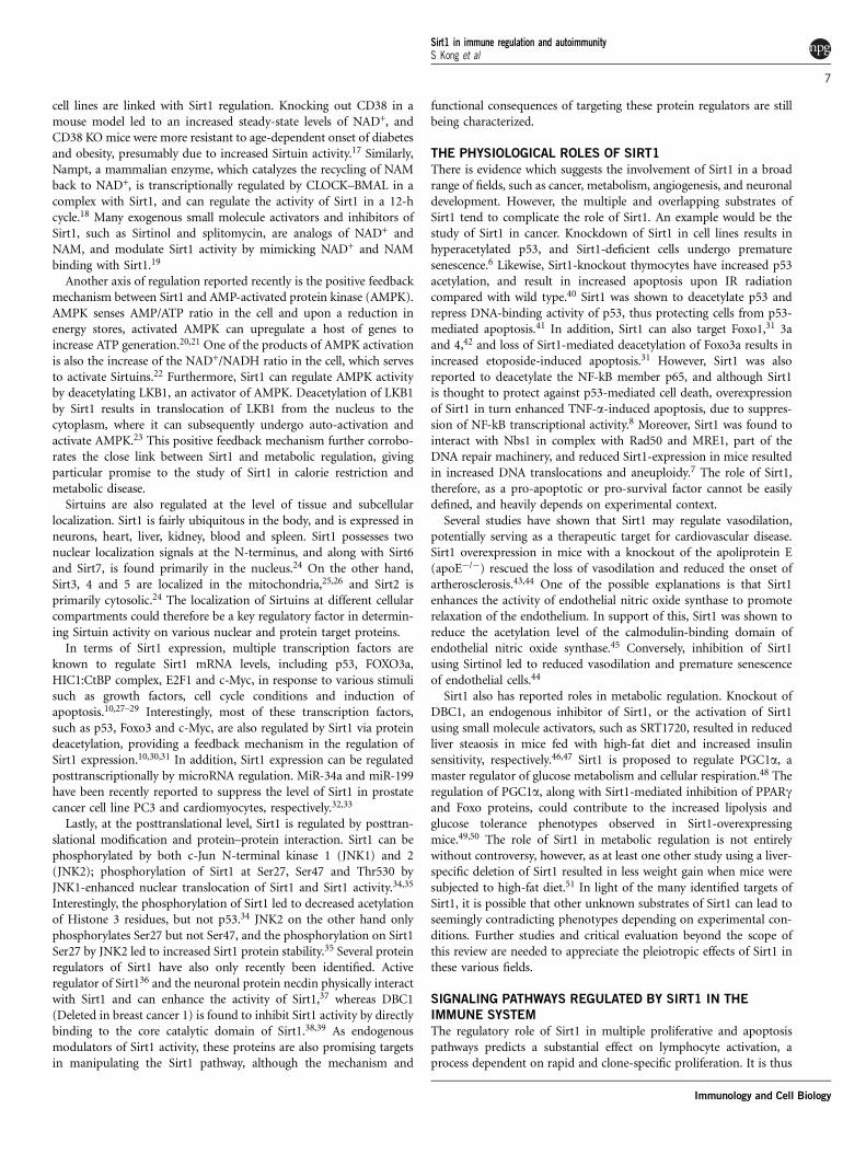
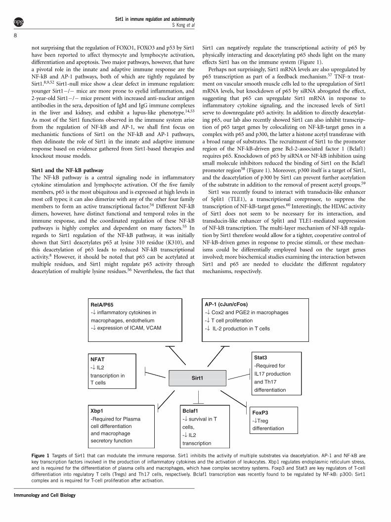
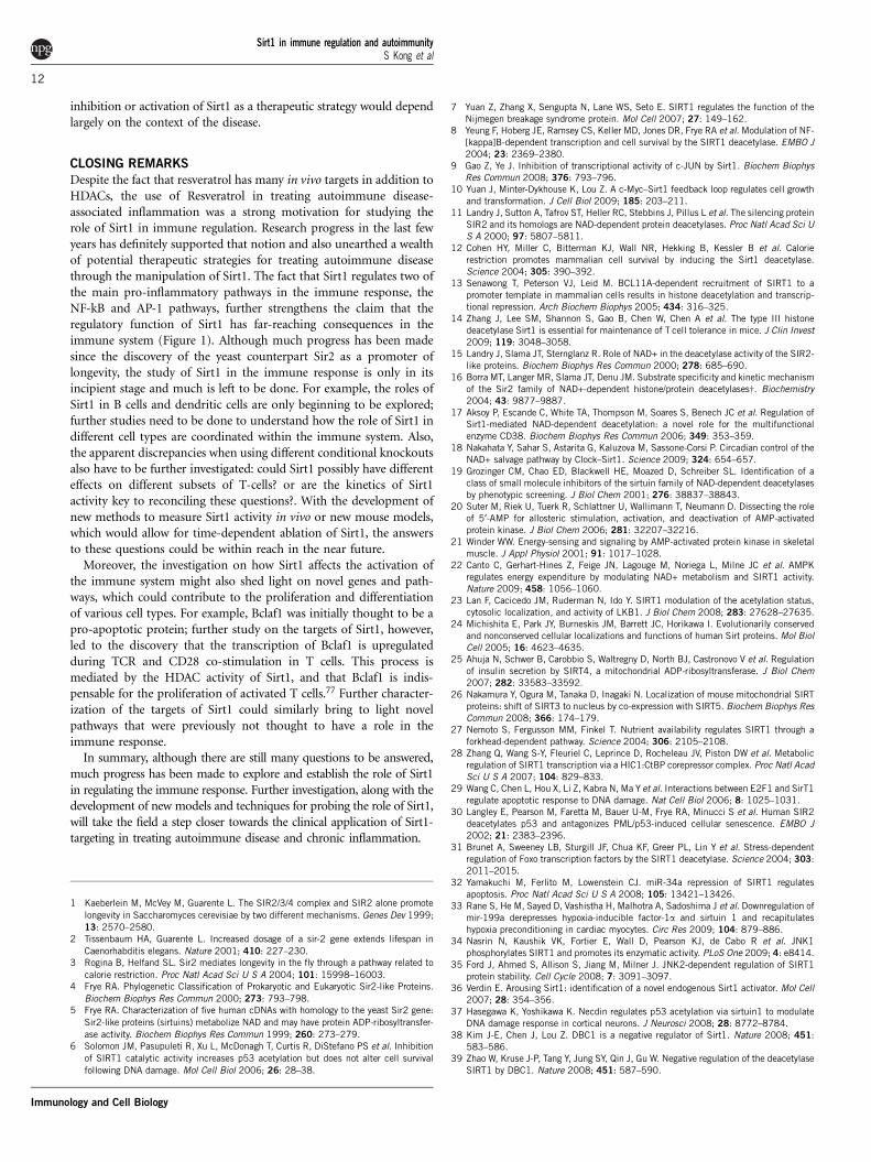
AP-1 (cJun/cFos)
-↓ Cox2 and PGE2 in macrophages
-↓ T cell proliferation
RelA/P65-↓ inflammatory cytokines in
macrophages, endothelium-↓ expression of ICAM, VCAM -↓ IL-2 production in T cells
Stat3NFAT-Required for
IL17 production-↓ IL2
transcription inand Th17T cellsdifferentiation
Xbp1 FoxP3Bclaf1
-Required for Plasma -↓ survival in Tcell differentiation
-↓Tregdifferentiationcells,
and macrophage -↓ IL2secretory function transcription
Sirt1
Figure 1 Targets of Sirt1 that can modulate the immune response. Sirt1 inhibits the activity of multiple substrates via deacetylation. AP-1 and NF-kB arekey transcription factors involved in the production of inflammatory cytokines and the activation of leukocytes. Xbp1 regulates endoplasmic reticulum stress,
and is required for the differentiation of plasma cells and macrophages, which have complex secretory systems. Foxp3 and Stat3 are key regulators of T-cell
differentiation into regulatory T cells (Tregs) and Th17 cells, respectively. Bclaf1 transcription was recently found to be regulated by NF-kB: p300: Sirt1
complex and is required for T-cell proliferation after activation.
Sirt1 in immune regulation and autoimmunityS Kong et al
8
Immunology and Cell Biology
Sirt1 interaction with AP-1Besides the NF-kB pathway, AP-1 transcriptional activity has anessential role in the activation of the immune response, particularlyin T cells. c-Jun and c-Fos heterodimers in particular are upregulatedshortly after T-cell activation to induce proliferation, interleukin (IL)-2 production and differentiation.61 (Figure 1) Using co-immuno-precipitation and acetylation assays, we showed that the c-Jun physi-cally interacts with the C-terminus of Sirt1 and is deacetylated by Sirt1in activated and anergic T cells. Conversely, c-Jun acetylation isincreased when overexpressed with p300, a histone acetyl tranferase,and this acetylation is brought back to basal level with the over-expression of Sirt1.14 Interaction between Sirt1 and AP-1 was alsoidentified in macrophages,52 where Sirt1 can interact with the leucinezipper domains of both c-Fos and c-Jun.9,52 Overexpression of Sirt1reduced the mRNA levels of Cox-2, a target gene of AP-1, in peritonealmacrophages, and the reduced production of prostaglandin E inmacrophages as a result of this suppression has broad implicationsin the inflammatory and tumoricidal functions of macrophages.
SIRT1, THE INNATE IMMUNE RESPONSE
AND INFLAMMATION
Vascular permeability and lymphocyte infiltrationThe suppression of NF-kB by Sirt1 on endothelial cells can have asubstantial role in enhancing systemic inflammation and lymphocyteinfiltration. Upon stimulation by TNF-a, endothelial cells undergovasodilation by expressing inducible nitric oxide synthase and IL-6,and upregulate adhesion molecules, such as ICAM. Both of thesephenomena can enhance the mobility and infiltration of leukocytes tothe site of inflammation, and both effects are mediated by NF-kBsignaling.62 Resveratrol treatment to activate Sirt1 in ApoE�/� miceand transgenic overexpression of Sirt1 in ApoE�/� mice were able toreduce the production of inflammatory cytokines and adhesionmolecules ICAM-1 and VCAM-1 without affecting vasodilation.43,44
Conversely, inhibition of Sirt1 in ApoE+/� strains did not increasephosphorylation and activity of endothelial nitric oxide synthase, butthe lack of Sirt1 activity led to increased production of reactive oxygenspecies, increased NF-kB activity and upregulated adhesion moleculesICAM-1 and VCAM-1 on the endothelium.45 Although the infiltra-tion of leukocytes during inflammation was not examined in thisstudy, Sirt1-null mice have increased lymphocyte infiltration in thecentral nervous system during induction of EAE.14 Thus, Sirt1 cansuppress pro-inflammatory phenotype in the endothelium and couldindirectly suppress the immune response in autoimmune diseases.
Sirt1 in regulation of macrophage functionsSirt1 has a direct regulatory role in macrophages, the most abundantcomponent of the innate immune system, and a main source of pro-inflammatory cytokines TNF-a, IL-6 and IL-1 secreted in response toinfection and inflammation. The inhibitory effect of Sirt1 on the NF-kBpathway has an essential role in suppressing the pro-inflammatoryphenotype of macrophages. In vitro, treating macrophages with Sirt1activators led to lower levels of inflammatory cytokines as well as ICAM-1surface expression.43 When rats were exposed to smoke extract, theNF-kB pathway was activated especially in macrophages, but Resver-atrol treatment attenuated the pro-inflammatory effect of cigarettesmoke.63 Knockdown of Sirt1 in RAW264.7 cell line, as well as inmouse intraperitoneal macrophages, also resulted in increased TNF-aproduction when stimulated with LPS.64 Additionally, myeloid-spe-cific knockout of Sirt1 (Mac-Sirt1 KO) led to hyperacetylation of p65,and when these mice were challenged with a high-fat diet, displayedhigh numbers of activated macrophages in the liver and kidney.65
Another signaling pathway that has been recently linked to inflam-mation is the unfolded protein response (UPR).66 Environmentalstress, such as nutrient deprivation and increased reactive oxygenspecies, can lead to disrupted endoplasmic reticulum function andincreased endoplasmic reticulum stress. As a result, the unfoldedprotein response leads to the activation of Inositol-requiring enzyme1, which splices X-box-binding protein 1 (Xbp1) into its functionalmessage and ultimately leads to suppressed global translation andincreased chaperone activity, or if the endoplasmic reticulum load isirresolvable, apoptosis.67 Xbp1 is critical for the function of highlysecretory cells, including hematopoietic cells, such as macrophagesand plasma cells.68,69 For example, Xbp1 is required for the matura-tion of B cells into antibody-secreting plasma cells, and the loss ofXbp1 in macrophages leads to impaired interferon b production.69
One of the ways Xbp1 is regulated besides alternative splicing isposttranslational acetylation and deacetylation by p300 and Sirt1,respectively.70 Reduced Sirt1 expression leads to increased expressionof Xbp1-target genes in MEFs, in response to endoplasmic reticulumstress, and increased resistance to apoptosis. By suppressing theunfolded protein response in macrophages by this mechanism, Sirt1could repress the production of Type I interferon and inhibit theactivation of the innate immune system (Figure 1).
On the other hand, Sirt1 can also maintain the function ofmacrophages, especially in a chronic inflammation setting, by inhibit-ing AP-1 transcription of Cox-2.52 The resulting decrease in PGE2levels due to the suppression of this pathway leads to improvedphagocytic and tumoricidal activities of peritoneal macrophages.It is probable, however, that the subpopulations of macrophagesinvolved in tumoricidal functions are not conventional M1 macro-phages present during an immune response.71 Further study is neededto understand the differential effects of Sirt1 on the dynamic regula-tion of NF-Kb, unfolded protein response and AP-1 pathways inmacrophages.
Sirt1: linking metabolism and inflammationThe fact that Sirt1 activation depends on NAD+ availability impliesthat the suppression of NF-kB and AP-1 pathways by Sirt1 is linked tothe metabolic status of the cell. Accumulation of adipose tissue directlyinduces inflammation by the secretion of pro-inflammatory cytokines,such as TNF-a, but activation of Sirt1 by calorie restriction, over-expression or administration of Sirt1 activators relieves the symptomsof obesity-associated inflammation, possibly by suppressing the NF-kB pathway.44,48,72 Similarly, deletion of DBC1, a suppressor of Sirt1,led to increased Sirt1 expression and activity in the mouse, and theincreased abundance of Sirt1 led to protection from liver steaosiswhen mice were challenged with a high-fat diet.73 These beneficialeffects of Sirt1 appear to be manifest only in nutritionally stressedanimals.
Zhang et al.74 reported that the administration of endotoxin, apotent inducer of inflammation, transiently lowered levels of ATP andSirt1 protein in leukocytes and liver. Recovery of ATP levels waspreceded by the induction of autophagy and was accompanied by thereappearance of Sirt1. In mice with liver-specific Sirt1 knockout, thisrecovery was delayed, consistent with the idea that Sirt1 has a role inautophagy regulation.75 Pretreatment of mice with Resveratrol pre-vented the decline in ATP. Hence, the link between metabolism andinflammation through Sirt1 appears to be bidirectional74 (Figure 1).
SIRT1 AND THE ADAPTIVE IMMUNE RESPONSE
The role of Sirt1 in the adaptive immune response has been primarilyfocused on the activation and differentiation of T cells. Sirt1 protein
Sirt1 in immune regulation and autoimmunityS Kong et al
9
Immunology and Cell Biology
levels are elevated in activated T cells and even more dramatically inanergic T cells in comparison with mature naıve T cells.14 However,Sirt1-null mice do not present with aberrant T or B cell numberscompared with wild-type littermates, indicating that Sirt1 might notbe critical for T- or B-cell development. The clonality or repertoire ofmature lymphocytes, however, have not been studied, so it remainspossible that the selection process of T or B cells might be affected inthe mutant even though cell numbers are not affected.
T-cell proliferationT cells from Sirt1-null mice are hyperproliferative and are activatedwithout CD28 co-stimulation. Although wild-type T cells require bothTCR and CD28 stimuli in vitro, TCR stimulation alone leads to asmuch proliferation in Sirt1�/� T cells as TCR/CD28 co-stimula-tion.14,58 This observation is also reproducible in vivo, as T cellsisolated from OVA-immunized Sirt1�/� mice have significantlyhigher proliferation upon OVA stimulation, and produce more IL-2,IFN-g and IL-5. OVA-specific antibodies in the sera were also higherin Sirt1�/� OVA-immunized mice, suggesting that Sirt1-null T cellsare more readily activated and undergo differentiation into cytokine-producing effector T cells.14
The hyperactivation of T cells results from the loss of suppression ofNF-kB and AP-1 transcriptional activity. Microarray data show thatSirt1�/� T cells upregulate multiple NF-kB- and AP-1-target genes24 h upon activation (Lee and Fang, unpublished data). In addition,c-Jun is hyperacetylated in Sirt1�/�-activated T cells. An interestingobservation, however, is that although the physical interactionbetween c-Jun and Sirt1 is increased when T cells are activated, thisphysical interaction is markedly enhanced in anergic T cells comparedwith activated and naıve T cells.14 This suggests that the regulation ofAP-1 by Sirt1 might be more complex and dependent on theintegration of other signaling pathways.
Besides directly regulating the activity of AP-1 and NF-kB tran-scription factors, Sirt1 can also modulate the accessibility of genesnecessary for T-cell proliferation and function. Bclaf1 is a proteininitially identified as a Bcl-2-binding protein, and has pro-apoptoticfunction in epithelial cells.76 However, a systemic knockout of Bclaf1in mice showed that it was dispensable for the induction of apoptosis,especially in lymphocytes by IR radiation, but surprisingly wasrequired for T-cell activation.77 Sirt1 is a suppressor of Bclaf1transcription, not only by suppressing NF-kB transcriptional activitybut also by localizing to the Bclaf1 locus, and deacetylating histonelysine residues, including H3K56 at the promoter region of Bclaf1. Thesuppression of Bclaf1 transcription by Sirt1 is key to controllinghyper-proliferation and aberrant activation of T cells, as the differencein Bclaf1 expression and histone deacetylation at the Bclaf1 locusbetween wild-type and Sirt1-null mice are unnoticeable in naıve,unactivated T cells.58
IL-2 provides a crucial signal for maintaining the viability andpromoting proliferation of activated T cells. IL-2 can signal throughthe phosphoinositide kinase/Akt pathway to suppress Foxo1, 3 and 4,preventing apoptosis in activated T –cells, which express the high-affinity IL-2 receptor CD25.78 Sirt1, however, may have a role in thisaxis of T-cell activation by deacetylating Foxo proteins and inhibitingFoxo1, 3 and 4 apoptotic signaling. Sirt1 has been shown to deacetylateFoxo family members, although the dynamics and regulation by Sirt1 inthe context of IL-2 signaling pathway has not been explicitly shown.
Sirt1 in T-cell differentiationSirt1 is implicated in the differentiation of activated T cells, althoughthe specific mechanisms are not clear. Kuroda et al.14 recently found
that basic leucine zipper, ATF-like (BATF) can specifically suppress thetranscription of Sirt1 but not other HDACs. CD8 T cells lacking BATFhad lower histone acetylation levels at the Tbet promoter, and lowerIFNg, IFNgR1 and IL12rb2 expression, presumably through higherexpression levels of Sirt1.79 Conversely, T cells from Sirt1-null miceproduce higher levels of IL-5 and IFNg when stimulated in vitro,further supporting the idea that Sirt1 could regulate the differentiationand effector function of T cells (Figure 1).
Sirt1 also has a potential role in mediating the differentiation ofT helper (Th) cells by suppressing Stat3 activity.80 Upon stimulationby cytokines, such as IL-6, IL-21 or IL-23, Stat3 is activated throughthe Jak–Stat pathway, and depending on the activation of other Statmembers, can induce transcriptional programs that mediate differ-entiation of CD4+ T cells into either Th2 or Th17 cells. The activationof Stat3 can also inhibit the activity of forkhead box P3 (Foxp3),suppressing the expansion of regulatory T cells.81 In addition, uponIL-6 stimulation, Stat3 can also upregulate the expression of chemo-kines, such as CCL5, enhancing the migration of T cell to the site ofinflammation.82 Hence, Stat3 is associated with a pro-inflammatoryT-cell phenotype. Interestingly, Sirt1 was recently found to deacetylateand suppress STAT3 activity in the liver, leading to improvedgluconeogenesis by relieving the STAT3 inhibition of FOXO1.80
Although not directly demonstrated in T cells, it is likely that thesame mechanism is in effect in T cells, and Sirt1 could regulate theactivity of Stat3 and hence the differentiation of CD4+ Th cells.Consistent with this idea, the loss of Stat3 in mice renders themmore resistant to the induction of EAE, and the reverse is observed inSirt1-null mice, which are more susceptible to the disease.14,83
Sirt1 was also reported to mediate the differentiation of CD4+
Th cells into Foxp3+ regulatory T cells (Tregs). Foxp3 protein is akey transcription factor induced during the differentiation of regula-tory T cells. Sirt1, along with the histone acetyl transferase p300, wasrecently found to regulate the acetylation status and stability ofFoxp3.84 Hyperacetylation of Foxp3 led to a decrease in polyubiquiti-nation and prolonged Foxp3 protein stability, whereas the addition ofNAM, a general Sirtuin inhibitor, led to reduced acetylation of Foxp3,promoting the ubiquitination and degradation of Foxp3. Mice withCD4-cre- or Foxp3-cre-driven deletion of Sirt1 did not have increasednumbers of natural Tregs, but the expression of Foxp3 and the suppres-sive function of Foxp3+ T cells appears to be increased.85 However, in thegermline Sirt1 knockout mice, no changes in both percentage and thesuppressive functions of natural Tregs were detected.27
The observations from systemic Sirt1 knockout mouse models andCD4-specific Sirt1 deletion seem to be contradictory; although theredefinitely needs to be more work done to fully appreciate the role ofSirt1 in the immune response, several possible explanations mightreconcile the seemingly opposing phenotypes in these two models.Firstly, T cells from Sirt1 conditional knockout mice still carry atruncated Sirt1 protein with a deletion of about 150 amino acidswithin the catalytic domain.86 Although this truncated mutant form ofSirt1 lacks deacetylase activity, it is not known whether it has adominant negative function. More importantly, it has been reportedthat mutant forms of Sirt1 lacking deacetylase activity are stillcompletely neuroprotective,87 implying that Sirt1 has functions inde-pendent of its deacetylase activity. Second, the systemic knockout ofSirt1 might point to an essential role of Sirt1 in other cell types in theimmune response, such as antigen presenting cells. For example, it wasmentioned earlier that Sirt1 deletion in macrophages resulted inpronounced repression of pro-inflammatory function.64 Althoughsystemic Sirt1 deletion results in autoimmunity and increased suscept-ibility to EAE, it is possible that antigen presenting cells could have a
Sirt1 in immune regulation and autoimmunityS Kong et al
10
Immunology and Cell Biology
major role in determining the strength and fate of T-cell response, andthis compensates for the lack of Sirt1 in T cells. Furthermore, theallograft model in this study differs slightly from the autoimmunecontext in previous studies. The ‘self versus nonself ’ difference betweenthese models might hint at an intriguing differential signaling outcomebased on MHC restriction or tolerance. This rationalization, however,does not explain the hyperproliferative response of T cells isolated fromSirt1-null mice in vitro, nor that adoptive transfer of purified T cellsfrom Sirt1-null mice into T cell-null mice facilitates EAE developmentinduced by MOG antigen.27 The fact that isolated T cells from Sirt1-null mice still exhibit hyperactivation points to a T-cell intrinsic role ofSirt1 on the immune response. Furthermore, other pathways that areregulated by Sirt1, such as the NF-kB pathway, AP-1 and STAT3 pointto Sirt1 being a suppressor of pro-inflammatory T-cell response.
Sirt1 in T-cell toleranceAs mentioned earlier, T cells lacking Sirt1 are hyperproliferative andcan be activated without CD28 co-stimulation. This suggests that Sirt1could have a role in maintaining T-cell tolerance. In fact, Sirt1 mRNA,as well as protein expression, was higher in anergic T cells than inactivated or naıve T cells.14 In addition, OT-II mice crossed withSirt1�/� mice could not be tolerized by OVA tail–vein injection, andwhen isolated T cells were stimulated with OVA in vitro, Sirt1�/� Tcells exhibit proliferation to the same degree as mice tolerized byphosphate-buffered saline. It is known that T cells that receive a weakTCR signal without CD28 co-stimulation normally undergo toleranceby specifically activating the NFAT pathway without activation ofNF-kB.88 Our lab recently found data that suggest an interaction betweenSirt1 and NFAT and the possible regulation of NFAT through acetylationto regulate IL-2 expression (Kim and Fang, unpublished data).
Sirt1 as a physiological target by the human immunodeficiencyvirus (HIV)Physiologically, Sirt1 is a target and mediator of HIV Tat proteinactivity.89 One of the characteristics of HIV infection is the upregula-tion of NF-kB activity and hyperactivation of CD4+ T cells.90 HIV Tatis one of the crucial viral genes to mediate viral RNA processing andmodulating host cellular signaling.91 It was recently shown that one ofthe ways Tat functions to promote viral replication is by inhibitingSirt1.89 Tat can bind to the catalytic domain of Sirt1 and block itsactivity. The long terminal repeat of HIV has two NF-kB-binding sites.With Tat-indced Sirt1 suppression, NF-kB is hyperacetylated andtranscriptional activity of both cellular proliferative genes and HIV-specific genes are enhanced. Moreover, Sirt1 can also deacetylate Tatprotein at Lysine 50, facilitating the recycling of Tat.92 Tat acetylationby p300 or GCN5 disables Tat from binding to trans-activatingresponse element motifs on viral RNA to initiate viral replication;instead, acetylated Tat is released from trans-activating responseelement and forms a complex with p300/Creb-binding protein-asso-ciated factor. Deacetylation of Lys50 by Sirt1 on the other hand allowsTat to re-associate with trans-activating response element viral RNAand resume replication.
The role of Sirt1 in B cellsThe role of Sirt1 in B cells has not been well studied. Nonetheless, it ispossible that Sirt1 or other Sirtuins might regulate the activation andmaturation of B cells in coordination with CD38 expression andNAD+ availability.17,93 The lupus-like symptoms in Sirt1 knockoutmice also suggest that Sirt1 might have a role in B-cell activation.14,53
However, further work needs to be done to elucidate the function ofSirt1 in regulating B-cell activation and differentiation.
SIRT1 IN AUTOIMMUNE DISEASE AND POSSIBLE THERAPIES
Given that Sirt1 negatively regulates multiple pro-inflammatory path-ways, it is not surprising that impaired Sirt1 activity leads to increasedinflammation and autoimmunity. When Sirt1 is deleted systemically,mice develop uveitis at an early age, although whether this is caused byan autoimmune-like mechanism or reduced immunity to bacterialinfection is unclear.53 Moreover, Sirt1-null mice about 1-year-old havehigher titers of anti-dsDNA and anti-nuclear antigen IgG and IgMimmunoglobulin compared with wild type, and accumulate immu-noglobulin complexes in the kidney and liver akin to the symptoms ofsystemic lupus erythmatosus.14,53 These results in the Sirt1-null mousemodel mirror the fact that in human patients, histone modificationdysregulation is associated with the incidence of systemic lupuserythmatosus, and hyperacetylation of Histone 3, a Sirt1 substrate,is associated with the increase of disease severity (r¼�0.889,P¼0.044).94 Unexpectedly, the incidence of disease is also associatedwith increased Sirt1 mRNA levels, although this could be due to aglobal loss of transcriptional regulation and hyperacetylation.
Sirt1�/� mice not only develop systemic lupus erythmatosus-likesymptoms spontaneously, they are also more susceptible to inducedEAE, an experimental model of the debilitating human disease multi-ple sclerosis. Sirt1�/� mice injected with MOG peptide developsignificantly more severe symptoms than wild type, and also experi-ence higher mortality rate.14 On the contrary, treating mice withResveratrol after MOG peptide injection led to decreased diseasesymptoms as well as inflammatory cell infiltration in the CNS. Allthese data suggest that activation of Sirt1 may be a successfultherapeutic strategy against autoimmune disease.
Pharmacological modulation of Sirt1 activity has been used as atherapeutic strategy in autoimmune disease settings. Resveratrol haslong been known as an anti-inflammatory compound and can alleviatevarious inflammatory diseases, such as rheumatoid arthritis and type Idiabetes.95,96 Treatment with resveratrol not only reduced pro-inflammatory cytokine production and lymphocyte activation butalso suppressed lymphocyte infiltration by reducing expression ofCCR6 in non-obese diabetic mice.96 Treatment with Resveratrol alsoreduced the amount of reactive oxygen species production and NF-kBactivation in rats exposed to cigarette smoke extract, reducing inflam-mation in a non-autoimmune setting.63 Although Resveratrol is nowknown to target a broad range of proteins, including AMPK97 andphosphoinositide kinase,97 the anti-inflammatory effects of Resveratrolare inhibited by the simultaneous administration of inhibitors of Sirt1,such as Sirtinol or splitomycin, suggesting that the benefits of Resver-atrol are at least partially conferred by the activation of Sirt1.63,98
Similarly, SRT2172, another Sirt1 activator, prevented neutrophilinfiltration in the lungs of a mouse model of chronic obstructivepulmonary disease, and improved exercise tolerance, demonstratingthat Sirt1 activation by small molecules not only ameliorates auto-immune disease but also other forms of chronic inflammation.98
However, not all diseases benefit from the activation of Sirt1. Asmentioned earlier, as Sirt1 suppresses pro-inflammatory lymphocytes,which are primarily driven by Th1 and Th17 cells, the activation ofSirt1 might have little benefit or might even worsen Th2-drivenimmune responses. A study using OVA/alum-induced asthma inmice demonstrated that inhibition of Sirt1 using Sirtinol reduced thenumber of infiltrating lymphocytes when compared with mock treat-ment.99 On the other hand, deletion of Sirt1 might lead to improvedimmune surveillance against pathogenic infection and nonself antigens.For example, Sirt1 is manipulated by HIV Tat protein to enhance viralreproduction in hyperactivated CD4+ T cells, and also CD4-specificdeletion of Sirt1 led to reduced cardiac allograft rejection. Thus the
Sirt1 in immune regulation and autoimmunityS Kong et al
11
Immunology and Cell Biology
inhibition or activation of Sirt1 as a therapeutic strategy would dependlargely on the context of the disease.
CLOSING REMARKS
Despite the fact that resveratrol has many in vivo targets in addition toHDACs, the use of Resveratrol in treating autoimmune disease-associated inflammation was a strong motivation for studying therole of Sirt1 in immune regulation. Research progress in the last fewyears has definitely supported that notion and also unearthed a wealthof potential therapeutic strategies for treating autoimmune diseasethrough the manipulation of Sirt1. The fact that Sirt1 regulates two ofthe main pro-inflammatory pathways in the immune response, theNF-kB and AP-1 pathways, further strengthens the claim that theregulatory function of Sirt1 has far-reaching consequences in theimmune system (Figure 1). Although much progress has been madesince the discovery of the yeast counterpart Sir2 as a promoter oflongevity, the study of Sirt1 in the immune response is only in itsincipient stage and much is left to be done. For example, the roles ofSirt1 in B cells and dendritic cells are only beginning to be explored;further studies need to be done to understand how the role of Sirt1 indifferent cell types are coordinated within the immune system. Also,the apparent discrepancies when using different conditional knockoutsalso have to be further investigated: could Sirt1 possibly have differenteffects on different subsets of T-cells? or are the kinetics of Sirt1activity key to reconciling these questions?. With the development ofnew methods to measure Sirt1 activity in vivo or new mouse models,which would allow for time-dependent ablation of Sirt1, the answersto these questions could be within reach in the near future.
Moreover, the investigation on how Sirt1 affects the activation ofthe immune system might also shed light on novel genes and path-ways, which could contribute to the proliferation and differentiationof various cell types. For example, Bclaf1 was initially thought to be apro-apoptotic protein; further study on the targets of Sirt1, however,led to the discovery that the transcription of Bclaf1 is upregulatedduring TCR and CD28 co-stimulation in T cells. This process ismediated by the HDAC activity of Sirt1, and that Bclaf1 is indis-pensable for the proliferation of activated T cells.77 Further character-ization of the targets of Sirt1 could similarly bring to light novelpathways that were previously not thought to have a role in theimmune response.
In summary, although there are still many questions to be answered,much progress has been made to explore and establish the role of Sirt1in regulating the immune response. Further investigation, along with thedevelopment of new models and techniques for probing the role of Sirt1,will take the field a step closer towards the clinical application of Sirt1-targeting in treating autoimmune disease and chronic inflammation.
1 Kaeberlein M, McVey M, Guarente L. The SIR2/3/4 complex and SIR2 alone promote
longevity in Saccharomyces cerevisiae by two different mechanisms. Genes Dev 1999;
13: 2570–2580.2 Tissenbaum HA, Guarente L. Increased dosage of a sir-2 gene extends lifespan in
Caenorhabditis elegans. Nature 2001; 410: 227–230.3 Rogina B, Helfand SL. Sir2 mediates longevity in the fly through a pathway related to
calorie restriction. Proc Natl Acad Sci U S A 2004; 101: 15998–16003.4 Frye RA. Phylogenetic Classification of Prokaryotic and Eukaryotic Sir2-like Proteins.
Biochem Biophys Res Commun 2000; 273: 793–798.5 Frye RA. Characterization of five human cDNAs with homology to the yeast Sir2 gene:
Sir2-like proteins (sirtuins) metabolize NAD and may have protein ADP-ribosyltransfer-
ase activity. Biochem Biophys Res Commun 1999; 260: 273–279.6 Solomon JM, Pasupuleti R, Xu L, McDonagh T, Curtis R, DiStefano PS et al. Inhibition
of SIRT1 catalytic activity increases p53 acetylation but does not alter cell survival
following DNA damage. Mol Cell Biol 2006; 26: 28–38.
7 Yuan Z, Zhang X, Sengupta N, Lane WS, Seto E. SIRT1 regulates the function of theNijmegen breakage syndrome protein. Mol Cell 2007; 27: 149–162.
8 Yeung F, Hoberg JE, Ramsey CS, Keller MD, Jones DR, Frye RA et al. Modulation of NF-[kappa]B-dependent transcription and cell survival by the SIRT1 deacetylase. EMBO J2004; 23: 2369–2380.
9 Gao Z, Ye J. Inhibition of transcriptional activity of c-JUN by Sirt1. Biochem BiophysRes Commun 2008; 376: 793–796.
10 Yuan J, Minter-Dykhouse K, Lou Z. A c-Myc–Sirt1 feedback loop regulates cell growthand transformation. J Cell Biol 2009; 185: 203–211.
11 Landry J, Sutton A, Tafrov ST, Heller RC, Stebbins J, Pillus L et al. The silencing proteinSIR2 and its homologs are NAD-dependent protein deacetylases. Proc Natl Acad Sci US A 2000; 97: 5807–5811.
12 Cohen HY, Miller C, Bitterman KJ, Wall NR, Hekking B, Kessler B et al. Calorierestriction promotes mammalian cell survival by inducing the Sirt1 deacetylase.Science 2004; 305: 390–392.
13 Senawong T, Peterson VJ, Leid M. BCL11A-dependent recruitment of SIRT1 to apromoter template in mammalian cells results in histone deacetylation and transcrip-tional repression. Arch Biochem Biophys 2005; 434: 316–325.
14 Zhang J, Lee SM, Shannon S, Gao B, Chen W, Chen A et al. The type III histonedeacetylase Sirt1 is essential for maintenance of T cell tolerance in mice. J Clin Invest2009; 119: 3048–3058.
15 Landry J, Slama JT, Sternglanz R. Role of NAD+ in the deacetylase activity of the SIR2-like proteins. Biochem Biophys Res Commun 2000; 278: 685–690.
16 Borra MT, Langer MR, Slama JT, Denu JM. Substrate specificity and kinetic mechanismof the Sir2 family of NAD+-dependent histone/protein deacetylasesw. Biochemistry2004; 43: 9877–9887.
17 Aksoy P, Escande C, White TA, Thompson M, Soares S, Benech JC et al. Regulation ofSirt1-mediated NAD-dependent deacetylation: a novel role for the multifunctionalenzyme CD38. Biochem Biophys Res Commun 2006; 349: 353–359.
18 Nakahata Y, Sahar S, Astarita G, Kaluzova M, Sassone-Corsi P. Circadian control of theNAD+ salvage pathway by Clock–Sirt1. Science 2009; 324: 654–657.
19 Grozinger CM, Chao ED, Blackwell HE, Moazed D, Schreiber SL. Identification of aclass of small molecule inhibitors of the sirtuin family of NAD-dependent deacetylasesby phenotypic screening. J Biol Chem 2001; 276: 38837–38843.
20 Suter M, Riek U, Tuerk R, Schlattner U, Wallimann T, Neumann D. Dissecting the roleof 5¢-AMP for allosteric stimulation, activation, and deactivation of AMP-activatedprotein kinase. J Biol Chem 2006; 281: 32207–32216.
21 Winder WW. Energy-sensing and signaling by AMP-activated protein kinase in skeletalmuscle. J Appl Physiol 2001; 91: 1017–1028.
22 Canto C, Gerhart-Hines Z, Feige JN, Lagouge M, Noriega L, Milne JC et al. AMPKregulates energy expenditure by modulating NAD+ metabolism and SIRT1 activity.Nature 2009; 458: 1056–1060.
23 Lan F, Cacicedo JM, Ruderman N, Ido Y. SIRT1 modulation of the acetylation status,cytosolic localization, and activity of LKB1. J Biol Chem 2008; 283: 27628–27635.
24 Michishita E, Park JY, Burneskis JM, Barrett JC, Horikawa I. Evolutionarily conservedand nonconserved cellular localizations and functions of human Sirt proteins. Mol BiolCell 2005; 16: 4623–4635.
25 Ahuja N, Schwer B, Carobbio S, Waltregny D, North BJ, Castronovo V et al. Regulationof insulin secretion by SIRT4, a mitochondrial ADP-ribosyltransferase. J Biol Chem2007; 282: 33583–33592.
26 Nakamura Y, Ogura M, Tanaka D, Inagaki N. Localization of mouse mitochondrial SIRTproteins: shift of SIRT3 to nucleus by co-expression with SIRT5. Biochem Biophys ResCommun 2008; 366: 174–179.
27 Nemoto S, Fergusson MM, Finkel T. Nutrient availability regulates SIRT1 through aforkhead-dependent pathway. Science 2004; 306: 2105–2108.
28 Zhang Q, Wang S-Y, Fleuriel C, Leprince D, Rocheleau JV, Piston DW et al. Metabolicregulation of SIRT1 transcription via a HIC1:CtBP corepressor complex. Proc Natl AcadSci U S A 2007; 104: 829–833.
29 Wang C, Chen L, Hou X, Li Z, Kabra N, Ma Y et al. Interactions between E2F1 and SirT1regulate apoptotic response to DNA damage. Nat Cell Biol 2006; 8: 1025–1031.
30 Langley E, Pearson M, Faretta M, Bauer U-M, Frye RA, Minucci S et al. Human SIR2deacetylates p53 and antagonizes PML/p53-induced cellular senescence. EMBO J2002; 21: 2383–2396.
31 Brunet A, Sweeney LB, Sturgill JF, Chua KF, Greer PL, Lin Y et al. Stress-dependentregulation of Foxo transcription factors by the SIRT1 deacetylase. Science 2004; 303:2011–2015.
32 Yamakuchi M, Ferlito M, Lowenstein CJ. miR-34a repression of SIRT1 regulatesapoptosis. Proc Natl Acad Sci U S A 2008; 105: 13421–13426.
33 Rane S, He M, Sayed D, Vashistha H, Malhotra A, Sadoshima J et al. Downregulation ofmir-199a derepresses hypoxia-inducible factor-1a and sirtuin 1 and recapitulateshypoxia preconditioning in cardiac myocytes. Circ Res 2009; 104: 879–886.
34 Nasrin N, Kaushik VK, Fortier E, Wall D, Pearson KJ, de Cabo R et al. JNK1phosphorylates SIRT1 and promotes its enzymatic activity. PLoS One 2009; 4: e8414.
35 Ford J, Ahmed S, Allison S, Jiang M, Milner J. JNK2-dependent regulation of SIRT1protein stability. Cell Cycle 2008; 7: 3091–3097.
36 Verdin E. Arousing Sirt1: identification of a novel endogenous Sirt1 activator. Mol Cell2007; 28: 354–356.
37 Hasegawa K, Yoshikawa K. Necdin regulates p53 acetylation via sirtuin1 to modulateDNA damage response in cortical neurons. J Neurosci 2008; 28: 8772–8784.
38 Kim J-E, Chen J, Lou Z. DBC1 is a negative regulator of Sirt1. Nature 2008; 451:583–586.
39 Zhao W, Kruse J-P, Tang Y, Jung SY, Qin J, Gu W. Negative regulation of the deacetylaseSIRT1 by DBC1. Nature 2008; 451: 587–590.
Sirt1 in immune regulation and autoimmunityS Kong et al
12
Immunology and Cell Biology
40 Cheng H-L, Mostoslavsky R, Saito Si, Manis JP, Gu Y, Patel P et al. Developmentaldefects and p53 hyperacetylation in Sir2 homolog (SIRT1)-deficient mice. Proc NatlAcad Sci U S A 2003; 100: 10794–10799.
41 Vaziri H, Dessain SK, Eaton EN, Imai S-I, Frye RA, Pandita TK et al.hSIR2SIRT1 functions as an NAD-dependent p53 deacetylase. Cell 2001; 107:149–159.
42 Motta MC, Divecha N, Lemieux M, Kamel C, Chen D, Gu W et al. Mammalian Sirt1represses forkhead transcription factors. Cell 2004; 116: 551–563.
43 Stein S, Schafer N, Breitenstein A, Besler C, Winnik S, Lohmann C et al. SIRT1reduces endothelial activation without affecting vascular function in ApoE�/� mice.Aging (Albany NY) 2010; 2: 353–360.
44 Zhang Q-j, Wang Z, Chen H-z, Zhou S, Zheng W, Liu G et al. Endothelium-specificoverexpression of class III deacetylase Sirt1 decreases atherosclerosis in apolipoproteinE-deficient mice. Cardiovasc Res 2008; 80: 191–199.
45 Mattagajasingh I, Kim C-S, Naqvi A, Yamamori T, Hoffman TA, Jung S-B et al. SIRT1promotes endothelium-dependent vascular relaxation by activating endothelial nitricoxide synthase. Proc Natl Acad Sci U S A 2007; 104: 14855–14860.
46 Escande C, Chini CC, Nin V, Dykhouse KM, Novak CM, Levine J et al. Deleted in breastcancer-1 regulates Sirt1 activity and contributes to high-fat diet-induced liver steatosisin mice. J Clin Invest 2010; 120: 545–558.
47 Milne JC, Lambert PD, Schenk S, Carney DP, Smith JJ, Gagne DJ et al. Small moleculeactivators of Sirt1 as therapeutics for the treatment of type 2 diabetes. Nature 2007;450: 712–716.
48 Rodgers JT, Lerin C, Haas W, Gygi SP, Spiegelman BM, Puigserver P. Nutrient control ofglucose homeostasis through a complex of PGC-1[alpha] and SIRT1. Nature 2005;434: 113–118.
49 Picard F, Kurtev M, Chung N, Topark-Ngarm A, Senawong T, Machado de Oliveira Ret al. Sirt1 promotes fat mobilization in white adipocytes by repressing PPAR-[gamma].Nature 2004; 429: 771–776.
50 Frescas D, Valenti L, Accili D. Nuclear trapping of the forkhead transcription factorFoxo1 via Sirt-dependent deacetylation promotes expression of glucogenetic genes.J Biol Chem 2005; 280: 20589–20595.
51 Li X, Zhang S, Blander G, Tse JG, Krieger M, Guarente L. SIRT1 deacetylates andpositively regulates the nuclear receptor LXR. Mol Cell 2007; 28: 91–106.
52 Zhang R, Chen HZ, Liu JJ, Jia YY, Zhang ZQ, Yang RF et al. SIRT1 suppresses activatorprotein-1 transcriptional activity and cyclooxygenase-2 expression in macrophages.J Biol Chem 2010; 285: 7097–7110.
53 Sequeira J, Boily G, Bazinet S, Saliba S, He X, Jardine K et al. Sirt1-null mice developan autoimmune-like condition. Exp Cell Res 2008; 314: 3069–3074.
54 Gilmore TD. Introduction to NF-[kappa]B: players, pathways, perspectives. Oncogene0000; 25: 6680–6684.
55 Hoffmann A, Baltimore D. Circuitry of nuclear factor kB signaling. Immunol Rev 2006;210: 171–186.
56 Chen L-f, Mu Y, Greene WC. Acetylation of RelA at discrete sites regulates distinctnuclear functions of NF-[kappa]B. EMBO J 2002; 21: 6539–6548.
57 Zhang HN, Li L, Gao P, Chen HZ, Zhang R, Wei YS et al. Involvement of the p65/RelAsubunit of NF-kappaB in TNF-alpha-induced SIRT1 expression in vascular smoothmuscle cells. Biochem Biophys Res Commun 2010; 397: 569–575.
58 Kong S, Kim S-J, Sandal B, Lee S-M, Gao B, Zhang DD et al. The Type III histonedeacetylase Sirt1 protein suppresses p300-mediated histone H3 lysine 56acetylation at Bclaf1 promoter to inhibit T cell activation. J Biol Chem 2011; 286:16967–16975.
59 Bouras T, Fu M, Sauve AA, Wang F, Quong AA, Perkins ND et al. SIRT1 deacetylationand repression of p300 involves lysine residues 1020/1024 within the cell cycleregulatory domain 1. J Biol Chem 2005; 280: 10264–10276.
60 Ghosh HS, Spencer JV, Ng B, McBurney MW, Robbins PD. Sirt1 interacts withtransducin-like enhancer of split-1 to inhibit nuclear factor kappaB-mediated transcrip-tion. Biochem J 2007; 408: 105–111.
61 Foletta V, Segal D, Cohen D. Transcriptional regulation in the immune system: all roadslead to AP-1. J Leukoc Biol 1998; 63: 139–152.
62 McHale JF, Harari OA, Marshall D, Haskard DO. Vascular endothelial cell expression ofICAM-1 and VCAM-1 at the onset of eliciting contact hypersensitivity in mice: evidencefor a dominant role of TNF-a. J Immunol 1999; 162: 1648–1655.
63 Yang S-R, Wright J, Bauter M, Seweryniak K, Kode A, Rahman I. Sirtuin regulatescigarette smoke-induced proinflammatory mediator release via RelA/p65 NF-kB inmacrophages in vitro and in rat lungs in vivo: implications for chronic inflammation andaging. Am J Physiol Lung Cell Mol Physiol 2007; 292: L567–L576.
64 Yoshizaki T, Schenk S, Imamura T, Babendure JL, Sonoda N, Bae EJ et al. SIRT1inhibits inflammatory pathways in macrophages and modulates insulin sensitivity. Am JPhysiol Endocrinol Metabol 2010; 298: E419–E428.
65 Schug TT, Xu Q, Gao H, Peres-da-Silva A, Draper DW, Fessler MB et al. Myeloid deletionof Sirt1 induces inflammatory signaling in response to environmental stress. Mol CellBiol 2010; 30: 4712–4721.
66 Hu P, Han Z, Couvillon AD, Kaufman RJ, Exton JH. Autocrine tumor necrosis factoralpha links endoplasmic reticulum stress to the membrane death receptor pathwaythrough IRE1{alpha}-mediated NF-{kappa}B activation and down-regulation of TRAF2expression. Mol Cell Biol 2006; 26: 3071–3084.
67 Calfon M, Zeng H, Urano F, Till JH, Hubbard SR, Harding HP et al. IRE1 couplesendoplasmic reticulum load to secretory capacity by processing the XBP-1 mRNA.Nature 2002; 415: 92–96.
68 Reimold AM, Iwakoshi NN, Manis J, Vallabhajosyula P, Szomolanyi-Tsuda E, GravalleseEM et al. Plasma cell differentiation requires the transcription factor XBP-1. Nature2001; 412: 300–307.
69 Zeng L, Liu Y-P, Sha H, Chen H, Qi L, Smith JA. XBP-1 couples endoplasmic reticulumstress to augmented IFN-b induction via a cis-acting enhancer in macrophages.J Immunol 2010; 185: 2324–2330.
70 Wang FM, Chen YJ, Ouyang HJ. Regulation of unfolded protein response modulatorXBP1 by acetylation and deacetylation. Biochem J 2010; 433: 245–252.
71 Mantovani A, Sozzani S, Locati M, Allavena P, Sica A. Macrophage polarization: tumor-associated macrophages as a paradigm for polarized M2 mononuclear phagocytes.Trends Immunol 2002; 23: 549–555.
72 Xu F, Gao Z, Zhang J, Rivera CA, Yin J, Weng J et al. Lack of Sirt1 (mammalian sirtuin1) activity leads to liver steatosis in the SIRT1+/- mice: a role of lipid mobilization andinflammation. Endocrinology 2010; 151: 2504–2514.
73 Escande C, Chini CCS, Nin V, Dykhouse KM, Novak CM, Levine J et al. Deleted in breastcancer–1 regulates SIRT1 activity and contributes to high-fat diet–induced liversteatosis in mice. J Clin Invest 2010; 120: 545–558.
74 Zhang Z, Lowry SF, Guarente L, Haimovich B. Roles of Sirt1 in the acute and restorativephases following induction of inflammation. J Biol Chem 2010; 285: 41391–41401.
75 Lee IH, Cao L, Mostoslavsky R, Lombard DB, Liu J, Bruns NE et al. A role for the NAD-dependent deacetylase Sirt1 in the regulation of autophagy. Proc Natl Acad Sci U S A2008; 105: 3374–3379.
76 Kasof GM, Goyal L, White E. Btf, a novel death-promoting transcriptional repressor thatinteracts with Bcl-2-related proteins. Mol Cell Biol 1999; 19: 4390–4404.
77 McPherson JP, Sarras H, Lemmers B, Tamblyn L, Migon E, Matysiak-Zablocki E et al.Essential role for Bclaf1 in lung development and immune system function. Cell DeathDiffer 2008; 16: 331–339.
78 Stahl M, Dijkers PF, Kops GJ, Lens SM, Coffer PJ, Burgering BM et al. The forkheadtranscription factor Foxo regulates transcription of p27Kip1 and Bim in response toIL-2. J Immunol 2002; 168: 5024–5031.
79 Kuroda S, Yamazaki M, Abe M, Sakimura K, Takayanagi H, Iwai Y. Basic leucine zippertranscription factor, ATF-like (BATF) regulates epigenetically and energetically effectorCD8 T-cell differentiation via Sirt1 expression. Proc Natl Acad Sci U S A 2011; 108:14885–14889.
80 Nie Y, Erion DM, Yuan Z, Dietrich M, Shulman GI, Horvath TL et al. STAT3 inhibition ofgluconeogenesis is downregulated by SirT1. Nat Cell Biol 2009; 11: 492–500.
81 Yang XO, Panopoulos AD, Nurieva R, Chang SH, Wang D, Watowich SS et al. STAT3regulates cytokine-mediated generation of inflammatory helper T cells. J Biol Chem2007; 282: 9358–9363.
82 McLoughlin RM, Jenkins BJ, Grail D, Williams AS, Fielding CA, Parker CR et al. IL-6trans-signaling via Stat3 directs Tcell infiltration in acute inflammation. Proc Natl AcadSci U S A 2005; 102: 9589–9594.
83 Liu X, Lee YS, Yu C-R, Egwuagu CE. Loss of Stat3 in CD4+ Tcells prevents developmentof experimental autoimmune diseases. J Immunol 2008; 180: 6070–6076.
84 van Loosdregt J, Vercoulen Y, Guichelaar T, Gent YY, Beekman JM, van Beekum O et al.Regulation of Treg functionality by acetylation-mediated Foxp3 protein stabilization.Blood 2010; 115: 965–974.
85 Beier UH, Wang L, Bhatti TR, Liu Y, Han R, Ge G et al. Sirtuin-1 targeting promotesFoxp3+ T-regulatory cell function and prolongs allograft survival. Mol Cell Biol 2011;31: 1022–1029.
86 Li H, Rajendran GK, Liu N, Ware C, Rubin BP, Gu Y. SirT1 modulates the estrogen-insulin-like growth factor-1 signaling for postnatal development of mammary gland inmice. Breast Cancer Res 2007; 9: R1.
87 Pfister JA, Ma C, Morrison BE, D’Mello SR. Opposing effects of sirtuins on neuronalsurvival: sirt1-mediated neuroprotection is independent of its deacetylase activity.PLoS One 2008; 3: e4090.
88 Macian F, GarcIa-Cozar F, Im S-H, Horton HF, Byrne MC, Rao A. Transcriptionalmechanisms underlying lymphocyte tolerance. Cell 2002; 109: 719–731.
89 Kwon H-S, Brent MM, Getachew R, Jayakumar P, Chen L-F, Schnolzer M et al. Humanimmunodeficiency virus type 1 Tat protein inhibits the Sirt1 deacetylase and induces Tcell hyperactivation. Cell Host Microbe 2008; 3: 158–167.
90 Stevenson M, Stanwick TL, Dempsey MP, Lamonica CA. HIV-1 replication is controlledat the level of T cell activation and proviral integration. EMBO J 1990; 9: 1551–1560.
91 Barboric M, Taube R, Nekrep N, Fujinaga K, Peterlin BM. Binding of Tat to TAR andrecruitment of positive transcription elongation factor b occur independently in bovineimmunodeficiency virus. J Virol 2000; 74: 6039–6044.
92 Pagans S, Pedal A, North BJ, Kaehlcke K, Marshall BL, Dorr A et al. SIRT1 regulatesHIV transcription via Tat deacetylation. PLoS Biol 2005; 3: e41.
93 Yasue T, Baba M, Mori S, Mizoguchi C, Uehara S, Takatsu K. IgG1 production by sIgD+splenic B cells and peritoneal B-1 cells in response to IL-5 and CD38 ligation. IntImmunol 1999; 11: 915–923.
94 Hu N, Qiu X, Luo Y, Yuan J, Li Y, Lei W et al. Abnormal histone modification patterns inlupus CD4+ T cells. J Rheumatol 2008; 35: 804–810.
95 Elmali N, Baysal O, Harma A, Esenkaya I, Mizrak B. Effects of resveratrol ininflammatory arthritis. Inflammation 2007; 30: 1–6.
96 Lee SM, Yang H, Tartar D, Gao B, Luo X, Ye S et al. Prevention and treatment ofdiabetes with resveratrol in a non-obese mouse model of type 1 diabetes. Diabetologia2011; 54: 1136–1146.
97 Frojdo S, Cozzone D, Vidal H, Pirola L. Resveratrol is a class IA phosphoinositide 3-kinase inhibitor. Biochem J 2007; 406: 511–518.
98 Nakamaru Y, Vuppusetty C, Wada H, Milne JC, Ito M, Rossios C et al. A proteindeacetylase Sirt1 is a negative regulator of metalloproteinase-9. FASEB J 2009; 23:2810–2819.
99 Kim SR, Lee KS, Park SJ, Min KH, Choe YH, Moon H et al. Involvement of sirtuin 1 inairway inflammation and hyperresponsiveness of allergic airway disease. J Allergy ClinImmunol 2010; 125: 449–460.
Sirt1 in immune regulation and autoimmunityS Kong et al
13
Immunology and Cell Biology