Embed Size (px)
Citation preview
1
Sirtuins, Mitochondria and the Melatonergic Pathway in Alzheimer’s Disease
Anderson G,1
Maes M.2-4
1
CRC Scotland & London, Eccleston Square, London, UK
2
Dept Psychiatry, Faculty of Medicine, Chulalongkorn University, Bangkok, Thailand.
3
Dept Psychiatry, Medical University Plovdiv, Plovdiv, Bulgaria.
4
IMPACT Research Center, Deakin University, Geelong, Australia.
Corresponding author
George Anderson,
Head of Research, CRC Scotland & London, UK.
Email: [email protected]
Preprints (www.preprints.org) | NOT PEER-REVIEWED | Posted: 27 February 2020 doi:10.20944/preprints202002.0396.v1
© 2020 by the author(s). Distributed under a Creative Commons CC BY license.
2
ABSTRACT
Alzheimer's disease (AD) has been the subject of extensive investigation as to its biological
underpinnings. However, this has produced little of therapeutic benefit or indeed provided any
accepted biomarkers that could tailor treatment. This chapter reviews data on the main
pathophysiologic processes that have been widely shown to be altered in AD, including circadian
dysregulation, mitochondrial dysfunction, gut dysbiosis, and immune-glia-platelet activation. It is
proposed that alterations in the gut microbiome, including gut dysbiosis and increased gut
permeability drive changes in mitochondrial function that are intimately associated with significant
variations in sirtuin expression. Both mitochondria-located and nucleus/cytoplasm located sirtuins
can act on mitochondrial function in different cells and body systems to co-ordinate the ageing-
associated changes that underpin AD. The sirtuins are therefore key aspect to a developmental
model of AD that is more 'holistic' in perspective, thereby providing a framework for the detection
of earlier biomarkers and more successful treatment for the heterogenous nature of AD
pathoetiology.
Preprints (www.preprints.org) | NOT PEER-REVIEWED | Posted: 27 February 2020 doi:10.20944/preprints202002.0396.v1

3
Keywords: Alzheimer's disease; sirtuins; mitochondria; leaky gut; inflammation; neuroimmune
Preprints (www.preprints.org) | NOT PEER-REVIEWED | Posted: 27 February 2020 doi:10.20944/preprints202002.0396.v1
4
INTRODUCTION
A wide array of biological factors and processes are associated with Alzheimer's disease (AD) risk
and/or pathophysiology. It is generally accepted that genetic factors and epigenetic processes over
the lifespan interact with ageing to drive the pathophysiological changes occurring in
neurodegeneration. Consequently, AD symptomatology may arise from an array of distinct
processes culminating in a common hub of neurodegeneration. This is highly suggestive of a
heterogenous set of pathways, if not distinct conditions. However, within such heterogeneity a
number of key processes seem to be important, including circadian dysregulation, mitochondrial
dysfunction, gut dysbiosis and increased gut permeability, as well as alterations in immune, glia and
platelet activity. Other factors commonly found in people with AD, including increased levels of
amyloid-β42 (Aβ42), hyperphosphorylated tau, ceramide and formaldehyde and decreased levels of
the short-chain fatty acid, butyrate, melatonin, pericytes and taurine may all arise as a consequence,
thereafter contributing to AD pathophysiology.
Circadian dysregulation is an important aspect of AD etiology [1] and pathophysiology [2]. This is
supported by work in preclinical models, where it is proposed that circadian dysregulation leads to
an increase in the oxidant/antioxidant ratio, thereby contributing to neuronal apoptotic susceptibility
[3]. The decrease in the night-time secretion of pineal gland melatonin in AD contributes to elevated
oxidative damage, although this seems a two-way interaction, as oxidative damage and associated
immune-inflammation contribute to the suppression of pineal melatonin secretion [4]. Orexin, the
wake-promoting factor, is also profoundly decreased in AD, contributing to the loss of a sleep-
active rhythm [5].
A large body of data also shows mitochondrial dysregulation in AD, with mitochondrial dysfunction
mooted as a viable AD treatment target [6], as in many other conditions [7,8]. Targeting
mitochondrial function in an AD preclinical model prevents classical AD indicants, viz tangle and
plaque formation [9]. Notably, the circadian rhythm drives changes in mitochondrial function and
mitochondrial rate-limiting enzymes [10], suggesting that circadian dysregulation impacts core
aspects of metabolic function. This is especially important in immune and glia cells, where night-
time melatonin switches metabolism in reactive cells from glycolysis to oxidative phosphorylation
(OXPHOS), and thereby from a reactive to quiescent state. Again, this is a two-way interaction as
mitochondria function and redox regulation can modulate circadian proteins and the circadian
rhythm [11,12].
Preprints (www.preprints.org) | NOT PEER-REVIEWED | Posted: 27 February 2020 doi:10.20944/preprints202002.0396.v1
5
Recent data indicates a role for gut dysbiosis and increased gut permeability across a wide array of
medical conditions [13-5], including AD [16]. Gut dysbiosis and gut permeability are also important
in the shift from mild cognitive impairment (MCI) to AD [17]. The gut contributes to AD via two
main mechanisms: 1) increased gut permeability leads to elevated levels of circulating
lipopolysaccharide (LPS) and exosomal high-mobility group box (HMGB)1 [18], with both LPS
and HMGB1 mediating their effects via toll-like receptor (TLR)4, in association with heightened
levels of oxidative stress and immune-inflammatory activity; 2) gut dysbiosis is invariably
associated with a decrease in the short-chain fatty acid, butyrate. Butyrate seals the gut barrier,
suppresses immune and glia cell reactivity and optimizes mitochondria functioning, including
within immune and glia cells, reviewed in [14]. These are the two important routes whereby the gut
interacts with AD etiology and pathophysiology, with relevance to a host of other diverse medical
presentations. It is also important to note that the gut is an integral aspect of the circadian rhythm.
An increased levels of inflammation, from the activation of immune and glia cells, has long been
associated with AD, highlighting the relevance of systemic processes and their interactions with
CNS regulation. Variations in the platelet activity are another systemic factor associated with the
circadian rhythm and showing alterations in AD and other neurodegenerative conditions [7],
including as arising from the regulation of mitochondrial function [19]. As with immune and glial
cells, platelet mitochondria can be regulated by histone deacetylase (HDAC) inhibitors, such as the
gut microbiome-derived, butyrate [20]. As such, platelets, like glia and immune cells, are another
important hub for gut-mitochondria interactions, with consequences that include the production of
thrombin and fibrin(ogen), both of which can drive neurodegenerative processes in AD [21-2], as
well as contribute to AD risk factors, such as cardiovascular disorders and ischaemic stroke [23-4],
as well as increasing the risk of cerebral amyloid angiopathy [25]. Overall, immunity, glia and
platelets contribute to AD pathophysiology partly via changes that are regulated by the circadian
rhythm and gut driven changes in mitochondrial function.
Other pathophysiological changes in AD may be linked to this, including the increased levels of
ceramide that are evident in AD and other neurodegenerative conditions. For example, increased gut
permeability leading to LPS and HMGB1 activation of microglia and the production of
peroxynitrite (ONOO-) and tumor necrosis factor (TNF)-α. ONOO- activates acidic
sphingomyelinase (aSMase) leading to long-chain ceramides, with ceramide and TNF-α decreasing
levels of daytime orexin and night-time melatonin, resulting in the loss of orexin and melatonin
optimization of mitochondrial OXPHOS and contributing to circadian dysregulation. Ceramide also
increases Aβ42 levels via the stabilization of beta-site amyloid precursor protein-cleaving enzyme
Preprints (www.preprints.org) | NOT PEER-REVIEWED | Posted: 27 February 2020 doi:10.20944/preprints202002.0396.v1
6
(BACE)1 [26]. The lower butyrate levels also contribute to suboptimal mitochondria functioning,
including in glia and immune cells, thereby dysregulating immune-inflammatory activity. Lower
butyrate levels also attenuate its suppression of ceramide. LPS and HMGB1, via TLR4, can also
regulate platelet activity as can ceramide and aSMase, whilst decreased levels of butyrate will
attenuate its inhibition of platelet activation, leading to increased levels of thrombin and
fibrin(ogen) in AD pathoetiology. Such data can be linked in an integrative, developmental model,
comprised of a gut-mitochondria-(platelet-immune-glia)-circadian axis, that incorporates an array of
diverse and previously disparate data on AD.
This chapter reviews data on the circadian dysregulation, mitochondrial dysfunction, gut dysbiosis,
and immune-glia-platelet activation in AD, proposing an important role for alterations in sirtuin
function in all of these processes and their interactions. First, we shall briefly review the sirtuins,
before integrating sirtuins in the processes and their interactions underpinning AD pathophysiology.
We then show that this can provide a more integrative model of AD, with future research and
treatment implications.
THE SIRTUINS
The sirtuins are a family of nicotinamide dinucleotide (NAD)+-dependent class III histone
deacetylases. The sirtuins are important to metabolic and circadian regulation, with epigenetic
impacts on a host of physiological and pathophysiological processes. Mammals have seven sirtuins
(SIRT1–7), with SIRT1, SIRT6 and SIRT7 being predominantly expressed in the nucleus, whilst
SIRT3, SIRT4 and SIRT5 are primarily mitochondria-expressed. SIRT2 is expressed in the
cytoplasm. The sirtuins regulate a wide array of core cellular processes, including mitochondrial
metabolism, DNA repair, adipocyte differentiation, neurogenesis, insulin sensitivity, and fatty acid
oxidation, as well as inflammation, and aging-associated processes. All of these processes have
been linked to AD pathophysiology.
SIRT1 has classically been referred to as the 'longevity protein', given its association with the
increased lifespan in yeast and rodents. Lower SIRT1 levels are evident in insulin-resistant cells,
with the induced cellular expression of SIRT1 improving insulin sensitivity. This is important in
AD, which has been referred to as type III diabetes, due to the role of type II diabetes in AD
pathoetiology [27]. SIRT1 also deacetylates peroxisome proliferator-activated receptor gamma
coactivator 1-alpha (PGC1α), which is commonly referred to as the mitochondria 'master regulator',
Preprints (www.preprints.org) | NOT PEER-REVIEWED | Posted: 27 February 2020 doi:10.20944/preprints202002.0396.v1
7
highlighting the important role of SIRT1 in mitochondrial metabolism. SIRT1 also deacetylates and
deactivates the pro-apoptotic protein, p53, with p53 being intimately linked to the effects of
hyperphosphorylated tau and Aβ42 in AD-associated neuronal death [28].
SIRT2 is more highly expressed in the brain and heart than other organs. SIRT2 has some similar
effects to SIRT1, including improving insulin sensitivity and deacetylating PGC1α. However,
SIRT2 can also have pro-apoptotic and pro-ageing effects, with the inhibition of SIRT2 in AD
preclinical models decreasing AD-like symptomatology [29]. However, recent data shows SIRT2 to
deacetylate the NLR family pyrin domain containing (NLRP)3 inflammasome, which the authors
propose to indicate a role in reversing, and not simply slowing, ageing [30].
SIRT3 is mitochondria matrix-located and has generated a lot or research interest due to the
suboptimal mitochondrial function that is evident in AD. SIRT3 increases mitochondrial respiration,
with its over-expression in cells leading to a decrease in reactive oxygen species (ROS) production.
SIRT3 deacetylates, activates and complexes with acetyl-coA synthetase (AceCS2), As acetyl-CoA
is an important component of the tricarboxylic acid (TCA) cycle and OXPHOS, the induction of
acetyl-CoA drives core aspects of mitochondrial metabolism. As acetyl-CoA is also a necessary co-
substrate for aralkylamine N-acetyltransferase (AANAT) and therefore the activation of the
mitochondrial melatonergic pathway, SIRT3 may be intimately linked with mitochondrial melatonin
regulation. As the effects of mitochondrial melatonergic pathway activity include the upregulation
of SIRT3 and superoxide dismutase (SOD)2 [31-2], the interactions of SIRT3 with melatonin and
mitochondrial metabolism may be of some importance in AD, as highlighted below.
SIRT4 seems to have greater catalytic activity for lipoyl- and biotinyl-lysine modifications,
compared to its deacetylation activity [33]. SIRT4 is a significant regulator of mitochondrial PDC,
and therefore of the conversion of pyruvate to acetyl-CoA, and thereby upregulating the TCA cycle,
OXPHOS and the mitochondrial melatonergic pathway. However, the increase in SIRT4 in dermal
fibroblasts is associated with photo-aged cellular senescence [34], whilst data in other cells
indicates its association with ageing-linked suboptimal mitochondrial function arising from a
decrease in mitophagy [35]. In contrast, the knockout of SIRT4 leads to decreased longevity, as
shown in drosophila [36]. Although modulating insulin resistance and susceptibility to diabetes,
SIRT4 has been relatively little investigated in AD [37]. The effects of SIRT4 are complicated by its
apparent diverse effects in different cell types and species [38] and clearly requires further
investigation as to how its interactions can be associated with such diverse effects.
Preprints (www.preprints.org) | NOT PEER-REVIEWED | Posted: 27 February 2020 doi:10.20944/preprints202002.0396.v1
8
SIRT5 has been little investigated in AD or AD-associated pathophysiological processes. There is
some evidence to indicate that SIRT5 may compensate the loss of SIRT3 in mitochondria in sepsis
models [39], suggesting impacts on mitochondria modulation of immune responses. Anti-ageing
type effects driven by SIRT1 and nuclear factor erythroid 2-related factor (Nrf)2 in C. Elegans
involve SIRT5 activation [40], paralleling the effects of SIRT1 in the deacetylation and activation of
mitochondria-located SIRT3. The suppression of SIRT5 attenuates mitochondrial ATP production,
as well as promoting AMP-activated protein kinase (AMPK) activation when under energy stress
[41]. SIRT5 suppresses pancreatic β-cells proliferation and insulin secretion, with increased SIRT5
levels evident in type II diabetes patients [42], suggesting a role for SIRT5 in the association of type
II diabetes with AD. SIRT5 desuccinylates and activates SOD1 [43] and mitochondria-located
SOD2 [44], indicating impacts on antioxidant defences. SIRT5 can also regulate PDC activity via
the suppression of pyruvate kinase [45]. Although little investigated in AD, SIRT5 has significant
impacts on AD-associated pathophysiological processes.
SIRT6 knockout in mice produces marked premature aging [46], whereas the over-expression of
SIRT6 extends murine male lifespan by ~20% [47]. The post-translational regulation of SIRT6 is
also important in the recruitment of poly(ADP-ribose) polymerase (PARP)1 to oxidative stress-
induced DNA breaks [48]. PARP1 levels are elevated in AD, with a prolonged rise in PARP1 levels
leading to NAD+, and therefore sirtuin, depletion, thereby inducing energy depletion and apoptosis
[49]. As the sirtuins are NAD+ dependent, oxidative stress-induced damage and PARP1 induction,
by using NAD+, will deplete all sirtuins. SIRT6 also has a number of neuroprotective functions,
with SIRT6 levels decreasing over age in association with rising levels of DNA damage [50]. These
authors also showed that the brain-specific loss of SIRT6 leads to an increase in the levels of
phosphorylated tau in preclinical models, as well as showing a suppression of SIRT6 mRNA and
protein in AD patients [50].
SIRT7 has been relatively little explored. SIRT7 expression is highest in the liver and spleen and
relatively low in the brain compared to other sirtuins. The SIRT7 knockout rodent shows
accelerated aging [51], with cellular data showing SIRT7 to regulate DNA repair and decrease
glycolytic metabolism. The association of SIRT7 with slowed aging would indicate that SIRT7
could have a role in AD pathophysiology, perhaps especially via non-CNS effects.
We now look in more detail as to the sirtuin roles in the different areas of AD research.
Preprints (www.preprints.org) | NOT PEER-REVIEWED | Posted: 27 February 2020 doi:10.20944/preprints202002.0396.v1
9
GUT, SIRTUINS AND ALZHEIMER'S DISEASE
Gut dysbiosis and gut permeability have been at the cutting edge of AD research over the past five
years [52], in association with an emphasis on wider systemic processes. Such work has
significantly and etiologically elaborated, if not challenged, the classical neuron-centric models of
AD, which have emphasized the role of central Aβ42 and hyperphosphorylated tau. Elevations in
low-level systemic inflammation are evident in AD [53], to which gut dysbiosis and increased gut
permeability can contribute. The gut may therefore be seen as an important aspect of a more
systemic 'holistic' perspective as to the biological underpinnings of AD. The gut has two primary
ways of modulating central and systemic processes relevant to AD pathophysiology: increased gut
permeability-mediated LPS and HMGB1, leading to TLR4 activation; and gut dysbiosis-driven
suppression of butyrate levels. Both of these processes can interact to drive many of the diverse
aspects of AD pathophysiology, including increased ceramide levels and glia and immune cell
activation as well as wider systemic processes that can be relevant to AD, including alterations in
platelet function.
Alterations in sirtuins seem important as to how gut-associated changes may modulate AD.
Probiotics efficacy in suppressing high-fat diet impacts on the gut microbiome and gut permeability
are mediated via the upregulation of SIRT1 in intestinal epithelial cells and the liver [54]. SIRT1
knockout, specifically in intestinal epithelial cells, highlights the importance of SIRT1 in the
regulation of gut dysbiosis and inflammatory activity [55]. These authors showed intestinal
epithelial cell SIRT1 to modulate nuclear factor kappa-light-chain-enhancer of activated B cells
(NF-κB) levels in association with heightened levels of spontaneous inflammation (colitis),
dramatic changes in the composition of the gut microbiome and significant alterations in bile acids,
coupled to significant changes in mitochondrial metabolism and gene expressions [55]. Colitis is an
AD risk factor [56], as are many other immune-mediated disorders. Wellman and colleagues also
showed that Paneth and goblet cells are significantly increased when SIRT1 is knocked out [55].
Goblet cells are classically associated with providing the protective mucus lining to the gut.
However, they also act as importance sensors through antigen presentation to dendritic cell
function, thereby influencing the patterning of the mucosal immune response [57]. As such, SIRT1
is a key regulator of not only gut permeability, but also adaptive structural changes in the gut that
can impact on mucosal immune regulation.
Such data on the role of gut SIRT1 in the regulation of the intestinal epithelial cells may be of some
Preprints (www.preprints.org) | NOT PEER-REVIEWED | Posted: 27 February 2020 doi:10.20944/preprints202002.0396.v1
10
note. There is a growing recognition as to the importance of the interactions of the gut microbiome,
intestinal epithelial cells and mucosal immunity in determining how the gut modulates such a wide
array of diverse medical conditions. Intestinal epithelial cell SIRT1 ko decreases the Lactobacillus
gut bacterial species and butyrate production [55]. Butyrate is an important positive regulator of
mitochondrial function, with consequences for immune and glia reactivity and thereby on a wide
array of systemic and central processes [58]. Sirtuin-driven changes in the gut can then impact on
wider body processes via alterations in butyrate production, with consequences in the mitochondria
of diverse body cells, including via sirtuin changes in these cells.
Decreased fecal butyrate correlates with elevated Aβ42 in MCI patients [17], indicating the
pathophysiological relevance of suppressed butyrate levels in prodromal AD. Butyrate's regulation
of mitochondrial function includes via PDC disinhibition [59], with PDC also being regulated by
the mitochondrial sirtuins [33,60]. SIRT3 interacts with PDC to increase its enzymatic activity
mediated by protein deacetylation [60]. This would suggest that gut SIRT1, via butyrate regulation,
may then impact on the regulation of mitochondrial metabolism via alterations in SIRT3 and
perhaps other mitochondrial sirtuins, in distant organs and tissues. As to whether butyrate's PDC
disinhibition involves the regulation of mitochondrial sirtuins in different cell types requires
investigation, including in glia and immune cells as well as in neurons.
Butyrate can act via its receptor as well as via HDAC inhibition. PDC disinhibition by butyrate not
only increases acetyl-CoA for the TCA cycle and OXPHOS, but also for the mitochondrial
melatonergic pathway [61]. The activation of the mitochondrial melatonergic pathway is proposed
to underpin the shift in reactive glia and immune cells to a more quiescent phenotype via the
autocrine effects of melatonin [14,62]. Melatonin can upregulate different sirtuins and may
contribute to butyrate's induction of sirtuins [63]. This will be important to determine as it would
suggest that a decrease in melatonin availability may attenuate the influence of gut microbiome-
derived butyrate on sirtuin induction and mitochondrial function in distant cells, including immune,
glia and CNS cells. For example, lower levels of serotonin as a precursor, or decreased 14-3-3
protein for the stabilization of AANAT or decreased acetyl-CoA induction will impact on the
melatonergic pathway and therefore may attenuate butyrate's modulation of mitochondria and
sirtuins. This would also suggest that other factors acting on such processes will regulate sirtuins
and mitochondria, such as ceramide's suppression of 14-3-3 proteins or the decreased serotonin that
is often evident in depression.
The interactions of mitochondrial sirtuins and the mitochondrial melatonergic pathway clearly
Preprints (www.preprints.org) | NOT PEER-REVIEWED | Posted: 27 February 2020 doi:10.20944/preprints202002.0396.v1
11
require further investigation. The above would suggest their mutual induction, with excess acetyl-
CoA production acting to negatively feedback on this via PDC inhibition. Recent work proposes
that mitochondrial sirtuins and the melatonergic pathway have been intimate partners over the
course of evolution [64], suggesting that mitochondrial sirtuins and melatonin are closely co-
ordinated in the regulation of mitochondrial function. Melatonin also increases SIRT1 in many cell
types, with the protective effects of melatonin against Aβ42 requiring the induction of SIRT1 [65].
These authors also showed that SIRT1 is required for melatonin's effects on mitochondria
biogenesis in an AD model. As SIRT1 contributes to the deacetylation and activation of SIRT3, this
would suggest melatonin effects on both cytoplasmic and mitochondrial sirtuins, with consequences
for mitochondrial function. Clearly, the effects of butyrate on the melatonergic pathway may have
significant impacts on sirtuins, which may be necessary for the cellular and mitochondrial effects of
butyrate.
Melatonin's regulation of mitochondria is partly driven by its induction of the circadian gene,
Bmal1 [66]. Bmal1 mediates exogenous melatonin's disinhibition of PDC, thereby allowing
melatonin to increase the TCA cycle and OXPHOS, as well as inducing the mitochondrial
melatonergic pathway. It is also of note that Bmal1 and SIRT1 can mutually induce each other, as
shown in different cell types [67-8], with SIRT1 deacetylating Bmal1 and increasing its activity
[67]. SIRT1 is therefore a component of the circadian rhythm with important interactions with other
circadian factors. This may be particularly important in reactive cells, including glia and immune
cells, as these cells are shifted to a quiescent state at night via pineal melatonin, thereby
dramatically suppressing the night-time immune response. By releasing pro-inflammatory
cytokines, required ongoing night-time inflammation suppresses pineal melatonin production,
thereby forming the immune-pineal axis [69]. Heightened levels of immune-inflammatory activity
and glia activation are core aspects of AD pathophysiology and will contribute to the circadian
dysregulation associated with AD [70]. Inflammation-driven suppression of pineal melatonin will
therefore attenuate melatonin's night-time suppression of immune and glia activation [71]. Future
research should clarify the specifics of melatonin, Bmal1 and SIRT1/3 interactions in the regulation
of glia and immune cells over the circadian rhythm.
SIRT1-6 can modulate immune and glial cell function, as shown for SIRT1 [72], SIRT2/3 [73],
SIRT4 [74], SIRT5 [75] SIRT6 [76]. As indicated above, such sirtuin-driven changes in
mitochondrial function and associated immune/glia reactivity thresholds and night-time immune
quiescence may be key aspects of the effects of gut microbiome-derived butyrate and pineal
melatonin in the pathoetiology of AD. As mitochondrial metabolism is a major determinant of
Preprints (www.preprints.org) | NOT PEER-REVIEWED | Posted: 27 February 2020 doi:10.20944/preprints202002.0396.v1
12
immune and glia cell function, including reactivity thresholds and phenotype [77], alterations in the
levels and regulation of sirtuin will modulate this key aspect of the longer-term course of AD
pathophysiology.
The relevance of butyrate in AD is also highlighted by its impact, via HDAC inhibition, in the
attenuation of hippocampus-associated memory impairments [78]. Notably, the main genetic
susceptibility factor for AD, the apolipoprotein (Apo)E4 allele, is associated with a decrease in
butyrate producing bacteria in AD patients [79]. As ApoE4 decreases mitochondria SIRT3 in
neurons [80], the interactions of butyrate and SIRT3 with ApoE4 in other cell types will be
important to determine. As such, decreases in gut microbiome-derived butyrate are correlated with
the main AD genetic risk factor, with effects that seem mediated via alterations in mitochondrial
sirtuins.
Butyrate and Ceramide
Heightened levels of circulating and central ceramide are evident in many neurodegenerative
conditions, including AD [7,81]. Ceramide mediates its detrimental effects via mitochondrial
dysfunction, and at higher levels via mitochondria-driven apoptosis. Ceramide also decreases 14-3-
3, suggesting that it may contribute to a decrease in the stabilization of AANAT, and therefore
decrease the activation of the melatonergic pathway [7]. Butyrate also decreases ceramide levels,
including via the addition of glucosyl moiety to ceramide, leading to the glucosylceramide [82],
with the administration of glucosylceramide to a preclinical AD model ameliorating memory
deficits [83]. As well as directly acting on mitochondria, butyrate attenuates the negative effects of
ceramide on mitochondrial function, including mitochondria circadian regulation by orexin and
melatonin. However, this may be of transient benefit in preventing AD pathophysiology, as
glucosylceramide is the precursor for ganglioside formation, with some gangliosides contributing to
AD pathophysiology via promotion of Aβ fibril formation in the presynaptic membrane in
ganglioside-enriched lipid domains [84]. As some gangliosides afford protection in AD models [85],
whilst some are detrimental [86], it will be important to determine how factors that differentially
regulate ganglioside formation interact with butyrate-induced glucosylceramide. Data indicates that
gangliosides at the core of Aβ plaques differ from those in the Aβ plaque periphery, suggesting
dynamic regulation of ganglioside in Aβ plaques [87]. The effects of butyrate in the intercellular
environment of such Aβ plaques will be important to determine.
Ceramide is a major determinant of immune and glia cell reactivity, including via the suppression of
Preprints (www.preprints.org) | NOT PEER-REVIEWED | Posted: 27 February 2020 doi:10.20944/preprints202002.0396.v1
13
mitochondrial OXPHOS [88-9]. Gut permeability-driven ceramide is further increased by the
suppression of butyrate in AD, with consequences for glia and immune cell reactivity. As ceramide
can drive down wake-promoting orexin and pineal melatonin, ceramide can also change the
circadian regulation of immune and glial cells, with both orexin and melatonin increasing OXPHOS
in these cells, reviewed in [7]. Ceramide, like the gut microbiome and sirtuins, also exhibits a
circadian rhythm [90-1], highlighting the relevance of ceramide to the alterations in the circadian
rhythm that occur over the course of AD.
Clearly, the role of sirtuins in modulating gut microbiome-derived butyrate will modulate ceramide
levels and effects in AD. This requires future investigation as some data indicates that under
conditions of ischaemia-reperfusion, SIRT3 deacetylates ceramide synthase, leading to an increase
in ceramide synthesis [92]. The interactions of the sirtuins with the regulation of ceramide at
different sites will be important to determine
Gut Permeability
As well as gut dysbiosis and associated changes in short-chain fatty acids, such as butyrate, an
increase in gut permeability is another aspect of altered gut function that impacts on wide
pathophysiological processes, including AD. Gut permeability arises from a slackening of intestinal
epithelial cell tight junctions, leading to the transfer of LPS from the gut to the general circulation.
Coupled to this is an increase in intestinal epithelial cell exosomes containing HMGB1, with both
LPS and HMGB1 triggering inflammatory responses via the TLR4 [18]. The activation of TLR2/4
in microglia increases inducible nitric oxide synthase (iNOS) and superoxide, which readily interact
to form ONOO- [93]. ONOO- is the major inducer of aSMase and ceramide [94]. As such, the
heightened levels of central ceramide that are evident across diverse CNS conditions, including
multiple sclerosis and AD [7,81], may be contributed to, if not driven by, increased gut
permeability.
As noted, ceramide is damaging to mitochondria via a number of mechanisms, including decreasing
mitochondrial Complex III, PDC and OXPHOS [92]. Recent work shows all factors and enzymes
of the melatonergic pathway to be expressed in mitochondria [64], including AANAT,
acetylserotonin methyltransferase (ASMT), 14-3-3 and acetyl-CoA, reviewed in [8]. 14-3-3 is
necessary for the stabilization of AANAT, whilst acetyl-CoA is a necessary co-substrate for AANAT
[95]. Exogenous melatonin increases OXPHOS, SIRT3 and SOD2, which is proposed to parallel the
Preprints (www.preprints.org) | NOT PEER-REVIEWED | Posted: 27 February 2020 doi:10.20944/preprints202002.0396.v1
14
effects of mitochondrial melatonin [32,96]. The mitochondria-located SIRT3 is protective against
ceramide effects in brain cells [97], indicating that any suppression of the mitochondrial
melatonergic pathway will sensitize cells to ceramide-induced suboptimal mitochondrial function
and decreased OXPHOS. This supported by data showing SIRT3 to suppress levels of tau
accumulation in AD patients as well as in preclinical models [98], paralleling the similar effects of
melatonin [99]. It is also of note that 14-3-3 suppression sensitizing cells to ceramide's apoptotic
effects [100]. Ceramide's suppression of the mitochondrial melatonergic pathway will decreases
melatonin's induction of, and interaction with, sirtuin-3 [64], thereby contributing to classical AD
pathophysiological changes, including tau-associated tangles [98] and Aβ-associated plaques [65].
This is given some support by data showing that cerebrospinal fluid level of total tau and brain
atrophy correlate with ceramide levels in AD patients [101].
The sirtuins are positive regulators of barrier maintenance, including in the gut, but also in the lung
and blood-brain barrier [102-4]. As well as being a secondary bile acid, lithocholic acid is a vitamin
D receptor agonist, with its effects at the vitamin D receptor in intestinal epithelial cells maintaining
the gut barrier via SIRT1 upregulation [103]. The association of decreased vitamin D with AD risk
has classically been seen as a consequence of vitamin D effects on immune cell function. However,
such data highlights vitamin D to be a significant regulator of gut permeability, with effects that are
mediated via an upregulation of SIRT1. There is a paucity of studies looking at the role of the other
sirtuins in the regulation of gut permeability, although data on the role of suboptimal mitochondrial
function and mitophagy in the gut permeability would indicate a role for the mitochondria-located
sirtuins in its regulation [104].
Stress, Gut and AD
Heightened levels of stress have long been associated with AD etiology and pathophysiology.
Classically, this was thought to be mediated by an increase in activation of the HPA axis and the
effects of heightened cortisol on neurons and neurogenesis, crystalized in Sapolsky's cortisol
cascade model and subsequent revisions [106]. Recent work indicates that stress quickly activates
hypothalamic and amygdala corticotropin releasing hormone (CRH), with CRH acting on mucosal
mast cells to increase TNFα, thereby increasing gut permeability [107]. Mucosal mast cell TNFα
increases gut permeability via the downregulation of SIRT1 [103]. This is likely to be of some
significance to AD pathophysiology, including as to how AD risk is increased by other medical
conditions, including recurrent depression, migraine and inflammatory bowel diseases [108-9]. As
well as via pathophysiological overlaps, the stress that is associated with such conditions will
Preprints (www.preprints.org) | NOT PEER-REVIEWED | Posted: 27 February 2020 doi:10.20944/preprints202002.0396.v1
15
contribute to alterations in the gut, in turn impacting on AD pathoetiology [110]. Such processes
underpin how stress may contribute to symptom exacerbations across such seemingly diverse
conditions. Stress-induced decreases in intestinal epithelial cell SIRT1 seems an important
component of such trans-diagnostic processes.
Alterations in SIRT1-6 have been associated with psychological stress in a wide array of preclinical
models [37,111-4], with the effects of long-term stress in the regulation of depression proposed to
be mediated by a decrease in SIRT3-SOD2 [115]. Much of this work has focussed on changes in
neurons, reflective of a neuron-centric model of many disorders that may be better framed within a
more holistic, developmental perspective of wider interacting systemic processes [58]. However, as
indicated above, the pathophysiology of stress may be intimately associated with immune activation
and gut permeability, with alterations in sirtuins at these sites contributing to, if not underpinning,
stress pathophysiology.
Gut and Platelets
There is a growing interest in platelet function in AD, especially as to whether platelets can provide
biomarkers that are relevant to diagnosis, progression and treatment [116]. Platelets can also be a
significant source of Aβ [117]. The transfer of platelets from an AD preclinical model to control
animals leads to breakdown of the BBB and microglia activation, with the authors proposing a role
for AD platelets in cerebral amyloid angiopathy [118]. Both aSMase and ceramide increase platelet
activation and thrombin formation [119], with aged platelets producing higher levels of aSMase and
ceramide [120], thereby potentially contributing to AD pathophysiology. LPS can activate platelets,
with low-dose LPS inducing an M1 phenotype in macrophages [121], indicating that gut
permeability may not only activate platelets but also decrease the threshold of transition to an M1-
type macrophage that can regulate platelet function. As butyrate shifts M1 macrophages to an anti-
inflammatory M2-like phenotype [122] its decrease in AD will contribute to alterations in platelet-
macrophage interactions. However, low-dose LPS activated platelets may also decrease immune
responses, which may be dependent on specific TLR4 ligands [123]. This requires clarification in
future research.
The raised levels of oxidized low-density lipoprotein (ox-LDL) in AD can also activate platelets via
TLR4 [124], indicating that the association of a high fat diet with AD risk may be mediated not only
via increased gut permeability [125], but also with concurrent platelet activation. HMGB1 is
Preprints (www.preprints.org) | NOT PEER-REVIEWED | Posted: 27 February 2020 doi:10.20944/preprints202002.0396.v1
16
released from, and can activate, platelets [126], suggesting that HMGB1 may modulate the
interactions of the gut and brain via platelets. Such processes may be relevant to AD pathoetiology.
Recent data shows AD platelets to have lower levels of adhesion and activation in AD patients at
initial diagnosis [127]. These authors showed that the alpha 7 nicotinic receptor (α7nAChR) levels
are significantly higher in AD platelets, with the α7nAChR contributing to the alterations in platelet
function in AD patients. The α7nAChR can be expressed on the plasma membrane and
mitochondrial membrane, with activation of the latter suppressing apoptotic processes in
mitochondria [128]. This could suggest that there is a compensatory upregulation of the α7nAChR
in AD platelets and requires further investigation. Other work shows that mitochondrial function is
significantly altered in AD platelets, with citrate synthase and Complex IV activity decreased,
compensated by increases in other Complexes [129]. Although platelet count positively correlates
with blood Aβ42 levels, platelet count is not correlated with central Aβ levels nor does the altered
mitochondrial function in AD platelets correlate with the blood Aβ levels [129].
Overall, significant changes occur in AD platelets, but it is still unclear as to the significance of this
in regard to AD diagnosis and management. Changes in platelets seem regulated by gut dysbiosis
and increased gut permeability.
Although not extensively investigated, SIRT1-3 can regulate platelet function [124,130-1]. The
relatively recent finding of an important role for acetylation in the regulation of platelet function has
highlighted the need for further research on platelet sirtuins [132]. The inhibition of platelet sirtuins
leads an aged-like phenotype [133], suggesting that some of the metabolic alterations in AD
platelets may be intimately linked to changes in the regulation of different sirtuins and their
interactions with the mitochondrial melatonergic pathway. It is also of note that aged platelets have
increased production of aSMase and ceramide [120], which may be important to AD
pathophysiology, including BBB breakdown and the early white matter loss that is evident in AD,
as in many other CNS disorders [7]. De novo ceramide synthesis, like decreased sirtuins, can
accelerate neuronal ageing [134]. The raised levels of ox-LDL in AD activate platelets via processes
that include the downregulation of SIRT1, with the addition of resveratrol, a SIRT1 inducer,
preventing platelet activation, aggregation and adhesion [124]. Transferring AD and MCI platelet
mitochondria into other cells to form a hybrid results in decreased SIRT1 coupled to metabolic
changes that are commonly seen in AD neurons [135]. Such data highlights the significant role of
altered mitochondrial metabolism in AD and the importance of sirtuin regulation, including from
the consequences of increased ceramide. Clearly, the interactions of raised levels of platelet
activating ligands, altered mitochondrial function, sirtuins and ceramide over the course of AD
requires investigation.
Preprints (www.preprints.org) | NOT PEER-REVIEWED | Posted: 27 February 2020 doi:10.20944/preprints202002.0396.v1
17
CIRCADIAN DYSREGULATION: MITOCHONDRIA, BUTYRATE AND CERAMIDE
There is a growing appreciation of the relevance of circadian dysregulation in AD, as shown
clinically and preclinically [1,3]. A decrease in the sleep-promoting effects of melatonin and the
wake promoting effects of orexin contribute to this [5], with effects mediated by alterations in
mitochondrial function. Gut microbiome-derived butyrate and ceramide differentially modulate the
mitochondrial changes over the circadian rhythm in AD.
Alterations in mitochondria functioning may be at the heart of the circadian rhythm, with the effects
of circadian melatonin mediated via Bmal1-SIRT1/3 [45] leading to PDC disinhibition and
activation of the mitochondrial melatonergic pathway across diverse cell types [96]. This may have
particular relevance in immune and glial cells [58]. As noted above, the switching of reactive cells
to a quiescent phenotype at night is driven by pineal melatonin-induced Bmal1-SIRT1/3, and the
switching to OXPHOS, coupled to an increase in PDC, the TCA cycle and acetyl-CoA [32,64].
Exogenous melatonin is taken up into mitochondria by active transport via the peptide transporters
(PEPT)1/2, and not only by passive diffusion as previously assumed [136]. The attenuated pineal
melatonin production in AD will therefore suppress this night-time dampening of immune activity
and the inflammatory nature of immune mitochondria. Such circadian processes may better explain
the widespread mitochondrial dysregulation that is evident in a host of diverse clinical
presentations, including depression, schizophrenia, endometriosis [137], multiple sclerosis [7], and
Parkinson's disease [15], all of which are associated with gut dysbiosis, circadian dysregulation,
mitochondrial dysfunction and heightened immune-inflammation.
Ceramide not only dysregulates mitochondrial function, but also inhibits and induces apoptosis in
the positive regulators of mitochondria OXPHOS, including melatonin and orexin neurons,
respectively. Orexin levels are dramatically decreased in AD [5], coupled to a decrease in pineal
melatonin. As such, the inhibition of orexin and melatonin by ceramide and pro-inflammatory
cytokines [138] will contribute to the circadian dysregulation in AD. Although melatonin and orexin
have opposing effects on wakefulness, data in sheep show orexin to increase pineal AANAT, and
therefore pineal melatonin production [139]. This requires investigation in humans, including as to
whether the orexin-induced melatonergic pathway activation occurs in mitochondria and whether
this is mediated by increases in acetyl-CoA and/or 14-3-3. This will be important to determine, as it
suggests that the dramatic orexin loss in AD may contribute to decreased pineal melatonin and the
Preprints (www.preprints.org) | NOT PEER-REVIEWED | Posted: 27 February 2020 doi:10.20944/preprints202002.0396.v1
18
associated circadian dysregulation of mitochondrial function and immune/glia reactivity that
ensues. Overall, gut permeability-mediated elevations in ceramide can be associated with circadian
dysregulation via the suboptimal mitochondrial functioning arising from ceramide and cytokine
suppression of daytime orexin and night-time melatonin, being driven by decreased PDC, acetyl-
CoA and mitochondrial melatonergic pathway activity (see Figure 1).
As butyrate converts ceramide to glucosylceramide, the decreased butyrate levels in AD will
contribute to heightened ceramide effects on mitochondria, including in immune and glial cells as
well as such mitochondrial effects underpinning the suppression of circadian melatonin and orexin
[7]. Interestingly, there is a circadian variation in platelets, which may be predominantly determined
by pineal melatonin [140], with the inhibition of pineal melatonin occurring prior to dysregulation
in wider sleep parameters in mild/moderate AD patients [141]. Notably, PDC disinhibition
significant attenuates platelet activation by known activators [142]. This indicates that the
utilization of pyruvate and increased production of acetyl-CoA in mitochondria decreases platelet
activation, suggesting that the potentiation of the mitochondrial melatonergic pathway may be a
relevant determinant of platelet activation threshold. Notably, 14-3-3ζ significantly modulates
platelet mitochondrial metabolic activity and thereby metabolic activation [143]. As 14-3-3ζ also
stabilizes AANAT, 14-3-3ζ regulation may be one way of modulating platelet function via the
mitochondrial melatonergic pathway. It requires investigation as to whether ceramide effects in
platelets are associated with a decrease in 14-3-3ζ and thereby with alterations in AANAT
stabilization and associated mitochondrial function as well as the role of the platelet mitochondrial
melatonergic pathway in the platelet circadian rhythm.
Sirtuins and the Circadian Rhythm
SIRT1 regulates both the central and peripheral clock genes [144]. The dimerization of the core
clock genes, CLOCK and BMAL1, drives the circadian expression of a wide array of genes,
including their own negative regulators, viz periods (PER) and cryptochromes (CRY). Likewise, the
accumulation of daytime PER and CRY, along with casein kinase 1 (CK1), repress their own
transcription. SIRT1 protein has a circadian rhythm, and promotes the circadian rhythm of Bmal1,
Per2, and Cry1, as well as PER2 deacetylation and degradation. There is a proposed negative
reciprocated interaction between PER2 and SIRT1 [145]. As a consequence of such circadian gene
regulation, decreased SIRT1 in AD will contribute to alterations in mitochondrial function and
associated changes in the activity of reactive cells, including immune and glial cells.
Preprints (www.preprints.org) | NOT PEER-REVIEWED | Posted: 27 February 2020 doi:10.20944/preprints202002.0396.v1
19
SIRT3 is also a powerful regulator of mitochondrial function, including via the modulation of
mitochondrial quality control [146]. SIRT3 also has a circadian rhythm, partly driven by an increase
in SIRT1 and its deacetylation of SIRT3, with both SIRT1 and SIRT3 having their circadian rhythm
regulated by NAD+ [147]. As such, nicotinamide phosphoribosyltransferase (NAMPT)-mediated
NAD+ biosynthesis as an important driver of the sirtuin circadian rhythm [148]. As the sirtuins may
be key regulators of AD pathophysiology, the regulation of NAD+ level is of some importance.
Consequently, oxidative stress-induced DNA damage and the induction of PARP1, by driving down
NAD+ availability, will desynchronize the complex interactions of gut, LPS, ceramide, butyrate,
circadian rhythm, inflammation and stress in AD pathophysiology [see Figure 1].
{INSERT Figure 1 about here}
INTEGRATING SIRTUINS INTO AD PATHOPHYSIOLOGY
As shown in Figure 1, a complex series of interactions across different organs and body systems
will contribute to AD risk and pathophysiology. Gut dysbiosis/permeability and its regulation of
mitochondrial function, especially in glia and immune cells, seems of particular importance. This is
a two-way interaction as glia and immune cells can modulate gut mitochondria to regulate gut
dysbiosis/permeability. Such homeostatic-like interactions of mitochondria across organs and body
systems is powerfully regulated by variations in the expression of different sirtuins. Sirtuins are an
integral aspect of mitochondrial function, including via their interactions with PDC, OXPHOS,
acetyl-CoA and the mitochondrial melatonergic pathway, especially in co-ordinating the role of the
circadian rhythm in its powerful modulation of mitochondrial function in immune and glial cells.
Gut permeability increases ceramide, which is further contributed to by a decrease in butyrate.
Preprints (www.preprints.org) | NOT PEER-REVIEWED | Posted: 27 February 2020 doi:10.20944/preprints202002.0396.v1
20
Changes in the gut are also relevant to platelet alterations in AD, and the potential role of platelets
in AD pathophysiology and AD risk factors, such as stroke. The main genetic susceptibility for AD,
the ApoE4 allele, may be having its impacts not only in the brain but also in the gut where it is
associated with a decrease in sirtuins. The contribution of stress in AD may also be via gut dysbiosis
and increased gut permeability, with stress interacting with ApoE4 in the regulation of emerging
cognitive deficits [149]. It would seem highly likely that such stress-ApoE4 interactions involve
changes in intestinal epithelial cell sirtuins, with consequences for sirtuins, Bmal1 and the
melatonergic pathway in the mitochondria of central and systemic cells, including glia and immune
cells. Such a perspective provides a physiological framework for a longer-term, developmental
model of AD that can readily incorporate the effects of diet and lifestyle.
The above model has a number of research and treatment implications.
FUTURE RESEARCH DIRECTIONS
Longer-term, prospective studies should provide information as to the lifestyle and diet interactions
with genetic and epigenetic factors in the etiology of AD. Clearly, the earlier a biomarker can be
detected, the more likely that an intervention will be successful. This may require a different
orientation of AD, whereby there is a focus on wider systemic physiology and a decreased emphasis
on the end-point CNS changes. It would seem clear that the interactions of the sirtuins with
mitochondrial Bmal1 and melatonin is important to this, especially in immune and glial cells.
Clearly further research is needed on how SIRT1-7 interact with wider cellular and intercellular
processes over the course of ageing. SIRT1 and SIRT3 have been most extensively investigated.
However, all sirtuins seem to have impacts on what seem core AD pathophysiological processes.
What sirtuins show alterations in intestinal epithelial cells under conditions of gut dysbiosis and
increased gut permeability?
Does butyrate induction of melatonin [61] contribute to the induction of different sirtuins, including
in immune and glia cells? This could give a more direct link of the gut to the sirtuin regulation of
distant cells and systems. This could also suggest that the autocrine effects of melatonin in
suppressing immune cell reactivity may involve sirtuin induction [62].
Preprints (www.preprints.org) | NOT PEER-REVIEWED | Posted: 27 February 2020 doi:10.20944/preprints202002.0396.v1
21
How do melatonin, Bmal1 and SIRT1/3 interact in the regulation of glia and immune cells over the
circadian rhythm?
Does the main AD susceptibility factor, ApoE4, contribute to sirtuin suppression in the gut, as well
as in CNS cells [80]? Given that decreased SIRT3 increases gut permeability [18], this could
suggest that ApoE4, as the main AD risk factor, may be at least partly acting via suppressed SIRT3
in intestinal epithelial cells and thereby via increased gut permeability and associated gut dysbiosis.
How does butyrate's conversion of ceramide to glucosylceramide regulate specific gangliosides,
given the opposing effects of different gangliosides on AD neuro-pathophysiology?
Increased ceramide seems to contribute to AD pathophysiology. However, under conditions of
ischaemia-reperfusion, SIRT3 deacetylates ceramide synthase, leading to an increase in ceramide
synthesis [92]. Clarification as to the interactions of the sirtuins with circadian rhythm of ceramide
synthesis is required, including the impact of different sirtuins on the ceramide/sphingosine-1-
phosphate ratio [81].
Does ceramide decrease mitochondrial 14-3-3, thereby inhibiting the stabilization of AANAT and
the activation of the mitochondrial melatonergic pathway?
How does ApoE4 interact with stress-induced decreases in SIRT1 in the regulation of gut
permeability? Would this parallel the data showing ApoE4 to potentiate the effects of military
trauma stress on cognitive dysfunction [149]?
How relevant are platelets to AD pathophysiology? Do butyrate and LPS regulate platelet sirtuins?
Does orexin increase pineal, and perhaps mitochondrial, melatonin via PDC disinhibition and the
upregulation of acetyl-CoA and/or 14-3-3?
Does resveratrol mediate its efficacy partly by increasing SIRT1 and SIRT3 in intestinal epithelial
cells?
TREATMENT IMPLICATIONS
Preprints (www.preprints.org) | NOT PEER-REVIEWED | Posted: 27 February 2020 doi:10.20944/preprints202002.0396.v1
22
Sodium butyrate
Preclinical AD models have shown benefits of probiotics in decreasing AD-like symptomatology,
including decreasing Aβ deposits, cognitive impairment, and microglia activation, as well as
lowering TNFα and IL-1β levels [150-1]. These effects seem mediated via an increase in butyrate.
This is supported by data showing the benefits of the early administration of sodium butyrate in the
prevention of AD-like symptoms and Aβ levels [150]. Likewise, the effects of chronic noise stress-
induced senescence are mediated via alterations in the gut microbiome [152], which may be
attenuated by sodium butyrate.
As noted above, the deleterious effects of ApoE4 may be partly mediated via its suppression of gut
butyrate levels [79], suggesting that the early screening of people for the ApoE4 allele should lead
to advice on how to optimize their gut microbiome, if not to take sodium butyrate supplements. This
may also be of importance for people with type II diabetes, with butyrate having utility in the
management of metabolic heart disease, with efficacy mediated via an increase in mitochondrial
ATP [153]. As noted, AD is sometimes referred to as type III diabetes, highlighting the overlapping
changes in metabolism as well as the potential utility of sodium butyrate. As indicated throughout,
the effects of butyrate include sirtuin upregulation [154]
Ketone Diet
Although originally developed to treat refractory epilepsy, the ketogenic diet has proved to have
utility across a number of medical conditions. The ketogenic diet aims to modulate metabolism,
with parallels to calorie restriction. Decreased butyrate in MCI inversely correlates with
cerebrospinal fluid levels of Aβ42, with the ketogenic diet having utility in reversing this [17]. This
is supported by other work showing the ketogenic diet to decrease cerebrospinal fluid levels of tau
[155] and to improve cognition in a mild AD and metabolic syndrome patient [156]. Although
larger controlled studies are clearly needed, it seems likely that the ketogenic diet will have some
utility in MCI and AD, with effects that include an increase in gut microbiome-derived butyrate.
Notably, the ketogenic diet improves mitochondrial biogenesis and bioenergetics via an increase in
PGC1α-SIRT3 [157]. This seem driven increased NAD+ within a couple of days [158], with these
authors showing the ketogenic diet to elevate the mRNA and protein levels of nuclear sirtuins. Such
changes are associated with a decrease in PARP1 and indicants of oxidative stress [158], suggesting
that the increase in NAD+ may arise partly as a consequence of a decrease in oxidative damage to
DNA and therefore a decrease in NAD+ utilization by PARP1.
Preprints (www.preprints.org) | NOT PEER-REVIEWED | Posted: 27 February 2020 doi:10.20944/preprints202002.0396.v1
23
Resveratrol
Resveratrol is a natural polyphenol, found at high levels in grapes and red wine, with efficacy
across a number of medical conditions via its capacity to increase SIRT1. Resveratrol, via SIRT1,
can decrease Aβ deposits and inflammation by increasing Th2 anti-inflammatory cytokines and
suppress neuronal apoptosis by p53 [159]. Resveratrol can also increase SIRT3 and antioxidant
enzymes, as shown in AD lymphocytes [160]. The effects of resveratrol, like the ketogenic diet, on
mitochondrial function also has parallels to calorie restriction [161]. Resveratrol disinhibits PDC,
thereby increasing acetyl-CoA and OXPHOS, as shown in the rodent cortex [162], indicating that it
will activate the mitochondrial melatonergic pathway.
Interestingly, resveratrol seems in a two-way interaction with the gut microbiome [163], with
preclinical data showing resveratrol to increase gut butyrate production [164]. As to whether this is
mediated by an increase in SIRT1 and SIRT3 in intestinal epithelial cells will be important to
determine.
Melatonin
It has been long appreciated that the antioxidant, anti-inflammatory and mitochondrial optimizing
effects of melatonin [165], would allow it to have clinical utility in AD. Melatonin decreases tau
levels and aggregation [99] as well as changing the composition of exosomes from cells challenged
with Aβ [166]. Importantly, melatonin resets immune and glia cells in a circadian manner via it
impact on mitochondrial function, including via its induction of, and two-way interactions with, the
sirtuins. Lower pineal gland volume and pineal calcification are evident in AD, being accompanied
by cognitive decline and dysregulated sleep circadian patterns [167]. Melatonin is cheap, readily
available and side-effect free and is markedly underused across a host of conditions, including AD
and MCI where it has shown efficacy [168]. The suppression of melatonin may be an important
early event in MCI and AD.
Taurine
Taurine is one of the most common free amino acids in the brain. Brain taurine levels are
significantly decreased in AD, thereby preventing taurine's binding to oligomeric Aβ and its
cognition-enhancing effects [169]. Taurine also attenuates Aβ42-induced mitochondrial dysfunction
Preprints (www.preprints.org) | NOT PEER-REVIEWED | Posted: 27 February 2020 doi:10.20944/preprints202002.0396.v1
24
via SIRT1 induction [170] as well as modulating the gut microbiome and its influence on the
immune system [171]. Clearly, the role of taurine in AD can be readily incorporated into the
integrated model above.
It should be noted that many factors that modulate AD risk or pathophysiology, including green
tea's epigallocatechin-3-gallate, exercise, zinc, walnut and sesame seeds, can activate the
melatonergic pathway and elevate sirtuin levels as well as modulate the gut microbiome [172].
CONCLUSION
AD may be better conceptualized as a lifespan developmental disorder, involving a complex series
of interactions across different organs and body systems, will contribute to AD risk and
pathophysiology. Gut dysbiosis/permeability and its regulation of mitochondrial function in glia and
immune cells is a crucial aspect of the biological underpinnings of AD. This involves a two-way
interaction, given that glia and immune cells can modulate gut mitochondria and thereby gut
dysbiosis/permeability. Alterations in the regulation of SIRT1-6 are integral to such gut-systemic
mitochondria interactions. A decrease in gut microbiome-derived butyrate and increase in gut
permeability-associated LPS will increase ceramide, with detrimental effects mediated via a
decrease in mitochondrial function and the mitochondrial melatonergic pathway. The importance of
a more holistic perspective is highlighted by the effects of the main AD susceptibility allele, ApoE4,
which impacts not only on CNS but also gut function, including via a decrease in sirtuins. Such a
perspective also readily incorporates the effects of stress and highlights the powerful role for the
circadian regulation of glia and immune cell mitochondrial function in the course of AD
pathophysiology. This provides a physiological framework of reference for a lifespan
developmental model of AD, which can readily incorporate the impacts of diet, stress and lifestyle
on biological systems rather than endpoint levels of brain Aβ42 and hyperphosphorylated tau.
ABBREVIATIONS
α7nAChR alpha 7 nicotinic receptor
AANAT aralkylamine N-acetyltransferase
Aβ amyloid-beta
AD Alzheimer's disease
Apo apolipoprotein
aSMase acidic sphingomyelinase
Preprints (www.preprints.org) | NOT PEER-REVIEWED | Posted: 27 February 2020 doi:10.20944/preprints202002.0396.v1
25
CRH corticotropin releasing hormone
CRY cryptochromes
HDAC histone deacetylase
HMGB high-mobility group box
LPS lipopolysaccharide
MCI mild cognitive impairment
NAD+ nicotinamide dinucleotide
NF-κB nuclear factor kappa-light-chain-enhancer of activated B cells
NLRP NLR family pyrin domain containing
Nrf nuclear factor erythroid 2-related factor
OXPHOS oxidative phosphorylation
ONOO- peroxynitrite
oxLDL oxidized low-density lipoprotein
PARP poly(ADP-ribose) polymerase
PER periods
PGC peroxisome proliferator-activated receptor gamma coactivator
SIRT sirtuin
SOD superoxide dismutase
TCA tricarboxylic acid
TLR toll-like receptor
TNF tumor necrosis factor
ACKNOWLEDGEMENT
Not Applicable
CONFLICTS OF INTEREST
Neither author has a conflict of interest
REFERENCES
[1]. Musiek, E.S., Bhimasani, M., Zangrilli, M.A., et al. (2018). Circadian Rest-Activity Pattern
Changes in Aging and Preclinical Alzheimer Disease. JAMA Neurol 75(5), 582-590. doi:
10.1001/jamaneurol.2017.4719.
[2]. Milán-Tomás, Á, Shapiro, C.M. (2018). Circadian Rhythms Disturbances in Alzheimer Disease:
Current Concepts, Diagnosis, and Management. Alzheimer Dis Assoc Disord 32(2), 162-171. doi:
10.1097/WAD.0000000000000243
Preprints (www.preprints.org) | NOT PEER-REVIEWED | Posted: 27 February 2020 doi:10.20944/preprints202002.0396.v1
26
[3]. LeVault, K.R., Tischkau, S.A., Brewer, G.J. (2016). Circadian Disruption Reveals a Correlation
of an Oxidative GSH/GSSG Redox Shift with Learning and Impaired Memory in an Alzheimer's
Disease Mouse Model. J Alzheimers Dis 49(2), 301-16. doi: 10.3233/JAD-150026.
[4]. Bedrosian, T.A., Nelson, R.J. (2012). Pro: Alzheimer's disease and circadian dysfunction:
chicken or egg Alzheimers Res Ther 4(4), 25. doi: 10.1186/alzrt128.
[5]. Oh, J., Eser, R.A., Ehrenberg, A.J., et al. (2019). Profound degeneration of wake-promoting
neurons in Alzheimer's disease. Alzheimers Dement 15(10), 1253-1263. doi:
10.1016/j.jalz.2019.06.3916.
[6]. Cenini, G., Voos, W. (2019). Mitochondria as Potential Targets in Alzheimer Disease Therapy:
An Update. Front Pharmacol 10, 902. doi: 10.3389/fphar.2019.00902.
[7]. Anderson, G., Rodriguez, M., Reiter, R.J. (2019). Multiple Sclerosis: Melatonin, Orexin and
Ceramide Interact with Platelet Activation, Coagulation Factors and Gut-Microbiome-derived
Butyrate in the Circadian Dysregulation of Mitochondria in Glia and Immune Cells. Int J Mol Sci
20(21), pii: E5500. doi: 10.3390/ijms20215500.
[8]. Anderson, G., Reiter, R.J. (2019). Glioblastoma:Role of Mitochondria N-
acetylserotonin/Melatonin Ratio in Mediating Effects of miR-451, Aryl Hydrocarbon Receptor and
in Co-ordinating Wider Biochemical Changes. Int J Tryptophan Res 2(1), 44-66. doi:
10.32794/mr11250011
[9]. Stefanova, N.A., Ershov, N.I., Kolosova, N.G. (2019). Suppression of Alzheimer's Disease-Like
Pathology Progression by Mitochondria-Targeted Antioxidant SkQ1: A Transcriptome Profiling
Study. Oxid Med Cell Longev 2019, 3984906. doi: 10.1155/2019/3984906.
[10]. Neufeld-Cohen, A., Robles, M.S., Aviram, R., et al. (2016). Circadian control of oscillations in
mitochondrial rate-limiting enzymes and nutrient utilization by PERIOD proteins. Proc Natl Acad
Sci U S A 113(12), E1673-82. doi: 10.1073/pnas.1519650113.
[11]. Del Olmo, M., Kramer, A., Herzel, H. (2019). A Robust Model for Circadian Redox
Oscillations. Int J Mol Sci 20(9), pii: E2368. doi: 10.3390/ijms20092368.
[12]. Rutter, J., Reick, M., Wu, L.C., McKnight, S.L. (2001). Regulation of clock and NPAS2 DNA
binding by the redox state of NAD cofactors. Science 293(5529), 510-4.
[13]. Seo, M., Anderson, G. (2019). Gut-Amygdala Interactions in Autism Spectrum Disorder:
Developmental Roles via regulating Mitochondria, Exosomes, Immunity and microRNAs. Curr
Pharm Des 25(41), 4344-4356. doi: 10.2174/1381612825666191105102545.
[14]. Anderson, G. (2019). Gut Dysbiosis Dysregulates Central and Systemic Homeostasis via
Decreased Melatonin and Suboptimal Mitochondria Functioning: Pathoetiological and
Pathophysiological Implications. Melatonin Res 2(2), 70-85; doi: 10.32794/mr11250022
[15]. Anderson, G., Seo, M., Berk, M., Carvalho, A.F., Maes, M. (2016). Gut Permeability and
Microbiota in Parkinson's Disease: Role of Depression, Tryptophan Catabolites, Oxidative and
Nitrosative Stress and Melatonergic Pathways. Curr Pharm Des 22(40), 6142-6151.
[16]. D'Argenio, V., Sarnataro, D. (2019). Microbiome Influence in the Pathogenesis of Prion and
Preprints (www.preprints.org) | NOT PEER-REVIEWED | Posted: 27 February 2020 doi:10.20944/preprints202002.0396.v1
27
Alzheimer's Diseases. Int J Mol Sci 20(19), pii: E4704. doi: 10.3390/ijms20194704.
[17]. Nagpal, R., Neth, B.J., Wang, S., Craft, S., Yadav, H. (2019). Modified Mediterranean-
ketogenic diet modulates gut microbiome and short-chain fatty acids in association with
Alzheimer's disease markers in subjects with mild cognitive impairment. EbioMedicine 47, 529-
542. doi: 10.1016/j.ebiom.2019.08.032.
[18]. Chen, Y., Sun, H., Bai, Y., Zhi, F. (2019). Gut dysbiosis-derived exosomes trigger hepatic
steatosis by transiting HMGB1 from intestinal to liver in mice. Biochem Biophys Res Commun
509(3), 767-772. doi: 10.1016/j.bbrc.2018.12.180.
[19]. Fišar Z, Jirák R, Zvěřová M, et al. (2019). Decreased platelet mito respiration in AD, but not
corr plasma AB Plasma amyloid beta levels and platelet mitochondrial respiration in patients with
Alzheimer's disease. Clin Biochem 72, 71-80. doi: 10.1016/j.clinbiochem.2019.04.003.
[20]. Peer, C.J., Hall, O.M., Sissung, T.M., et al. (2018). A population pharmacokinetic/toxicity
model for the reduction of platelets during a 48-h continuous intravenous infusion of the histone
deacetylase inhibitor belinostat. Cancer Chemother Pharmacol 82(3), 565-570. doi:
10.1007/s00280-018-3631-7.
[21]. Merlini, M., Rafalski, V.A., Rios Coronado, P.E., et al. (2019). Fibrinogen Induces Microglia-
Mediated Spine Elimination and Cognitive Impairment in an Alzheimer's Disease Model. Neuron.
101(6), 1099-1108.e6. doi: 10.1016/j.neuron.2019.01.014.
[22]. Grammas, P., Martinez, J.M. (2014). Targeting thrombin: an inflammatory neurotoxin in
Alzheimer's disease. J Alzheimers Dis 42(4), S537-44. doi: 10.3233/JAD-141557.
[23]. Yu, J.H., Han, K., Park, S., et al. (In press). Incidence and Risk Factors for Dementia in Type 2
Diabetes Mellitus: A Nationwide Population-Based Study in Korea. Diabetes Metab J doi:
10.4093/dmj.2018.0216.
[24]. De Luca, C., Virtuoso, A., Maggio, N., Papa, M. (2017) Neuro-Coagulopathy: Blood
Coagulation Factors in Central Nervous System Diseases. Int J Mol Sci 18(10), pii: E2128. doi:
10.3390/ijms18102128.
[25]. Goulay, R., Mena Romo, L., Hol, E.M., Dijkhuizen, R.M. (In press). From Stroke to
Dementia: a Comprehensive Review Exposing Tight Interactions Between Stroke and Amyloid-β
Formation. Transl Stroke Res doi: 10.1007/s12975-019-00755-2.
[26]. Puglielli, L., Ellis, B.C., Saunders, A.J., Kovacs, D.M. (2003). Ceramide stabilizes beta-site
amyloid precursor protein-cleaving enzyme 1 and promotes amyloid beta-peptide biogenesis. J Biol
Chem 278(22), 19777-83.
[27]. Kandimalla, R., Thirumala, V., Reddy, P.H. (2017). Is Alzheimer's disease a Type 3 Diabetes?
A critical appraisal. Biochim Biophys Acta Mol Basis Dis 1863(5), 1078-1089. doi:
10.1016/j.bbadis.2016.08.018.
[28]. Jazvinšćak Jembrek, M., Slade, N., Hof, P.R., Šimić, G. (2018). The interactions of p53 with
tau and Aß as potential therapeutic targets for Alzheimer's disease. Prog Neurobiol 168, 104-127.
doi: 10.1016/j.pneurobio.2018.05.001.
[29]. Diaz-Perdigon, T., Belloch, F.B., Ricobaraza, A, et al. (2020). Early sirtuin 2 inhibition
Preprints (www.preprints.org) | NOT PEER-REVIEWED | Posted: 27 February 2020 doi:10.20944/preprints202002.0396.v1
28
prevents age-related cognitive decline in a senescence-accelerated mouse model.
Neuropsychopharmacology 45(2), 347-357. doi: 10.1038/s41386-019-0503-8.
[30]. He, M., Chiang, H.H., Luo, H., et al. (2020). An Acetylation Switch of the NLRP3
Inflammasome Regulates Aging-Associated Chronic Inflammation and Insulin Resistance. Cell
Metab pii: S1550-4131(20)30009-7. doi: 10.1016/j.cmet.2020.01.009.
[31]. Anderson, G. (In press). The effects of melatonin on signaling pathways and molecules
involved in glioma: Melatonin and glioblastoma: pathophysiology and treatment. Fundam Clin
Pharmacol doi: 10.1111/fcp.12538.
[32]. Anderson, G. (2019). Daytime orexin and night-time melatonin regulation of mitochondria
melatonin: roles in circadian oscillations systemically and centrally in breast cancer
symptomatology. Melatonin Res 2(4), 1-8; doi: 10.32794/mr11250037
[33]. Mathias, R.A., Greco, T.M., Oberstein, A., et al. (2014). Sirtuin 4 is a lipoamidase regulating
pyruvate dehydrogenase complex activity. Cell 159(7), 1615-25. doi: 10.1016/j.cell.2014.11.046.
[34]. Lang A, Grether-Beck S, Singh M, et al. (2016). MicroRNA-15b regulates mitochondrial
ROS production and the senescence-associated secretory phenotype through sirtuin 4/SIRT4. Aging
(Albany NY) 8(3), 484-505.
[35]. Lang, A., Anand, R., Altinoluk-Hambüchen, S., et al. (2017). SIRT4 interacts with OPA1 and
regulates mitochondrial quality control and mitophagy. Aging (Albany NY) 9(10), 2163-2189. doi:
10.18632/aging.101307.
[36]. Parik, S., Tewary, S., Ayyub, C., Kolthur-Seetharam, U. (2018). Loss of mitochondrial SIRT4
shortens lifespan and leads to a decline in physical activity. J Biosci 43(2), 243-247.
[37]. Betsinger, C.N., Cristea, I.M. (2019). Mitochondrial Function, Metabolic Regulation, and
Human Disease Viewed through the Prism of Sirtuin 4 (SIRT4) Functions. J Proteome Res 18(5),
1929-1938. doi: 10.1021/acs.jproteome.9b00086.
[38]. Min, Z., Gao, J., Yu, Y. (2019). The Roles of Mitochondrial SIRT4 in Cellular Metabolism.
Front Endocrinol (Lausanne) 9, 783. doi: 10.3389/fendo.2018.00783.
[39]. Heinonen, T., Ciarlo, E., Le Roy, D., Roger, T. (2019). Impact of the Dual Deletion of the
Mitochondrial Sirtuins SIRT3 and SIRT5 on Anti-microbial Host Defenses. Front Immunol 10,
2341. doi: 10.3389/fimmu.2019.02341.
[40]. Dehghan, E., Goodarzi, M., Saremi, B., Lin, R., Mirzaei, H. (2019). Hydralazine targets
cAMP-dependent protein kinase leading to sirtuin1/5 activation and lifespan extension in C.
elegans. Nat Commun ;10(1), 4905. doi: 10.1038/s41467-019-12425-w.
[41]. Zhang, M., Wu, J., Sun, R., et al. (2019). SIRT5 deficiency suppresses mitochondrial ATP
production and promotes AMPK activation in response to energy stress. PLoS One 14(2),
e0211796. doi: 10.1371/journal.pone.0211796.
[42]. Ma, Y., Fei, X. (2018). SIRT5 regulates pancreatic β-cell proliferation and insulin secretion in
type 2 diabetes. Exp Ther Med 16(2), 1417-1425. doi: 10.3892/etm.2018.6301.
[43]. Lin, Z.F., Xu, H.B., Wang, J.Y., et al. (2013). SIRT5 desuccinylates and activates SOD1 to
Preprints (www.preprints.org) | NOT PEER-REVIEWED | Posted: 27 February 2020 doi:10.20944/preprints202002.0396.v1
29
eliminate ROS. Biochem Biophys Res Commun 441(1), 191-5. doi: 10.1016/j.bbrc.2013.10.033.
[44]. Liu L, Peritore C, Ginsberg J, et al. (2015). Protective role of SIRT5 against motor deficit and
dopaminergic degeneration in MPTP-induced mice model of Parkinson's disease. Behav Brain Res
281, 215-21. doi: 10.1016/j.bbr.2014.12.035.
[45]. Wang, F., Wang, K., Xu, W., et al. (2017). SIRT5 Desuccinylates and Activates Pyruvate
Kinase M2 to Block Macrophage IL-1β Production and to Prevent DSS-Induced Colitis in Mice.
Cell Rep 19(11), 2331-2344. doi: 10.1016/j.celrep.2017.05.065.
[46]. Lombard, D.B., Schwer, B., Alt, F.W., Mostoslavsky, R. (2008). SIRT6 in DNA repair,
metabolism and ageing. J Intern Med 263(2), 128-41. doi: 10.1111/j.1365-2796.2007.01902.x.
[47]. Kanfi Y, Naiman S, Amir G, et al. (2012). The sirtuin SIRT6 regulates lifespan in male mice.
Nature 483(7388), 218-21. doi: 10.1038/nature10815.
[48]. Van Meter, M., Simon, M., Tombline, G., et al. (2016). JNK Phosphorylates SIRT6 to
Stimulate DNA Double-Strand Break Repair in Response to Oxidative Stress by Recruiting PARP1
to DNA Breaks. Cell Rep 16(10), 2641-2650. doi: 10.1016/j.celrep.2016.08.006.
[49]. Martire, S., Mosca, L., d'Erme, M. (2015). PARP-1 involvement in neurodegeneration: A
focus on Alzheimer's and Parkinson's diseases. Mech Ageing Dev 146-148, 53-64. doi:
10.1016/j.mad.2015.04.001.
[50]. Kaluski S, Portillo M, Besnard A, et al. (2017). Neuroprotective Functions for the Histone
Deacetylase SIRT6. Cell Rep 18(13), 3052-3062. doi: 10.1016/j.celrep.2017.03.008.
[51]. Vazquez, B.N., Thackray, J.K., Simonet, N.G., et al. (2016). SIRT7 promotes genome integrity
and modulates non-homologous end joining DNA repair. EMBO J 35(14), 1488-503. doi:
10.15252/embj.201593499.
[52]. Anderson, G., Maes, M. The Gut-Brain Axis: The Role of Melatonin in Linking Psychiatric,
Inflammatory and Neurodegenerative Conditions. Adv Integrat Med 2(1), 31-37.
[53]. Alexandraki, K.I., Apostolopoulos, N.V., Adamopoulos, C., et al. (2019). Differential
Expression of Apoptotic and Low-Grade Inflammatory Markers in Alzheimer Disease Compared to
Diabetes Mellitus Type 1 and 2. J Appl Lab Med 3(6), 1003-1013. doi: 10.1373/jalm.2018.027623.
[54]. Jang, H.M., Han, S.K., Kim, J.K, et al. (2019). Lactobacillus sakei Alleviates High-Fat-Diet-
Induced Obesity and Anxiety in Mice by Inducing AMPK Activation and SIRT1 Expression and
Inhibiting Gut Microbiota-Mediated NF-κB Activation. Mol Nutr Food Res 63(6), e1800978. doi:
10.1002/mnfr.201800978.
[55]. Wellman, A.S., Metukuri, M.R., Kazgan, N., et al. (2017). Intestinal Epithelial Sirtuin 1
Regulates Intestinal Inflammation During Aging in Mice by Altering the Intestinal Microbiota.
Gastroenterology 153(3), 772-786. doi: 10.1053/j.gastro.2017.05.022.
[56]. Caini S, Bagnoli S, Palli D, et al. (2016). Total and cancer mortality in a cohort of ulcerative
colitis and Crohn's disease patients: The Florence inflammatory bowel disease study, 1978-2010.
Dig Liver Dis 48(10), 1162-7. doi: 10.1016/j.dld.2016.07.008.
[57]. Knoop, K.A., Newberry, R.D. (2018). Goblet cells: multifaceted players in immunity at
Preprints (www.preprints.org) | NOT PEER-REVIEWED | Posted: 27 February 2020 doi:10.20944/preprints202002.0396.v1
30
mucosal surfaces. Mucosal Immunol 11(6), 1551-1557. doi: 10.1038/s41385-018-0039-y.
[58]. Anderson, G., Maes, M. (In press). Gut Dysbiosis Dysregulates Central and Systemic
Homeostasis via Suboptimal Mitochondrial Function: Assessment, Treatment and Classification
Implications. Curr Top Med Chem doi: 10.2174/1568026620666200131094445.
[59]. Naia L, Cunha-Oliveira T, Rodrigues J, et al. (2017). Histone Deacetylase Inhibitors Protect
Against Pyruvate Dehydrogenase Dysfunction in Huntington's Disease. J Neurosci 37(10), 2776-
2794. doi: 10.1523/JNEUROSCI.2006-14.2016.
[60]. Ozden O, Park SH, Wagner BA, et al. (2014). SIRT3 deacetylates and increases pyruvate
dehydrogenase activity in cancer cells. Free Radic Biol Med 76, 163-172. doi:
10.1016/j.freeradbiomed.2014.08.001.
[61]. Jin CJ, Engstler AJ, Sellmann C, et al. (2016). Sodium butyrate protects mice from the
development of the early signs of non-alcoholic fatty liver disease: role of melatonin and lipid
peroxidation. Br J Nutr 23, 1-12.
[62]. Muxel SM, Pires-Lapa MA, Monteiro AW, et al. (2012). NF-κB drives the synthesis of
melatonin in RAW 264.7 macrophages by inducing the transcription of the arylalkylamine-N-
acetyltransferase (AA-NAT) gene. PLoS One 7(12) e52010. doi: 10.1371/journal.pone.0052010.
[63]. Liu, L., Chen, H., Jin, J., et al. (2019). Melatonin ameliorates cerebral ischemia/reperfusion
injury through SIRT3 activation. Life Sci 239, 117036. doi: 10.1016/j.lfs.2019.117036.
[64]. Reiter, R.J., Tan, D.X., Rosales-Corral, S., Galano, A., Jou, M.J., Acuna-Castroviejo, D.
(2018). Melatonin Mitigates Mitochondrial Meltdown: Interactions with SIRT3. Int J Mol Sci 19(8),
pii: E2439. doi: 10.3390/ijms19082439.
[65]. Ansari Dezfouli, M., Zahmatkesh, M., Farahmandfar, M., Khodagholi, F. Melatonin protective
effect against amyloid β-induced neurotoxicity mediated by mitochondrial biogenesis; involvement
of hippocampal Sirtuin-1 signaling pathway. Physiol Behav 204, 65-75. doi:
10.1016/j.physbeh.2019.02.016.
[66]. Beker, M.C., Caglayan, B., Caglayan, A.B., et al. (2019). Interaction of melatonin and Bmal1
in the regulation of PI3K/AKT pathway components and cellular survival. Sci Rep 9(1), 19082. doi:
10.1038/s41598-019-55663-0.
[67]. Wang Y, Lv D, Liu W, et al. (2018). Disruption of the Circadian Clock Alters Antioxidative
Defense via the SIRT1-BMAL1 Pathway in 6-OHDA-Induced Models of Parkinson's Disease. Oxid
Med Cell Longev 2018, 4854732. doi: 10.1155/2018/4854732.
[68]. Zhang J, Liu J, Zhu K, et al. (2016). Effects of BMAL1-SIRT1-positive cycle on estrogen
synthesis in human ovarian granulosa cells: an implicative role of BMAL1 in PCOS. Endocrine
53(2), 574-84. doi: 10.1007/s12020-016-0961-2.
[69]. Markus, R.P., Fernandes, P.A., Kinker, G.S., da Silveira Cruz-Machado, S., Marçola, M.
(2018). Immune pineal axis - acute inflammatory responses coordinate melatonin synthesis by
pinealocytes and phagocytes. Br J Pharmacol 175(16), 3239-3250. doi: 10.1111/bph.14083.
[70]. Homolak, J., Mudrovčić, M., Vukić, B., Toljan, K. (2018). Circadian Rhythm and Alzheimer's
Disease. Med Sci (Basel) 6(3), pii: E52. doi: 10.3390/medsci6030052.
Preprints (www.preprints.org) | NOT PEER-REVIEWED | Posted: 27 February 2020 doi:10.20944/preprints202002.0396.v1
31
[71]. Anderson, G., Maes, M. (2017). How Immune-inflammatory Processes Link CNS and
Psychiatric Disorders: Classification and Treatment Implications. CNS Neurol Disord Drug Targets
16(3), 266-278. doi: 10.2174/1871527315666161122144659.
[72]. Ma N, Deng TT, Wang Q, et al. (2019). Erythromycin Regulates Cigarette Smoke-Induced
Proinflammatory Mediator Release Through Sirtuin 1-Nuclear Factor κB Axis in Macrophages and
Mice Lungs. Pathobiology 86(5-6), 237-247. doi: 10.1159/000500628.
[73]. Heinonen, T., Ciarlo, E., Rigoni, E., et al. (2019). Dual Deletion of the Sirtuins SIRT2 and
SIRT3 Impacts on Metabolism and Inflammatory Responses of Macrophages and Protects From
Endotoxemia. Front Immunol 10, 2713. doi: 10.3389/fimmu.2019.02713.
[74]. Tao, J., Zhang, J., Ling, Y., McCall, C.E., Liu, T.F. (2018). Mitochondrial Sirtuin 4 Resolves
Immune Tolerance in Monocytes by Rebalancing Glycolysis and Glucose Oxidation Homeostasis.
Front Immunol 9, 419. doi: 10.3389/fimmu.2018.00419.
[75]. Heinonen, T., Ciarlo, E., Le Roy, D., Roger, T. (2019). Impact of the Dual Deletion of the
Mitochondrial Sirtuins SIRT3 and SIRT5 on Anti-microbial Host Defenses. Front Immunol 10,
2341. doi: 10.3389/fimmu.2019.02341.
[76]. Arsiwala, T., Pahla, J., van Tits, L.J., et al. (2020). Sirt6 deletion in bone marrow-derived cells
increases atherosclerosis - Central role of macrophage scavenger receptor 1. J Mol Cell Cardiol 139,
24-32. doi: 10.1016/j.yjmcc.2020.01.002.
[77]. De Biasi, S., Simone, A.M., Bianchini, E., et al. (2019). Mitochondrial functionality and
metabolism in T cells from progressive multiple sclerosis patients. Eur J Immunol 49(12), 2204-
2221. doi: 10.1002/eji.201948223
[78]. Topuz, R.D., Gunduz, O., Tastekin, E., Karadag, H.C. (In press). Effects of Hippocampal
Histone Acetylation and HDAC Inhibition on Spatial Learning and Memory in the Morris Water
Maze in Rats. Fundam Clin Pharmacol doi: 10.1111/fcp.12512.
[79]. Tran, T.T.T., Corsini, S., Kellingray, L., et al. (2019). APOE genotype influences the gut
microbiome structure and function in humans and mice: relevance for Alzheimer's disease
pathophysiology. FASEB J 33(7), 8221-8231. doi: 10.1096/fj.201900071R.
[80]. Yin, J., Nielsen, M., Carcione, T., Li, S., Shi, J. (2019). Apolipoprotein E regulates
mitochondrial function through the PGC-1α-sirtuin 3 pathway. Aging (Albany NY) 11(23), 11148-
11156. doi: 10.18632/aging.102516.
[81]. Czubowicz, K., Jęśko, H., Wencel, P., Lukiw, W.J., Strosznajder, R.P. (2019). The Role of
Ceramide and Sphingosine-1-Phosphate in Alzheimer's Disease and Other Neurodegenerative
Disorders. Mol Neurobiol 56(8), 5436-5455. doi: 10.1007/s12035-018-1448-3.
[82]. Yamane, M., Yamane, S. (2007). The induction of colonocyte differentiation in CaCo-2 cells
by sodium butyrate causes an increase in glucosylceramide synthesis in order to avoid apoptosis
based on ceramide. Arch Biochem Biophys 459(2), 159-68.
[83]. Lee, Y., Oliynyk, S., Jung, J.C., Han, J.J., Oh, S. (2013). Administration of glucosylceramide
ameliorated the memory impairment in aged mice. Evid Based Complement Alternat Med 2013,
824120. doi: 10.1155/2013/824120.
Preprints (www.preprints.org) | NOT PEER-REVIEWED | Posted: 27 February 2020 doi:10.20944/preprints202002.0396.v1
32
[84]. Matsubara, T., Yasumori, H., Ito, K., Shimoaka, T., Hasegawa, T., Sato, T. (2018). Amyloid-β
fibrils assembled on ganglioside-enriched membranes contain both parallel β-sheets and turns. J
Biol Chem 293(36), 14146-14154. doi: 10.1074/jbc.RA118.002787.
[85]. Shin MK, Choi MS, Chae HJ, et al. (2019). Ganglioside GQ1b ameliorates cognitive
impairments in an Alzheimer's disease mouse model, and causes reduction of amyloid precursor
protein. Sci Rep 9(1), 8512. doi: 10.1038/s41598-019-44739-6.
[86]. Dukhinova, M., Veremeyko, T., Yung, A.W.Y., et al. (2019). Fresh evidence for major brain
gangliosides as a target for the treatment of Alzheimer's disease. Neurobiol Aging 77, 128-143. doi:
10.1016/j.neurobiolaging.2019.01.020.
[87]. Michno, W., Wehrli, P.M., Zetterberg, H., Blennow, K., Hanrieder, J. (2019). GM1 locates to
mature amyloid structures implicating a prominent role for glycolipid-protein interactions in
Alzheimer pathology. Biochim Biophys Acta Proteins Proteom 1867(5), 458-467. doi:
10.1016/j.bbapap.2018.09.010.
[88]. Serbulea, V., Upchurch, C.M., Ahern, K.W., et al. (2018). Macrophages sensing oxidized
DAMPs reprogram their metabolism to support redox homeostasis and inflammation through a
TLR2-Syk-ceramide dependent mechanism. Mol Metab 7, 23-34. doi:
10.1016/j.molmet.2017.11.002.
[89]. Scheiblich, H., Schlütter, A., Golenbock, D.T., et al. (2017). Activation of the NLRP3
inflammasome in microglia: the role of ceramide. J Neurochem 143(5), 534-550. doi:
10.1111/jnc.14225.
[90]. Parkar, S.G., Kalsbeek, A., Cheeseman, J.F. (2019). Potential Role for the Gut Microbiota in
Modulating Host Circadian Rhythms and Metabolic Health. Microorganisms 7(2), pii: E41. doi:
10.3390/microorganisms7020041.
[91]. Jang, Y.S., Kang,Y.J., Kim, T.J., Bae, K. (2012). Temporal expression profiles of ceramide and
ceramide-related genes in wild-type and mPer1/mPer2 double knockout mice. Mol Biol Rep 9(4),
4215-21. doi: 10.1007/s11033-011-1207-2.
[92]. Novgorodov SA, Riley CL, Keffler JA, et al. (2016). SIRT3 Deacetylates Ceramide
Synthases: IMPLICATIONS FOR MITOCHONDRIAL DYSFUNCTION AND BRAIN INJURY. J
Biol Chem 291(4), 1957-73. doi: 10.1074/jbc.M115.668228.
[93]. Tse, J.K.Y. (2017). Gut Microbiota, Nitric Oxide, and Microglia as Prerequisites for
Neurodegenerative Disorders. ACS Chem Neurosci 8(7), 1438-1447. doi:
10.1021/acschemneuro.7b00176.
[94]. Castillo, S.S., Levy, M., Thaikoottathil, J.V., Goldkorn, T. (2007). Reactive nitrogen and
oxygen species activate different sphingomyelinases to induce apoptosis in airway epithelial cells.
Exp Cell Res 313(12), 2680-6.
[95]. De Angelis J, Gastel J, Klein DC, Cole PA. Kinetic analysis of the catalytic mechanism of
serotonin N-acetyltransferase (EC 2.3.1.87). J Biol Chem. 1998 Jan 30;273(5):3045-50.
[96]. Reiter, R.J., Sharma, R., Ma, Q., et al. (2019). Inhibition of Mitochondrial Pyruvate
Dehydrogenase Kinase: A Proposed Mechanism by Which Melatonin Causes Cancer Cells to
Preprints (www.preprints.org) | NOT PEER-REVIEWED | Posted: 27 February 2020 doi:10.20944/preprints202002.0396.v1
33
Overcome Aerobic Glycolysis, Limit Tumor Growth and Reverse Insensitivity to Chemotherapy.
Melatonin Res 2(3), 105-119; doi: 10.32794/mr11250033
[97]. Novgorodov, S.A., Voltin, J.R., Gooz, M.A., et al. (2018). Acid sphingomyelinase promotes
mitochondrial dysfunction due to glutamate-induced regulated necrosis. J Lipid Res 59(2), 312-329.
doi: 10.1194/jlr.M080374.
[98]. Li S, Yin J, Nielsen M, et al. (2019). Sirtuin 3 Mediates Tau Deacetylation. J Alzheimers Dis.
69(2), 355-362. doi: 10.3233/JAD-190014.
[99]. Das, R., Balmik, A.A., Chinnathambi, S. (2020). Effect of Melatonin on Tau aggregation and
Tau-mediated cell surface morphology. Int J Biol Macromol. 2020, pii: S0141-8130(19)39787-9.
doi: 10.1016/j.ijbiomac.2020.01.296.
[100]. Pozuelo-Rubio, M. (2010). Proteomic and biochemical analysis of 14-3-3-binding proteins
during C2-ceramide-induced apoptosis. FEBS J 277(16), 3321-42. doi: 10.1111/j.1742-
4658.2010.07730.x.
[101]. Barupal, D.K., Baillie, R., Fan, S., et al. (2019). Sets of coregulated serum lipids are
associated with Alzheimer's disease pathophysiology. Alzheimers Dement (Amst) 11, 619-627. doi:
10.1016/j.dadm.2019.07.002.
[102]. Zhang, Y., Cui, G., Wang, Y., Gong, Y., Wang, Y. (2019). SIRT1 activation alleviates brain
microvascular endothelial dysfunction in peroxisomal disorders. Int J Mol Med 44(3), 995-1005.
doi: 10.3892/ijmm.2019.4250.
[103]. Yao, B., He, J., Yin, X., et al. (2019). The protective effect of lithocholic acid on the intestinal
epithelial barrier is mediated by the vitamin D receptor via a SIRT1/Nrf2 and NF-κB dependent
mechanism in Caco-2 cells. Toxicol Lett 316, 109-118. doi: 10.1016/j.toxlet.2019.08.024.
[104]. Fu, C., Hao, S., Xu, X., et al. (2019). Activation of SIRT1 ameliorates LPS-induced lung
injury in mice via decreasing endothelial tight junction permeability. Acta Pharmacol Sin 40(5),
630-641. doi: 10.1038/s41401-018-0045-3.
[105]. Cao, S., Zhang, Q., Wang, C., et al. (2018). LPS challenge increased intestinal permeability,
disrupted mitochondrial function and triggered mitophagy of piglets.
Innate Immun 24(4), 221-230. doi: 10.1177/1753425918769372.
[106]. Landfield, P.W., Blalock, E.M., Chen, K.C., Porter, N.M. A new glucocorticoid hypothesis of
brain aging: implications for Alzheimer's disease. Curr Alzheimer Res 4(2), 205-12.
[107]. Vanuytsel T, van Wanrooy S, Vanheel H, et al. (2014). Psychological stress and corticotropin-
releasing hormone increase intestinal permeability in humans by a mast cell-dependent mechanism.
Gut 63(8), 1293-9. doi: 10.1136/gutjnl-2013-305690.
[108]. Gałecki, P., Talarowska, M., Anderson, G., Berk, M., Maes, M. (2015). Mechanisms
underlying neurocognitive dysfunctions in recurrent major depression. Med Sci Monit 21, 1535-47.
doi: 10.12659/MSM.893176.
[109]. Morton, R.E., St John, P.D., Tyas, S.L. (2019). Migraine and the risk of all-cause dementia,
Alzheimer's disease, and vascular dementia: A prospective cohort study in community-dwelling
older adults. Int J Geriatr Psychiatry 34(11), 1667-1676. doi: 10.1002/gps.5180.
Preprints (www.preprints.org) | NOT PEER-REVIEWED | Posted: 27 February 2020 doi:10.20944/preprints202002.0396.v1
34
[110]. Anderson, G. (2019). Integrating Pathophysiology in Migraine: Role of the Gut Microbiome
and Melatonin. Curr Pharm Des 25(33), 3550-3562. doi: 10.2174/1381612825666190920114611.
[111]. Maity, S., Muhamed, J., Sarikhani, M., et al. (2020). Sirtuin 6 deficiency transcriptionally up-
regulates TGF-β signaling and induces fibrosis in mice. J Biol Chem 295(2), 415-434. doi:
10.1074/jbc.RA118.007212.
[112]. Shen J, Li Y, Qu C, et al. (2019). The enriched environment ameliorates chronic
unpredictable mild stress-induced depressive-like behaviors and cognitive impairment by activating
the SIRT1/miR-134 signaling pathway in hippocampus. J Affect Disord 248, 81-90. doi:
10.1016/j.jad.2019.01.031.
[113]. Spadari, R.C., Cavadas, C., de Carvalho, A.E.T.S., et al. (2018). Role of Beta-adrenergic
Receptors and Sirtuin Signaling in the Heart During Aging, Heart Failure, and Adaptation to Stress.
Cell Mol Neurobiol 38(1), 109-120. doi: 10.1007/s10571-017-0557-2.
[114]. Muñoz-Cobo, I., Belloch, F.B., Díaz-Perdigón, T., Puerta, E., Tordera, R.M. (2017). SIRT2
inhibition reverses anhedonia in the VGLUT1+/- depression model. Behav Brain Res 335, 128-131.
doi: 10.1016/j.bbr.2017.07.045.
[115]. Xie, X., Shen, Q., Cao, L., et al. (2019). Depression caused by long-term stress regulates
premature aging and is possibly associated with disruption of circadian rhythms in mice. Physiol
Behav 199, 100-110. doi: 10.1016/j.physbeh.2018.11.011.
[116]. Pluta, R., Ułamek-Kozioł, M. (2019). Lymphocytes, Platelets, Erythrocytes, and Exosomes as
Possible Biomarkers for Alzheimer's Disease Clinical Diagnosis. Adv Exp Med Biol 1118, 71-82.
doi: 10.1007/978-3-030-05542-4_4.
[117]. Inyushin, M., Zayas-Santiago, A., Rojas, L., Kucheryavykh, Y., Kucheryavykh, L. (2019).
Platelet-generated amyloid beta peptides in Alzheimer's disease and glaucoma. Histol Histopathol
34(8), 843-856. doi: 10.14670/HH-18-111.
[118]. Kniewallner, K.M., Foidl, B.M., Humpel, C. (2018). Platelets isolated from an Alzheimer
mouse damage healthy cortical vessels and cause inflammation in an organotypic ex vivo brain slice
model. Sci Rep 8(1), 15483. doi: 10.1038/s41598-018-33768-2.
[119]. Münzer, P., Borst, O., Walker, B., et al. (2014). Acid sphingomyelinase regulates platelet cell
membrane scrambling, secretion, and thrombus formation. Arterioscler Thromb Vasc Biol 34(1),
61-71. doi: 10.1161/ATVBAHA.112.300210.
[120]. McVey, M.J., Kim, M., Tabuchi, A., et al. (2017). Acid sphingomyelinase mediates murine
acute lung injury following transfusion of aged platelets. Am J Physiol Lung Cell Mol Physiol
312(5), L625-L637. doi: 10.1152/ajplung.00317.2016.
[121]. Carestia, A., Mena, H.A., Olexen, C.M., et al. (2019). Platelets Promote Macrophage
Polarization toward Pro-inflammatory Phenotype and Increase Survival of Septic Mice. Cell Rep
28(4), 896-908.e5. doi: 10.1016/j.celrep.2019.06.062.
[122]. Wang, Z., Zhang, X., Zhu, L., et al. (2019). Inulin alleviates inflammation of alcoholic liver
disease via SCFAs-inducing suppression of M1 and facilitation of M2 macrophages in mice. Int
Immunopharmacol 78, 106062. doi: 10.1016/j.intimp.2019.106062.
Preprints (www.preprints.org) | NOT PEER-REVIEWED | Posted: 27 February 2020 doi:10.20944/preprints202002.0396.v1
35
[123]. Hally, K.E., La Flamme, A.C., Harding, S.A., Larsen, P.D.(2018). Platelets regulate
leucocyte responses to Toll-like receptor stimulation. Clin Transl Immunology 7(7), e1036. doi:
10.1002/cti2.1036.
[124]. Sun, J., Zhang, M., Chen, K., et al. (2018). Suppression of TLR4 activation by resveratrol is
associated with STAT3 and Akt inhibition in oxidized low-density lipoprotein-activated platelets.
Eur J Pharmacol 836, 1-10. doi: 10.1016/j.ejphar.2018.08.014.
[125]. Bacchetti, T., Vignini, A., Giulietti, A., et al. (2015). Higher Levels of Oxidized Low Density
Lipoproteins in Alzheimer's Disease Patients: Roles for Platelet Activating Factor Acetyl Hydrolase
and Paraoxonase-1. J Alzheimers Dis 46(1), 179-86. doi: 10.3233/JAD-143096.
[126]. Wu, H., Li, R., Pei, L.G., et al. (2018). Emerging Role of High Mobility Group Box-1 in
Thrombosis-Related Diseases. Cell Physiol Biochem 47(4), 1319-1337. doi: 10.1159/000490818.
[127]. Wiest, I., Wiemers, T., Kraus, M.J., et al. (2019). Multivariate Platelet Analysis Differentiates
Between Patients with Alzheimer's Disease and Healthy Controls at First Clinical Diagnosis. J
Alzheimers Dis 71(3), 993-1004. doi: 10.3233/JAD-190574.
[128]. Gergalova, G., Lykhmus, O., Kalashnyk, O., et al. (2012). Mitochondria express α7 nicotinic
acetylcholine receptors to regulate Ca2+ accumulation and cytochrome c release: study on isolated
mitochondria. PLoS One 7(2), e31361. doi: 10.1371/journal.pone.0031361.
[129]. Fišar, Z., Hansíková, H., Křížová, J., et al. (2019). Activities of mitochondrial respiratory
chain complexes in platelets of patients with Alzheimer's disease and depressive disorder.
Mitochondrion. 48, 67-77. doi: 10.1016/j.mito.2019.07.013.
[130]. Hayashi, H., Cherpokova, D., Martinod, K., et al. (2017). Sirt3 deficiency does not affect
venous thrombosis or NETosis despite mild elevation of intracellular ROS in platelets and
neutrophils in mice. PLoS One 12(12), e0188341. doi: 10.1371/journal.pone.0188341.
[131]. Moscardó, A., Vallés, J., Latorre, A., Jover, R., Santos, M.T. (2015). The histone deacetylase
sirtuin 2 is a new player in the regulation of platelet function. J Thromb Haemost 13(7), 1335-44.
doi: 10.1111/jth.13004.
[132]. Latorre, A., Moscardó, A.(2016). Regulation of Platelet Function by
Acetylation/Deacetylation Mechanisms. Curr Med Chem 23(35), 3966-3974.
[133]. Kumari, S., Chaurasia, S.N., Nayak, M.K., Mallick, R.L., Dash, D.(2015). Sirtuin Inhibition
Induces Apoptosis-like Changes in Platelets and Thrombocytopenia. J Biol Chem 290(19), 12290-9.
doi: 10.1074/jbc.M114.615948.
[134]. Granzotto A, Bomba M, Castelli V, et al. (2019). Inhibition of de novo ceramide biosynthesis
affects aging phenotype in an in vitro model of neuronal senescence. Aging (Albany NY) 11(16),
6336-6357. doi: 10.18632/aging.102191.
[135]. Silva, D.F., Selfridge, J.E., Lu, J., et al. (2013). Bioenergetic flux, mitochondrial mass and
mitochondrial morphology dynamics in AD and MCI cybrid cell lines. Hum Mol Genet 22(19),
3931-46. doi: 10.1093/hmg/ddt247.
[136]. Huo, X., Wang, C., Yu, Z., et al. (2017). Human transporters, PEPT1/2, facilitate melatonin
Preprints (www.preprints.org) | NOT PEER-REVIEWED | Posted: 27 February 2020 doi:10.20944/preprints202002.0396.v1
36
transportation into mitochondria of cancer cells: An implication of the therapeutic potential. J Pineal
Res 62(4). doi: 10.1111/jpi.12390.
[137]. Anderson, G. (2019). Endometriosis Pathoetiology and Pathophysiology: Roles of Vitamin A,
Estrogen, Immunity, Adipocytes, Gut Microbiome and Melatonergic Pathway on Mitochondria
Regulation. Biomol Concepts 10(1), 133-149. doi: 10.1515/bmc-2019-0017.
[138]. Pontes, G.N., Cardoso, E.C., Carneiro-Sampaio, M.M., Markus, R.P. (2007). Pineal
melatonin and the innate immune response: the TNF-alpha increase after cesarean section
suppresses nocturnal melatonin production. J Pineal Res 43(4), 365-71.
[139]. Kirsz, K., Szczęsna, M., Biernat, W., Molik, E., Zięba, D.A. (2019). Involvement of orexin A
in nocturnal melatonin secretion into the cerebrospinal fluid and the blood plasma in seasonal
sheep. Gen Comp Endocrinol 113304. doi: 10.1016/j.ygcen.2019.113304.
[140]. Brunkhorst, R., Pfeilschifter, W., Rajkovic, N., et al. (2019). Diurnal regulation of
sphingolipids in blood. Biochim Biophys Acta Mol Cell Biol Lipids 1864(3), 304-311. doi:
10.1016/j.bbalip.2018.12.001.
[141]. Manni, R., Cremascoli, R., Perretti, C., et al. (2019). Evening melatonin timing secretion in
real life conditions in patients with Alzheimer disease of mild to moderate severity. Sleep Med 63,
122-126. doi: 10.1016/j.sleep.2019.04.018.
[142]. Nayak, M.K., Dhanesha, N., Doddapattar, P., et al. (2018). Dichloroacetate, an inhibitor of
pyruvate dehydrogenase kinases, inhibits platelet aggregation and arterial thrombosis. Blood Adv
2(15), 2029-2038. doi: 10.1182/bloodadvances.2018022392.
[143]. Schoenwaelder SM, Darbousset R, Cranmer SL, et al. (2016). 14-3-3ζ regulates the
mitochondrial respiratory reserve linked to platelet phosphatidylserine exposure and procoagulant
function. Nat Commun 7, 12862. doi: 10.1038/ncomms12862.
[144]. Xu, J., Jackson, C.W., Khoury, N., Escobar, I., Perez-Pinzon, M.A. (2018). Brain SIRT1
Mediates Metabolic Homeostasis and Neuroprotection. Front Endocrinol (Lausanne) 9, 702. doi:
10.3389/fendo.2018.00702.
[145]. Wang, R.H., Zhao, T., Cui, K., et al. (2016). Negative reciprocal regulation between Sirt1 and
Per2 modulates the circadian clock and aging. Sci Rep 6, 28633. doi: 10.1038/srep28633.
[146]. Meng, H., Yan, W.Y., Lei, Y.H., et al. (2019). SIRT3 Regulation of Mitochondrial Quality
Control in Neurodegenerative Diseases. Front Aging Neurosci 11, 313. doi:
10.3389/fnagi.2019.00313.
[147]. Peek, C.B., Affinati, A.H., Ramsey, K.M., et al. (2013). Circadian clock NAD+ cycle drives
mitochondrial oxidative metabolism in mice. Science 342(6158), 1243417. doi:
10.1126/science.1243417.
[148]. Ramsey, K.M., Yoshino, J., Brace, C.S., et al. (2009). Circadian clock feedback cycle through
NAMPT-mediated NAD+ biosynthesis. Science 324(5927), 651-4. doi: 10.1126/science.1171641.
[149]. Averill, L.A., Abdallah, C.G., Levey, D.F., et al. (2019). Apolipoprotein E gene
polymorphism, posttraumatic stress disorder, and cognitive function in older U.S. veterans: Results
from the National Health and Resilience in Veterans Study. Depress Anxiety 36(9), 834-845. doi:
Preprints (www.preprints.org) | NOT PEER-REVIEWED | Posted: 27 February 2020 doi:10.20944/preprints202002.0396.v1
37
10.1002/da.22912.
[150]. Fernando, W.M.A.D.B., Martins, I.J., Morici, M., et al., (In press). Sodium Butyrate Reduces
Brain Amyloid-β Levels and Improves Cognitive Memory Performance in an Alzheimer's Disease
Transgenic Mouse Model at an Early Disease Stage. J Alzheimers Dis doi: 10.3233/JAD-190120.
[151]. Sun, J., Xu, J., Yang, B., et al., (2020). Effect of Clostridium butyricum against Microglia-
Mediated Neuroinflammation in Alzheimer's Disease via Regulating Gut Microbiota and
Metabolites Butyrate. Mol Nutr Food Res 64(2), e1900636. doi: 10.1002/mnfr.201900636.
[152]. Cui, B., Su, D., Li, W., et al., (2018). Effects of chronic noise exposure on the microbiome-
gut-brain axis in senescence-accelerated prone mice: implications for Alzheimer's disease. J
Neuroinflammation 15(1), 190. doi: 10.1186/s12974-018-1223-4.
[153]. Panagia, M., He, H., Baka, T., et al., (2020). Increasing mitochondrial ATP synthesis with
butyrate normalizes ADP and contractile function in metabolic heart disease. NMR Biomed e4258.
doi: 10.1002/nbm.4258.
[154]. Chriett, S., Dąbek, A., Wojtala, M., et al., (2019). Prominent action of butyrate over β-
hydroxybutyrate as histone deacetylase inhibitor, transcriptional modulator and anti-inflammatory
molecule. Sci Rep 9(1), 742. doi: 10.1038/s41598-018-36941-9.
[155]. Neth, B.J., Mintz. A., Whitlow, C., et al., (2020). Modified ketogenic diet is associated with
improved cerebrospinal fluid biomarker profile, cerebral perfusion, and cerebral ketone body uptake
in older adults at risk for Alzheimer's disease: a pilot study. Neurobiol Aging 86, 54-63. doi:
10.1016/j.neurobiolaging.2019.09.015.
[156]. Morrill, S.J., Gibas, K.J. (2019). Ketogenic diet rescues cognition in ApoE4+ patient with
mild Alzheimer's disease: A case study. Diabetes Metab Syndr 13(2), 1187-1191. doi:
10.1016/j.dsx.2019.01.035.
[157]. Hasan-Olive, M.M., Lauritzen, K.H., Ali, M., et al., (2019). A Ketogenic Diet Improves
Mitochondrial Biogenesis and Bioenergetics via the PGC1α-SIRT3-UCP2 Axis. Neurochem Res
44(1), 22-37. doi: 10.1007/s11064-018-2588-6.
[158]. Elamin, M., Ruskin, D.N., Masino, S.A., Sacchetti, P. (2018). Ketogenic Diet Modulates
NAD+-Dependent Enzymes and Reduces DNA Damage in Hippocampus. Front Cell Neurosci 12,
263. doi: 10.3389/fncel.2018.00263.
[159]. Mendes, P.F.S., Varela, E.L.P., Monteiro, M.C., et al. (2018). Neuroprotective Mechanisms of
Resveratrol in Alzheimer's Disease: Role of SIRT1. Oxid Med Cell Longev 2018, 8152373. doi:
10.1155/2018/8152373.
[160]. Cosín-Tomàs, M., Senserrich, J., Arumí-Planas, M., et al. (2019). Role of Resveratrol and
Selenium on Oxidative Stress and Expression of Antioxidant and Anti-Aging Genes in
Immortalized Lymphocytes from Alzheimer's Disease Patients. Nutrients 11(8), pii: E1764. doi:
10.3390/nu11081764.
[161]. Gabandé-Rodríguez, E., Gómez de Las Heras, M., Mittelbrunn, M. (2019). Control of
Inflammation by Calorie Restriction Mimetics: On the Crossroad of Autophagy and Mitochondria.
Cells 9(1), pii: E82. doi: 10.3390/cells9010082.
Preprints (www.preprints.org) | NOT PEER-REVIEWED | Posted: 27 February 2020 doi:10.20944/preprints202002.0396.v1
38
[162]. Khoury, N., Xu, J., Stegelmann, S.D., et al., (2019). Resveratrol Preconditioning Induces
Genomic and Metabolic Adaptations within the Long-Term Window of Cerebral Ischemic
Tolerance Leading to Bioenergetic Efficiency. Mol Neurobiol 56(6), 4549-4565. doi:
10.1007/s12035-018-1380-6.
[163]. Hu, Y., Chen, D., Zheng, P., et al., (2019). The Bidirectional Interactions between Resveratrol
and Gut Microbiota: An Insight into Oxidative Stress and Inflammatory Bowel Disease Therapy.
Biomed Res Int 2019, 5403761. doi: 10.1155/2019/5403761.
[164]. Alrafas, H.R., Busbee, P.B., Nagarkatti, M., Nagarkatti, P.S. (2019). Resveratrol modulates
the gut microbiota to prevent murine colitis development through induction of Tregs and
suppression of Th17 cells. J Leukoc Biol 106(2), 467-480. doi: 10.1002/JLB.3A1218-476RR.
[165]. Anderson, G., Mae,s M. (2015). Pharmaceutical and nutrition benefits in Alzheimer's disease
via convergence on the Melatoninergic Pathways. In “Frontiers in Clinical Drug Research-
Alzheimer Disorder” (Atta-Ur-Rahmanr, Ed..), Vol 78, pp.50-127. Bentham Press e-books
[166]. Ozansoy, M., Ozansoy, M.B., Yulug, B., et al., (In press). Melatonin affects the release of
exosomes and tau-content in in vitro amyloid-beta toxicity model. J Clin Neurosci pii: S0967-
5868(19)31646-7. doi: 10.1016/j.jocn.2019.11.046.
[167]. Song, J. (2019). Pineal gland dysfunction in Alzheimer's disease: relationship with the
immune-pineal axis, sleep disturbance, and neurogenesis. Mol Neurodegener 14(1), 28. doi:
10.1186/s13024-019-0330-8.
[168]. Wade AG, Farmer M, Harari G, et al., (2014). Add-on prolonged-release melatonin for
cognitive function and sleep in mild to moderate Alzheimer's disease: a 6-month, randomized,
placebo-controlled, multicenter trial. Clin Interv Aging 9, 947-61. doi: 10.2147/CIA.S65625.
[169]. Jang H, Lee S, Choi SL, et al. (2017). Taurine Directly Binds to Oligomeric Amyloid-β and
Recovers Cognitive Deficits in Alzheimer Model Mice. Adv Exp Med Biol 975(1), 233-241. doi:
10.1007/978-94-024-1079-2_21.
[170]. Sun Q, Hu H, Wang W, et al. (2014). Taurine attenuates amyloid β 1-42-induced
mitochondrial dysfunction by activating of SIRT1 in SK-N-SH cells. Biochem Biophys Res
Commun 447(3), 485-9. doi: 10.1016/j.bbrc.2014.04.019.
[171]. Fang H, Meng F, Piao F, et al, (2019). Effect of Taurine on Intestinal Microbiota and Immune
Cells in Peyer's Patches of Immunosuppressive Mice. Adv Exp Med Biol 1155, 13-24. doi:
10.1007/978-981-13-8023-5_2.
[172]. Anderson, G., Maes, M. (2015). Pharmaceutical and Nutritional Benefits in Alzheimer's
Disease via Convergence on the Melatoninergic Pathways. “In FCDR-Alzheimer Disorder” ((Atta-
Ur-Rahmanr, Ed.), Vol. 4, pp. 50-127. Bentham Science Publications.
Preprints (www.preprints.org) | NOT PEER-REVIEWED | Posted: 27 February 2020 doi:10.20944/preprints202002.0396.v1
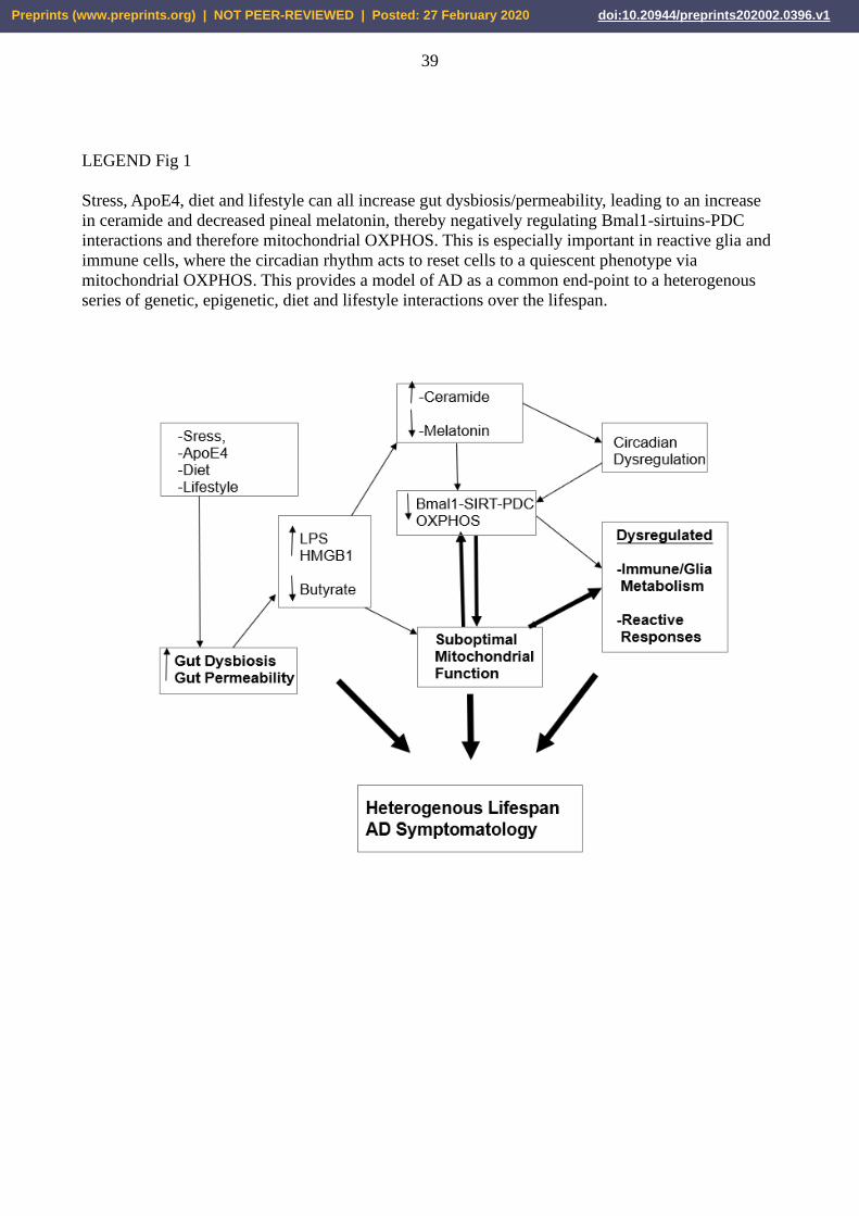
39
LEGEND Fig 1
Stress, ApoE4, diet and lifestyle can all increase gut dysbiosis/permeability, leading to an increase
in ceramide and decreased pineal melatonin, thereby negatively regulating Bmal1-sirtuins-PDC
interactions and therefore mitochondrial OXPHOS. This is especially important in reactive glia and
immune cells, where the circadian rhythm acts to reset cells to a quiescent phenotype via
mitochondrial OXPHOS. This provides a model of AD as a common end-point to a heterogenous
series of genetic, epigenetic, diet and lifestyle interactions over the lifespan.
Preprints (www.preprints.org) | NOT PEER-REVIEWED | Posted: 27 February 2020 doi:10.20944/preprints202002.0396.v1


![Activation of Nrf2 Pathway Contributes to …...NF-κB,p53andNrf2inbraincells[14,15].Amongtheseven sirtuins,Sirtuin1 (SIRT1)hasbeenreportedtobeinvolvedin promoting longevity in various](https://img.pdfslide.net/doc/110x75/5fc15a05ed4fb114a500e06b/activation-of-nrf2-pathway-contributes-to-nf-bp53andnrf2inbraincells1415amongtheseven.jpg)

![University of Zurich · the intrinsic (mitochondria-initiated) pathway, which converge in type II cells [1-3]. The extrinsic pathway requires that upon binding of a death ligand to](https://img.pdfslide.net/doc/110x75/608c59c03296430b7f29e887/university-of-zurich-the-intrinsic-mitochondria-initiated-pathway-which-converge.jpg)
![Archives of Biochemistry and Biophysics - cityu.edu.hk · pathway and mitochondria-mediated intrinsic pathway [13,14]. The former pathway is initiated mainly by binding of death re-](https://img.pdfslide.net/doc/110x75/5d1951c288c993b3668b912b/archives-of-biochemistry-and-biophysics-cityueduhk-pathway-and-mitochondria-mediated.jpg)