Embed Size (px)
Citation preview

Site-Specific Antibody Functionalization Using Tetrazine−StyreneCycloadditionBenjamin J. Umlauf,† Kalie A. Mix,‡,§ Vanessa A. Grosskopf,† Ronald T. Raines,‡,#,§ and Eric V. Shusta*,†
†Department of Chemical and Biological Engineering, ‡Department of Biochemistry, and #Department of Chemistry, University ofWisconsin−Madison, Madison, Wisconsin 53706, United States
*S Supporting Information
ABSTRACT: Biologics, such as antibody−drug conjugates, are becoming mainstreamtherapeutics. Consequently, methods to functionalize biologics without disrupting theirnative properties are essential for identifying, characterizing, and translating candidatebiologics from the bench to clinical practice. Here, we present a method for site-specific,carboxy-terminal modification of single-chain antibody fragments (scFvs). ScFvs displayedon the surface of yeast were isolated and functionalized by combining intein-mediatedexpressed protein ligation (EPL) with inverse electron-demand Diels−Alder (IEDDA)cycloaddition using a styrene−tetrazine pair. The high thiol concentration required totrigger EPL can hinder the subsequent chemoselective ligation reactions; therefore, theEPL reaction was used to append styrene to the scFv, limiting tetrazine exposure todamaging thiols. Subsequently, the styrene-functionalized scFv was reacted with tetrazine-conjugated compounds in an IEDDA cycloaddition to generate functionalized scFvs thatretain their native binding activity. Rapid functionalization of yeast surface-derived scFv ina site-directed manner could find utility in many downstream laboratory and preclinicalapplications.
■ INTRODUCTION
Antibodies are a rapidly growing class of therapeutic agentswith significant clinical success.51 In addition, imaging,diagnostics, and separation technologies often employ antibod-ies due to the high specificity and affinity for their cognateantigens. Functionalization of antibodies with chemical probessuch as fluorophores,1,2 small-molecule drugs,3−5 or otherbiomolecules6−8 is used to customize these reagents for manyapplications. Still, a growing number of studies indicate that it isessential to append these probes in a site-specific manner thatdoes not disrupt antibody function.3,6,9−12
We previously developed a method for site-specific, carboxy-terminal antibody modification by employing yeast surfacedisplay in combination with expressed protein ligation(EPL).13,14 In this system, the carboxy-terminus of the scFvis fused to an engineered, non-self-cleavable intein (202−08)and expressed on the surface of yeast cells. Our laboratorypreviously generated 202−08 by evolving the Mxe GyrA inteinto improve yeast surface display of scFv-intein fusions.14−17,36
Addition of a thiol, 2-mercaptoethanesulfonic acid (MESNA),liberates the scFv from the yeast surface and activates inteinsplicing to undergo transthioesterification and produce acarboxy-terminal thioester. Subsequently, a cysteine (Cys)amide is used to link the scFv to a Cys-modified probe ofinterest via an amide bond. Of note, this system enables rapidprotein modification specifically at the carboxy-terminus of thereleased protein, without requiring protein purification.14,36 Asa result, the yeast display EPL system is ideal for the rapid,high-throughput functionalization of antibodies.
While other systems for site-specific labeling includingFLASH, CLIP tags, and genetic code expansion (GCE) canalso facilitate antibody functionalization, non-self-cleavinginteins are beneficial in that they exhibit traceless appendageof the functional group upon EPL.13,14,36,38,39 Although one canappend a variety of functional groups to proteins using EPL,including post-translationally modified peptides,13 noncanon-ical amino acids,18 and biophysical probes,19 it can beadvantageous to employ the expanding catalog of bioorthog-onal reagents to append peptides, fluorophores, nanoparticles,and purification-enhancing moieties.45−48 To this end, wepreviously employed EPL with surface-displayed proteins toappend an azide as a reactive handle for copper-catalyzedazide−alkyne cycloaddition (CuAAC).14 Although the rapidrate of this reaction makes it useful for many in vitroapplications, CuAAC can have more limited utility in vivodue to the oxidative stress induced by Cu(I) and cross-reactivity of ascorbate with biological nucleophiles.20,21 Addi-tionally, the multicomponent nature of the reaction, requiring acopper catalyst, activating ligand, and reducing agent inaddition to the azide and alkyne reagents often requiresoptimization to apply the reaction to different molecules andconditions. Further, the high concentration of thiols can impaircertain CuAAC reactions.22−24,37 As an alternative, strainedcyclooctynes (e.g., DBCO) could be used in copper free
Received: February 13, 2018Revised: April 9, 2018Published: April 25, 2018
Article
pubs.acs.org/bcCite This: Bioconjugate Chem. 2018, 29, 1605−1613
© 2018 American Chemical Society 1605 DOI: 10.1021/acs.bioconjchem.8b00114Bioconjugate Chem. 2018, 29, 1605−1613

reactions with azides. A DBCO-cysteine reagent cannot beeffectively used for the EPL-mediated scFv modificationbecause strained cyclooctynes interact with thiols at concen-trations that are orders of magnitude lower than those used inthe EPL reaction.25 Instead, we have successfully used EPL tofirst append an azide that is subsequently reacted with aDBCO-conjugated probe to modify scFvs.36 However, certainapplications such as in vivo targeting with an azide-modifiedscFv, followed by a DBCO probe would be limited sincestrained cyclooctynes suffer from poor bioavailability.52
In order to address the aforementioned chemical compati-bility issues with the yeast display EPL system, and to expandits utility, we explored the possibility of replacing Cu-catalyzedand strain-based biorthogonal reactions with an inverseelectron-demand Diels−Alder (IEDDA) cycloaddition reaction.Styrene was shown to be inert to millimolar thiolconcentration, enabling EPL driven carboxy-terminal modifica-tion of scFv with styrene. Subsequently, a styrene-modifiedscFv can be covalently linked to tetrazine-containing probes viaIEDDA cycloaddition, and the functionalized scFvs retain theirantigen-binding capacity.
■ RESULTSStyrene is Compatible with Both EPL and IEDDA
Reactions. The reaction of a tetrazine with trans-cyclooctene(TCO) has become a well-established and useful tool inchemical biology as a result of its aqueous, rapid, and two-component nature.26,27 For compatibility with yeast surfacedisplay EPL, either tetrazine or TCO must be inert to high thiolconcentrations (Figure 1, Steps A−C). To measure stability inthe presence of free thiols, trans-cyclooctenol was incubatedwith FmocCysOH. After only 4 h, the trans-cyclooctenol hadisomerized completely to the unreactive cis isomer (Figure 2a).The dienophile in an IEDDA cycloaddition can also beactivated by electron-donating groups instead of strain.28 Onesuch activated alkene, 4-aminostyrene,29 possesses an aminogroup to both activate the alkene by donating electrons into thearyl system and act as a handle for derivatization. To test thecompatibility of styrene with a high concentration of thiols, 4-aminostyrene was incubated with FmocCysOH and in contrastto trans-cyclooctenol, no degradation of the styrene wasdetectable after 12 h (Figure 2b). NMR spectroscopy wasnext used to examine the reaction kinetics of 4-acetamidostyr-ene (4) with a phenyltetrazine (Figure S1).27 The second-orderrate constant was found to be k = (4.0 ± 0.1) × 10−3 M−1 s−1.Tetrazine is Not Compatible with a Yeast Display EPL
Reaction. While a styrene appeared to be an appropriatedienophile for IEDDA in the context of a yeast display EPLreaction, the feasibility of using tetrazine in an EPL reaction wasalso explored. The tetrazine would have to withstandincubation with yeast and thiol for it to be compatible withthe yeast display EPL reaction (one-pot reaction of Figure 1,Steps A−C) since it has been reported that a reduced tetrazineis no longer capable of an IEDDA reaction.30 Incubation of atetrazine with live yeast cells resulted in a significant decrease inreactive tetrazine as measured by tetrazine absorbance (Figure2c), indicating that the tetrazine is destroyed, sequestered, orreduced by the yeast cells.30 Tetrazine was also affected bycommon reducing agents used in intein-mediated EPLreactions (Figure 2d). Thiol-based reducing agents, including2-mercaptoethanol, dithiothreitol, and MESNA, all drovetetrazine reduction. Incubation of a tetrazine with tris(2-carboxyethyl)phosphine (TCEP) at only 4 mM, the minimal
amount required to release scFv from the yeast surface, alsoresulted in tetrazine reduction. Given that tetrazine reductionwould hinder an IEDDA reaction with a styrene-probe reactant(Figure 1, Step D), its use in the EPL stages of the conjugationscheme was not pursued further.
Styrene Modification of scFv by EPL Followed byIEDDA Cycloaddition with a Tetrazine. Based on theresults above, we chose to modify the scFv with the styrenereagent using EPL, and then derivatize the modified scFv with atetrazine probe after removal of yeast and reducing agents(Figure 1). To test the strategy, an scFv that binds tofluorescein (4-4-20) was used. 4-4-20 scFv−intein fusionprotein was displayed on the yeast surface, and the yeast waspelleted to remove growth medium. The yeast cells wereresuspended, and 50 mM of MESNA was added, followed by 5mM Cys−PEG3−styrene (3) to promote the EPL reaction.
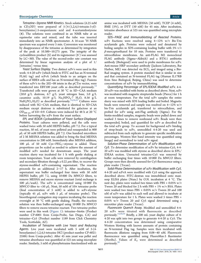
Figure 1. Route for the site-specific functionalization of a yeastsurface-displayed scFv. A four-step process is used to functionalize thecarboxy-terminus of an scFv by coupling expressed protein ligation(EPL) and inverse electron-demand Diels−Alder (IEDDA) cyclo-addition. Step A is the release of the scFv from the yeast surface bythiol−disulfide exchange with MESNA. Step B is the cleavage of theintein and formation of a thioester with MESNA at the carboxy-terminus. Step C is reaction of the thioester with Cys-PEG3-styrene(3). Steps A−C occur in a one-pot reaction with yeast, MESNA andCys-PEG3-styrene (3). After removal of yeast and MESNA, Step D isthe IEDDA conjugation between scFv−styrene and a tetrazine-containing probe. Inset: structures of two tetrazine probes used forIEDDA cycloaddition.
Bioconjugate Chemistry Article
DOI: 10.1021/acs.bioconjchem.8b00114Bioconjugate Chem. 2018, 29, 1605−1613
1606

The EPL reaction proceeded for 3−18 h at room temperaturewith gentle shaking. MESNA released the Aga2p fusion andtriggered intein cleavage, yielding an scFv with a carboxy-terminal thioester (Figure 1, Steps A and B). Subsequently, theCys−PEG3−styrene reacted with the scFv-thioester to yield anscFv modified at its carboxy-terminus with styrene (Figure 1,Step C). After removal of yeast cells by pelleting, sterile-filtering
through 0.22 μm spin columns, and dissolution in MESNA viabuffer exchange with 10 000 Da MWCO filters, styrene-modified scFv was reacted with tetrazine−biotin in an IEDDAreaction to generate scFv functionalized with carboxy-terminalbiotin (Figure 1, Step D). Western blotting demonstrated thatbiotin functionalization of scFv was dependent on both styrenemodification and reaction with tetrazine−biotin (Figure 3a).
Figure 2. Stability of candidate reagents for EPL/IEDDA. (a) NMR spectra of vinyl protons of trans-cyclooctenol over the course of 4 h ofincubation with FmocCysOH. (b) NMR spectra of 4-aminostyrene, FmocCysOH, and the reaction mixture after a 12 h incubation. (c) Absorbanceat 525 nm of 5 mM tetrazine-amine with or without 100 μL of yeast cell culture incubated for 45 min. Mean and standard deviation are plotted froma minimum of three replicates. Inset: images showing the loss of the characteristic pink color of a tetrazine in the presence of yeast cells (*p < 0.01using two tailed t-test). (d) Absorbance at 525 nm of a 5 mM tetrazine−amine solution containing various reducing agents used for yeast surfacecleavage and EPL incubated for 45 min. Mean and standard deviation are plotted from a minimum of three replicates. Inset: images demonstrate lossof the characteristic pink color of a tetrazine (&p < 0.01 using ANOVA).
Figure 3. Functionalization of 4-4-20 scFv by EPL followed by IEDDA. (A) Western blot examining the biotin modification of a 4-4-20 scFv with anEPL reaction followed by reaction with 5 mM tetrazine−biotin overnight at 30 °C with gentle shaking. Total released and intein-cleaved scFv (α-FLAG) and functionalized scFv (α-Biotin) were probed. (B) Western blot showing EPL/IEDDA reaction yields as a function of MESNAconcentration. α-FLAG detection is indicative of total antibody available for functionalization while α-biotin detection is used to compare thecombined EPL and IEDDA efficiency. (C) Fraction of 4-4-20 scFv released from yeast that is biotinylated through combined EPL/IEDDA reactions.Total scFv released from yeast and the fraction of this scFv that was biotinylated as determined by binding to streptavidin beads were quantified byWestern blotting with α-FLAG antibody. The mean and standard deviation for a minimum of three replicates is plotted. 78% ± 10% of the total scFvis modified using the combined EPL/IEDDA reaction (p < 0.05, t-test).
Bioconjugate Chemistry Article
DOI: 10.1021/acs.bioconjchem.8b00114Bioconjugate Chem. 2018, 29, 1605−1613
1607

MESNA concentrations of 200 mM yielded the most robustcombined EPL/IEDDA reaction as measured by the maximalamount of biotin-functionalized scFv (Figure 3b). Theefficiency of the combined EPL/IEDDA reactions, in termsof the fraction of scFv released from the yeast surface that wassuccessfully biotinylated, was 78% ± 10% (Figure 3c).ScFv Modification with Styrene−Tetrazine EPL/IEDDA
Yields Functional scFvs. Next, styrene-modified scFvs wereconjugated to a fluorophore in an IEDDA reaction withtetrazine−Cy5 (Figure 1, Step D and inset). As demonstratedfor the 4-4-20 scFv, antibody-associated Cy5 fluorescence
required both styrene and tetrazine reactants (Figure 4a). Totest that the scFv antigen-binding region retains functionfollowing modification, 4-4-20−Cy5 was applied to microtiterwells coated with antigen in the form of FITC-dextran. Therewas a 50-fold increase in Cy5 signal for 4-4-20−Cy5 comparedwith an antibody that does not bind fluorescein, scFvA,49
indicating that the Cy5-modified 4-4-20 retained antigen-binding capability (Figure 4b). To confirm that antigen bindingwas not affected by EPL/IEDDA, the antigen binding affinitiesfor unmodified 4-4-20 and 4-4-20−Cy5 scFvs were measured,and the Kd values for 4-4-20−Cy5 (1.52 ± 0.05 nM) and
Figure 4. Evaluation of 4-4-20 scFv function after EPL/IEDDA with styrene-tetrazine. (a) 4-4-20 scFv-associated Cy5 signal after EPL/IEDDAmodification with Cys−PEG3−styrene (3) and tetrazine−Cy5. Unreacted tetrazine-Cy5 is removed from the scFv fraction using buffer exchange andthe remaining Cy5 fluorescence measured (&p < 0.01 using ANOVA). The mean and standard deviation for a minimum of three replicates for eachgroup are plotted. (B) 4-4-20 and scFvA modified with Cy5 are incubated with immobilized FITC−dextran antigen. Unbound scFv was removedand wells quantified for Cy5 signal (*p < 0.01, t-test). The mean and standard deviation for a minimum of three replicates is plotted. (C)Equilibrium binding analysis by fluorescence quench affinity titration was used to compare the binding affinity of 20 nM unmodified 4-4-20 or Cy5-modified 4-4-20. Calculated Kd values (mean ± SD) are 1.65 ± 0.04 nM and 1.52 ± 0.05 nM, respectively (p = 0.38 using nonpaired, two-tailed ttest).
Figure 5. Internalization of Cy5-labeled scFv into rat brain endothelial (RBE4) cells. (a) Representative flow cytometry dot plot after RBE4 cellinternalization of scFvA−Cy5 (blue) or 4-4-20−Cy5 (red), with sample gate used for quantification. (b) Quantification of Cy5-positive cells acrossthree independent experiments for each group, with mean and standard deviation plotted (*p < 0.01, t-test). (c) Fluorescence microscopy images ofRBE4 cells after allowing for internalization of scFvA−Cy5 (red). Cells were fixed, permeabilized and stained for a Myc tag on the scFv (green).Nuclei were stained with Hoechst 33342 (blue) (d) RBE4 cells treated with scFvA−styrene that was not reacted with tetrazine−Cy5 and is stainedas in panel F. (e) RBE4 cells not treated with any scFv, and stained as in panel c. Scale bars = 20 μm.
Bioconjugate Chemistry Article
DOI: 10.1021/acs.bioconjchem.8b00114Bioconjugate Chem. 2018, 29, 1605−1613
1608

unmodified 4-4-20 scFv (1.65 ± 0.04 nM) were indistinguish-able (Figure 3c; p = 0.38, nonpaired, two tailed t-test).As further verification of scFv functionalization and to
demonstrate the utility of the EPL/IEDDA approach for cell-based assays, scFvA, a brain endothelial cell binding andinternalizing antibody,49 was functionalized with Cy5. ThescFvA−Cy5 conjugate was then used in flow cytometry andimmunofluorescence based internalization assays. Rat brainendothelial cells (RBE4) were incubated with scFvA−Cy5 orcontrol 4-4-20−Cy5, for 1 h at 37 °C to allow for scFvinternalization. Internalization was quantified using flowcytometry, with 96% ± 3% of RBE4 cells having internalizedscFvA−Cy5, whereas only ∼1% of cells internalized 4-4-20scFv−Cy5 (Figure 5a and b), indicating a specific interaction ofscFvA−Cy5 with RBE4 cells. The internalization of scFvA wasalso evaluated using fluorescence microscopy. ScFvA−Cy5 wasfound localized in intracellular puncta (Figure 5c) as previouslydescribed.49 Staining patterns of scFvA−Cy5 and unmodifiedscFvA appear similar (Figure 5d), and a strong colocalizationbetween antimyc and Cy5 signal was observed (Figure 5c)indicating internalization of intact scFvA−Cy5 conjugates.
■ DISCUSSIONWe have described a facile method for functionalizing yeast-displayed scFvs at their carboxy-terminus by combining EPLand IEDDA reactions. EPL modification presented challengesfor the subsequent IEDDA reactions because of the highconcentration of sulfur nucleophiles required for the EPLreaction.23 As a result, TCO was not an effective dienophile inour system due to its sensitivity to thiol reduction, but styrenewas stable under the high thiol concentrations required of EPL.Styrenes are known to undergo cycloaddition with tetra-zines;27,28,31,32 and have been reported for the bioconjugationof DNA.31 However, taking advantage of the inertness ofstyrene to thiols in an EPL/IEDDA protein-labeling system hasnot yet been described previously. One shortcoming is that therate of reaction between styrene and tetrazine (Figure S1, (4.0± 0.1) × 10−3 M−1 s−1) is quite slow compared to the reportedreaction of TCO and tetrazine ((1−10) × 103 M−1 s−1).27
Using the system described here, the lowered reaction rate wasovercome by adding an excess of tetrazine and performing thereaction at 30 °C.22 Further tuning the electronics of eachreagent could potentially enhance the reaction rates, andthereby enhance functionalization of low concentrations ofstyrene-modified proteins.29,33 For instance, the rate could beaccelerated by incorporation of electron-donating groups onthe aryl ring of the styrene reagent, and by using dipyridyl-tetrazine in place of phenyltetrazine.There exist several constraints with respect to the tetrazine
moiety in the yeast display EPL/IEDDA system. Both live yeastcells and reducing agents, including the non-thiol-based TCEP,resulted in reduction and subsequent inactivation of a tetrazine.To circumvent this problem, Cys−PEG3−styrene (3) ratherthan Cys−PEG3−tetrazine was used as the EPL reagent. It maybe possible, however, to modify the scFv with Cys−PEG3−tetrazine if reducing reagents were removed post-reaction andtetrazine reoxidized. A recent study described incubation oftetrazines with methylene blue or using intense red light toreoxidize a reduced tetrazine and restore reactivity.30 Both ofthese routes would, however, require additional steps; and thusthe addition of styrene, rather than tetrazine, by EPL ispreferable in this system. Given the relative differences in sizebetween an scFv and styrene (∼30 kDa versus ∼100 Da), a
PEG spacer between the cysteine amide used for EPL and thestyrene moiety was added. In the experiments described herewith biotin or fluorophore modification, the relatively small sizeof the tetrazine-probe is unlikely to cause major sterichindrance. Nevertheless, in applications that seek to modifythe scFv−styrene with a tetrazine-probe of larger size (e.g.,nanoparticulate substrates), the PEG spacer will likely have abeneficial impact.34,35
Another important factor in method optimization was theconcentration of MESNA employed in the EPL reaction.Interestingly, there exists an optimal EPL concentration ofMESNA at 200 mM for maximum yields from the IEDDAreaction, despite the fact that additional scFv is released fromthe yeast surface when up to 800 mM MESNA is used. Whileone might expect a plateau rather than optimum in MESNAconcentration, the presence of an optimum could be due tohigh concentrations of MESNA preventing the S- to N-acylswitch required for EPL modification with the styrene handle,or a result of competition between MESNA and cysteine fornucleophilic attack on the intein-generated thioester. At thisoptimum, we observed 78% ± 10% modification of 4-4-20 scFvusing the EPL/IEDDA protocol, which is sufficient fordownstream applications as demonstrated here.Finally, the utility of the yeast display EPL/IEDDA antibody
modification method was demonstrated using a variety offluorescence-based assays. Modification of 4-4-20 scFv withstyrene enabled carboxy-terminal conjugation to tetrazine−Cy5without affecting antigen binding.40,41 Such site-specificmodification distal to the binding site can provide a distinctadvantage over nonspecific, amine- or thiol-based antibodyfunctionalization protocols that often result in a largepercentage of inactive antibody because probe functionalizationoccurs near to or within the antigen-binding domain. Theutility of this method was also demonstrated using brainendothelial cell internalization assays with scFvA−Cy5,42 wherethe scFvA−Cy5 conjugate retains its ability to internalize intoRBE4 cells. We believe that the facile, modular, site-directedantibody-labeling protocol demonstrated herein is a powerfulmeans to facilitate the development and assessment ofantibodies and biologics for laboratory and preclinical use.
■ MATERIALS AND METHODSConditions. All procedures were performed in air at
ambient temperature (∼22 °C) and pressure (1.0 atm) unlessindicated otherwise.
Chemical Synthesis. Materials. Unless noted otherwise,reagents and solvents were from Sigma−Aldrich (Milwaukee,WI) and were used without further purification. Reagent-gradesolvents: acetonitrile, dichloromethane (DCM), tetrahydrofur-an (THF), and triethylamine (TEA) were dried over a columnof alumina and were removed from a dry still under an inertatmosphere. Flash column chromatography was performedwith 40−63 Å silica (230−400 mesh) from Silicycle (QuebecCity, Canada), and thin-layer chromatography was performedwith EMD 250 μm silica gel 60 F254 plates.
Solvent Removal. The phrase “concentrated under reducedpressure” refers to the removal of solvents and other volatilematerials using a rotary evaporator at water aspirator pressure(<20 Torr) while maintaining a water bath below 40 °C.Residual solvent was removed from samples at high vacuum(<0.1 Torr).
Compound 1. Boc-S-tert-butylthio-L-cysteine (500 mg, 1.6mmol) from Chem-Impex International (Wood Dale, IL) was
Bioconjugate Chemistry Article
DOI: 10.1021/acs.bioconjchem.8b00114Bioconjugate Chem. 2018, 29, 1605−1613
1609

dissolved in THF (5 mL). N-Hydroxysuccinimide (186 mg, 1.6mmol) and N,N′-dicyclohexylcarbodiimide (DCC; 363 mg, 1.7mmol) were added, and the resulting solution was stirredovernight. The reaction mixture was filtered, and the filtrate wasconcentrated under reduced pressure. The residue wasdissolved in DCM (15 mL). 4,7,10-Trioxa-1,13-tridecanedi-amine (0.9 mL, 4.3 mmol) was added, and the resultingsolution was stirred overnight. The reaction mixture wasfiltered, and the filtrate was concentrated under reducedpressure. The residue was purified by reverse-phase HPLC on aC18 column using a gradient of water−acetonitrile containingtrifluoroacetic acid (0.1% v/v) to yield compound 1 as a clearoil (64 mg, 10% for 2 steps).
Compound 2. 4-Aminostyrene (100 mg, 0.8 mmol) wasdissolved in DCM (8 mL). Succinic anhydride (84 mg, 0.84mmol) and TEA (0.24 mL, 1.7 mmol) were added, and theresulting solution was stirred overnight. The reaction mixturewas concentrated under reduced pressure. The residue wasdissolved in EtOAc and washed twice with 1 M HCl. Theorganic layer was dried over Na2SO4(s) and concentrated underreduced pressure to yield compound 2 as a white solid (99 mg,54%). 1H NMR (500 MHz, CD3OD, δ): 7.52 (d, 2H, J = 8.6Hz), 7.37 (d, 2H, J = 8.6 Hz), 6.65−6.71 (dd, 1H, J = 10.9 Hz,17.7 Hz), 5.71 (d, 1H, J = 17.6 Hz), 5.15 (d, 1H, J = 11.0 Hz),2.66 (s, 4H). 13C NMR (125 MHz, CD3OD, δ): 176.4, 172.8,139.6, 137.6, 137.5, 134.7, 127.6, 120.9, 32.3, 30.0. HRMS−ESI(m/z): [M − H]− calcd for C12H13NO3, 218.0823; found,218.0821.
Cys(StBu)−PEG3−styrene (3). Compound 1 (361 mg, 0.71mmol) was dissolved in DCM (7 mL). Compound 2 (156 mg,
0.71 mmol), N-hydroxysuccinimide (82 mg, 0.71 mmol), andDCC (146 mg, 0.71 mmol) were added, and the resultingsolution was stirred overnight. The reaction mixture wasfiltered, and then concentrated under reduced pressure. Theresidue was dissolved in acetonitrile and purified by reverse-phase HPLC on a C18 column using a gradient of water−acetonitrile containing trifluoroacetic acid (0.1% v/v). Theresidue was then dissolved in 4.0 M HCl in dioxane, and theresulting solution was stirred for 1 h. The solution was spargedwith N2(g) for 10 min to remove HCl and then concentratedunder reduced pressure to yield compound 3 as a white solid(61 mg, 12% for 2 steps).
4-Acetamidostyrene (4). 4-Aminostyrene (100 mg, 0.84mmol) was dissolved in DCM (8.4 mL). Acetyl chloride (0.18mL, 0.84 mmol) and TEA (0.24 mL, 1.68 mmol) were added,and the resulting solution was stirred overnight. The reactionmixture was concentrated under reduced pressure, and theresidue was dissolved in EtOAc. The solution was washed twicewith 1 M HCl and twice with saturated aqueous NaHCO3. Theorganic layer was dried over Na2SO4(s), and then concentratedunder reduced pressure. The residue was purified further bychromatography on silica gel, eluting with 1:1 EtOAc/hexanesto yield compound 4 as a white solid (39 mg, 29%). 1H NMR(500 MHz, CDCl3, δ): 7.47 (d, 2H, J = 8.6 Hz), 7.37 (d, 2H, J= 8.5 Hz), 7.14 (s, 1H), 6.70−6.64 (dd, 1H, J = 10.9, 17.6 Hz),5.68 (d, 2H, J = 17.6 Hz), 5.20 (d, 2H, J = 10.9 Hz), 2.19 (s,3H). 13C NMR (125 MHz, CDCl3, δ): 168.1, 137.4, 136.1,133.7, 126.8, 119.7, 113.1, 24.7. HRMS−ESI+ (m/z): [M + H]+
calcd for C10H11NO, 162.0913; found, 162.0912.Styrene and trans-Cyclooctene Stability. Stock solutions
were prepared by dissolving FmocCysOH, 4-acetamidostyrene(4), and trans-cyclooctenol in CD3OD at a concentration of200 mM. The solutions were combined in an NMR tube to givean equimolar ratio and mixed, and the tube was insertedimmediately into an NMR spectrometer. A 16-scan 1H NMRspectrum was acquired every 60 min.
Bioconjugate Chemistry Article
DOI: 10.1021/acs.bioconjchem.8b00114Bioconjugate Chem. 2018, 29, 1605−1613
1610
Tetrazine−Styrene NMR Kinetics. Stock solutions (6.25 mMin CD3OD) were prepared of 5-[4-(1,2,4,5-tetrazin-3-yl)-benzylamino]-5-oxopentanoic acid and 4-acetamidostyrene(4). The solutions were combined in an NMR tube at anequimolar ratio and mixed, and the tube was insertedimmediately into an NMR spectrometer. A 16-scan 1H NMRspectrum was acquired every 5 min. Conversion was monitoredby disappearance of the tetrazine as determined by integrationof the peak at 10.300−10.275 ppm. The integrity of thecycloaddition product (5) and its regioisomer (5′) was assessedby LC−MS. The value of the second-order rate constant wasdetermined by linear regression analysis of a plot of 1/[tetrazine] versus time.Yeast Surface Display. Two distinct scFv’s were used in this
work: 4-4-20 scFv (which binds to FITC and has an N-terminalFLAG tag) and scFvA (which binds to an antigen on thesurface of RBE4 cells and has an N-terminal Myc tag). Fusionsof these scFv’s to the 202−08 intein in the pCTre vector, weretransfected into EBY100 yeast cells as described previously.14
Transfected cells were grown at 30 °C in SD−CAA medium(20.0 g/L dextrose, 6.7 g/L yeast nitrogen base, 5.0 g/Lcasamino acids, 10.19 g/L Na2HPO4·7 H2O, 8.56 g/LNaH2PO4·H2O) as described previously.14,36,43 Cultures wereinduced with SG−CAA medium, that is identical to SD-CAAmedium except dextrose is substituted for galactose at anOD600 nm of 0.8−0.9. Cultures were induced for 48 h at 20 °Cbefore harvesting the scFv from the yeast surface.EPL and IEDDA Cycloaddition of Yeast Surface-Displayed
Proteins. Yeast cultures were pelleted, and the cells werewashed twice with 50 mM HEPES buffer, pH 7.2. For a typicalreaction, 50 mL of yeast were pelleted and resuspended in 800μL of 50 mM HEPES buffer, pH 7.2. One hundred microlitersof 2 M MESNA solution was added to bring the final MESNAconcentration to 200 mM. Immediately after MESNA addition,100 μL of 50 mM Cys−PEG3−styrene is added. Theseproportions can be scaled as needed to achieve the amount ofmodified scFv needed for downstream assays. The yeastreaction slurry was incubated for 1 h with gentle shaking atroom temperature. Yeast cells were removed by centrifugationand secondary filtration through a 0.22 μm filter, to recover thestyrene-modified scFv-containing supernatant. The reactionproceeds for an additional 2−17 h. After incubation, thesupernatant was buffer exchanged four times with 50 mMHEPES buffer, pH 7.2, using 10 000 Da MWCO filters, toremove MESNA and excess styrene reactant (total exchange is500 μL/wash). The scFv is concentrated using 10 000 DaMWCO filter to <50 μL. Next, 50 mM of 10× tetrazine probe(final concentration of 5 mM) is added to scFv-styrene(typically 45 μL scFv with 5 μL of tetrazine probe). Thebioorthogonal conjugation step requires incubating the reactionovernight at 30 °C with gentle shaking. Finally, the reactionsolution was then buffer-exchanged using 10 000 Da MWCOfilters to remove excess tetrazine reactant. Two tetrazine probeswere used in this work (Figure 1): tetrazine−biotin (Productnumber CP-6001 from Conju-Probe, San Diego, CA) andtetrazine−Cy5 (Product number 1189 from Click ChemistryTools, Scottsdale, AZ).Incubation of Tetrazine with Live Yeast and Reducing
Agents. Live yeast were incubated with 5 mM of 3-(4-benzylamino)-1,2,4,5-tetrazine HCl (product number CP-6021-25MG from Conju-probe). After 45 min yeast was pellet andtetrazine absorbance was quantified at 525 nm using microplatereader. Similarly, 5 mM of phenyltetrazine functionlized with an
amine was incubated with MESNA (50 mM), TCEP (4 mM),BME (5%), or DTT (50 nM) for 45 min. After incubation,tetrazine absorbance at 525 nm was quantified using microplatereader.
SDS−PAGE and Immunoblotting of Reacted Proteins.scFv fractions were resolved using 4−12% w/v Bis-Trisacrylamide gels. Proteins were reduced and denatured byboiling samples in SDS-containing loading buffer with 5% v/vβ-mercaptoethanol for 10 min. Proteins were transferred tonitrocellulose membranes. An anti-FLAG M2 monoclonalFLAG antibody (Sigma−Aldrich) and a BTN.1 antibiotinantibody (Biolegend) were used to probe membranes for scFv.Anti-mouse HRP secondary antibody (Jackson Laboratory, BarHarbor, ME) was detected via ECL, and imaged using a Bio-Rad imaging system. A protein standard that is similar in sizeand that contained an N-terminal FLAG tag (Human IL17RBfrom Sino Biological, Beijing, China) was used to determineconcentrations of scFv by immunoblotting.
Quantifying Percentage of EPL/IDEAA Modified scFv. 4-4-20 scFv was modified with biotin as described above. Next, scFvwas incubated with magnetic streptavidin beads (Pierce) for 1 hat room temperature. For the total scFv samples the entireslurry was mixed with SDS loading buffer and boiled. Magneticbeads were removed and sample was resolved on 4−12% w/vbis-Tris acrylamide gel, transferred to nitrocellulose, andprobed for scFv using anti-Flag antibody (Sigma). For thebiotin-modified samples, magnetic beads were pulled down andwashed 3 times to remove nonbound scFv. Beads were thenresuspended, boiled, and quantified for scFv as described forthe total scFv group. To account for background scFv bindingof strep-beads to scFv, unmodified 4-4-20 was used andsubtracted from each replicate to generate specific modificationpercentages. Western blot band intensity was quantified usingImageJ and normalized to total scFv signal.
Solution-Phase Determination of scFv Modification withCy5. To determine modification of scFv by tetrazine-Cy5, 4-4-20 scFv was modified with styrene as described above in EPL/IDEAA section. Unreacted tetrazine-Cy5 was removed bybuffer exchanging four times with 10 000 Da MWCO filters.Groups were then directly assessed for Cy5 fluorescence using aplate reader (Tecan).
Solid-Phase Determination of scFv Modification with Cy5.4-4-20 and scFvA were modified with Cy5 using the approachdescribed above. FITC-dextran was immobilized onto maxi-sorp ELISA plates (Nunc) by O.N. incubation at 4 °C. Thenext day, plates were washed two times with PBS + 0.05% w/vTween 20 and blocked for 2 h with PBS + 1% w/v BSA. Plateswere washed two times PBS + 0.05% w/v Tween 20 and 100nM of scFv was added to each well and allowed to incubate atroom temperature for 1 h. Plates were washed 5 times PBS +0.05% w/v Tween 20 and Cy5 signal determined using amicrotiter plate reader (Tecan).
Fluorescein Quench Assay. Modified and unmodified 4-4-20 scFv were titrated with fluorescein as describedpreviously.14,40,44 Briefly, a 200 mL yeast display culture of 4-4-20 was split into two groups to generate 4-4-20 ± Cy5. The4-4-20 concentration was determined using comparativeWestern blotting with known amounts of protein containingan N-terminal Flag tag. Samples were then incubated withfluorescein dilutions ranging from 0.08−40 nM. Fluoresceinfluorescence was quantified using FluoroMax-3 fluorimeter(Horiba). Values of Kd were determined as describedpreviously.50
Bioconjugate Chemistry Article
DOI: 10.1021/acs.bioconjchem.8b00114Bioconjugate Chem. 2018, 29, 1605−1613
1611

Flow Cytometry. RBE4 cells were cultured on collagen typeI-coated tissue culture flasks in 45% v/v Alpha MinimumEssential Medium, 45% v/v Ham’s F10 medium, 10% v/v fetalcalf serum, 100 mg/L streptomycin, 100 000 units/L penicillinG, 0.3 g/L Geneticin, and 1 μg/L basic fibroblast growth factor(bFGF) as described previously.42 Cells were incubated withscFvA-Cy5 or unmodified scFvA (0.5 μM) for 1 h at 37 °C and5% CO2. Cells were washed three times with PBS (5 minwashes) and trypsinized for 5 min at 37 °C and 5% CO2.Cultures were diluted 1:1 with serum-containing growthmedium to quench trypsin, and the cells were pelleted bycentrifugation. The cell pellet was resuspended in PBScontaining 10 mM EDTA. Internalized fluorescence wasmeasured with a BD FACSCaliber cytometer by quantifying10 000 events/group using software from FlowJo (Ashland,OR).Fluorescence Microscopy. RBE4 cells were cultured on glass
coverslips.42 RBE4 cells were incubated with modified orunmodified scFvA (2 μM) for 1 h at 37 °C and 5% CO2. Cellswere then washed three times with PBS, fixed in 4% v/vparaformaldehyde for 10 min, and permeabilized with 0.1% v/vTriton X-100. Permeabilized RBE4 cells were stained with9E10 anti-Myc antibody (1:200), goat anti-mouse-AF488antibody (1:200), and Hoechst 33342 (1:800). Slides werewashed, mounted, and imaged on a Nikon upright fluorescencemicroscope.
■ ASSOCIATED CONTENT*S Supporting InformationThe Supporting Information is available free of charge on theACS Publications website at DOI: 10.1021/acs.bioconj-chem.8b00114.
Disappearance of a tetrazine in the presence of 4-acetamidostyrene, 1H and 13C NMR spectra, as well asLC−MS chromatograms of synthetic compounds used inthis study (PDF)
■ AUTHOR INFORMATIONCorresponding Author*E-mail: [email protected]. Phone: (608) 265-5103. Fax:(608) 262-5434.
ORCIDBenjamin J. Umlauf: 0000-0002-1882-6092Present Address§Department of Chemistry, Massachusetts Institute of Tech-nology, 77 Massachusetts Avenue, Cambridge, Massachusetts02139, United States.
NotesThe authors declare no competing financial interest.
■ ACKNOWLEDGMENTSK.A.M. was supported by Molecular Biosciences Training GrantT32 GM007215 (NIH) and a fellowship from the University ofWisconsin−Madison College of Agricultural and Life Sciences.This work was funded by National Science Foundation grantCBET1403350.
■ REFERENCES(1) Pleiner, T., Bates, M., Trakhanov, S., Lee, C.-T., Schliep, J. E.,Chug, H., Bohning, M., Stark, H., Urlaub, H., and Gorlich, D. (2015)
Nanobodies: Site-specific labeling for super-resolution imaging, rapidepitope-mapping and native protein complex isolation. eLife 4, e11349.(2) Devaraj, N. K., Upadhyay, R., Haun, J. B., Hilderbrand, S. A., andWeissleder, R. (2009) Fast and sensitive pretargeted labeling of cancercells through a tetrazine/trans-cyclooctene cycloaddition. Angew.Chem., Int. Ed. 48, 7013−7016.(3) Casi, G., Huguenin-Dezot, N., Zuberbuhler, K., Scheuermann, J.,and Neri, D. (2012) Site-specific traceless coupling of potent cytotoxicdrugs to recombinant antibodies for pharmacodelivery. J. Am. Chem.Soc. 134, 5887−5892.(4) Parslow, A. C., Parakh, S., Lee, F.-T., Gan, H. K., and Scott, A. M.(2016) Antibody−drug conjugates for cancer therapy. Biomedicines 4,14−20.(5) Sievers, E. L., and Senter, P. D. (2013) Antibody−drugconjugates in cancer therapy. Annu. Rev. Med. 64, 15−29.(6) Bernardes, G. J. L., Steiner, M., Hartmann, I., Neri, D., and Casi,G. (2013) Site-specific chemical modification of antibody fragmentsusing traceless cleavable linkers. Nat. Protoc. 8, 2079−2089.(7) Xiao, H., Woods, E. C., Vukojicic, P., and Bertozzi, C. R. (2016)Precision glycocalyx editing as a strategy for cancer immunotherapy.Proc. Natl. Acad. Sci. U. S. A. 113, 10304−10309.(8) Schaefer, G., Haber, L., Crocker, L. M., Shia, S., Shao, L.,Dowbenko, D., Totpal, K., Wong, A., Lee, C. V., Stawicki, S., Clark, R.,et al. (2011) A two-in-one antibody against HER3 and EGFR hassuperior inhibitory activity compared with monospecific antibodies.Cancer Cell 20, 472−486.(9) Junutula, J. R., Raab, H., Clark, S., Bhakta, S., Leipold, D. D.,Weir, S., Chen, Y., Simpson, M., Tsai, S. P., Dennis, M. S., et al. (2008)Site-specific conjugation of a cytotoxic drug to an antibody improvesthe therapeutic index. Nat. Biotechnol. 26, 925−932.(10) Strop, P., Delaria, K., Foletti, D., Witt, J. M., Hasa-Moreno, A.,Poulsen, K., Casas, M. G., Dorywalska, M., Farias, S., Pios, A., et al.(2015) Site-specific conjugation improves therapeutic index ofantibody drug conjugates with high drug loading. Nat. Biotechnol. 33,694−696.(11) Marlind, J., Kaspar, M., Trachsel, E., Sommavilla, R., Hindle, S.,Bacci, C., Giovannoni, L., and Neri, D. (2008) Antibody-mediateddelivery of interleukin-2 to the stroma of breast cancer stronglyenhances the potency of chemotherapy. Clin. Cancer Res. 14, 6515−6524.(12) Gutbrodt, K. L., Schliemann, C., Giovannoni, L., Frey, K., Pabst,T., Klapper, W., Berdel, W. E., and Neri, D. (2013) Antibody-baseddelivery of interleukin-2 to neovasculature has potent activity againstacute myeloid leukemia. Sci. Transl. Med. 5, 201ra118.(13) Muir, T. W., Sondhi, D., and Cole, P. A. (1998) Expressedprotein ligation: A general method for protein engineering. Proc. Natl.Acad. Sci. U. S. A. 95, 6705−6710.(14) Marshall, C. J., Agarwal, N., Kalia, J., Grosskopf, V. A., McGrath,N. A., Abbott, N. L., Raines, R. T., and Shusta, E. V. (2013) Facilechemical functionalization of proteins through intein-linked yeastdisplay. Bioconjugate Chem. 24, 1634−1644.(15) Shusta, E. V., Holler, P. D., Kieke, M. C., Kranz, D. M., andWittrup, K. D. (2000) Directed evolution of a stable scaffold for T-cellreceptor engineering. Nat. Biotechnol. 18, 754−759.(16) Huang, D., Gore, P. R., and Shusta, E. V. (2008) Increasingyeast secretion of heterologous proteins by regulating expression ratesand post-secretory loss. Biotechnol. Bioeng. 101, 1264−1275.(17) Tillotson, B. J., Lajoie, J. M., and Shusta, E. V. (2015) Yeastdisplay-based antibody affinity maturation using detergent-solubilizedcell lysates. Methods Mol. Biol. 1319, 65−78.(18) Valiyaveetil, F. I., Sekedat, M., MacKinnon, R., and Muir, T. W.(2004) Glycine as a D-amino acid surrogate in the K+-selectivity filter.Proc. Natl. Acad. Sci. U. S. A. 101, 17045−17049.(19) Flavell, R. R., Kothari, P., Bar-Dagan, M., Synan, M.,Vallabhajosula, S., Friedman, J. M., Muir, T. W., and Ceccarini, G.(2008) Site-specific 18F-labeling of the protein hormone leptin using ageneral two-step ligation procedure. J. Am. Chem. Soc. 130, 9106−9112.
Bioconjugate Chemistry Article
DOI: 10.1021/acs.bioconjchem.8b00114Bioconjugate Chem. 2018, 29, 1605−1613
1612

(20) Brewer, G. J. (2010) Risks of copper and iron toxicity duringaging in humans. Chem. Res. Toxicol. 23, 319−326.(21) Reihl, O., Lederer, M. O., and Schwack, W. (2004)Characterization and detection of lysine−arginine cross-links derivedfrom dehydroascorbic acid. Carbohydr. Res. 339, 483−491.(22) Hong, V., Presolski, S. I., Ma, C., and Finn, M. G. (2009)Analysis and optimization of copper-catalyzed azide−alkyne cyclo-addition for bioconjugation. Angew. Chem., Int. Ed. 48, 9879−9883.(23) McKay, C. S., and Finn, M. G. (2014) Click chemistry incomplex mixtures: Bioorthogonal bioconjugation. Chem. Biol. 21,1075−1101.(24) Presolski, S. I., Hong, V. P., and Finn, M. G. (2011) Copper-catalyzed azide−alkyne click chemistry for bioconjugation. Curr.Protoc. Chem. Biol. 3, 153−162.(25) Beatty, K. E., Fisk, J. D., Smart, B. P., Lu, Y. Y., Szychowski, J.,Hangauer, M. J., Baskin, J. M., Bertozzi, C. R., and Tirrell, D. A. (2010)Live-cell imaging of cellular proteins by a strain-promoted azide−alkyne cycloaddition. ChemBioChem 11, 2092−2095.(26) Taylor, M. T., Blackman, M. L., Dmitrenko, O., and Fox, J. M.(2011) Design and synthesis of highly reactive dienophiles for thetetrazine−trans-cyclooctene ligation. J. Am. Chem. Soc. 133, 9646−9649.(27) Blackman, M. L., Royzen, M., and Fox, J. M. (2008) Tetrazineligation: Fast bioconjugation based on inverse-electron-demandDiels−Alder reactivity. J. Am. Chem. Soc. 130, 13518−13519.(28) Wijnen, J. W., Zavarise, S., and Engberts, J. B. F. N. (1996)Substituent effects on an inverse electron demand hetero Diels−Alderreaction in aqueous solution and organic solvents: Cycloaddition ofsubstituted styrenes to di(2-pyridyl)-1,2,4,5-tetrazine. J. Org. Chem. 61,2001−2005.(29) Knall, A.-C., Hollauf, M., and Slugovc, C. (2014) Kinetic studiesof inverse electron demand Diels−Alder reactions (iEDDA) ofnorbornenes and 3,6-dipyridin-2-yl-1,2,4,5-tetrazine. Tetrahedron Lett.55, 4763−4766.(30) Zhang, H., Trout, W. S., Liu, S., Andrade, G. A., Hudson, D. A.,Scinto, S. L., Dicker, K. T., Li, Y., Lazouski, N., Rosenthal, J., et al.(2016) Rapid bioorthogonal chemistry turn-on through enzymatic orlong wavelength photocatalytic activation of tetrazine ligation. J. Am.Chem. Soc. 138, 5978−5983.(31) Rieder, U., and Luedtke, N. W. (2014) Alkene−tetrazineligation for imaging cellular DNA. Angew. Chem. 126, 9322−9326.(32) Sauer, J., Heldmann, D. K., Hetzenegger, J., Krauthan, J., Sichert,H., and Schuster, J. (1998) 1,2,4,5-Tetrazine: Synthesis and reactivityin [4 + 2] cycloadditions. Eur. J. Org. Chem. 1998, 2885−2896.(33) Knall, A.-C., and Slugovc, C. (2013) Inverse electron demandDiels−Alder (iEDDA)-initiated conjugation: A (high) potential clickchemistry scheme. Chem. Soc. Rev. 42, 5131−5142.(34) Brown, K. C. (2010) Peptidic tumor targeting agents: The roadfrom phage display peptide selections to clinical applications. Curr.Pharm. Des. 16, 1040−1054.(35) Li, S., Gray, B. P., McGuire, M. J., and Brown, K. C. (2011)Synthesis and biological evaluation of a peptide−paclitaxel conjugatewhich targets the integrin αvβ6. Bioorg. Med. Chem. 19, 5480−5489.(36) Marshall, C. J., Grosskopf, V. A., Moehling, T. J., Tillotson, B. J.,Wiepz, G. J., Abbott, N. L., Raines, R. T., and Shusta, E. V. (2015) Anevolved Mxe GyrA intein for enhanced production of fusion proteins.ACS Chem. Biol. 10, 527−538.(37) Lin, P.-C., Ueng, S.-H., Tseng, M.-C., Ko, J.-L., Huang, K.-T.,Yu, S.-C., Adak, A. K., Chen, Y.-J., and Lin, C.-C. (2006) Site-specificprotein modification through Cu(I)-catalyzed 1,2,3-triazole formationand Its implementation in protein microarray fabrication. Angew.Chem. 118, 4392−4396.(38) Sydor, J. R., Mariano, M., Sideris, S., and Nock, S. (2002)Establishment of intein-mediated protein ligation under denaturingconditions: C-Terminal labeling of a single-chain antibody for biochipscreening. Bioconjugate Chem. 13, 707−712.(39) Albertsen, L., Shaw, A. C., Norrild, J. C., and Strømgaard, K.(2013) Recombinant production of peptide C-terminal α-amides usingan engineered intein. Bioconjugate Chem. 24, 1883−1894.
(40) Boder, E. T., Midelfort, K. S., and Wittrup, K. D. (2000)Directed evolution of antibody fragments with monovalent femtomo-lar antigen-binding affinity. Proc. Natl. Acad. Sci. U. S. A. 97, 10701−10705.(41) Wang, X. X., and Shusta, E. V. (2005) The use of scFv-displaying yeast in mammalian cell surface selections. J. Immunol.Methods 304, 30−42.(42) Shindo, A., Maki, T., Mandeville, E. T., Liang, A. C., Egawa, N.,Itoh, K., Itoh, N., Borlongan, M., Holder, J. C., Chuang, T. T.,McNeish, J. D., Tomimoto, H., Lok, J., Lo, E. H., and Arai, K. (2016)Astrocyte-derived pentraxin 3 supports blood−brain barrier integrityunder acute phase of stroke. Stroke 47, 1094−1100.(43) Zhang, X., Wang, X. X., and Shusta, E. V. (2014) Creation andevaluation of a single-chain antibody tetramer that targets brainendothelial cells. AIChE J. 60, 1245−1252.(44) Tillotson, B. J., Cho, Y. K., and Shusta, E. V. (2013) Cells andcell lysates: A direct approach for engineering antibodies againstmembrane proteins using yeast surface display. Methods 60, 27−37.(45) Elias, D. R., Cheng, Z., and Tsourkas, A. (2010) An intein-mediated site-specific click conjugation strategy for improved tumortargeting of nanoparticle systems. Small 6, 2460−2468.(46) Gupta, S. S., Kuzelka, J., Singh, P., Lewis, W. G., Manchester, M.,and Finn, M. G. (2005) Accelerated bioorthogonal conjugation: Apractical method for the ligation of diverse functional molecules to apolyvalent virus scaffold. Bioconjugate Chem. 16, 1572−1579.(47) Bundy, B. C., and Swartz, J. R. (2010) Site-specificincorporation of p-propargyloxyphenylalanine in a cell-free environ-ment for direct protein−protein click conjugation. Bioconjugate Chem.21, 255−263.(48) Li, X., Patterson, J. T., Sarkar, M., Pedzisa, L., Kodadek, T.,Roush, W. R., and Rader, C. (2015) Site-specific dual antibodyconjugation via engineered cysteine and selenocysteine residues.Bioconjugate Chem. 26, 2243−2248.(49) Wang, X. X., Cho, Y. K., and Shusta, E. V. (2007) Mining a yeastlibrary for brain endothelial cell-binding antibodies. Nat. Methods 4,143−145.(50) Burns, M. L., Malott, T. M., Metcalf, K. J., Hackel, B. J., Chan, J.R., and Shusta, E. V. (2014) Directed evolution of brain-derivedneurotrophic factor for improved folding and expression inSaccharomyces cerevisiae. Appl. Environ. Microbiol. 80, 5732−5742.(51) Carter, P. J., and Lazar, G. A. (2017) Next generation antibodydrugs: Pursuit of the ‘high-hanging fruit’. Nat. Rev. Drug Discovery 17,197−223.(52) Chang, P. V., Prescher, J. A., Sletten, E. M., Baskin, J. M., Miller,I. A., Agard, N. J., Lo, A., and Bertozzi, C. R. (2010) Copper-free clickchemistry in living animals. Proc. Natl. Acad. Sci. U. S. A. 107, 1821−1826.
Bioconjugate Chemistry Article
DOI: 10.1021/acs.bioconjchem.8b00114Bioconjugate Chem. 2018, 29, 1605−1613
1613