Embed Size (px)
Citation preview

NANO EXPRESS Open Access
Size-regulated group separation of CoFe2O4nanoparticles using centrifuge and their magneticresonance contrast propertiesJongeun Kang1,2, Hyunseung Lee1, Young-Nam Kim1, Areum Yeom1, Heejeong Jeong1, Yong Taik Lim2
and Kwan Soo Hong1,2*
Abstract
Magnetic nanoparticle (MNP)-based magnetic resonance imaging (MRI) contrast agents (CAs) have been the subject ofextensive research over recent decades. The particle size of MNPs varies widely and is known to influence theirphysicochemical and pharmacokinetic properties. There are two commonly used methods for synthesizing MNPs,organometallic and aqueous solution coprecipitation. The former has the advantage of being able to control the particlesize more effectively; however, the resulting particles require a hydrophilic coating in order to be rendered water soluble.The MNPs produced using the latter method are intrinsically water soluble, but they have a relatively wide particle sizedistribution. Size-controlled water-soluble MNPs have great potential as MRI CAs and in cell sorting and labelingapplications. In the present study, we synthesized CoFe2O4 MNPs using an aqueous solution coprecipitation method.The MNPs were subsequently separated into four groups depending on size, by the use of centrifugation at differentspeeds. The crystal shapes and size distributions of the particles in the four groups were measured and confirmed bytransmission electron microscopy and dynamic light scattering. Using X-ray diffraction analysis, the MNPs were found tohave an inverse spinel structure. Four MNP groups with well-selected semi-Gaussian-like diameter distributions wereobtained, with measured T2 relaxivities (r2) at 4.7 T and room temperature in the range of 60 to 300 mM−1s−1,depending on the particle size. This size regulation method has great promise for applications that requirehomogeneous-sized MNPs made by an aqueous solution coprecipitation method. Any group of the CoFe2O4 MNPscould be used as initial base cores of MRI T2 CAs, with almost unique T2 relaxivity owing to size regulation. Themethodology reported here opens up many possibilities for biosensing applications and disease diagnosis.
Keywords: Magnetic nanoparticles; Magnetic resonance imaging; Relaxivity; Particle size regulation
PACS: 75.75.Fk, 78.67.Bf, 61.46.Df
BackgroundMagnetic resonance imaging (MRI) is a powerful diagnosticmodality for noninvasive in vivo imaging due to its highresolution, lack of exposure to radiation, superior soft tissuecontrast, and large image window. However, it has less sen-sitivity than nuclear medicine and fluorescence imagingwhen monitoring small tissue lesions and molecular or cel-lular activities [1]. Contrast agents (CAs) can improve thecontrast and specificity in particular target regions of MR
images, and these are widely used to produce brighter anddarker areas with T1 and T2 CAs, respectively. T2 CAs,mainly based on iron oxide magnetic nanoparticles(MNPs), provide dark contrast in T2- or T2*-weighted(T2*-W) MR images depending on the T2 relaxivity of r2and the MNP concentration in the region of interest [2].Superparamagnetic iron oxide (SPIO) nanoparticles withdiameters of 50 to 150 nm are thus the most commonlyused MNPs in a variety of biomedical applications such asMRI contrast agents, induction of local hyperthermia, ma-nipulation of cell membranes, biosensors, cell labeling andtracking, and drug targeting and delivery [3-8].SPIO particles have different physicochemical and bio-
logical properties, depending on the particle size and
* Correspondence: [email protected] for MR Research, Korea Basic Science Institute, Cheongwon 363-883,South Korea2Graduate School of Analytical Science and Technology, Chungnam NationalUniversity, Daejeon 305-764, South Korea
© 2013 Kang et al.; licensee Springer. This is an Open Access article distributed under the terms of the Creative CommonsAttribution License (http://creativecommons.org/licenses/by/2.0), which permits unrestricted use, distribution, and reproductionin any medium, provided the original work is properly cited.
Kang et al. Nanoscale Research Letters 2013, 8:376http://www.nanoscalereslett.com/content/8/1/376

coating material, including MR T2 relaxivity r2 [9], cell la-beling efficiency [10], cell cytotoxicity [11], and in vivopharmacokinetics such as blood half-life and biodistribution[12]. Therefore, strategies by which uniform-sized biocom-patible MNPs with long circulation times can be producedare highly sought after for nanomedical applications.There are two commonly used methods for synthesiz-
ing MNPs, organometallic [13] and aqueous solutioncoprecipitation [14]. In the organometallic approach, theparticle size can be easily controlled [15]; however, theMNPs are only soluble in nonpolar and moderately polarorganic solvents. This brings about the requirement forhydrophilic and biocompatible polymer coating to makethem soluble enough for in vivo uses [16-18]. On theother hand, the aqueous solution coprecipitation methodresults in nanoparticles that are intrinsically water-soluble;however, the particle size distribution is relatively wide,resulting in nonuniform contrast in T2- or T2*-W MR im-ages. Size-controlled water-soluble nanoparticles providethe possibility to achieve uniform functionalization of theirsurfaces with other imaging probes such as fluorescentdyes and radiolabeled probes or with targeting moleculessuch as antibodies, peptides, and genes, as well as thera-peutics [18,19]. Several reports are available regarding thesize regulation of MNPs synthesized by coprecipitation, in-cluding a temperature-controlled coprecipitation methodthat requires specialized equipment and a piezoelectricnozzle method [20,21]. These processes are either highlycomplex or relatively ineffective owing to the require-ment for a high level of control over parameters such astemperature during the synthesis. In addition, the pro-duced particles still have an inadequate size distribution.The piezoelectric nozzle method is more effective forcontrolling the size; however, this technique requires spe-cialized equipment such as a piezoelectric transducer and afrequency amplifier.To address these issues, a facile method for controlling
the MNP core size via the coprecipitation process is intro-duced here. Initially, we synthesized CoFe2O4 nanoparticlesusing an aqueous solution coprecipitation method andthen separated the particles into four groups depending ontheir size by employing a variety of centrifugation speeds.The physicochemical properties of the four groups weresubsequently evaluated. The size distribution was assessedby transmission electron microscopy (TEM) and dynamiclight scattering (DLS), crystallographic confirmation wascarried out by X-ray diffraction (XRD), the water protonT2 relaxation rate (R2) versus Co/Fe concentration wasevaluated, and MR image contrast was measured at 4.7 T.
MethodsSynthesis of CoFe2O4 nanoparticlesThe CoFe2O4 MNPs were synthesized by an aqueous so-lution coprecipitation method reported previously [14].
Initially, the reagents, 0.5 M FeCl3·6H2O (≥98%; Sigma-Aldrich, Tokyo, Japan) and 0.25 M CoCl2·6H2O (99% to102%; Sigma-Aldrich), were mixed in an aqueous solu-tion, giving a Co/Fe ratio of 1:2. The reaction mixturewas stirred vigorously for 6 h in boiling distilled waterwith 1 M NaOH (96%; Junsei, Tokyo, Japan), and then,the resulting dark brown suspension was centrifuged at1,771 × g. The precipitate was dissolved in a 2-M HNO3 so-lution with stirring for 20 min and then centrifuged againat 1,771 × g. The resulting precipitate was dissolved in0.5 M Fe(NO3)3 (≥98%; Sigma-Aldrich) and stirred vigor-ously for 30 min at 100°C. After the reaction, centrifugationat 1,771 × g and redispersion in distilled water were per-formed three times. Finally, the suspension was dissolved inwater and stored at room temperature until further use.
Size selection of MNPs and synthesis of SiO2-coated MNPsAs the synthesized MNPs had a broad size distributionbetween 5 and 300 nm, they were separated depending ontheir size by stepwise centrifugation. A high-speed vacuumcentrifuge system was used (SUPRA 25K; Hanil Scimed,Gangneung, Korea), with five different speeds of 1,771 × g,2,767 × g, 11,068 × g, 24,903 × g, and 35,860 × g in order toseparate the synthesized particles into four groups. Firstly,aggregated particles were removed by down-sinking with1,771 × g for 1 h. The remaining mixture was centrifuged at35,860 × g for 1 h, and then, the suspended solution wasremoved. Resuspension of the bottom layer provided theinitial MNP solution. This was then centrifuged at 2,767 ×g, 11,068 × g, and 24,903 × g for 1 h, with the bottom layercollected as groups A, B, and C, respectively. The firstsuspended solution remaining after centrifugation at24,903 × g was labeled as group D. The MNPs of group Cwere selected for SiO2 coating for further applications. SiO2
coating was done as follows: the MNPs of group Cwere stabilized with polyvinylpyrrolidone (PVP) todisperse them homogeneously, and then, tetraethoxysilanesolution was polymerized on the surface of PVP-stabilizedCoF2O4 MNPs by adding ammonia solution as a catalystto form SiO2 coating on the MNPs. The volume ratio ofthe ammonia solution was 4.2% to control the SiO2 shellthickness of the final SiO2-coated MNPs in this process.
MNP characterizationThe crystal shapes and structures of the synthesized MNPsin each group, in addition to the SiO2-coated MNPs, weremeasured and confirmed by TEM (Tecnai G2 F30, FEI,Hillsboro, OR, USA) and XRD (XPERT MPD, Philips,Amsterdam, The Netherlands). The XRD patterns werecompared with a typical XRD spectrum of a CoFe2O4
crystal. The hydrodynamic diameter distribution of theparticles was measured by DLS (UPA-150l, Microtrac,Montgomeryville, PA, USA), and the size distributionwas verified from the TEM images.
Kang et al. Nanoscale Research Letters 2013, 8:376 Page 2 of 7http://www.nanoscalereslett.com/content/8/1/376

In order to compare T2 relaxivities (r2) of the four groupsand the SiO2-coated MNPs, the T2 relaxation times weremeasured against the Co/Fe concentration in a rangebelow 1 mM Fe using a spin-echo pulse sequence (multi-spin multi-echo) on a 4.7-T animal MRI system (Biospec47/40; Bruker, Karlsruhe, Germany). The amount of Co/Fein each group was measured using an inductively coupledplasma atomic emission spectrometry system (Optima4300DV, PerkinElmer, Waltham, MA, USA). For the MRIexperiment, the MNPs were sampled at four different Co/Fe concentrations of 1.0, 0.75, 0.5, and 0.25 mM Co/Fe indistilled water in 250-μl microtubes. The MRI parametersused were as follows: TE/TR = 10/10,000 ms, number ofscans = 2, slice thickness = 1 mm, FOV = 5 × 5 cm2, num-ber of slices = 1. T2 contrast differences depending on Feconcentration for the separated groups were also comparedin T2-W MR images.
Results and discussionThe MNPs synthesized by the coprecipitation method werefound to have an extremely broad size distribution [14].This characteristic would likely result in nonuniform con-trast in MR images. The purpose of the present study wasto overcome this limitation by separating the different sizesof particles by centrifugation. After the initial removal of
aggregates, the nanoparticles were sequentially centrifugedat speeds 2,767 × g, 11,068 × g, 24,903 × g, and 35,860 × g,producing groups A, B, C, and D, respectively. As shown inthe TEM images in Figure 1, the centrifugation processresulted in four groups containing particles relativelyuniform in size. The mean diameters measured fromapproximately 100 randomly selected particles from eachgroup were found to be 24.2 ± 3.6, 20.0 ± 3.6, 15.8 ± 3.6,and 10.5 ± 2.4 nm for groups A, B, C, and D, respectively.As the rotational speed increased, the MNP diametersdecreased, with significant differences between adjacentgroups (P < 0.01). The hydrodynamic diameter distribu-tions of the MNPs in the four groups were Gaussian-like,with values of 65.5 ± 14.0, 38.9 ± 9.1, 23.1 ± 6.0, and 18.5 ±4.4 nm (Figure 2a) and volume ratios of 29%, 48%, 13%,and 10% for groups A to D, respectively. Further, from themeasured volume ratios in Figure 2a, the highest MNPvolume was observed for group B; groups C and D couldalso provide an adequate quantity of uniform-sized MNPsfor use in applications that require very small sized(approximately 10 nm) MNPs. The amount of synthesizedMNPs from group D was approximately 0.5 g, which couldbe easily scaled-up using a larger reaction vessel.The mean diameter of the MNPs, as measured by
TEM and DLS, decreased as the centrifugation speed
Figure 1 TEM images of the four MNP groups. The TEM images show that the particles were well dispersed and size-regulated according tothe group. The mean diameters for the four groups were 24.2 ± 3.6, 20.0 ± 3.6, 15.8 ± 3.6, and 10.5 ± 2.4 nm, for groups a to d, respectively.
Kang et al. Nanoscale Research Letters 2013, 8:376 Page 3 of 7http://www.nanoscalereslett.com/content/8/1/376

decreased (Figure 2b), indicating that the MNP particlessynthesized by the coprecipitation method were wellseparated and clearly resolved into the four groups by thedifferent centrifugation speeds.Using the organometallic method reported by others,
the particle size of MNPs can be easily controlled, witha narrower diameter distribution achievable in compari-son to the combined coprecipitation and centrifugationmethods described here. However, the amount of MNPsthat can be synthesized in a single process is quite small,and these have the added disadvantage of being hydro-phobic. A coating is therefore necessary in order to ren-der these MNPs hydrophilic and to enable them to beused for functions such as drug loading, targeting, orimaging probes (PET or fluorescence). Even though thesize distribution of MNPs synthesized by the coprecipi-tation method was large, huge amounts of size-controlledMNPs were obtained by combining the method with a
simple centrifugation process. Figure 3a shows the XRDresults obtained for the four groups of CoFe2O4 MNPs. Allgroups can be seen to exhibit the same peaks, which matchwell with the standard Fe3O4 XRD pattern (JCPDS 75–0030). The mean particle size (D) can be calculated by thefull-width at half-maximum (FWHM) and the area/heightratio (β) of the XRD peaks with instrumental correction,using the equation D = Kλ / β × cosθ, where K is theScherrer constant, λ is the wavelength, β is the FWHM (inradians), and θ is the peak angular position [22,23]. TheXRD information gave crystallite sizes of 14.9, 13.2, 12.1,and 7.3 nm (Figure 3). As MNPs synthesized by copreci-pitation may contain some iron oxide crystals, the particlesize calculated from the TEM images was larger than thatfrom the XRD data (Figure 3b).The size-dependent MR contrast (T2 relaxivity) of the
MNPs was measured on a 4.7-T MRI system. Figure 4ashows the dependence of the T2 relaxation rate (R2, s
−1)
Figure 2 Relative size distributions of separated MNP groups and correlation between DLS and TEM results. Relative size distributions ofseparated MNP groups in aqueous solution measured by DLS (a) and a graph showing correlation between DLS and TEM results (b). The meanDLS diameters for the four groups, A to D, were 65.5 ± 14.0, 38.9 ± 9.1, 23.1 ± 6.0, and 18.5 ± 4.4 nm, respectively, with relative volumes of 29%(A), 49% (B), 12% (C), and 10% (D) as measured by integration of the DLS spectra.
Figure 3 A stack plot of XRD patterns of MNPs and size calculation. The nanoparticles were well crystallized, and the peaks are in accordance withthe typical CoFe2O4 XRD spectrum in which the main peaks are (111), (220), (311), (400), (511), and (440) (a). The mean diameters of the crystal particlescalibrated from signal width for the four groups from A to D were 14.9, 13.2, 12.1, and 7.3 nm, respectively (b).
Kang et al. Nanoscale Research Letters 2013, 8:376 Page 4 of 7http://www.nanoscalereslett.com/content/8/1/376
on the MNPs of the four groups. The T2 relaxation ratewas increased with increased Co/Fe concentration, andthe T2 relaxivities (r2) for the groups were measuredfrom the slopes of the data. The r2 values were found tobe 302 ± 9, 268 ± 8, 179 ± 5, and 66 ± 4 mM−1s−1 forgroups A, B, C, and D, respectively (Figure 4b). Thesevalues are comparable to those in the study of Joshi et al.[24], in which the T2 relaxivity of cobalt ferrite nanostruc-tures synthesized by the thermal decomposition methodwas reported to be 110 to 301 mM−1s−1 depending on theparticle size (6 to 15 nm). Figure 4c shows an MRIphantom image with the four groups depending on the
Co/Fe concentration measured on the 4.7-T MRI system.The increase in MR T2 negative contrast was shown todepend on both the particle diameter and the Co/Fe con-centration, indicating that a well-controlled contrast witheach size-selected group of MNPs could be obtained. Theparticle size dependence of T2 relaxivity was in accordancewith other reports [25,26], in which T2 spin-spin relaxationis affected by mass magnetization depending on the mag-netic particle size in the range lower than approximately 1μm. This demonstrates that each group of MNPs could beused for specific applications depending on the particlediameter. One concern regarding these as-prepared MNPs
Figure 4 Calculated T2 relaxation rates and relaxivity and representative MR image for the four groups. Concentration-dependent T2relaxation rates (1/T2) (a), calculated T2 relaxivity r2 (b) for the four groups at 4.7 T (200 MHz for protons), and representative MR image (c) for thefour groups depending on the Co/Fe concentration. The slopes of the fitted lines provide the T2 relaxivity (r2) at the concentration of 1 mM foreach group; the values are 302 ± 9, 268 ± 8, 179 ± 5, and 66 ± 4 mM−1s−1 for groups A, B, C, and D, respectively. A representative T2-weightedMR image (TE/TR = 10/10,000 ms, slice thickness = 2 mm, number of scans = 2), obtained by a conventional spin-echo pulse sequence on a 4.7-TMRI system, from the samples with four different Co/Fe concentrations (0.25, 0.5, 0.75, and 1.0 mM) for the groups A to D is shown (c). The signaldecrease due to T2 negative contrast is higher with increasing particle size and increasing Co/Fe concentration, especially for group A, which is inaccordance with the result shown in (a).
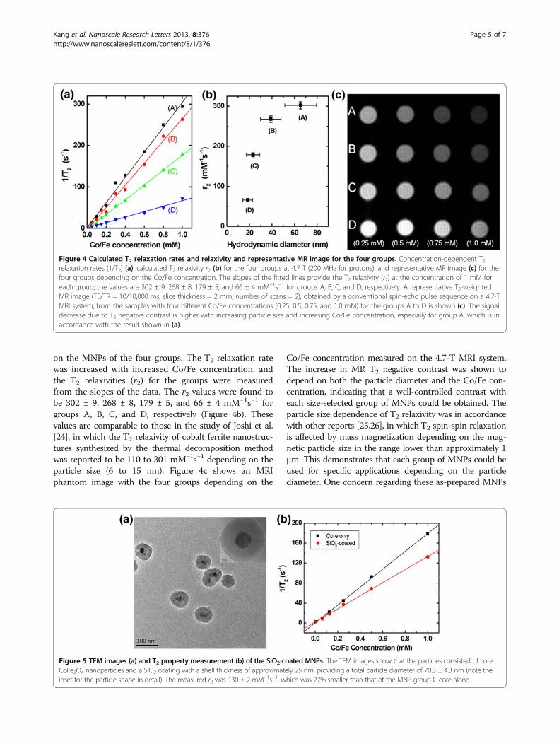
Figure 5 TEM images (a) and T2 property measurement (b) of the SiO2-coated MNPs. The TEM images show that the particles consisted of coreCoFe2O4 nanoparticles and a SiO2 coating with a shell thickness of approximately 25 nm, providing a total particle diameter of 70.8 ± 4.3 nm (note theinset for the particle shape in detail). The measured r2 was 130 ± 2 mM−1s−1, which was 27% smaller than that of the MNP group C core alone.
Kang et al. Nanoscale Research Letters 2013, 8:376 Page 5 of 7http://www.nanoscalereslett.com/content/8/1/376

is that they are not stable to variations in pH. This is aproblem that needs to be overcome if they are to besuccessfully employed in vivo. We therefore investigatedthe coating of the MNPs with a stable and biocompatiblematerial such as SiO2 to enhance stability and avoid poten-tial toxic effects on cells (Figure 5) [19]. The T2 relaxivityof the SiO2-coated MNPs made from group C was 130 ± 2mM−1s−1 (Figure 5b), which was approximately 27% lowerthan that of the original core particles. Group C wasselected for SiO2 coating in order to get final SiO2-coatedSPIO MNPs with a diameter of 50 to 100 nm and witha moderate T2 relaxivity value. The SiO2 coating wouldfacilitate the addition of therapeutic and targeting func-tions such as drugs and antibodies to the MNPs, enablingthem to serve as both imaging agents and a therapeuticcarrier species.There have been several reports on Fe3O4-based MNPs
with a narrow size distribution made by the coprecipitationmethod. Lee et al. used a piezoelectric nozzle [20], which,despite effectively controlling the particle size, requiresspecialized equipment and many steps. Jiang et al.employed a coprecipitation methodology using urea, whichprovided SPIO MNPs with a narrow size distribution [27].The average diameter of these MNPs could be adjustedfrom 8 to 50 nm depending on the decomposition of ureain the ferrite solution; however, they required additionaldextran coating in order to make them water soluble.In the present study, the use of centrifugation in combin-ation with the coprecipitation method enabled effectiveregulation of the size of the MNPs without the require-ment for a specialist. A large quantity of each size of parti-cles could be produced, overcoming many of theshortcomings of the coprecipitation method.
ConclusionsA simple centrifugation technique was combined with acoprecipitation method in aqueous solution in order toobtain four groups of CoFe2O4 MNPs. These were success-fully produced in large quantities, with different diametersand MRI T2 relaxivity values and narrow size distributions,depending on the centrifugation speed. The obtainedMNPs had a strong size-dependent MRI T2 contrast withT2 relaxivities between 302 and 66 mM−1s−1, providing aselection of particles from which the most appropriate for aspecific application could be chosen. In the present study,the particles of group C were selected for additional SiO2
coating. This was to demonstrate the potential of theseMNPs to be used for in vivo applications where they wouldrequire a long blood half-life, in addition to biocompatibi-lity. Each of the groups of CoFe2O4 MNPs could be used asthe initial base cores of MRI T2 contrast agents, withalmost unique T2 relaxivity due to the size regulation. Thisopens up many possibilities for biosensing applications anddisease diagnosis.
AbbreviationsCAs: Contrast agents; DLS: Dynamic light scattering; FWHM: Full-width athalf-maximum; MNP: Magnetic nanoparticle; MRI: Magnetic resonanceimaging; SPIO: Superparamagnetic iron oxide; TEM: Transmission electronmicroscopy; XRD: X-ray diffraction.
Competing interestsThe authors declare that they have no competing interests.
Authors’ contributionsJK, YTL, and KSH designed the experiments. JK, HL, AY, and Y-NK performedthe experiments. JK, Y-NK, and HJ analyzed the data. JK, HL, AY, and HJ madethe figures. JK and KSH wrote the manuscript. All authors read and approvedthe final manuscript.
AcknowledgementsThis work was supported by grants from the Korean Ministry of Education,Science and Technology (2011–0029263); the Korea Health Technology R&DProject, Ministry of Health and Welfare (A111499); and the CAP (PBC066)funded by the Korea Research Council of Fundamental Science andTechnology (KRCF).
Received: 23 June 2013 Accepted: 23 August 2013Published: 3 September 2013
References1. Judenhofer MS, Wehrl HF, Newport DF, Catana C, Siegel SB, Becker M,
Thielscher A, Kneilling M, Lichy MP, Eichner M, Klingel K, Reischl G, Widmaier S,Rocken M, Nutt RE, Machulla HJ, Uludag K, Cherry SR, Claussen CD, Pichler BJ:Simultaneous PET-MRI: a new approach for functional and morphologicalimaging. Nat Med 2008, 14:459–465.
2. Lu AH, Salabas EL, Schuth F: Magnetic nanoparticles: synthesis,protection, functionalization, and application. Angew Chem Int EdEngl 2007, 46:1222–1244.
3. Tanaka K, Narita A, Kitamura N, Uchiyama W, Morita M, Inubushi T, Chujo Y:Preparation for highly sensitive MRI contrast agents using core/shelltype nanoparticles consisting of multiple SPIO cores with thin silicacoating. Langmuir 2010, 26:11759–11762.
4. Artan Y, Haider MA, Langer DL, van der Kwast TH, Evans AJ, Yang Y,Wernick MN, Trachtenberg J, Yetik IS: Prostate cancer localization withmultispectral MRI using cost-sensitive support vector machines andconditional random fields. IEEE Trans Image Process 2010, 19:2444–2455.
5. Bennewitz MF, Lobo TL, Nkansah MK, Ulas G, Brudvig GW, Shapiro EM:Biocompatible and pH-sensitive PLGA encapsulated MnO nanocrystalsfor molecular and cellular MRI. ACS Nano 2011, 5:3438–3446.
6. Chertok B, Moffat BA, David AE, Yu F, Bergemann C, Ross BD, Yang VC: Ironoxide nanoparticles as a drug delivery vehicle for MRI monitoredmagnetic targeting of brain tumors. Biomaterials 2008, 29:487–496.
7. Branca RT, Cleveland ZI, Fubara B, Kumar CS, Maronpot RR, Leuschner C,Warren WS, Driehuys B: Molecular MRI for sensitive and specific detectionof lung metastases. Proc Natl Acad Sci USA 2010, 107:3693–3697.
8. Sonvico F, Mornet S, Vasseur S, Dubernet C, Jaillard D, Degrouard J,Hoebeke J, Duguet E, Colombo P, Couvreur P: Folate-conjugated iron oxidenanoparticles for solid tumor targeting as potential specific magnetichyperthermia mediators: synthesis, physicochemical characterization, andin vitro experiments. Bioconjug Chem 2005, 16:1181–1188.
9. Tromsdorf UI, Bigall NC, Kaul MG, Bruns OT, Nikolic MS, Mollwitz B, Sperling RA,Reimer R, Hohenberg H, Parak WJ, Forster S, Beisiegel U, Adam G, Weller H: Sizeand surface effects on the MRI relaxivity of manganese ferrite nanoparticlecontrast agents. Nano Lett 2007, 7:2422–2427.
10. Thorek DL, Tsourkas A: Size, charge and concentration dependent uptakeof iron oxide particles by non-phagocytic cells. Biomaterials 2008,29:3583–3590.
11. Matuszewski L, Persigehl T, Wall A, Schwindt W, Tombach B, Fobker M,Poremba C, Ebert W, Heindel W, Bremer C: Cell tagging with clinicallyapproved iron oxides: feasibility and effect of lipofection, particle size,and surface coating on labeling efficiency. Radiology 2005, 235:155–161.
12. Chouly C, Pouliquen D, Lucet I, Jeune JJ, Jallet P: Development ofsuperparamagnetic nanoparticles for MRI: effect of particle size, chargeand surface nature on biodistribution. J Microencapsul 1996, 13:245–255.
Kang et al. Nanoscale Research Letters 2013, 8:376 Page 6 of 7http://www.nanoscalereslett.com/content/8/1/376

13. Yu WW, Falkner JC, Yavuz CT, Colvin VL: Synthesis of monodisperse ironoxide nanocrystals by thermal decomposition of iron carboxylate salts.Chem Commun 2004, 0:2306–2307.
14. Hasegawa KR, Saw T: Particle-size distribution of CoFe2O4 formed by thecoprecipitation method. J Appl Phys 1967, 38:4707–4712.
15. Jana NR, Chen Y, Peng X: Size- and shape-controlled magnetic (Cr, Mn,Fe, Co, Ni) oxide nanocrystals via a simple and general approach.Chem Mater 2004, 16:3931–3935.
16. Lee HY, Lee SH, Xu C, Xie J, Lee JH, Wu B, Koh AL, Wang X, Sinclair R, Wang SX,Nishimura DG, Biswal S, Sun S, Cho SH, Chen X: Synthesis and characterizationof PVP-coated large core iron oxide nanoparticles as an MRI contrast agent.Nanotechnology 2008, 19:165101–165106.
17. Mulder WJ, Strijkers GJ, van Tilborg GA, Griffioen AW, Nicolay K: Lipid-basednanoparticles for contrast-enhanced MRI and molecular imaging.NMR Biomed 2006, 19:142–164.
18. Laurent S, Forge D, Port M, Roch A, Robic C, van der Elst L, Muller RN:Magnetic iron oxide nanoparticles: synthesis, stabilization, vectorization,physicochemical characterizations, and biological applications. Chem Rev2008, 108:2064–2110.
19. Yoon TJ, Yu KN, Kim E, Kim JS, Kim BG, Yun SH, Sohn BH, Cho MH, Lee JK,Park SB: Specific targeting, cell sorting, and bioimaging with smartmagnetic silica core-shell nanomaterials. Small 2006, 2:209–215.
20. Lee SJ, Jeong JR, Shin SC, Kim JC, Kim JD: Synthesis and characterizationof superparamagnetic maghemite nanoparticles prepared bycoprecipitation technique. J Magn Magn Mater 2004, 282:147–150.
21. Kim YI, Kim D, Lee CS: Synthesis and characterization of CoFe2O4magnetic nanoparticles prepared by temperature-controlledcoprecipitation method. Physica B 2003, 337:42–53.
22. Ibrahim MM, Zhao J, Seehra MS: Determination of particle size distributionin an Fe2O3-based catalyst using magnetometry and X-ray diffraction.J Mater Res 1992, 7:1856–1860.
23. Crosa M, Boero V, Angela MF: Determination of mean crystallite dimensionsfrom X-ray diffraction peak profiles: a comparative analysis of synthetichematites. Clays Clay Miner 1999, 47:742–747.
24. Joshi HM, Lin YP, Aslam M, Prasad PV, Schultz-Sikma EA, Edelman R,Meade T, Dravid VP: Effects of shape and size of cobalt ferritenanostructures on their MRI contrast and thermal activation. J PhysChem C 2009, 113:17761–17767.
25. Jun Y, Huh YM, Choi J, Lee JH, Song HT, Kim S, Yoon S, Kim KS, Shin JS, Suh JS,Cheon J: Nanoscale size effect of magnetic nanocrystals and their utilizationfor cancer diagnosis via magnetic resonance imaging. J Am Chem Soc 2005,127:5732–5733.
26. Shapiro EM, Skrtic S, Sharer K, Hill JM, Dunbar CE, Koretsky AP: MRI detectionof single particles for cellular imaging. Proc Natl Acad Sci USA 2004,101:10901–10906.
27. Jiang W, Yang HC, Yang SY, Horng HE, Hung JC, Chen YC, Hong CY:Preparation and properties of superparamagnetic nanoparticles withnarrow size distribution and biocompatible. J Magn Magn Mater 2004,283:210–214.
doi:10.1186/1556-276X-8-376Cite this article as: Kang et al.: Size-regulated group separation ofCoFe2O4 nanoparticles using centrifuge and their magnetic resonancecontrast properties. Nanoscale Research Letters 2013 8:376.
Submit your manuscript to a journal and benefi t from:
7 Convenient online submission
7 Rigorous peer review
7 Immediate publication on acceptance
7 Open access: articles freely available online
7 High visibility within the fi eld
7 Retaining the copyright to your article
Submit your next manuscript at 7 springeropen.com
Kang et al. Nanoscale Research Letters 2013, 8:376 Page 7 of 7http://www.nanoscalereslett.com/content/8/1/376