Embed Size (px)
Citation preview
Skeletal Morphogenesis of the Vertebral Columnof the Miniature Hylid Frog Acris crepitans,With Comments on Anomalies
L. Analıa Pugener* and Anne M. Maglia
Department of Biological Sciences, Missouri University of Science and Technology, Rolla, Missouri 65409
ABSTRACT Although the vertebral columns of anu-rans have received much study in the last 150 years,few detailed descriptions exist of the skeletal morpho-genesis of this anatomical unit. Herein, the ontogeny ofthe vertebral skeleton of the hylid frog Acris crepitansis described based on cleared and double-stained speci-mens, radiographs, and 3D reconstructions generatedfrom synchrotron microCT scans. The adult axial for-mula is 1-7-1-1, and the vertebral centra are epichordaland procoelous. The neural arches are nonimbricate,and there is a medial articulation between the laminaeof Presacrals I and II. Free ribs are absent. The sacraldiapophyses are uniform in width or slightly expandeddistally. The urostyle is slender, round in cross section,and about equal in length to the presacral region. Pre-sacral vertebrae are the first to form, developing in acephalic-to-caudal sequence. However, development andgrowth are decoupled and growth is fastest initially inthe posterior presacrals and sacrum. In addition, thereis a time lag between the formation of the presacral/sac-ral region and the postsacral region. More than 8.5% ofthe specimens examined have vertebral anomalies, andabout 50% display small variants from the typical verte-bral column morphology. However, these malformationsdo not seem to have been so severe as to have affectedsurvival. J. Morphol. 270:52–69, 2009. � 2008 Wiley-Liss, Inc.
KEY WORDS: Anura; Hylidae; Acris; vertebral column;skeleton; morphogenesis; anomalies
The anuran vertebral column has attracted theinterest of morphologists for more than 150 years.One of the most important contributions to ourunderstanding of the anuran vertebral column isthat of Nicholls (1916). His views on the structureof the column (particularly on the morphogenesisof the vertebral centra), and its significance as abasis for classification, generated great interestand stimulated research for decades (e.g., Noble,1922, 1931; Mookerjee, 1931; Mookerjee and Das,1939; Ritland, 1955; Griffiths, 1963; Kluge andFarris, 1969; Trueb, 1973). Other aspects of thevertebral column have also been investigated. Forexample, in leptodactylid frogs Lynch (1969, 1971)defined three types of cervical cotyles, which havebeen applied extensively to other anurans (e.g.,Lynch, 1973; Trueb, 1973; Sanchiz, 1984; Canna-tella, 1985; Clarke, 1988; Baez and Basso, 1996).
Noble (1931), Tihen (1960), Lynch (1973), Canna-tella (1985), and Cannatella and Trueb (1988a),among others, discussed the fusion of the atlasand second vertebra. The length, orientation, andshape of the transverse processes, and the pres-ence of free ribs in larvae and/or adults have alsoreceived wide attention (e.g., Zweifel, 1956; Klugeand Farris, 1969; Lynch, 1971; Cannatella andTrueb, 1988a,b; Clarke, 1988; Duellman andTrueb, 1994; Baez and Basso, 1996; da Silva, 1998;Maglia, 1998; Blanco and Sanchiz, 2000; Pramuk,2002, 2006; Fabrezi, 2006). In addition, the fusionbetween the sacrum and urostyle has beenregarded as a diagnostic feature of some anurans(Nicholls, 1916; Noble, 1922; Tihen, 1960; Trueb,1971; Estes and Reig, 1973; Lynch, 1973; Canna-tella, 1985; Cannatella and Trueb, 1988a,b; Baezand Trueb, 1997; Baez and Pugener, 2003; Frostet al., 2006).
A survey of the literature reveals that deform-ities among natural frog populations have beendocumented since the late 1800s. Interestingly,early reports were concerned almost exclusivelywith the vertebral column. Among the first contri-butions are publications by Howes (1886, 1893)and Adolphi (1892, 1895), describing vertebralfusions in pelobatids, bufonids, and ranids. Taylor(1942), Schiromany (1950), Tihen (1959), Holman(1963), Lynch (1965), Madej (1965), and Sanchizand Perez (1974) described anuran sacral fusionsand other vertebral column malformations in avariety of taxa. A more extensive article is that ofTrueb (1977), who described intrapopulation varia-tions in the osteology of Hyla lanciformis. Herfindings suggest that, unlike the skull and limbs,
Contract grant sponsor: NSF; Contract grant number: DBI-0445752; Contract grant sponsor: MDC Wildlife Collectors; Contractgrant numbers: 11903, 13515.
*Correspondence to: L. Analıa Pugener, Department of BiologicalSciences, Missouri University of Science and Technology, 105Schrenk Hall, 400 West 11th Street, Rolla, MO 65409-1120.E-mail: [email protected] or [email protected]
Published online 22 October 2008 inWiley InterScience (www.interscience.wiley.com)DOI: 10.1002/jmor.10665
JOURNAL OF MORPHOLOGY 270:52–69 (2009)
� 2008 WILEY-LISS, INC.
the vertebral column seems to be a highly variablearchitectural unit. Moreover, Trueb (1977) recom-mended that morphological descriptions and dis-cussions of variability and anomalies should beassessed in light of ontogenetic development.
Acris crepitans, commonly known as NorthernCricket Frogs, are small anurans residing instreams, rivers, and wetlands of the eastern halfof the United States and northeastern Mexico(Duellman, 2001; McCallum and Trauth, 2003;Gray et al., 2005a). Despite being members of thetree frog family Hylidae, A. crepitans are not arbo-real, but rather, are semiaquatic. These frogs havereceived much attention owing to reports of popu-lation declines and malformations (e.g., Greenwellet al., 1996; Brodman and Kilmurry, 1998; Heme-sath, 1998; Moriarty, 1998; Hammerson and Livo,1999; Gray, 2000; Lipps, 2000; Johnson et al.,2001; McCallum and Trauth, 2003; Gray andBrown, 2005; Gray et al., 2005b; and Irwin, 2005,to name a few). Yet, only recently has the adultskeletal anatomy of A. crepitans been thoroughlystudied (Maglia et al., 2007). Maglia et al. (2007)reported that these miniature anurans exhibit sev-eral novel morphologies and a large number ofosteological abnormalities, several of which occurin the vertebral column. The skeletal and larvaldevelopment of A. crepitans has yet to be investi-gated. Herein, we describe the skeletal ontogeny ofthe vertebral column of A. crepitans to understandfurther the malformations described by Magliaet al. (2007). We compare the normal vertebral de-velopment to that of other frogs (and in particularhylids), and we discuss malformations observed inthe context of vertebral anomalies known fromother species.
MATERIALS AND METHODS
We examined the vertebral columns of 49 premetamorphicand 80 postmetamorphic A. crepitans [Dumeril and Bibron(1841) (Table 1)]. Specimens from 14 other hylid species werealso examined (Table 2). A developmental series of 38 specimensrepresenting Gosner (1960) Stages 32 through adult werestaged, measured, eviscerated, and cleared and double-stainedfor cartilage and bone following methods adapted from Taylorand van Dyke (1985). Additional postmetamorphic and adultspecimens were examined using X-rays. The radiographs weretaken at The University of Kansas Natural History Museumand Biodiversity Research Center using a Picker Hot Shot TFI805D radiographic system operating at 30 kV, with exposuretimes of 90 s.Measurements of snout–vent length (SVL) were taken using
electronic digital calipers, accurate to 0.03 mm, prior to clearingand staining or image processing. Angle of orientation of thetransverse processes and sacral diapophyses were measured fol-lowing the methods of Trueb (1977). When referring to trans-verse processes/diapophyses, we consider the length to be themedial–lateral extension and the width to be the anterior–pos-terior extension. Specimens were considered to be adults if theywere 20 mm in SVL (the size at sexual maturity reported byHulse et al., 2001) or larger. Cleared and stained specimensand radiographs were examined with the aid of an Olympus
SZX12 stereo microscope equipped with a camera lucida and a5-megapixel digital camera.
A three-dimensional reconstruction of the vertebral column ofan adult A. crepitans was generated using data obtained viasynchrotron microCT at the beamline 2-BM of the AdvancedPhoton Source of the Argonne National Laboratory with the fol-lowing settings: energy 5 13.3 kiloelectron volts (keV); lens 51.253 objective; and time 5 470 ms. (For a detailed explanationof the 2-BM fast microtomography system, please see de Carloet al., 2006.) A series of 900 slice images was used to generatethe reconstruction with the aid of the ImageJ and 3D Doctor�software packages. An interactive version of the 3D image isavailable for examination in the MorphologyNet web-basedlibrary of anatomical reconstructions (www.morphologynet.org;Leopold et al., 2005), under number MN 008. Specimens usedin this study are deposited in the herpetological collections ofthe Natural History Museum and Biodiversity Research Centerat The University of Kansas (KU). Thirteen specimens (KU303230-42) were examined previously in the postmetamorphicosteology study of Maglia et al. (2007).
Vertebrae are designated via Roman numerals in the orderthey first appear in ontogeny, starting with the first morphologi-cal discrete vertebra visible, Presacral I, or the atlas. VertebraIX is generally referred to as the sacrum. Postsacral verte-brae—when distinguishable as separate elements prior to fus-ing to form the urostyle—are designated in an anterior-to-poste-rior sequence using Arabic numerals. The neural arches of allpresacral vertebrae, except the atlas, and the sacrum bear lat-erally oriented processes known as diapophyses, or transverseprocesses. Here, the term transverse process is used in refer-ence to the processes of the presacral vertebrae, and the termdiapophysis is restricted to the processes of the sacrum.
The term coccyx has been used extensively as a synonym ofurostyle (e.g., Ritland, 1955; Lynch, 1973; Trueb, 1973; Canna-tella, 1985; Cannatella and Trueb, 1988a,b; Clarke, 1988;Wiens, 1989; Duellman and Trueb, 1994; Baez and Basso, 1996;Wild, 1997; Zug et al., 2001; Frost et al., 2006). Coccyx isdefined as a small, bony element at the base of the vertebralcolumn, consisting of several fused, rudimentary caudal verte-brae (Pugener, 2002). A coccyx is present in the tail-less apes(Kent, 1987) and in anurans (Pugener, 2002). A comparable os-seous structure called pygostyle also occurs in birds (Baumel,1979). The coccyx of anurans fuses synostotically to an unseg-mented ventral structure, the hypochord, to form the urostyle(Mookerjee, 1931; Griffiths, 1963; Branham and List, 1979;Maglia and Pugener, 1998; Pugener, 2002; Rockova and Rocek,2005). Therefore, the coccyx and urostyle are only partially ho-mologous structures, and the use of these terms as synonymsshould be avoided.
RESULTS
The notochord and vertebral column are themain longitudinal structural elements of the bodythat provide support for the head and viscera. Thenotochord is a rod of fibrous connective tissue sur-rounding a core of fluid-filled cells that lies dorsalto the digestive system and directly ventral to thespinal cord. The notochord is present during earlydevelopment in all anurans, but gives way to thevertebral column after metamorphosis.
The vertebral column is a metameric, semiflexi-ble, arched bar located in the dorsal part of thetrunk, and is formed by a series of bony vertebrae.The vertebral column provides suspension for theappendicular skeleton and protection for the spinalnerve cord. In anurans, the vertebral column is di-vided into three regions, namely, presacral, sacral,
A. CREPITANS AXIAL DEVELOPMENT 53
Journal of Morphology
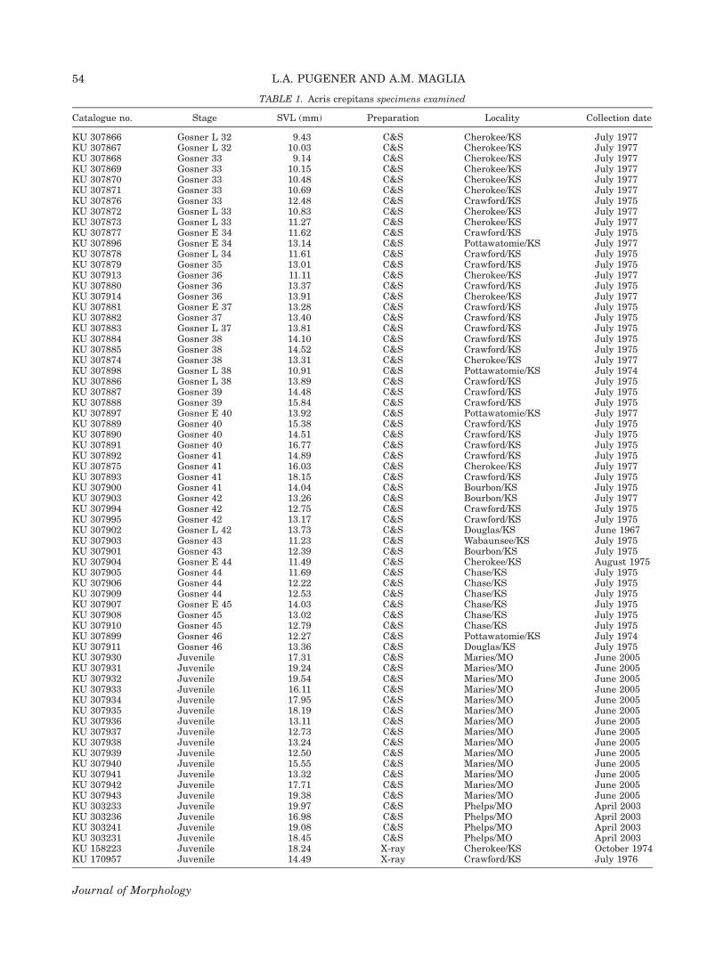
TABLE 1. Acris crepitans specimens examined
Catalogue no. Stage SVL (mm) Preparation Locality Collection date
KU 307866 Gosner L 32 9.43 C&S Cherokee/KS July 1977KU 307867 Gosner L 32 10.03 C&S Cherokee/KS July 1977KU 307868 Gosner 33 9.14 C&S Cherokee/KS July 1977KU 307869 Gosner 33 10.15 C&S Cherokee/KS July 1977KU 307870 Gosner 33 10.48 C&S Cherokee/KS July 1977KU 307871 Gosner 33 10.69 C&S Cherokee/KS July 1977KU 307876 Gosner 33 12.48 C&S Crawford/KS July 1975KU 307872 Gosner L 33 10.83 C&S Cherokee/KS July 1977KU 307873 Gosner L 33 11.27 C&S Cherokee/KS July 1977KU 307877 Gosner E 34 11.62 C&S Crawford/KS July 1975KU 307896 Gosner E 34 13.14 C&S Pottawatomie/KS July 1977KU 307878 Gosner L 34 11.61 C&S Crawford/KS July 1975KU 307879 Gosner 35 13.01 C&S Crawford/KS July 1975KU 307913 Gosner 36 11.11 C&S Cherokee/KS July 1977KU 307880 Gosner 36 13.37 C&S Crawford/KS July 1975KU 307914 Gosner 36 13.91 C&S Cherokee/KS July 1977KU 307881 Gosner E 37 13.28 C&S Crawford/KS July 1975KU 307882 Gosner 37 13.40 C&S Crawford/KS July 1975KU 307883 Gosner L 37 13.81 C&S Crawford/KS July 1975KU 307884 Gosner 38 14.10 C&S Crawford/KS July 1975KU 307885 Gosner 38 14.52 C&S Crawford/KS July 1975KU 307874 Gosner 38 13.31 C&S Cherokee/KS July 1977KU 307898 Gosner L 38 10.91 C&S Pottawatomie/KS July 1974KU 307886 Gosner L 38 13.89 C&S Crawford/KS July 1975KU 307887 Gosner 39 14.48 C&S Crawford/KS July 1975KU 307888 Gosner 39 15.84 C&S Crawford/KS July 1975KU 307897 Gosner E 40 13.92 C&S Pottawatomie/KS July 1977KU 307889 Gosner 40 15.38 C&S Crawford/KS July 1975KU 307890 Gosner 40 14.51 C&S Crawford/KS July 1975KU 307891 Gosner 40 16.77 C&S Crawford/KS July 1975KU 307892 Gosner 41 14.89 C&S Crawford/KS July 1975KU 307875 Gosner 41 16.03 C&S Cherokee/KS July 1977KU 307893 Gosner 41 18.15 C&S Crawford/KS July 1975KU 307900 Gosner 41 14.04 C&S Bourbon/KS July 1975KU 307903 Gosner 42 13.26 C&S Bourbon/KS July 1977KU 307994 Gosner 42 12.75 C&S Crawford/KS July 1975KU 307995 Gosner 42 13.17 C&S Crawford/KS July 1975KU 307902 Gosner L 42 13.73 C&S Douglas/KS June 1967KU 307903 Gosner 43 11.23 C&S Wabaunsee/KS July 1975KU 307901 Gosner 43 12.39 C&S Bourbon/KS July 1975KU 307904 Gosner E 44 11.49 C&S Cherokee/KS August 1975KU 307905 Gosner 44 11.69 C&S Chase/KS July 1975KU 307906 Gosner 44 12.22 C&S Chase/KS July 1975KU 307909 Gosner 44 12.53 C&S Chase/KS July 1975KU 307907 Gosner E 45 14.03 C&S Chase/KS July 1975KU 307908 Gosner 45 13.02 C&S Chase/KS July 1975KU 307910 Gosner 45 12.79 C&S Chase/KS July 1975KU 307899 Gosner 46 12.27 C&S Pottawatomie/KS July 1974KU 307911 Gosner 46 13.36 C&S Douglas/KS July 1975KU 307930 Juvenile 17.31 C&S Maries/MO June 2005KU 307931 Juvenile 19.24 C&S Maries/MO June 2005KU 307932 Juvenile 19.54 C&S Maries/MO June 2005KU 307933 Juvenile 16.11 C&S Maries/MO June 2005KU 307934 Juvenile 17.95 C&S Maries/MO June 2005KU 307935 Juvenile 18.19 C&S Maries/MO June 2005KU 307936 Juvenile 13.11 C&S Maries/MO June 2005KU 307937 Juvenile 12.73 C&S Maries/MO June 2005KU 307938 Juvenile 13.24 C&S Maries/MO June 2005KU 307939 Juvenile 12.50 C&S Maries/MO June 2005KU 307940 Juvenile 15.55 C&S Maries/MO June 2005KU 307941 Juvenile 13.32 C&S Maries/MO June 2005KU 307942 Juvenile 17.71 C&S Maries/MO June 2005KU 307943 Juvenile 19.38 C&S Maries/MO June 2005KU 303233 Juvenile 19.97 C&S Phelps/MO April 2003KU 303236 Juvenile 16.98 C&S Phelps/MO April 2003KU 303241 Juvenile 19.08 C&S Phelps/MO April 2003KU 303231 Juvenile 18.45 C&S Phelps/MO April 2003KU 158223 Juvenile 18.24 X-ray Cherokee/KS October 1974KU 170957 Juvenile 14.49 X-ray Crawford/KS July 1976
54 L.A. PUGENER AND A.M. MAGLIA
Journal of Morphology
and postsacral. In A. crepitans, the presacralregion consists of eight discrete vertebrae; the sac-ral region is composed of a single vertebra, andthe postsacral region is formed by the urostyle.
A typical anuran presacral vertebra consists oftwo essential parts, a dorsal neural arch and aventral centrum (see Fig. 1). The neural arch isformed by a pair of pedicels—which form first dur-
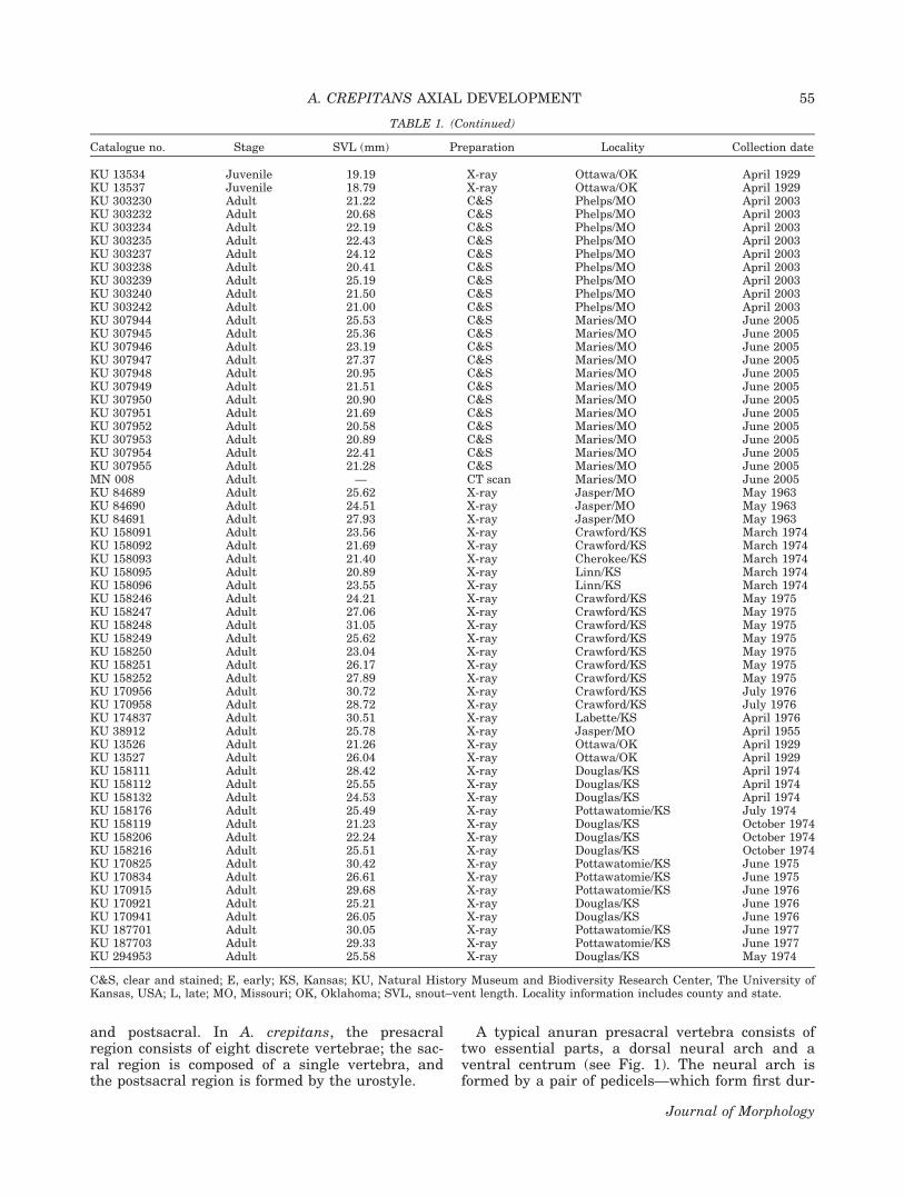
TABLE 1. (Continued)
Catalogue no. Stage SVL (mm) Preparation Locality Collection date
KU 13534 Juvenile 19.19 X-ray Ottawa/OK April 1929KU 13537 Juvenile 18.79 X-ray Ottawa/OK April 1929KU 303230 Adult 21.22 C&S Phelps/MO April 2003KU 303232 Adult 20.68 C&S Phelps/MO April 2003KU 303234 Adult 22.19 C&S Phelps/MO April 2003KU 303235 Adult 22.43 C&S Phelps/MO April 2003KU 303237 Adult 24.12 C&S Phelps/MO April 2003KU 303238 Adult 20.41 C&S Phelps/MO April 2003KU 303239 Adult 25.19 C&S Phelps/MO April 2003KU 303240 Adult 21.50 C&S Phelps/MO April 2003KU 303242 Adult 21.00 C&S Phelps/MO April 2003KU 307944 Adult 25.53 C&S Maries/MO June 2005KU 307945 Adult 25.36 C&S Maries/MO June 2005KU 307946 Adult 23.19 C&S Maries/MO June 2005KU 307947 Adult 27.37 C&S Maries/MO June 2005KU 307948 Adult 20.95 C&S Maries/MO June 2005KU 307949 Adult 21.51 C&S Maries/MO June 2005KU 307950 Adult 20.90 C&S Maries/MO June 2005KU 307951 Adult 21.69 C&S Maries/MO June 2005KU 307952 Adult 20.58 C&S Maries/MO June 2005KU 307953 Adult 20.89 C&S Maries/MO June 2005KU 307954 Adult 22.41 C&S Maries/MO June 2005KU 307955 Adult 21.28 C&S Maries/MO June 2005MN 008 Adult — CT scan Maries/MO June 2005KU 84689 Adult 25.62 X-ray Jasper/MO May 1963KU 84690 Adult 24.51 X-ray Jasper/MO May 1963KU 84691 Adult 27.93 X-ray Jasper/MO May 1963KU 158091 Adult 23.56 X-ray Crawford/KS March 1974KU 158092 Adult 21.69 X-ray Crawford/KS March 1974KU 158093 Adult 21.40 X-ray Cherokee/KS March 1974KU 158095 Adult 20.89 X-ray Linn/KS March 1974KU 158096 Adult 23.55 X-ray Linn/KS March 1974KU 158246 Adult 24.21 X-ray Crawford/KS May 1975KU 158247 Adult 27.06 X-ray Crawford/KS May 1975KU 158248 Adult 31.05 X-ray Crawford/KS May 1975KU 158249 Adult 25.62 X-ray Crawford/KS May 1975KU 158250 Adult 23.04 X-ray Crawford/KS May 1975KU 158251 Adult 26.17 X-ray Crawford/KS May 1975KU 158252 Adult 27.89 X-ray Crawford/KS May 1975KU 170956 Adult 30.72 X-ray Crawford/KS July 1976KU 170958 Adult 28.72 X-ray Crawford/KS July 1976KU 174837 Adult 30.51 X-ray Labette/KS April 1976KU 38912 Adult 25.78 X-ray Jasper/MO April 1955KU 13526 Adult 21.26 X-ray Ottawa/OK April 1929KU 13527 Adult 26.04 X-ray Ottawa/OK April 1929KU 158111 Adult 28.42 X-ray Douglas/KS April 1974KU 158112 Adult 25.55 X-ray Douglas/KS April 1974KU 158132 Adult 24.53 X-ray Douglas/KS April 1974KU 158176 Adult 25.49 X-ray Pottawatomie/KS July 1974KU 158119 Adult 21.23 X-ray Douglas/KS October 1974KU 158206 Adult 22.24 X-ray Douglas/KS October 1974KU 158216 Adult 25.51 X-ray Douglas/KS October 1974KU 170825 Adult 30.42 X-ray Pottawatomie/KS June 1975KU 170834 Adult 26.61 X-ray Pottawatomie/KS June 1975KU 170915 Adult 29.68 X-ray Pottawatomie/KS June 1976KU 170921 Adult 25.21 X-ray Douglas/KS June 1976KU 170941 Adult 26.05 X-ray Douglas/KS June 1976KU 187701 Adult 30.05 X-ray Pottawatomie/KS June 1977KU 187703 Adult 29.33 X-ray Pottawatomie/KS June 1977KU 294953 Adult 25.58 X-ray Douglas/KS May 1974
C&S, clear and stained; E, early; KS, Kansas; KU, Natural History Museum and Biodiversity Research Center, The University ofKansas, USA; L, late; MO, Missouri; OK, Oklahoma; SVL, snout–vent length. Locality information includes county and state.
A. CREPITANS AXIAL DEVELOPMENT 55
Journal of Morphology
ing development—and a pair of laminae. The ante-rior and posterior ends of each neural arch pedicelare indented to form anterior and posterior inter-vertebral notches; thus, when two vertebrae artic-ulate, the anterior notch of one vertebra and theposterior notch of the other create a bilateral inter-vertebral space. These spaces accommodate thespinal nerves as they emerge from the spinalcanal. The neural arch laminae cover the spinalcord dorsally, thereby enclosing the neural canal.The lateral margin of each lamina rests dorsal tothe superior end of the pedicel, and the medial endfuses to the medial end of the opposite lamina.Usually, in the area of contact of both laminae, aneural spine develops.
Each vertebra possesses six apophyses, fourzygapophyses and two transverse processes. Thezygapophyses consist of paired processes at thecephalic (5prezygapophyses) and caudal(5postzygapophyses) ends of the neural arch. Thearticular facets of the prezygapophyses of one ver-tebra face dorsomedially and articulate with thelateroventrally facing facets of the postzygapophy-ses of the preceding vertebra. Zygapophyses func-tion as interlocking structures between adjacentvertebrae, and limit dorsoventral flexion and lat-eral movement of the column in the trunk region.Transverse processes project on each side from thepoint where the lamina joins the pedicel. The pro-cesses of Presacrals II–IV are longer than those ofthe last four presacral vertebrae, and provide sur-faces for the attachment of the muscles that origi-nate mainly from the head, the scapula, and thesuprascapula. The transverse processes of Presac-rals V–VIII are subequal in length and servemainly for the attachment of muscles from thecaudal region of the vertebral column.
The vertebral bodies, or centra, functionallyreplace the notochord. In A. crepitans, the verte-bral centra are procoelous and epichordal. Procoe-lous defines a centrum that is concave at the ante-rior end. A procoelous centrum bears a condyle onthe posterior end for the articulation with the pos-terior adjacent vertebral centrum, forming a ball-and-socket joint that allows extensive motion inmost directions. Epichordal defines a centrumthat originates from ossification of the dorsal por-tion of the perichordal tube, as opposed to ossifica-tion of the entire perichordal tube (5perichordalcondition).
The atlas and sacral vertebra are similar, for themost part, to a typical presacral vertebra. The
Fig. 1. Schematic representation of Presacral Vertebrae Iand II of anurans, lateral view. Gray denotes cartilage; whitedenotes bone.
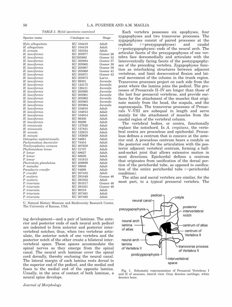
TABLE 2. Hylid specimens examined
Species name Catalogue no. Stage
Hyla alboguttata KU 104418 AdultH. alboguttata KU 104419 AdultH. armata KU 163334 AdultH. lanciformis KU 205977 Gosner 26H. lanciformis KU205982 Gosner 33H. lanciformis KU 205984 Gosner 37H. lanciformis KU 205965 Gosner 38H. lanciformis KU 205967 Gosner 39H. lanciformis KU 205969 Gosner 41H. lanciformis KU 205971 Gosner 42H. lanciformis KU 205973 LarvaH. lanciformis KU 99301 JuvenileH. lanciformis KU 124175 JuvenileH. lanciformis KU 126411 JuvenileH. lanciformis KU 205960 JuvenileH. lanciformis KU 205961 JuvenileH. lanciformis KU 205962 JuvenileH. lanciformis KU 205963 JuvenileH. lanciformis KU 205964 JuvenileH. lanciformis KU 104910 AdultH. lanciformis KU 104913 AdultH. lanciformis KU 104914 AdultH. lanciformis KU 99300 AdultH. lanciformis KU 99299 AdultH. minuscula KU 127820 AdultH. minuscula KU 127821 AdultH. minuta KU 122915 AdultH. minuta KU 182928 AdultOsteopilus septentrionalis KU 84661 AdultPachymedusa dacnicolor KU 78454 AdultTrachycephalus coriacea KU 207658 AdultPhyllomedusa lemur KU 31707 AdultP. lemur KU 68627 AdultP. lemur KU 68628 AdultP. lemur KU 101815 AdultPlectrohyla glandulosa KU 209699 AdultP. matudai KU 59948 AdultPseudacris crucifer KU 84668 AdultP. crucifer KU 207433 AdultP. ocularis KU 291049 Gosner 46P. ocularis KU 291052 AdultP. triseriata KU 201017 Gosner 46P. triseriata KU 291021 Gosner 46P. triseriata KU 98318 AdultP. triseriata KU 207491 AdultP. triseriata KU 207492 Adult
U, Natural History Museum and Biodiversity Research Center,The University of Kansas, USA.
56 L.A. PUGENER AND A.M. MAGLIA
Journal of Morphology
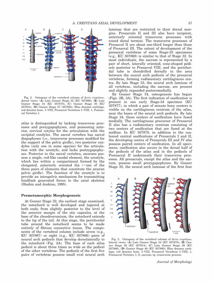
atlas is distinguished by lacking transverse proc-esses and prezygapophyses, and possessing ante-rior, cervical cotyles for the articulation with theoccipital condyles. The sacral vertebra has sacraldiapophyses (i.e., transverse processes modified forthe support of the pelvic girdle), two posterior con-dyles (only one in some species) for the articula-tion with the urostyle, and lacks postzygapophy-ses. Posterior to the sacral vertebra, anurans pos-sess a single, rod-like caudal element, the urostyle,which lies within a compartment formed by theelongated, anteriorly oriented ilia (5one of thethree pairs of elements that constitute the anuranpelvic girdle). The function of the urostyle is toprovide an intrapelvic mechanism for transmittinghindlimb generated forces to the axial skeleton(Shubin and Jenkins, 1995).
Premetamorphic Morphogenesis
At Gosner Stage 32, the earliest stage examined,the notochord is well developed and tapered atboth ends; from slightly posterior to the level ofthe anterior margin of the otic capsules, at thebase of the chondrocranium, the notochord extendsto the tip of the tail. At this stage, the perichordaltube around the notochord seems to be madeentirely of fibrous connective tissue. The compo-nents of the vertebral column include seven (e.g.,KU 307867) or eight (e.g., KU 307866) pairs ofneural arch pedicels that develop dorsolaterally tothe notochord (Fig. 2A). The base of each atlaspedicel is about three times as wide as the pedicelof the other vertebrae. The pedicels of the first fivepairs of vertebrae possess small oval neural arch
laminae that are restricted to their dorsal mar-gins. Presacrals II and III also have incipient,anteriorly oriented transverse processes withround distal termini. The transverse processes ofPresacral II are about one-third longer than thoseof Presacral III. The extent of development of thepresacral vertebrae of some Stage-33 specimens(e.g., KU 307868) is similar to that of Stage 32. Inmost individuals, the sacrum is represented by apair of short, laterally oriented, cone-shaped pedi-cels posterior to Presacral VIII, and the perichor-dal tube is chondrified dorsally in the areabetween the neural arch pedicels of the presacralvertebrae, forming rudimentary cartilaginous cen-tra. By late Stage 33, the neural arch laminae ofall vertebrae, including the sacrum, are presentand slightly expanded posteromedially.
By Gosner Stage 34, osteogenesis has begun(Figs. 2B, 3A). The first indication of ossification ispresent in one early Stage-34 specimen (KU307877), in which a pair of minute bony centers isvisible on the cartilaginous centrum of the atlas,near the bases of the neural arch pedicels. By lateStage 34, these centers of ossification have fusedmedially. The cartilaginous precursor of PresacralII also has a rudimentary centrum consisting oftwo centers of ossification that are fused at themidline. In KU 307878, in addition to the coa-lesced central ossifications of Presacrals I and II,the developing centra of Presacrals III and IV alsopossess paired centers of ossification. In all speci-mens, ossification also occurs in the dorsal half ofthe pedicels of the atlas and in the pedicels ofPostsacral II underneath their transverse proc-esses. All presacrals, except the atlas and the sac-rum, possess small prezygapophyses. By GosnerStage 35, the neural arch laminae of the first four
Fig. 2. Ontogeny of the vertebral column of Acris crepitans,dorsal views. (A) Late Gosner Stage 32 (KU 307866), (B) LateGosner Stage 34 (KU 307878), (C) Gosner Stage 36 (KU307914), (D) Gosner Stage 37 (307882). Blue denotes cartilage;red denotes bone. I–VIII, Presacral Vertebrae I–VIII; 1, Postsac-ral Vertebra 1.
Fig. 3. Ontogeny of the vertebral column of Acris crepitans,lateral views. (A) Late Gosner Stage 34 (KU 307878), (B) Gos-ner Stage 36 (KU 307914), (C) Late Gosner Stage 38 (KU307886), (D) Gosner Stage 40 (KU 307890). Blue denotes carti-lage; red denotes bone. I–VIII, Presacral Vertebrae I–VIII; 1,Postsacral Vertebra 1; S, sacrum; tp, transverse process.
A. CREPITANS AXIAL DEVELOPMENT 57
Journal of Morphology
presacrals are expanded medially; growth of thelaminae is more significant in Presacrals II–IVthan in the atlas. The transverse processes of Pre-sacral II remain about one-third longer than thoseof Presacral III.
Stage 36 is characterized by the development ofPostsacral 1 and the hypochord, and by a signifi-cant increase in the amount of ossification of theneural arches (Figs. 2C, 3B). Postsacral 1 is pres-ent in all Stage-36 specimens examined, andemerges as small, paired neural arch pedicels pos-terior to the sacrum. The hypochord is presentonly in some of the specimens examined (e.g., KU307914), and is first seen as a thin sliver of carti-lage about one-third longer than the length of theatlantal centrum. The hypochord originates paral-lel to, and beneath, the notochord, although not indirect contact with it. The anterior end of thehypochord lies at the level of the neural arches ofPostsacral 1. The neural arches of all presacralvertebrae and the sacrum are ossified. Ossificationextends from the upper portion of each neuralarch pedicel to the lateral aspect of the lamina,being more widespread in the atlas and less exten-sive in the sacrum. Each atlantal neural arch pedi-cel has a thin, but well-developed, cartilaginouscervical cotyle that occupies slightly less than halfof the anterior margin of the pedicel. PresacralsI–VI bear small postzygapophyses, although theystill do not articulate with the prezygapophyses.The transverse processes of Presacrals II and IIIare about equal in length, and their proximal por-tions are ossified. In addition, in some specimens(e.g., KU 307880, KU 307913) Presacrals IV–VIhave small transverse processes, whereas in otherspecimens (e.g., KU 186096) the transverse proc-esses are present in all presacral vertebrae. In allcases, the newly developed transverse processesare knob-like in dorsal view and dorsoventrallyelongate in lateral view; the transverse processesof Presacral IV are slightly more developed thanthose of the posterior vertebrae. The anterior andposterior margins of the cartilaginous vertebralcentra are clearly concave and convex, respec-tively. The degree of ossification of the centravaries among specimens; in some, such as in KU307913, the centra are ossified in all presacral ver-tebrae and the sacrum, whereas in others (e.g.,KU 307914) only the centra of the first three pre-sacral vertebrae are ossified.
By Stage 37, the neural arch laminae of Presac-ral IV-sacrum fuse at the midline (Fig. 2D). Thetransverse processes of Presacral III have length-ened, and are about one-third longer than thetransverse processes of Presacral II. The trans-verse processes of Presacral IV also are longer—slightly shorter than that of the transverse proc-esses of Presacral II—and their proximal ends areossified. The transverse processes of Presacrals V–VIII remain small and knob-like, and ossification
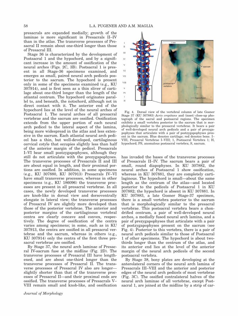
has invaded the bases of the transverse processesof Presacrals II–IV. The sacrum bears a pair ofsmall, round diapophyses. In KU 307882, theneural arches of Postsacral 1 show ossification,whereas in KU 307881, they are completely carti-laginous. The hypochord is small—about the samelength as the centrum of the atlas—and locatedposterior to the pedicels of Postsacral 1 in KU307882; the hypochord is absent in KU 307881. InKU 307883, a late Gosner Stage-37 specimen,there is a small vertebra posterior to the sacrumthat is morphologically similar to the presacralvertebrae. This postsacral vertebra bears a chon-drified centrum, a pair of well-developed neuralarches, a medially fused neural arch lamina, and apair of prezygapophyses that articulate with a pairof postzygapophyses present on the sacrum (seeFig. 4). Posterior to this vertebra, there is a pair ofneural arch pedicels similar to those of Postsacral1 of other specimens. The hypochord is about two-thirds longer than the centrum of the atlas, andits anterior end lies at the level of the anteriormargin of the neural arch pedicels of the secondpostsacral vertebra.
By Stage 38, bony plates are developing at theanterolateral corners of the neural arch lamina ofPresacrals III–VIII and the anterior and posterioredges of the neural arch pedicels of most vertebrae(Fig. 3C). The ossified contralateral halves of theneural arch laminae of all vertebrae, except Post-sacral 1, are joined at the midline by a strip of car-
Fig. 4. Dorsal view of the vertebral column of late GosnerStage 37 (KU 307883) Acris crepitans and (inset) close-up pho-tograph of the sacral and postsacral regions. The specimenexhibits a small vertebra posterior to the sacrum that is mor-phologically similar to the presacral vertebrae. It bears a pairof well-developed neural arch pedicels and a pair of prezyga-pophyses that articulate with a pair of postzygapophyses pres-ent in the sacrum. Blue denotes cartilage; red denotes bone. I–VIII, Presacral Vertebrae I–VIII; 1, Postsacral Vertebra 1; H,hypochord; PS, anomalous postsacral vertebra; S, sacrum.
58 L.A. PUGENER AND A.M. MAGLIA
Journal of Morphology
tilage. The prezygapophyses and postzygapophysesare cartilaginous, but articulate with each other,thereby providing more stability to the developingvertebral column. The distal ends of the transverseprocesses of Presacrals II and III are slightlyexpanded distally. In some Stage-38 and in allStage-39 specimens, the hypochord has begun toossify at the midline. The right and left counter-parts of Postsacral 1 remain dorsally and ventrallyseparated, but each side has an anterior cartilagi-nous cotyle. In one late Gosner Stage-38 specimen(KU 307886), the central portion of the neuralarch lamina of Presacral IV has failed to form.
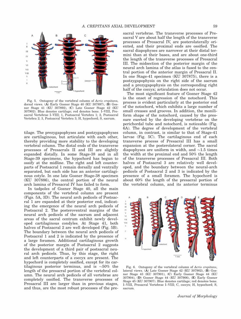
In tadpoles of Gosner Stage 40, all the maincomponents of the vertebral column are present(Figs. 5A, 3D). The neural arch pedicels of Postsac-ral 1 are expanded at their posterior end, indicat-ing the emergence of the neural arch pedicels ofPostsacral 2. The posteroventral margins of theneural arch pedicels of the sacrum and adjacentareas of the sacral centrum exhibit newly devel-oped cartilaginous condyles. At Stage 41, bothhalves of Postsacral 2 are well developed (Fig. 5B).The boundary between the neural arch pedicels ofPostsacral 1 and 2 is indicated by the presence ofa large foramen. Additional cartilaginous growthof the posterior margin of Postsacral 2 suggeststhe development of a third pair of postsacral neu-ral arch pedicels. Thus, by this stage, the rightand left counterparts of a coccyx are present. Thehypochord is completely ossified, except for its car-tilaginous posterior terminus, and is �50% thelength of the presacral portion of the vertebral col-umn. The neural arch pedicels of all vertebrae arecompletely ossified. The transverse processes ofPresacral III are larger than in previous stages,and thus, are the most robust processes of the pre-
sacral vertebrae. The transverse processes of Pre-sacral V are about half the length of the transverseprocesses of Presacral IV, are posterolaterally ori-ented, and their proximal ends are ossified. Thesacral diapophyses are narrower at their distal ter-mini than at their bases, and are about one-thirdthe length of the transverse processes of PresacralIII. The midsection of the posterior margin of theneural arch lamina of the atlas is fused to the cen-tral portion of the anterior margin of Presacral II.In one Stage-41 specimen (KU 307875), there is apostzygapophysis on the right side of the sacrumand a prezygapophysis on the corresponding righthalf of the coccyx; articulation does not occur.
The most significant feature of Gosner Stage 42is the onset of regression of the notochord. Thisprocess is evident particularly at the posterior endof the notochord, which exhibits a large number ofsmall creases and grooves. In addition, the monili-form shape of the notochord, caused by the pres-sure exerted by the developing vertebrae on theperichordal tube and notochord, is noticeable (Fig.6A). The degree of development of the vertebralcolumn, in contrast, is similar to that of Stage-41larvae (Fig. 5C). The cartilaginous end of eachtransverse process of Presacral III has a smallexpansion at the posterolateral corner. The sacraldiapophyses are uniform in width, and �1.5 timesthe width at the proximal end and 50% the lengthof the transverse processes of Presacral III. Bothhalves of Postsacral 3 are relatively well devel-oped, and the boundary between the neural-archpedicels of Postsacral 2 and 3 is indicated by thepresence of a small foramen. The hypochord isabout 60% the length of the presacral portion ofthe vertebral column, and its anterior terminus
Fig. 6. Ontogeny of the vertebral column of Acris crepitans,lateral views. (A) Late Gosner Stage 42 (KU 307902), (B) Gos-ner Stage 43 (KU 307901), (C) Early Gosner Stage 44 (KU307904), (D) Gosner Stage 44 (KU 307906), (E) Early GosnerStage 45 (KU 307907). Blue denotes cartilage; red denotes bone.I–VIII, Presacral Vertebrae I–VIII; C, coccyx; H, hypochord; S,sacrum.
Fig. 5. Ontogeny of the vertebral column of Acris crepitans,dorsal views. (A) Early Gosner Stage 40 (KU 307897), (B) Gos-ner Stage 41 (KU 307892), (C) Late Gosner Stage 42 (KU307902). Blue denotes cartilage; red denotes bone. I–VIII, Pre-sacral Vertebrae I–VIII; 1, Postsacral Vertebra 1; 2, PostsacralVertebra 2; 3, Postsacral Vertebra 3; H, hypochord; S, sacrum.
A. CREPITANS AXIAL DEVELOPMENT 59
Journal of Morphology
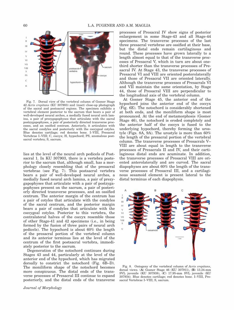
lies at the level of the neural arch pedicels of Post-sacral 1. In KU 307903, there is a vertebra poste-rior to the sacrum that, although small, has a mor-phology closely resembling that of the presacralvertebrae (see Fig. 7). This postsacral vertebrabears a pair of well-developed neural arches, amedially fused neural arch lamina, a pair of prezy-gapophyses that articulate with a pair of postzyga-pophyses present on the sacrum, a pair of posteri-orly directed transverse processes, and an ossifiedcentrum. The anterior margin of the centrum hasa pair of cotyles that articulate with the condylesof the sacral centrum, and the posterior marginbears a pair of condyles that articulate with thecoccygeal cotyles. Posterior to this vertebra, thecontralateral halves of the coccyx resemble thoseof other Stage-41 and 42 specimens (i.e., in beingformed by the fusion of three pairs of neural archpedicels). The hypochord is about 60% the lengthof the presacral portion of the vertebral columnand its anterior terminus lies at the level of thecentrum of the first postsacral vertebra, immedi-ately posterior to the sacrum.
Degeneration of the notochord continues duringStages 43 and 44, particularly at the level of theanterior end of the hypochord, which has migrateddorsally to constrict the notochord (Fig. 6B–D).The moniliform shape of the notochord becomesmore conspicuous. The distal ends of the trans-verse processes of Presacral III continue to expandposteriorly, and the distal ends of the transverse
processes of Presacral IV show signs of posteriorenlargement in some Stage-43 and all Stage-44specimens. The transverse processes of the lastthree presacral vertebrae are ossified at their base,but the distal ends remain cartilaginous andround. These processes have grown laterally to alength almost equal to that of the transverse proc-esses of Presacral V, which in turn are about one-third shorter than the transverse processes of Pre-sacral IV. At Stage 43, the transverse processes ofPresacral VI and VIII are oriented posterolaterallyand those of Presacral VII are oriented laterally.Although the transverse processes of Presacrals VIand VII maintain the same orientation, by Stage44, those of Presacral VIII are perpendicular tothe longitudinal axis of the vertebral column.
At Gosner Stage 45, the anterior end of thehypochord joins the anterior end of the coccyx(Fig. 6E). The notochord is considerably shortenedat both ends, and the moniliform shape is mostpronounced. At the end of metamorphosis (GosnerStage 46), the notochord is eroded completely andthe anterior half of the coccyx is fused to theunderlying hypochord, thereby forming the uros-tyle (Figs. 8A, 9A). The urostyle is more than 60%the length of the presacral portion of the vertebralcolumn. The transverse processes of Presacrals V–VIII are about equal in length to the transverseprocesses of Presacrals II and IV, and their carti-laginous distal ends are acuminate. In addition,the transverse processes of Presacral VIII are ori-ented anterolaterally and are curved. The sacraldiapophyses are about 60% the length of the trans-verse processes of Presacral III, and a cartilagi-nous sesamoid element is present lateral to thedistal terminus of each diapophysis.
Fig. 8. Ontogeny of the vertebral column of Acris crepitans,dorsal views. (A) Gosner Stage 46 (KU 307911), (B) 13.24-mmSVL juvenile (KU 307938), (C) 17.95-mm SVL juvenile (KU307934). Blue denotes cartilage; red denotes bone. I–VIII, Pre-sacral Vertebrae I–VIII; S, sacrum.
Fig. 7. Dorsal view of the vertebral column of Gosner Stage42 Acris crepitans (KU 307903) and (inset) close-up photographof the sacral and postsacral regions. The specimen exhibits avertebral element posterior to the sacrum that bears a pair ofwell-developed neural arches, a medially fused neural arch lam-ina, a pair of prezygapophyses that articulate with the sacralpostzygapophyses, a pair of posteriorly directed transverse proc-esses, and an ossified centrum. Anteriorly, it articulates withthe sacral condyles and posteriorly with the coccygeal cotyles.Blue denotes cartilage; red denotes bone. I–VIII, PresacralVertebrae I–VIII; C, coccyx; H, hypochord; PS, anomalous post-sacral vertebra; S, sacrum.
60 L.A. PUGENER AND A.M. MAGLIA
Journal of Morphology
Postmetamorphic Morphogenesis
In young postmetamorphic specimens of about13-mm SVL, the face or articular facet of eachprezygapophysis and postzygapophysis is cartilagi-nous, whereas the opposite side of the face orcounterface is ossified (Figs. 8B, 9B). By this stage,the distal ends of the transverse processes of thelast four presacral vertebrae have developed smalltear-shaped expansions. The area of contactbetween the neural arch pedicels and the vertebralcentra is cartilaginous. The coccyx is completelyfused to the hypochord, but a suture on either sideis clearly discernible. The combined neural archesof the postsacral vertebrae form a prominent, lon-gitudinal ridge on the anterodorsal aspect of theurostyle; the dorsal ends of the neural arches,
however, are not fused at the midline. A pair oflarge spinal nerve foramina pierces the lateralsides of the urostyle at the base of the longitudinalridge, close to the anterior margin of the urostyle.The second pair of spinal nerve foramina is no lon-ger present.
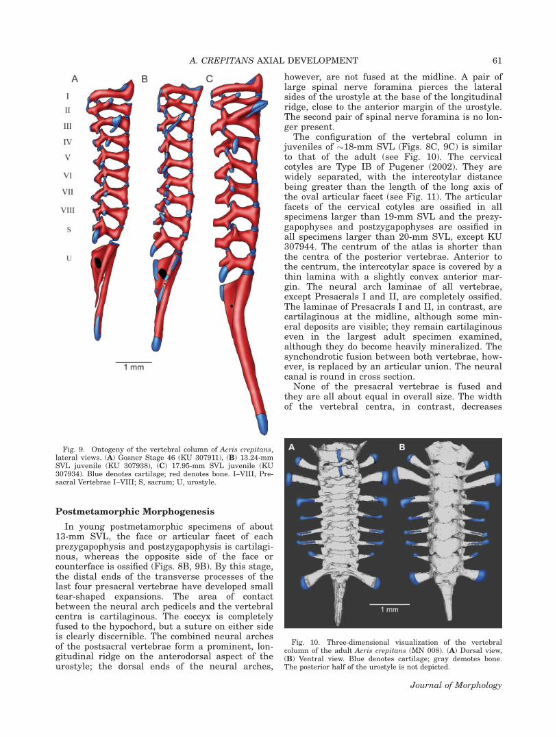
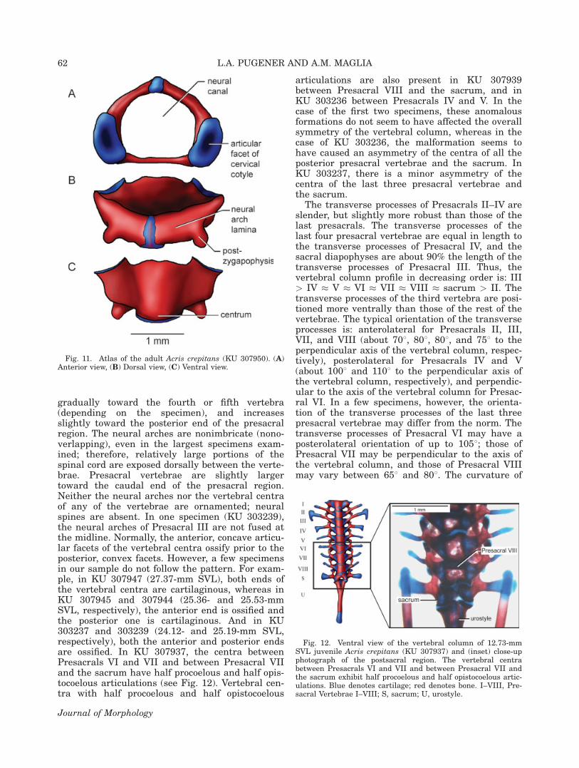
The configuration of the vertebral column injuveniles of �18-mm SVL (Figs. 8C, 9C) is similarto that of the adult (see Fig. 10). The cervicalcotyles are Type IB of Pugener (2002). They arewidely separated, with the intercotylar distancebeing greater than the length of the long axis ofthe oval articular facet (see Fig. 11). The articularfacets of the cervical cotyles are ossified in allspecimens larger than 19-mm SVL and the prezy-gapophyses and postzygapophyses are ossified inall specimens larger than 20-mm SVL, except KU307944. The centrum of the atlas is shorter thanthe centra of the posterior vertebrae. Anterior tothe centrum, the intercotylar space is covered by athin lamina with a slightly convex anterior mar-gin. The neural arch laminae of all vertebrae,except Presacrals I and II, are completely ossified.The laminae of Presacrals I and II, in contrast, arecartilaginous at the midline, although some min-eral deposits are visible; they remain cartilaginouseven in the largest adult specimen examined,although they do become heavily mineralized. Thesynchondrotic fusion between both vertebrae, how-ever, is replaced by an articular union. The neuralcanal is round in cross section.
None of the presacral vertebrae is fused andthey are all about equal in overall size. The widthof the vertebral centra, in contrast, decreases
Fig. 10. Three-dimensional visualization of the vertebralcolumn of the adult Acris crepitans (MN 008). (A) Dorsal view,(B) Ventral view. Blue denotes cartilage; gray demotes bone.The posterior half of the urostyle is not depicted.
Fig. 9. Ontogeny of the vertebral column of Acris crepitans,lateral views. (A) Gosner Stage 46 (KU 307911), (B) 13.24-mmSVL juvenile (KU 307938), (C) 17.95-mm SVL juvenile (KU307934). Blue denotes cartilage; red denotes bone. I–VIII, Pre-sacral Vertebrae I–VIII; S, sacrum; U, urostyle.
A. CREPITANS AXIAL DEVELOPMENT 61
Journal of Morphology
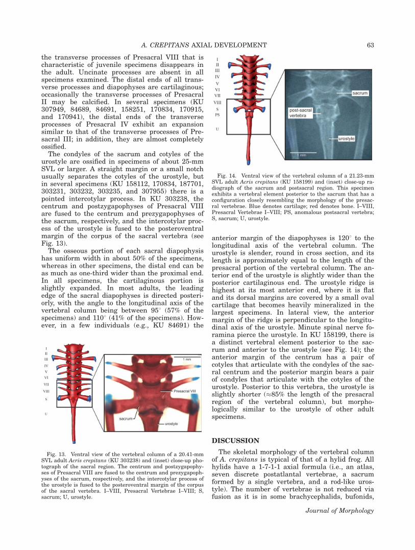
gradually toward the fourth or fifth vertebra(depending on the specimen), and increasesslightly toward the posterior end of the presacralregion. The neural arches are nonimbricate (nono-verlapping), even in the largest specimens exam-ined; therefore, relatively large portions of thespinal cord are exposed dorsally between the verte-brae. Presacral vertebrae are slightly largertoward the caudal end of the presacral region.Neither the neural arches nor the vertebral centraof any of the vertebrae are ornamented; neuralspines are absent. In one specimen (KU 303239),the neural arches of Presacral III are not fused atthe midline. Normally, the anterior, concave articu-lar facets of the vertebral centra ossify prior to theposterior, convex facets. However, a few specimensin our sample do not follow the pattern. For exam-ple, in KU 307947 (27.37-mm SVL), both ends ofthe vertebral centra are cartilaginous, whereas inKU 307945 and 307944 (25.36- and 25.53-mmSVL, respectively), the anterior end is ossified andthe posterior one is cartilaginous. And in KU303237 and 303239 (24.12- and 25.19-mm SVL,respectively), both the anterior and posterior endsare ossified. In KU 307937, the centra betweenPresacrals VI and VII and between Presacral VIIand the sacrum have half procoelous and half opis-tocoelous articulations (see Fig. 12). Vertebral cen-tra with half procoelous and half opistocoelous
articulations are also present in KU 307939between Presacral VIII and the sacrum, and inKU 303236 between Presacrals IV and V. In thecase of the first two specimens, these anomalousformations do not seem to have affected the overallsymmetry of the vertebral column, whereas in thecase of KU 303236, the malformation seems tohave caused an asymmetry of the centra of all theposterior presacral vertebrae and the sacrum. InKU 303237, there is a minor asymmetry of thecentra of the last three presacral vertebrae andthe sacrum.
The transverse processes of Presacrals II–IV areslender, but slightly more robust than those of thelast presacrals. The transverse processes of thelast four presacral vertebrae are equal in length tothe transverse processes of Presacral IV, and thesacral diapophyses are about 90% the length of thetransverse processes of Presacral III. Thus, thevertebral column profile in decreasing order is: III> IV � V � VI � VII � VIII � sacrum > II. Thetransverse processes of the third vertebra are posi-tioned more ventrally than those of the rest of thevertebrae. The typical orientation of the transverseprocesses is: anterolateral for Presacrals II, III,VII, and VIII (about 708, 808, 808, and 758 to theperpendicular axis of the vertebral column, respec-tively), posterolateral for Presacrals IV and V(about 1008 and 1108 to the perpendicular axis ofthe vertebral column, respectively), and perpendic-ular to the axis of the vertebral column for Presac-ral VI. In a few specimens, however, the orienta-tion of the transverse processes of the last threepresacral vertebrae may differ from the norm. Thetransverse processes of Presacral VI may have aposterolateral orientation of up to 1058; those ofPresacral VII may be perpendicular to the axis ofthe vertebral column, and those of Presacral VIIImay vary between 658 and 808. The curvature of
Fig. 12. Ventral view of the vertebral column of 12.73-mmSVL juvenile Acris crepitans (KU 307937) and (inset) close-upphotograph of the postsacral region. The vertebral centrabetween Presacrals VI and VII and between Presacral VII andthe sacrum exhibit half procoelous and half opistocoelous artic-ulations. Blue denotes cartilage; red denotes bone. I–VIII, Pre-sacral Vertebrae I–VIII; S, sacrum; U, urostyle.
Fig. 11. Atlas of the adult Acris crepitans (KU 307950). (A)Anterior view, (B) Dorsal view, (C) Ventral view.
62 L.A. PUGENER AND A.M. MAGLIA
Journal of Morphology
the transverse processes of Presacral VIII that ischaracteristic of juvenile specimens disappears inthe adult. Uncinate processes are absent in allspecimens examined. The distal ends of all trans-verse processes and diapophyses are cartilaginous;occasionally the transverse processes of PresacralII may be calcified. In several specimens (KU307949, 84689, 84691, 158251, 170834, 170915,and 170941), the distal ends of the transverseprocesses of Presacral IV exhibit an expansionsimilar to that of the transverse processes of Pre-sacral III; in addition, they are almost completelyossified.
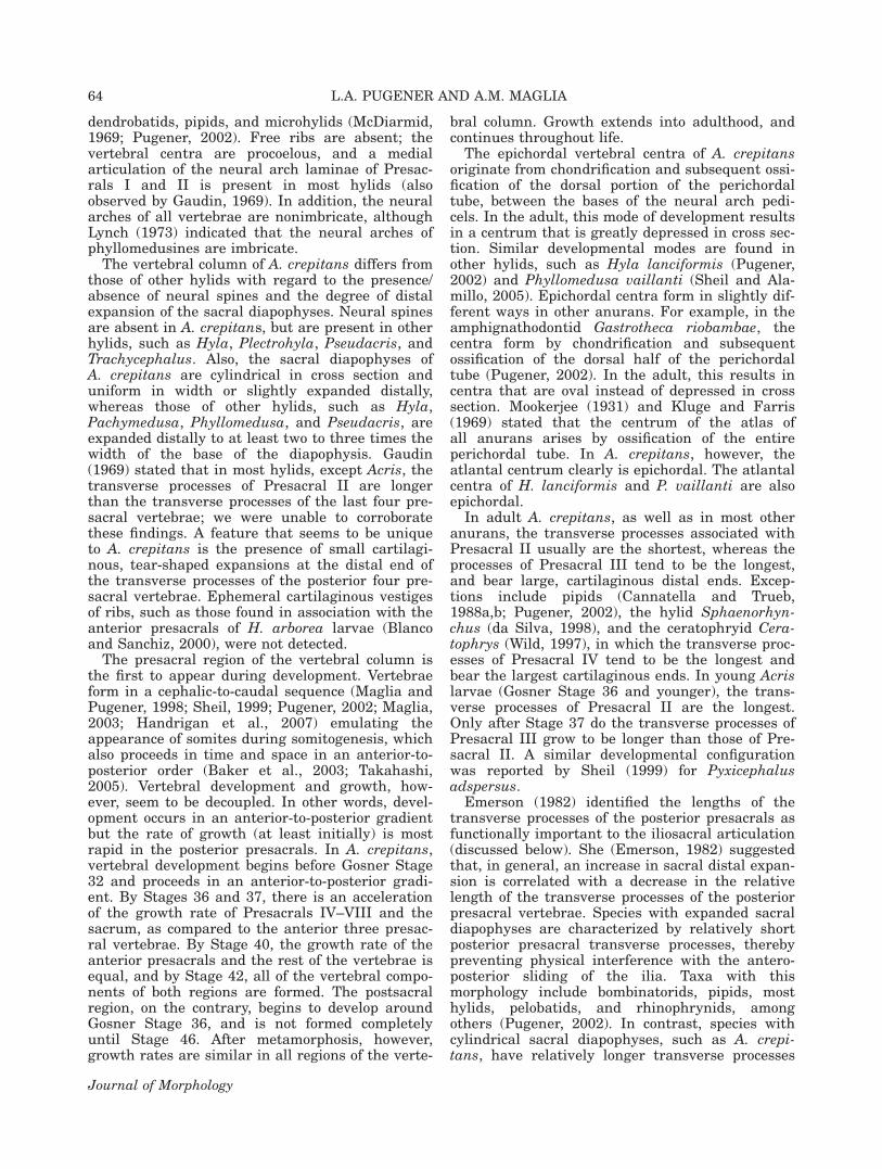
The condyles of the sacrum and cotyles of theurostyle are ossified in specimens of about 25-mmSVL or larger. A straight margin or a small notchusually separates the cotyles of the urostyle, butin several specimens (KU 158112, 170834, 187701,303231, 303232, 303235, and 307955) there is apointed intercotylar process. In KU 303238, thecentrum and postzygapophyses of Presacral VIIIare fused to the centrum and prezygapophyses ofthe sacrum, respectively, and the intercotylar proc-ess of the urostyle is fused to the posteroventralmargin of the corpus of the sacral vertebra (seeFig. 13).
The osseous portion of each sacral diapophysishas uniform width in about 50% of the specimens,whereas in other specimens, the distal end can beas much as one-third wider than the proximal end.In all specimens, the cartilaginous portion isslightly expanded. In most adults, the leadingedge of the sacral diapophyses is directed posteri-orly, with the angle to the longitudinal axis of thevertebral column being between 958 (57% of thespecimens) and 1108 (41% of the specimens). How-ever, in a few individuals (e.g., KU 84691) the
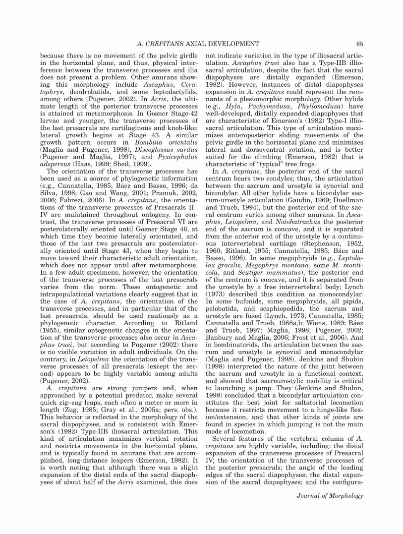
anterior margin of the diapophyses is 1208 to thelongitudinal axis of the vertebral column. Theurostyle is slender, round in cross section, and itslength is approximately equal to the length of thepresacral portion of the vertebral column. The an-terior end of the urostyle is slightly wider than theposterior cartilaginous end. The urostyle ridge ishighest at its most anterior end, where it is flatand its dorsal margins are covered by a small ovalcartilage that becomes heavily mineralized in thelargest specimens. In lateral view, the anteriormargin of the ridge is perpendicular to the longitu-dinal axis of the urostyle. Minute spinal nerve fo-ramina pierce the urostyle. In KU 158199, there isa distinct vertebral element posterior to the sac-rum and anterior to the urostyle (see Fig. 14); theanterior margin of the centrum has a pair ofcotyles that articulate with the condyles of the sac-ral centrum and the posterior margin bears a pairof condyles that articulate with the cotyles of theurostyle. Posterior to this vertebra, the urostyle isslightly shorter (�85% the length of the presacralregion of the vertebral column), but morpho-logically similar to the urostyle of other adultspecimens.
DISCUSSION
The skeletal morphology of the vertebral columnof A. crepitans is typical of that of a hylid frog. Allhylids have a 1-7-1-1 axial formula (i.e., an atlas,seven discrete postatlantal vertebrae, a sacrumformed by a single vertebra, and a rod-like uros-tyle). The number of vertebrae is not reduced viafusion as it is in some brachycephalids, bufonids,
Fig. 13. Ventral view of the vertebral column of a 20.41-mmSVL adult Acris crepitans (KU 303238) and (inset) close-up pho-tograph of the sacral region. The centrum and postzygapophy-ses of Presacral VIII are fused to the centrum and prezygapoph-yses of the sacrum, respectively, and the intercotylar process ofthe urostyle is fused to the posteroventral margin of the corpusof the sacral vertebra. I–VIII, Presacral Vertebrae I–VIII; S,sacrum; U, urostyle.
Fig. 14. Ventral view of the vertebral column of a 21.23-mmSVL adult Acris crepitans (KU 158199) and (inset) close-up ra-diograph of the sacrum and postsacral region. This specimenexhibits a vertebral element posterior to the sacrum that has aconfiguration closely resembling the morphology of the presac-ral vertebrae. Blue denotes cartilage; red denotes bone. I–VIII,Presacral Vertebrae I–VIII; PS, anomalous postsacral vertebra;S, sacrum; U, urostyle.
A. CREPITANS AXIAL DEVELOPMENT 63
Journal of Morphology
dendrobatids, pipids, and microhylids (McDiarmid,1969; Pugener, 2002). Free ribs are absent; thevertebral centra are procoelous, and a medialarticulation of the neural arch laminae of Presac-rals I and II is present in most hylids (alsoobserved by Gaudin, 1969). In addition, the neuralarches of all vertebrae are nonimbricate, althoughLynch (1973) indicated that the neural arches ofphyllomedusines are imbricate.
The vertebral column of A. crepitans differs fromthose of other hylids with regard to the presence/absence of neural spines and the degree of distalexpansion of the sacral diapophyses. Neural spinesare absent in A. crepitans, but are present in otherhylids, such as Hyla, Plectrohyla, Pseudacris, andTrachycephalus. Also, the sacral diapophyses ofA. crepitans are cylindrical in cross section anduniform in width or slightly expanded distally,whereas those of other hylids, such as Hyla,Pachymedusa, Phyllomedusa, and Pseudacris, areexpanded distally to at least two to three times thewidth of the base of the diapophysis. Gaudin(1969) stated that in most hylids, except Acris, thetransverse processes of Presacral II are longerthan the transverse processes of the last four pre-sacral vertebrae; we were unable to corroboratethese findings. A feature that seems to be uniqueto A. crepitans is the presence of small cartilagi-nous, tear-shaped expansions at the distal end ofthe transverse processes of the posterior four pre-sacral vertebrae. Ephemeral cartilaginous vestigesof ribs, such as those found in association with theanterior presacrals of H. arborea larvae (Blancoand Sanchiz, 2000), were not detected.
The presacral region of the vertebral column isthe first to appear during development. Vertebraeform in a cephalic-to-caudal sequence (Maglia andPugener, 1998; Sheil, 1999; Pugener, 2002; Maglia,2003; Handrigan et al., 2007) emulating theappearance of somites during somitogenesis, whichalso proceeds in time and space in an anterior-to-posterior order (Baker et al., 2003; Takahashi,2005). Vertebral development and growth, how-ever, seem to be decoupled. In other words, devel-opment occurs in an anterior-to-posterior gradientbut the rate of growth (at least initially) is mostrapid in the posterior presacrals. In A. crepitans,vertebral development begins before Gosner Stage32 and proceeds in an anterior-to-posterior gradi-ent. By Stages 36 and 37, there is an accelerationof the growth rate of Presacrals IV–VIII and thesacrum, as compared to the anterior three presac-ral vertebrae. By Stage 40, the growth rate of theanterior presacrals and the rest of the vertebrae isequal, and by Stage 42, all of the vertebral compo-nents of both regions are formed. The postsacralregion, on the contrary, begins to develop aroundGosner Stage 36, and is not formed completelyuntil Stage 46. After metamorphosis, however,growth rates are similar in all regions of the verte-
bral column. Growth extends into adulthood, andcontinues throughout life.
The epichordal vertebral centra of A. crepitansoriginate from chondrification and subsequent ossi-fication of the dorsal portion of the perichordaltube, between the bases of the neural arch pedi-cels. In the adult, this mode of development resultsin a centrum that is greatly depressed in cross sec-tion. Similar developmental modes are found inother hylids, such as Hyla lanciformis (Pugener,2002) and Phyllomedusa vaillanti (Sheil and Ala-millo, 2005). Epichordal centra form in slightly dif-ferent ways in other anurans. For example, in theamphignathodontid Gastrotheca riobambae, thecentra form by chondrification and subsequentossification of the dorsal half of the perichordaltube (Pugener, 2002). In the adult, this results incentra that are oval instead of depressed in crosssection. Mookerjee (1931) and Kluge and Farris(1969) stated that the centrum of the atlas ofall anurans arises by ossification of the entireperichordal tube. In A. crepitans, however, theatlantal centrum clearly is epichordal. The atlantalcentra of H. lanciformis and P. vaillanti are alsoepichordal.
In adult A. crepitans, as well as in most otheranurans, the transverse processes associated withPresacral II usually are the shortest, whereas theprocesses of Presacral III tend to be the longest,and bear large, cartilaginous distal ends. Excep-tions include pipids (Cannatella and Trueb,1988a,b; Pugener, 2002), the hylid Sphaenorhyn-chus (da Silva, 1998), and the ceratophryid Cera-tophrys (Wild, 1997), in which the transverse proc-esses of Presacral IV tend to be the longest andbear the largest cartilaginous ends. In young Acrislarvae (Gosner Stage 36 and younger), the trans-verse processes of Presacral II are the longest.Only after Stage 37 do the transverse processes ofPresacral III grow to be longer than those of Pre-sacral II. A similar developmental configurationwas reported by Sheil (1999) for Pyxicephalusadspersus.
Emerson (1982) identified the lengths of thetransverse processes of the posterior presacrals asfunctionally important to the iliosacral articulation(discussed below). She (Emerson, 1982) suggestedthat, in general, an increase in sacral distal expan-sion is correlated with a decrease in the relativelength of the transverse processes of the posteriorpresacral vertebrae. Species with expanded sacraldiapophyses are characterized by relatively shortposterior presacral transverse processes, therebypreventing physical interference with the antero-posterior sliding of the ilia. Taxa with thismorphology include bombinatorids, pipids, mosthylids, pelobatids, and rhinophrynids, amongothers (Pugener, 2002). In contrast, species withcylindrical sacral diapophyses, such as A. crepi-tans, have relatively longer transverse processes
64 L.A. PUGENER AND A.M. MAGLIA
Journal of Morphology
because there is no movement of the pelvic girdlein the horizontal plane, and thus, physical inter-ference between the transverse processes and iliadoes not present a problem. Other anurans show-ing this morphology include Ascaphus, Cera-tophrys, dendrobatids, and some leptodactylids,among others (Pugener, 2002). In Acris, the ulti-mate length of the posterior transverse processesis attained at metamorphosis. In Gosner Stage-42larvae and younger, the transverse processes ofthe last presacrals are cartilaginous and knob-like;lateral growth begins at Stage 43. A similargrowth pattern occurs in Bombina orientalis(Maglia and Pugener, 1998), Discoglossus sardus(Pugener and Maglia, 1997), and Pyxicephalusadspersus (Haas, 1999; Sheil, 1999).
The orientation of the transverse processes hasbeen used as a source of phylogenetic information(e.g., Cannatella, 1985; Baez and Basso, 1996; daSilva, 1998; Gao and Wang, 2001; Pramuk, 2002,2006; Fabrezi, 2006). In A. crepitans, the orienta-tions of the transverse processes of Presacrals II–IV are maintained throughout ontogeny. In con-trast, the transverse processes of Presacral VI areposterolaterally oriented until Gosner Stage 46, atwhich time they become laterally orientated, andthose of the last two presacrals are posterolater-ally oriented until Stage 43, when they begin tomove toward their characteristic adult orientation,which does not appear until after metamorphosis.In a few adult specimens, however, the orientationof the transverse processes of the last presacralsvaries from the norm. These ontogenetic andintrapopulational variations clearly suggest that inthe case of A. crepitans, the orientation of thetransverse processes, and in particular that of thelast presacrals, should be used cautiously as aphylogenetic character. According to Ritland(1955), similar ontogenetic changes in the orienta-tion of the transverse processes also occur in Asca-phus truei, but according to Pugener (2002) thereis no visible variation in adult individuals. On thecontrary, in Leiopelma the orientation of the trans-verse processes of all presacrals (except the sec-ond) appears to be highly variable among adults(Pugener, 2002).
A. crepitans are strong jumpers and, whenapproached by a potential predator, make severalquick zig–zag leaps, each often a meter or more inlength (Zug, 1985; Gray et al., 2005a; pers. obs.).This behavior is reflected in the morphology of thesacral diapophyses, and is consistent with Emer-son’s (1982) Type-IIB iliosacral articulation. Thiskind of articulation maximizes vertical rotationand restricts movements in the horizontal plane,and is typically found in anurans that are accom-plished, long-distance leapers (Emerson, 1982). Itis worth noting that although there was a slightexpansion of the distal ends of the sacral diapoph-yses of about half of the Acris examined, this does
not indicate variation in the type of iliosacral artic-ulation. Ascaphus truei also has a Type-IIB illio-sacral articulation, despite the fact that the sacraldiapophyses are distally expanded (Emerson,1982). However, instances of distal diapophysesexpansion in A. crepitans could represent the rem-nants of a plesiomorphic morphology. Other hylids(e.g., Hyla, Pachymedusa, Phyllomedusa) havewell-developed, distally expanded diapophyses thatare characteristic of Emerson’s (1982) Type-I illio-sacral articulation. This type of articulation maxi-mizes anteroposterior sliding movements of thepelvic girdle in the horizontal plane and minimizeslateral and dorsoventral rotation, and is bettersuited for the climbing (Emerson, 1982) that ischaracteristic of ‘‘typical’’ tree frogs.
In A. crepitans, the posterior end of the sacralcentrum bears two condyles; thus, the articulationbetween the sacrum and urostyle is synovial andbicondylar. All other hylids have a bicondylar sac-rum-urostyle articulation (Gaudin, 1969; Duellmanand Trueb, 1994), but the posterior end of the sac-ral centrum varies among other anurans. In Asca-phus, Leiopelma, and Notobatrachus the posteriorend of the sacrum is concave, and it is separatedfrom the anterior end of the urostyle by a continu-ous intervertebral cartilage (Stephenson, 1952,1960; Ritland, 1955; Cannatella, 1985; Baez andBasso, 1996). In some megophryids (e.g., Leptola-lax gracilis, Megophrys montana, some M. monti-cola, and Scutiger mammatus), the posterior endof the centrum is concave, and it is separated fromthe urostyle by a free intervertebral body; Lynch(1973) described this condition as monocondylar.In some bufonids, some megophryids, all pipids,pelobatids, and scaphiopodids, the sacrum andurostyle are fused (Lynch, 1973; Cannatella, 1985;Cannatella and Trueb, 1988a,b; Wiens, 1989; Baezand Trueb, 1997; Maglia, 1998; Pugener, 2002;Banbury and Maglia, 2006; Frost et al., 2006). Andin bombinatorids, the articulation between the sac-rum and urostyle is synovial and monocondylar(Maglia and Pugener, 1998). Jenkins and Shubin(1998) interpreted the nature of the joint betweenthe sacrum and urostyle in a functional context,and showed that sacrourostylic mobility is criticalto launching a jump. They (Jenkins and Shubin,1998) concluded that a bicondylar articulation con-stitutes the best joint for saltatorial locomotionbecause it restricts movement to a hinge-like flex-ion/extension, and that other kinds of joints arefound in species in which jumping is not the mainmode of locomotion.
Several features of the vertebral column of A.crepitans are highly variable, including: the distalexpansion of the transverse processes of PresacralIV; the orientation of the transverse processes ofthe posterior presacrals; the angle of the leadingedges of the sacral diapophyses; the distal expan-sion of the sacral diapophyses; and the configura-
A. CREPITANS AXIAL DEVELOPMENT 65
Journal of Morphology
tion of the intercotylar space of the urostyle. Inter-estingly, two of these structures that are highlyvariable in A. crepitans (i.e., the angle between theanterior edge of the sacral diapophyses and thelongitudinal axis of the vertebral column and theorientation of the transverse processes of PresacralVI) showed the least amount of variation inTrueb’s (1977) study of Hyla lanciformis. In addi-tion, 11 of the A. crepitans we examined havesome vertebral anomalies. Many genetic and envi-ronmental factors can affect somite formation(Baker et al., 2003), producing different defects invertebrae (Pourquie and Kusumi, 2001; Turnpennyet al., 2003; Kusumi and Turnpenny, 2007). Thenature and extent of the malformations depend onthe stage of somitogenesis in which the perturba-tion occurs (Rivard et al., 1979), and may consistof vertebral fusion or failure of vertebral forma-tion, among others (Pourquie and Kusumi, 2001;Dunwoodie et al., 2002; Kusumi and Turnpenny,2007). Three Acris specimens examined displaybarely noticeable anomalies, including neural archlaminae that did not develop incomplete midlineossification, and minor vertebral asymmetries. Theremaining eight specimens, on the other hand,show a host of more significant malformations, asdiscussed below.
Three A. crepitans specimens exhibit a malfor-mation that, to our knowledge, has not beenreported for any other anuran species. The malfor-mation consists of vertebral centra with half pro-coelous (the typical condition) and half opistocoe-lous articulations. Mookerjee (1931) and Griffiths(1963) proposed that the kind of articulation of thecentrum is determined by the nature of the inter-vertebral cartilage and its association to the cen-trum. In A. crepitans, however, there is no evi-dence of intervertebral cartilages, and the develop-ing centra present a procoelous morphology whenthey first form. This developmental pattern couldbe explained in one of two ways. Either the inter-vertebral cartilage fuses to the centrum early indevelopment or the intervertebral cartilage isabsent and the articular condyle is generated fromthe centrum. Considering our limited understand-ing of the development of the articular condyle ona normal vertebral centrum of A. crepitans, we arehesitant to suggest a scenario for the origin of thearticular anomalies.
In one adult A. crepitans, the centrum and post-zygapophyses of Presacral VIII are fused to thecentrum and prezygapophyses of the sacrum,respectively, and the intercotylar process of theurostyle is fused to the posteroventral margin ofthe corpus of the sacral vertebra. In some anurans(e.g., the bufonids Dendrophryniscus brevipollica-tus, Nectophryne afra, and Osornophryne guaca-mayo and all species of the pipid Hymenochirus),the last presacral vertebra, the sacrum, and theurostyle are fused to form a compound structure
called the synsacrum (Pugener, 2002). In matureindividuals of these species, sutures delineatingthe limits between the incorporated elements arenot visible. However, the occurrence of a synsa-crum usually can be recognized by the presenceof: 1) more than one pair of spinal nerve foraminapiercing the ventrolateral sides of the sacralstructure; 2) sacral diapophyses with broad proxi-mal ends (because they originate from two pairsof modified, subequal transverse processes); and 3)a pair of bony plates (5webbings) connectingthe posteromedial margins of the diapophyses withthe lateral margins of the anterior portion of theurostyle. The fusions observed in Acris clearly donot represent a synsacrum. Rather, they are likelythe consequence of hyperossification or a devel-opmental anomaly, and almost certainly had anadverse effect on midtrunk flexion and jumpingability. Similar anomalous fusions have been re-ported for a wide range of anurans, includingalytids (Sanchiz and Perez, 1974), bombinatorids(Madej, 1965), bufonids (Taylor, 1942; Holman,1963; Lynch, 1973; Pugener, 2002), dendrobatids(Holman, 1963), hylids, leptodactylids, microhylids(Pugener, 2002), pelobatids (Holman, 1963; Kluge,1966), pipids (Pugener, 2002), and ranids (Holman,1963).
Although the sacrum typically lacks postzyga-pophyses, and the urostyle is devoid of prezyga-pophyses, one Acris tadpole has a postzygapophy-sis on the right side of the sacrum and a prezyga-pophysis on the corresponding right half of thecoccyx. These zygapophyses, however, do not artic-ulate with one another. A similar malformationwas reported in Ascaphus truei and Megophrysnasuta (Pugener, 2002). Three Acris examinedhave a discrete vertebral element posterior to thesacrum. In two of the specimens, the postsacralvertebra articulates with the urostyle posteriorly;the third specimen is a young larva in which thecaudal neural arches and hypochord are barelydeveloped. The presence of a postsacral vertebra isnot uncommon in anurans. It has been observed inBombina bombina and B. variegata (Madej, 1965),B. orientalis (pers. obs.), Notobatrachus degiustoi(Baez and Basso, 1996), Microhyla ornata(Pugener, 2002), and Rana pipiens (Holman, 1963).Moreover, in the fossil yEopelobates anthracinusthere are two discrete postsacral vertebrae ante-rior to the urostyle (Spinar, 1972). In addition, Ste-phenson (1952) reported that in Leiopelma (char-acterized by the presence of nine presacral verte-brae), the sacral diapophyses form occasionally onthe ninth vertebra, in which case the tenth verte-bra articulates with the urostyle (instead of beingfused to it).
yTriadobatrachus massinoti, a salientian fromthe Early Triassic of Madagascar that has beeninterpreted as the sister group of anurans, has sixpostsacral vertebrae (Griffiths, 1963; Estes and
66 L.A. PUGENER AND A.M. MAGLIA
Journal of Morphology
Reig, 1973; Rage and Rocek, 1989). In this taxon,the first caudal vertebra has a pair of short trans-verse processes, whereas the posterior vertebraeare ring-like and become sequentially smaller pos-teriorly. Salamanders also possess caudal verte-brae, although the number is highly variable(Wake, 1966), and may consist of 20 to more than100 (Duellman and Trueb, 1994). These vertebraeexhibit a gradual reduction in the sizes of theirtransverse processes and zygapophyses, but suchstructures are never entirely absent. Supernumer-ary caudal vertebrae also occur in the larvae ofsome megophryid anurans (Griffiths, 1963; Haaset al., 2006; Handrigan and Wassersug, 2007; Han-drigan et al., 2007). The normal occurrence of post-sacral vertebrae in the tadpoles of these frogs con-stitutes an adaptation for their riparian lifestyle,and the vertebral elements disappear during meta-morphosis through apoptosis, leaving only theurostyle in the adult (Handrigan et al., 2007). Thepresence of caudal vertebrae in megophryidsseems to be a homoplastic reversal to a more an-cestral condition (Handrigan et al., 2007). Theoccasional formation of a postsacral vertebra in A.crepitans and other anurans could suggest reten-tion of the genetic and developmental mechanismsneeded to form vertebral elements posterior to thesacrum (as it does in megophryids) or it might rep-resent the anomalous formation of an extra somite.During somitogenesis, certain molecular or envi-ronmental factors may induce the formation ofadditional somites (Veini and Bellairs, 1986; Royet al., 1999; Baker et al., 2003). Under these cir-cumstances, the embryo has an extra vertebralprecursor, while the ‘‘normal’’ somites and verte-bral precursors would resemble those of typicalembryos. Interestingly, in frogs with a vertebraposterior to the sacrum the morphology of theurostyle does not seem to differ from the config-uration typically found in normal members of thespecies.
The preceding description and discussion demon-strate that the vertebral column is highly variablein A. crepitans. More than 8.5% of the specimensexamined have vertebral anomalies (11 of a totalsample of 129 specimens), and about 50% displaysmall, normal variants from the typical morphol-ogy. High malformation and variation rates arenot exclusive to Acris. Even a cursory perusal ofthe literature (see introductory comments) is suffi-cient to indicate that vertebral deformities are fre-quent among anurans. Moreover, the percentagesof vertebral anomalies observed in A. crepitansconcur with those reported for others species. Forexample, Madej (1965) observed a 10% rate (of1,368 specimens) of vertebral column anomalies inBombina, and Trueb (1977) found 9.4% of 53 speci-mens with vertebral malformations and over 50%deviating from the typical morphology in Hyla.Most vertebral column malformations, however, do
not seem to be severe enough to affect survival.Moreover, it seems that the vertebral column ishighly plastic in its development, and that frogsare able to survive a wide range of vertebral varia-bility and anomaly.
ACKNOWLEDGMENTS
The authors gratefully acknowledge AndrewCampbell (University of Oklahoma) for the X-rayof specimens. They are also indebted to LindaTrueb (The University of Kansas) for the loan ofcomparative material under her care, and JohnCampbell and Morgan Schiermeier (Missouri Uni-versity of Science and Technology) for their helpwith an early draft of this article. Francesco deCarlo and Jake Socha (Argonne National Labora-tory) provided invaluable assistance in the gener-ation of the microCT scans and Charles Huber(Missouri University of Science and Technology)in the construction of the three-dimensional visu-alization. The manuscript was greatly improvedby comments from Linda Trueb and ChristopherSheil.
LITERATURE CITED
Adolphi H. 1892. Ueber variationen der Spinalnerven und derWirbelsaule anurer Amphibien. I. (Bufo variabilis Pall.). Mor-phol Jahrb 19:45–56.
Adolphi H. 1895. Ueber variationen der Spinalnerven und derWirbelsaule anurer Amphibien. II. (Pelobates fuscus Wagl.und Rana esculenta L.). Morphol Jahrb 22:449–490.
Baez AM, Basso NG. 1996. The earliest known frogs of theJurassic of South America: Review and cladistic appraisalof their relationships. In: Arratia G, editor. Contributions ofSouthern South America to Vertebrate Paleontology. MunchnerGeowissenschaftliche Abhandlungen, Reihe A, Geologie undPalaontologie, Munchen: Verlag Dr. Friedrich Pfeil. pp 131–158.
Baez AM, Pugener LA. 2003. Ontogeny of a new Palaeogenepipid frog from southern South America and xenopodino-morph evolution. Zool J Linn Soc 139:439–476.
Baez AM, Trueb L. 1997. Redescription of the Paleogene Shela-nia pascuali from Patagonia and its bearing on the relation-ships of fossil and Recent pipid frogs. Sci Pap Univ KansasNat Hist Mus 4:1–41.
Baker RE, Schnell S, Maini PK. 2003. Formation of vertebralprecursors: Past models and future predictions. J Theor Med5:23–35.
Banbury B, Maglia AM. 2006. Skeletal development of the Mex-ican spadefoot, Spea multiplicata (Anura: Pelobatidae). JMorphol 139:439–476.
Baumel JJ. 1979. Osteologia. In: Baumel JJ, King AS, LucasAM, Breazile JE, Evans HE, editors. Nomina AnatomicaAvium: An Annotated Anatomical Dictionary of Birds. NewYork: Academic Press. pp 53–121.
Blanco MJ, Sanchiz FB. 2000. Evolutionary mechanisms of ribloss in anurans: A comparative developmental approach.J Morphol 240:49–75.
Branham AE, List JC. 1979. Development of the urostyle dur-ing metamorphosis in five species of anurans. J Morphol159:311–330.
Brodman R, Kilmurry M. 1998. Status of amphibians in north-western Indiana. In: Lannoo MJ, editor. Status and Conserva-tion of Midwestern Amphibians. Iowa City, IA: University ofIowa Press. pp 125–136.
A. CREPITANS AXIAL DEVELOPMENT 67
Journal of Morphology
Cannatella DC. 1985. A phylogeny of primitive frogs (Archaeo-batrachians). PhD Dissertation. Lawrence, KS: The Univer-sity of Kansas.
Cannatella DC, Trueb L. 1988a. Evolution of pipoid frogs: Mor-phology and phylogenetic relationships of Pseudhymenochi-rus. J Herpetol 22:439–456.
Cannatella DC, Trueb L. 1988b. Evolution of pipoid frogs: Inter-generic relationships of the aquatic frog family Pipidae(Anura). Zool J Linn Soc 94:1–38.
Clarke BT. 1988. Evolutionary relationships of the discoglossoidfrogs—Osteological evidence. PhD Dissertation. London: BritishMuseum (Natural History) and City of London Polytechnic.
da Silva HR. 1998. Phylogenetic relationships of the FamilyHylidae with emphasis on the relationships within the Sub-family Hylinae (Amphibia: Anura). PhD Dissertation. Law-rence, KS: The University of Kansas.
de Carlo F, Xiao XH, Tieman B. 2006. X-ray tomography sys-tem, automation and remote access at beamline 2-BM of theAdvanced Photon Source. In: Bonse U, editor. Developmentsin X-Ray tomography V, Proceedings of the SPIE. The Inter-national Society for Optical Engineering. p 6318.
Duellman WE. 2001. Hylid Frogs of Middle America, 2nd ed.Ithaca, NY: Society for the Study of Amphibians and Reptiles.
Duellman WE, Trueb L. 1994. Biology of Amphibians, 2nd ed.Baltimore, MD: The Johns Hopkins University Press.
Dumeril AMC, Bibron G. 1841. Erpetologie Generale ou HistoireNaturelle Complete des Reptiles, Vol. 8. Paris: Librairie Roret.
Dunwoodie SL, Clements M, Sparrow DB, Sa X, Conlon RA,Beddington SP. 2002. Axial skeleton defects caused by muta-tion in the spondylocostal dysplasia/pudgy gene DII3 areassociated with disruption of the segmentation clock withinthe presomitc mesoderm. Development 129:1795–1806.
Emerson SB. 1982. Frog postcranial morphology: Identificationof a functional complex. Copeia 1982:603–613.
Estes R, Reig OA. 1973. The early fossil record of frogs, areview of the evidence. In: Vial JL, editor. Evolutionary Biol-ogy of the Anurans, Contemporary Research on Major Prob-lems. Columbia, MO: University of Missouri Press. pp 11–63.
Fabrezi M. 2006. Morphological evolution of Ceratophryinae(Anura, Neobatrachia). J Zool Syst Evol Res 44:153–166.
Frost DR, Grant T, Faivovich J, Bain RH, Haas A, HaddadCFB, de Sa RO, Channing A, Wilkinson M, Donnellan SC,Raxworthy CJ, Campbell JA, Blotto BL, Moler P, Drewes RC,Nussbaum RA, Lynch JD, Green DM, Wheeler WC. 2006. Theamphibian tree of life. Bull Am Mus Nat Hist 297:1–371.
Gao K-Q, Wang Y. 2001. Mesozoic anurans from Liaoning prov-ince, China, and phylogenetic relationships of archaeobatra-chian anuran clades. J Vertebr Paleontol 21:460–476.
Gaudin AJ. 1969. A comparative study of the osteology and evo-lution of the Holarctic Tree Frogs: Hyla, Pseudacris, Acris,and Limnaoedus. PhD Dissertation. Los Angeles, CA: Univer-sity of Southern California.
Gosner KL. 1960. A simplified table for staging anuran embryosand larvae with notes on identification. Herpetologica 16:183–190.
Gray RH. 2000. Historical occurrence of malformations in thecricket frog, Acris crepitans, in Illinois. Trans Ill State AcadSci 93:279–284.
Gray RH, Brown LE. 2005. Decline of northern cricket frogs(Acris crepitans). In: Lannoo MJ, editor. Amphibian Declines:The Conservation Status of United States Species. Berkeley,CA: University of California Press. pp 47–54.
Gray RH, Brown LE, Blackburn L. 2005a. Acris crepitansBaird, 1854(b). In: Lannoo MJ, editor. Amphibian Declines:The Conservation Status of United States Species. Berkeley,CA: University of California Press. pp 47–54.
Gray RH, Brown LE, Blackburn L. 2005b. Family Hylidae. In:Lannoo MJ, editor. Amphibian Declines: The ConservationStatus of United States Species. Berkeley, CA: University ofCalifornia Press. pp 441–443.
Greenwell M, Beasley V, Brown LE. 1996. Cricket frog research:A mysterious decline of the cricket frog. Aquaticus 26:48–54.
Griffiths I. 1963. The phylogeny of the Salientia. Biol Rev38:241–292.
Haas A. 1999. Larval and metamorphic skeletal development inthe fast-developing frog Pyxicephalus adspersus (Anura, Rani-dae). Zoomorphology 119:23–35.
Haas A, Hertwing S, Das I. 2006. Extreme tadpoles: The mor-phology of the fossorial megophryid larva, Leptobrachellamjobergi. Zoology 109:26–42.
Hammerson GA, Livo LJ. 1999. Conservation status of thenorthern cricket frog (Acris crepitans) in Colorado and adja-cent areas in the northwestern edge of its range. HerpetolRev 30:70–80.
Handrigan GR, Wassersug RJ. 2007. The metamorphic fate ofsupernumerary caudal vertebrae in South Asian litter frogs(Anura: Megophryidae). J Anat 211:271–279.
Handrigan GR, Haas A, Wassersug RJ. 2007. Bony-tailed tad-poles: The supernumerary caudal vertebrae in larval mego-phryids (Anura). Evol Dev 9:190–202.
Hemesath LM. 1998. Iowa’s frog and toad survey, 1991-1994.In: Lannoo,MJ, editor. Status and Conservation of Midwest-ern Amphibians. Iowa City, IA: University of Iowa Press. pp206–216.
Holman JA. 1963. Anuran sacral fusions and the status of thePliocene genus Anchylorana Taylor. Herpetologica 19:160–166.
Howes GB. 1886. On some abnormalities of the frog’s vertebralcolumn, R. temporaria. Anat Anz 1:277–281.
Howes GB. 1893. Notes on variation and development of thevertebral and limb skeleton of the Amphibia. Proc Zool SocLond 19:268–278.
Hulse AC, McCoy CJ, Censky E. 2001. Amphibians and Reptilesof Pennsylvania and the Northeast. Ithaca, NY: ComstockPublishing Associates.
Irwin JT. 2005. Overwintering in northern cricket frogs. In:Lannoo MJ, editor. Amphibian Declines: The ConservationStatus of United States Species. Berkeley, CA: University ofCalifornia Press. pp 55–58.
Jenkins FH Jr, Shubin NH. 1998. Prosalirus bitis and the anu-ran caudopelvic mechanism. J Vertebr Paleontol 18:495–510.
Johnson PTJ, Lunde KB, Haight RW, Bowerman J, BlausteinAR. 2001. Ribeiroia ondatrae (Trematoda: Digenea) infectioninduces severe limb malformations in western toads (Bufoboreas). Can J Zool 79:370–379.
Kent GC. 1987. Comparative Anatomy of the Vertebrates.Dubuque, IA: Wm. C. Brown Publishers. 681 pp.
Kluge AG. 1966. A new pelobatine frog from the lower Mioceneof South Dakota with a discussion of the evolution of the Sca-phiopus-Spea complex. Contrib Sci 133:1–26.
Kluge AG, Farris JS. 1969. Quantitative phyletics and the evo-lution of anurans. Syst Zool 18:1–32.
Kusumi K, Turnpenny PD. 2007. Formation errors of the verte-bral column. J Bone Joint Surg Am 89:64–71.
Leopold J, Maglia M, Hoeft T. 2005. Interactive anatomy online:The MorphologyNet� Digital Library of Anatomy. IEEEPotentials 24:39–41.
Lipps GJ Jr. 2000. A survey of the distribution of Blanchard’sCricket Frog, Acris crepitans blanchardi, in Northern Ohio.Toledo and Columbus, OH: Reports to the Toledo ZoologicalSociety and Ohio Biological Survey.
Lynch JD. 1965. Sacral fusions in some Neotropical Rana.Trans Ill State Acad Sci 58:195–199.
Lynch JD. 1969. Evolutionary relationships and osteology of thefrog family Leptodactylidae. PhD Dissertation. Lawrence, KS:The University of Kansas.
Lynch JD. 1971. Evolutionary relationships, osteology, and zoo-geography of leptodactyloid frogs. Misc Publ Mus Nat HistUniv Kansas 53:1–238.
Lynch JD. 1973. The transition from archaic to advanced frogs.In: Vial JL, editor. Evolutionary Biology of the Anurans, Con-temporary Research on Major Problems. Columbia, MO: Uni-versity of Missouri Press. pp 133–182.
Madej Z. 1965. Variations in the sacral region of the spine inBombina bombina (Linnaeus, 1761) and Bombina variegata
68 L.A. PUGENER AND A.M. MAGLIA
Journal of Morphology
(Linnaeus, 1758) (Salientia, Discoglossidae). Acta Biol Craco-viensia Zoologia 8:186–197.
Maglia AM. 1998. Phylogenetic relationships of extant peloba-toid frogs (Anura: Pelobatidae): Evidence from adult morphol-ogy. Sci Pap Univ Kansas Nat Hist Mus 10:1–19.
Maglia AM. 2003. Skeletal development of Pelobates cultripesand a comparison of the osteogenesis of pelobatid frogs(Anura: Pelobatidae). Sci Pap Univ Kansas Nat Hist Mus30:1–13.
Maglia AM, Pugener LA. 1998. Skeletal development and adultosteology of Bombina orientalis (Anura: Bombinatoridae).Herpetologica 54:344–363.
Maglia AM, Pugener LA, Mueller JM. 2007. Skeletal morphol-ogy and adult ontogeny of Acris crepitans (Anura: Hylidae): Acase of miniaturization in frogs. J Morphol 268:193–282.
McCallum ML, Trauth SE. 2003. A forty-year museum study ofnorthern cricket frog (Acris crepitans) abnormalities inArkansas: Upward trends and distributions. J Wildl Dis39:522–528.
McDiarmid RW. 1969. Comparative morphology and evolutionof the Neotropical Frog Genera Atelopus, Dendrophryniscus,Melonophryniscus, Oreophrynella, and Brachycephallus. PhDDissertation. Los Angeles, CA: University of Southern Cali-fornia.
Mookerjee HK. 1931. On the development of the vertebral col-umn in Anura. Philos Trans R Soc Lond B 219:165–196.
Mookerjee HK, Das SK. 1939. Further investigations on thedevelopment of the vertebral column in Salientia (Anura).J Morphol 64:167–209.
Moriarty JJ. 1998. Status of amphibians in Minnesota. In:Lannoo MJ, editor. Status and Conservation of MidwesternAmphibians. Iowa City, IA: University of Iowa Press. pp 166–168.
Nicholls GC. 1916. The structure of the vertebral column inanura phaneroglossa and its importance as a basis of classifi-cation. Proc Linn Soc Lond Zool 128:80–92.
Noble GK. 1922. The phylogeny of the Salientia. I. The osteol-ogy and the thigh musculature; their bearing on classificationand phylogeny. Bull Am Mus Nat Hist 46:1–87.
Noble GK. 1931. The Biology of the Amphibia. New York, NY:McGraw-Hill Book Co.
Pourquie O, Kusumi K. 2001. When body segmentation goeswrong. Clin Genet 60:409–416.
Pramuk JB. 2002. Combined evidence and cladistic relation-ships of West Indian toads (Anura: Bufonidae). HerpetolMonogr 16:121–151.
Pramuk JB. 2006. Phylogeny of South American Bufo (Anura:Bufonidae) inferred from combined evidence. Zool J Linn Soc146:407–452.
Pugener LA. 2002. The vertebral column and spinal nerves ofanurans. PhD Dissertation. Lawrence, KS: The University ofKansas.
Pugener LA, Maglia AM. 1997. Osteology and skeletal develop-ment of Discoglossus sardus (Anura: Discoglossidae). J Mor-phol 233:267–286.
Rage JC, Rocek Z. 1989. Redescription of Triadobatrachus mas-sinoti (Piveteau, 1936), and anuran amphibian from the EarlyTriassic. Palaeontographica 206:1–16.
Ritland RM. 1955. Studies on the post-cranial morphology ofAscaphus truei. I. Skeleton and spinal nerves. J Morphol97:119–177.
Rivard CH, Narbaitz R, Uhthoff HK. 1979. Time of induction ofcongenital vertebral malformations in human and mouseembryo. Orthop Rev 8:135–139.
Rockova H, Rocek Z. 2005. Development of the pelvis and poste-rior part of the vertebral column in the Anura. J Anat206:17–35.
Roy MN, Prince VE, Ho RK. 1999. Heat shock produces periodicsomitic disturbances in the zebrafish embryo. Mech Dev85:27–34.
Sanchiz FB. 1984. Analisis filogenetico de la tribu Alytini(Anura, Discoglossidae) mediante el estudio de su morfoes-tructura osea. In: Hemmer H, Alcover JA, editors. Historia
Biologica del Ferreret (Life History of the Mallorcan MidwifeToad). Mallorca, Spain: Editorial Moll. pp 61–108.
Sanchiz FB, Perez PJ. 1974. Frecuencia de anomalias oseas enla poblacion de Discoglossus pictus (Anura, Discoglossidae) deCampos (Asturias). Bol Est Central Ecol 6:69–77.
Schiromany PA. 1950. Skeletal abnormalities in Rana tigrinaDaud. Sci Cult Calcutta 16:256–257.
Sheil CA. 1999. Osteology and skeletal development of Pyxice-phalus adspersus (Anura: Ranidae: Raninae). J Morphol 240:49–75.
Sheil CA, Alamillo H. 2005. Osteology and skeletal developmentof Phyllomedusa vaillanti (Anura: Hylidae: Phyllomedusinae)and a comparison of this arboreal species with a terrestrialmember of the genus. J Morphol 265:343–368.
Shubin NH, Jenkins FA Jr. 1995. An early Jurassic jumpingfrog. Nature 377:49–52.
Spinar ZV. 1972. Tertiary Frogs from Central Europe. Prague:Academia, Publishing House of the Czechoslovak Academy ofSciences.
Stephenson EM. 1952. The vertebral column and appendicularskeleton of Leiopelma hochstetteri Fitzinger. Trans R Soc N Z74:415–446.
Stephenson EM. 1960. The skeletal characters of Leiopelmahamiltoni McCulloch, with particular reference to the effectsof heterochrony on the genus. Trans R Soc N Z 88:473–488.
Takahashi Y. 2005. Common mechanisms for boundary forma-tion in somitogenesis and brain development: Shaping the‘chic’ chick. Int J Dev Biol 49:223–230.
Taylor EH. 1942. Extinct toads and frogs from the upper Plio-cene deposits of Meade County, Kansas. Univ Kans Sci Bull10:199–235.
Taylor WR, van Dyke GC. 1985. Revised procedure for stainingand clearing small fishes and other vertebrates for bone andcartilage study. Cybium 9:107–111.
Tihen JA. 1959. An interesting vertebral anomaly in a toad,Bufo cognatus. Herpetologica 15:29–30.
Tihen JA. 1960. Two new genera of African bufonids, withremarks on the phylogeny of related genera. Copeia 1960:225–233.
Trueb L. 1971. Phylogenetic relationships of certain Neotropicaltoads with the description of a new genus (Anura: Bufonidae).Contrib Sci Los Angeles Co Mus Nat Hist 216:1–40.
Trueb L. 1973. Bones, frogs, and evolution. In: Vial JL, editor.Evolutionary Biology of the Anurans, Contemporary Researchon Major Problems. Columbia, MO: University of MissouriPress. pp 65–132.
Trueb L. 1977. Osteology and anuran systematics: Intrapopula-tional variation in Hyla lanciformis. Syst Zool 26:165–184.
Turnpenny PD, Whittock N, Duncan J, Dunwoodie S, KusumiK, Ellard S. 2003. Novel mutations in DLL3, a somitogenesisgene encoding a ligand for the Notch signalling pathwaycause a consistent pattern of abnormal vertebral segmenta-tion in spondylocostal dysostosis. J Med Genet 40:333–339.
Veini JJ, Bellairs R. 1986. Heat shock effects in chick embryos.In: Bellairs R, Ede DA, Lash JW, editors. Somites in Develop-ing Embryos. New York, NY: Plenum Press. pp 135–145.
Wake DB. 1966. Comparative osteology and evolution of thelungless salamanders, Family Plethodontidae. Mem So CalifAcad Aci 4:1–111.
Wiens JJ. 1989. Ontogeny of the skeleton of Spea bombifrons(Anura: Pelobatidae). J Morphol 202:29–51.
Wild ER. 1997. Description of the adult skeleton and develop-mental osteology of the hyperossified horned frog Ceratophryscornuta (Anura: Leptodactylidae). J Morphol 232:169–206.
Zug GR. 1985. Anuran locomotion fatigue and jumping perform-ance. Herpetologica 41:188–194.
Zug GR, Vitt LJ, Caldwell JP. 2001. Herpetology. An Introduc-tory Biology of Amphibians and Reptiles. San Diego: Aca-demic Press.
Zweifel RG. 1956. Two pelobatid frogs from the Tertiary ofNorth America and their relationships to fossil and recentforms. Am Mus Novit 1762:1–45.
A. CREPITANS AXIAL DEVELOPMENT 69
Journal of Morphology