Embed Size (px)
Citation preview

1
Skeletal Muscle Tissue
Chapter 10
Introduction • Muscles are responsible for movement
– Contraction & relaxation • Muscles make up 40 – 50 % of a human’s
total body weight
Characteristics of Muscle Tissue
• Excitability – Can receive & respond to stimuli
• Contractility – Can shorten & thicken
• Extensibility – Can be stretched
• Elasticity – Can return to its original shape
Functions of Muscle Tissue
• Motion • Maintenance of posture • Heat production
Kinds of Muscle Tissue
• Skeletal Muscle • Cardiac Muscle • Smooth Muscle
The Muscular System
• Muscle tissue = all contractile tissue • Muscular system =
– Skeletal muscle tissue – Connective tissue
2
Human Anatomy, 3rd edition Prentice Hall, © 2001
Fascia of Skeletal Muscle
• Epimysium – Wraps muscle
• Perimysium – Divides muscle into
bundles (fasciculi) • Endomysium
– Separates each muscle cell
Human Anatomy, 3rd edition Prentice Hall, © 2001
Fascia of Skeletal Muscle
Human Anatomy, 3rd edition Prentice Hall, © 2001
Tendons & Aponeuroses
• Epimysium • Perimysium • Endomysium
Human Anatomy, 3rd edition Prentice Hall, © 2001
Tendon Sheaths
Human Anatomy, 3rd edition Prentice Hall, © 2001
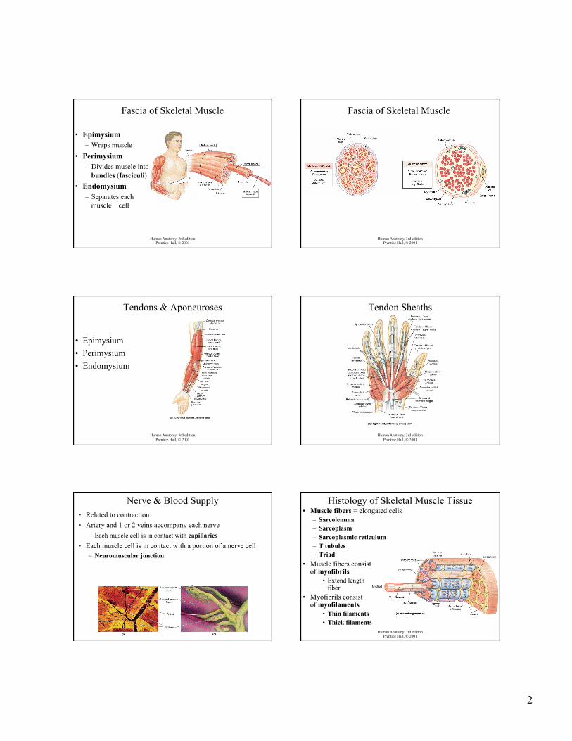
Nerve & Blood Supply • Related to contraction • Artery and 1 or 2 veins accompany each nerve
– Each muscle cell is in contact with capillaries • Each muscle cell is in contact with a portion of a nerve cell
– Neuromuscular junction
Human Anatomy, 3rd edition Prentice Hall, © 2001
Histology of Skeletal Muscle Tissue • Muscle fibers = elongated cells
– Sarcolemma – Sarcoplasm – Sarcoplasmic reticulum – T tubules – Triad
• Muscle fibers consist of myofibrils
• Extend length of fiber
• Myofibrils consist of myofilaments
• Thin filaments • Thick filaments
3
Human Anatomy, 3rd edition Prentice Hall, © 2001
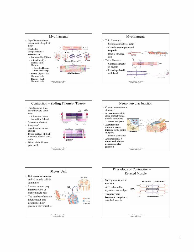
Myofilaments • Myofilaments do not
extend entire length of fiber.
• Stacked in compartments = sarcomeres – Partitioned by Z lines – A band (dark) –
contains thick filaments
• Includes H zone, zone of overlap
– I band (light) – thin filaments only
– H zone – thick filaments only Human Anatomy, 3rd edition
Prentice Hall, © 2001
Myofilaments • Thin filaments
– Composed mostly of actin – Contain tropomyosin and
troponin – Double-stranded
coil • Thick filaments
– Composed mostly of myosin
– Rod-shaped (tail) with head
Human Anatomy, 3rd edition Prentice Hall, © 2001
Contraction – Sliding Filament Theory • Thin filaments slide
inward toward the H zone – Z lines are drawn
toward the A band • Sarcomere shortens • Lengths of
myofilaments do not change
• Cross bridges of thick filaments connect with actin
• Width of the H zone gets smaller
Human Anatomy, 3rd edition Prentice Hall, © 2001
Neuromuscular Junction • Contraction requires a
stimulus • An axon comes into
close contact with a muscle membrane – Motor end plate
• Acetylcholine transmits nerve impulse to the motor end plate – Initiates contraction
• Axon terminal + motor end plate = neuromuscular junction
Human Anatomy, 3rd edition Prentice Hall, © 2001
Motor Unit • Def. – motor neuron
and all muscle cells it stimulates
• 1 motor neuron may innervate few or many muscle cells
• The number of muscle fibers/motor unit determines how precise a movement is.
Human Anatomy, 3rd edition Prentice Hall, © 2001
Physiology of Contraction – Relaxed Muscle
• Sarcoplasm is low in
calcium • ATP is bound to
myosin cross bridges • Tropomyosin-
troponin complex is attached to actin
4
Physiology of Contraction – Stimulation of Muscle
• Nerve impulse reaches motor end plate • Neuron releases ACh • Electrical charge travels along sarcolemma • Electrical charge travels down T tubules • Electrical charge travels to S.R. • S.R. releases calcium into sarcoplasm
Physiology of Contraction – Activation of Myosin
• Calcium binds to troponin • Cross bridges form • Calcium acts as an enzyme
– Breaks down ATP to ADP + P • Myosin cross bridges move • Sarcomere shortens • Muscle shortens
Physiology of Contraction – Relaxation of Muscle
• Nerve impulse ends • ACh is broken down by acetylcholinesterase • Calcium is actively transported back into S.R. • ADP + P = ATP
– Binds to cross bridges • Myosin cross bridges separate from actin • Binding sites on actin are covered • Thin myofilaments slip back to resting position • Sarcomeres return to resting length • Muscle fiber returns to resting length
Human Anatomy, 3rd edition Prentice Hall, © 2001
Physiology of Contraction
Fast, Slow, and Intermediate Muscles • Duration of contraction varies with function • Fast muscles (white)
– More extensive SR – Lack myoglobin – Fewer capillaries
• Slow muscles (red) – Smaller fibers – More capillaries – Lots of myoglobin
Disorders
• Fibrosis • Fibrositis • Fibromyalgia • Muscular dystrophy • Myasthenia gravis
5
Classifications in the Muscular System
How Skeletal Muscles Produce Movement
• Exert force on tendons • Attached to articulating bones forming
a joint • When muscle contracts, one bone
moves toward the other • Attachments
– Origin = attachment to stationary bone – Insertion = attachment to moveable bone – Belly = fleshy portion of muscle between
tendons
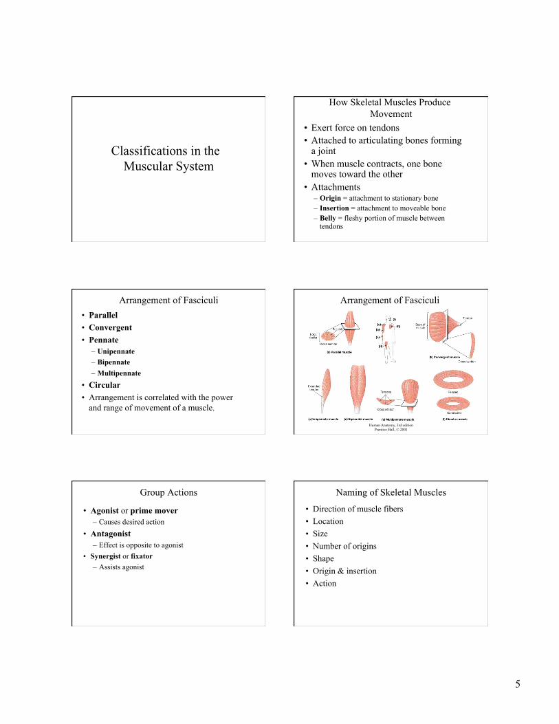
Arrangement of Fasciculi • Parallel • Convergent • Pennate
– Unipennate – Bipennate – Multipennate
• Circular • Arrangement is correlated with the power
and range of movement of a muscle.
Human Anatomy, 3rd edition Prentice Hall, © 2001
Arrangement of Fasciculi
Group Actions
• Agonist or prime mover – Causes desired action
• Antagonist – Effect is opposite to agonist
• Synergist or fixator – Assists agonist
Naming of Skeletal Muscles
• Direction of muscle fibers • Location • Size • Number of origins • Shape • Origin & insertion • Action