Embed Size (px)
Citation preview

1
1Skeletal System
LEARNING OBJECTIVES
1.Recognizeandusetermsrelatedtotheanatomyandphysiologyoftheskeletalsystem. 2.Recognizeandusetermsrelatedtothepathologyoftheskeletalsystem. 3.Recognizeandusetermsrelatedtotheproceduresfortheskeletalsystem. 4.Listanddescribefivefunctionsoftheskeletalsystem. 5.Explainthedifferencebetweencompactandspongybone. 6.Classifybonesaccordingtosizeandshape. 7.Identifythegeneralfeaturesofalongbone. 8.Explaintheprocessbywhichlongbonesgrowinlength. 9.Explainthedifferencebetweentheaxialandappendicularskeletons.10.Identifythebonesoftheskull.11.Identifythestructuralfeaturesofvertebrae.12.Listanddescribethedivisionsofthevertebralcolumn.13.Describethestructuralfeaturesofthesternumandribs.14.Identifythepartsofthepectoralgirdle.15.Identifythebonesoftheupperextremities.16.Identifythepartsofthepelvicgirdle.17.Identifythebonesofthelowerextremities.18.Listanddescribethedifferenttypesofjoints.19.Describewaysinwhichtheagingofanindividualaffectstheskeletalsystem.20.Identifypathologyrelatedtotheskeletalsystem.
CHAPTER OUTLINE
INTRODUCTION TO THE SKELETAL SYSTEMOverview of the Skeletal SystemFunctions of the Skeletal SystemStructure of Bone TissueClassification of BonesGeneral Features of a Long BoneBone MarkingsBone Development and GrowthBone Growth in LengthDivisions of the SkeletonBones of the Axial SkeletonSkullHyoid BoneVertebral ColumnThoracic Cage
Bones of the Appendicular SkeletonPectoral GirdleUpper ExtremityPelvic GirdleLower ExtremityLigaments and BursaeDiarthrosesAging of the Skeletal SystemTraumaFracturesSprain/Strain and Dislocation/SubluxationSetting FracturesSKELETAL SYSTEM REVIEW
Module_B_2622_Chapter 1_main.indd 1 10/8/2015 4:57:29 PM
To protect the rights of the author(s) and publisher we inform you that this PDF is an uncorrected proof for internal business use only by the author(s), editor(s), reviewer(s), Elsevier and typesetter Toppan Best-set. It is not allowed to publish this proof online or in print. This proof copy is the copyright property of the publisher and is confidential until formal publication.

2 CHAPTER 1 Skeletal System
CHAPTER AT A GLANCE
Use this list of key word parts and terms to assess your knowledge. Check off the ones you have mastered.AnatomyandPhysiology appendicular skeleton articulation axial skeleton depression process bursa
cartilage diaphysis epiphysis epiphyseal plate ligament osteoblast
osteoclast osteocyte osteon synarthrosis vertebrae
KeyWordPartsPREFIXES SUFFIXES COMBINING FORMS dia- -al arthr/o dys- -centesis burs/o endo-, end- -clasis chondr/o epi- -desis cost/o inter- -ectomy dactyl/o peri- -graphy myel/o poly -itis oste/o syn- -listhesis prosthes/o
-malacia spondyl/o -osis syndesm/o -pexy vertebr/o -plasty -sarcoma -scopy -y
KeyTerms arthrocentesis arthrodesis arthroplasty arthroscopy bursitis carpal tunnel syndrome (CTS) chondrosarcoma costochondritis
herniated intervertebral disk osteoarthritis (OA) osteoclasis osteomalacia osteomyelitis osteoporosis pathologic fractures polydactyly
rheumatoid arthritis (RA) spinal stenosis spondylosis spondylolisthesis subluxation syndactyly
INTRODUCTION TO THE SKELETAL SYSTEM
The skeletal system consists of the bones and the cartilage, ligaments, and tendons associated with the bones. It accounts for about 20% of the body weight. Bones are rigid structures that form the framework for the body. People often think of bones as dead, dry, inert pipes and plates because that is how they are seen in the laboratory. In reality, the living bones in our bodies contain active tissues that consume nutrients, require a blood supply, use oxygen and discharge waste products in metabolism, and change shape or remodel in response to variations in mechanical stress. The skeletal system is strong but lightweight. It is well adapted for the functions it must perform. It is a master-piece of design.
OVERVIEW OF THE SKELETAL SYSTEM
Functions of the Skeletal SystemThe skeletal system gives form and shape to the body. Without the skeletal components, we would appear as big “blobs” inefficiently “oozing” around on the ground. Besides contributing to shape and form, our bones perform several other functions and play an important role in homeostasis.
SupportBones provide a rigid framework that supports the soft organs of the body. Bones support the body against the pull of gravity, and the large bones of the lower limbs support the trunk when standing.
Module_B_2622_Chapter 1_main.indd 2 10/8/2015 4:57:29 PM
To protect the rights of the author(s) and publisher we inform you that this PDF is an uncorrected proof for internal business use only by the author(s), editor(s), reviewer(s), Elsevier and typesetter Toppan Best-set. It is not allowed to publish this proof online or in print. This proof copy is the copyright property of the publisher and is confidential until formal publication.

CHAPTER 1 Skeletal System 3
ProtectionThe skeleton protects the soft body parts. The fused bones of the cranium surround the brain to make it less vulnerable to injury. The vertebrae surround and protect the spinal cord. The bones of the rib cage help protect the heart and lungs in the thorax.
MovementBones provide sites for muscle attachment. Bones and muscles work together as simple mechanical lever systems to produce body movement.
StorageThe intercellular matrix of bone contains large amounts of calcium salts, the most important being calcium phosphate. Calcium is necessary for vital metabolic processes. When blood calcium levels decrease below normal, calcium is released from the bones so that there will be an adequate supply for metabolic needs. When blood calcium levels are increased, the excess calcium is stored in the bone matrix. Storage and release are dynamic processes that go on almost continually.
Blood Cell FormationBlood cell formation, called hematopoiesis (hee-mat-oh-poy-EE-sis), takes place mostly in the red marrow of bones. Red marrow is found in the cavities of most bones in an infant. With age, it is largely replaced by yellow marrow for fat storage. In the adult, red marrow is limited to the spongy bone in the skull, ribs, sternum, clavicles, vertebrae, and
pelvis. Red marrow functions in the formation of red blood cells, white blood cells, and blood platelets.
Structure of Bone TissueThere are two types of bone tissue: compact and spongy. As the names imply, the two types differ in density, or how tightly the tissue is packed together. Three types of cells contribute to bone homeostasis: osteoblasts, osteoclasts, and osteocytes. Osteoblasts are bone-forming cells, osteoclasts resorb or break down bone, and osteocytes are mature bone cells. An equilibrium between osteoblasts and osteoclasts maintains bone tissue.
Compact BoneThe microscopic unit of compact bone is known as the osteon (haversian system). The osteon consists of a central canal called the osteonic (haversian) canal, which is sur-rounded by concentric rings (lamellae) of hard, calcified matrix. Between the rings of matrix, the bone cells (osteo-cytes) are located in spaces called lacunae. Small channels (canaliculi) radiate from the lacunae to the osteonic (haver-sian) canal to provide passageways through the hard matrix. In compact bone the haversian systems are packed tightly together to form what appears to be a solid mass. The osteonic canals contain blood vessels that are parallel to the long axis of the bone. These blood vessels interconnect, by way of perforating (Volkmann) canals, with vessels on the surface of the bone. The microscopic structure of compact bone is illustrated in Figure 1-1.
Trabeculae ofspongy bone
Haversian canal
Volkmann’s canal
Lacuna
Osteocyte
Canaliculi
Periosteum
Lamellae ofan osteon
Osteon
Figure1-1 Structure of compact and spongy bone. Note the osteons packed together for compact bone and trabeculae of spongy bone.
Module_B_2622_Chapter 1_main.indd 3 10/8/2015 4:57:30 PM
To protect the rights of the author(s) and publisher we inform you that this PDF is an uncorrected proof for internal business use only by the author(s), editor(s), reviewer(s), Elsevier and typesetter Toppan Best-set. It is not allowed to publish this proof online or in print. This proof copy is the copyright property of the publisher and is confidential until formal publication.

4 CHAPTER 1 Skeletal System
Osteoporosis: Osteoporosis is a bone disorder caused bydecreasedosteoblastactivity.Itischaracterizedbylossoftheorganic matrix, collagenous fibers, and minerals in the bonetissue.Peoplewithosteoporosisaresusceptibletodeformitiesof thevertebralcolumnand fracturesbecause thebonesaretoo weak to support the weight of the body. OsteoporosisoccursmostfrequentlyinpostmenopausalCaucasianwomen.Factors that influence its occurrence are aging, malnutrition,lackofexercise,andhormoneimbalance.Supplementalestro-genaftermenopausemaybeofbenefit,andexerciseisalwaysimportantinmaintainingbonestrength.
Epiphyseal plate: Theepiphysealplatesof specific longbonesossifyatpredictable times.Radiologists frequentlycandeter-mineayoungperson’sagebyexaminingtheepiphysealplatestoseewhethertheyhaveossified.Adifferencebetweenboneageandchronologicagemayindicatesometypeofmetabolicdysfunction.
Mastoiditis:Themastoidaircellsareseparatedfromthecranialcavitybyonlya thinpartitionofbone.Amiddleear infectionthat spreads to the mastoid air cells (mastoiditis) is seriousbecausethereisdangerthattheinfectionwillspreadfromtheaircellstothemembranesaroundthebrain.
Sinus problems: The bones with paranasal sinuses are thefrontal, thesphenoid,theethmoid,andthetwomaxillae.Thesinusesarelinedwithmucousmembranesthatarecontinuouswiththenasalcavity.Allergiesandinfectionscauseinflamma-tionofthemembranes,whichresultsinsinusitis.Theswollenmembranes may reduce drainage from the sinuses so thatpressure within the cavities increases, resulting in sinusheadaches.
Soft spots:Thebonesintheskullofanewbornarenotcompletelyjoinedtogetherbutareseparatedbyfibrousmembranes.Thesix large areas of membranes are called fontanels, or softspots.Theanterior fontanel ison the topof thehead,at thejunctionofthefrontalandparietalbones.Theposteriorfontanelisatthejunctionoftheoccipitalandparietalbones.Oneachsideoftheheadthereisamastoid(posterolateral)fontanelnearthe mastoid region of the temporal bone and a sphenoid(anterolateral)fontaneljustsuperiortothesphenoidbone.
Abnormal spinal curvatures: An abnormally exaggeratedlumbarcurvatureiscalledlordosis,orswayback.Thisisoftenseeninpregnantwomenastheyadjusttotheirchangingcenterofgravity.Anincreasedroundnessofthethoraciccurvatureiskyphosis, or hunchback. This is frequently seen in elderlypeople.Abnormalside-to-sidecurvatureisscoliosis.Abnormalcurvatures may interfere with breathing and other vitalfunctions.
Yes and no:Theatlasholdsuptheskullandpermitsyoutonod“yes.”Theaxisallowsyoutorotateyourheadfromsidetosidetoindicate“no.”
Marrow biopsy:Thesternumisfrequentlyusedforaredmarrowbiopsy because it is accessible. The sample for biopsy is
obtained by performing a sternal puncture, in which a largeneedleisinsertedintothesternumtoremoveasampleofredbonemarrow.
Fractured clavicle:Theclavicleisthemostfrequentlyfracturedboneinthebodybecauseittransmitsforcesfromthearmtothetrunk.Theforcefromfallingontheshoulderoroutstretchedarmisoftensufficienttofracturetheclavicle.
Tennis elbow: Tennis elbow is an inflammation of the tissuessurroundingthelateralepicondyleofthehumerus.Sixmusclesthat control movement of the hand attach in this region,and repeated contraction of these muscles irritates theattachments. The medical term for tennis elbow is lateral epicondylitis.
Pelvic outlet and childbirth: The female pelvis is shaped toaccommodate childbearing. Because the fetus must passthroughthepelvicoutlet,thephysiciancarefullymeasuresthisopening to make sure there is enough room. The distancebetweenthetwoischialspinesisagoodindicationofthesizeof the pelvic outlet. If the opening is too small, a cesareandeliveryisindicated.
Broken hip:Elderlypeople,particularly thosewithosteoporosis,are susceptible to “breaking a hip.” The femur is a weight-bearingbone,andwhenitisweakened,itcannotsupporttheweightofthebodyandtheneckofthefemurfracturesunderthestress.Insteadofsaying,“Grandmafellandbrokeherhip,”often it ismoreappropriate to say, “Grandmabrokeherhip,thenfell.”
Bunion:Poorlyfittedshoesmaycompressthetoessothatthereisalateraldeviationofthebigtoetowardthesecondtoe.Whenthisoccurs,abursaandcallus format the jointbetweenthefirstmetatarsalandproximalphalanx.Thiscreatesabunion.
Gout: Gout was commonly known as the disease of the kingsbecause itwasbelieved tobecausedbya richdietandfinewines. Gout is an equal-opportunity disease, however, andoccursacrosstheentirepopulation.Arichdietandfinewinesmaycontribute to thedisease,but theyarenot thedefinitivecause.Gout is causedby theexcessiveaccumulationofuricacidthatformsneedle-likecrystalswithinthejoint,producingpain and inflammation. The great toe is the most commonlyaffectedjoint.Thedisorderisdiagnosedbyaspiratingjointfluidand observing the crystals under the microscope. Althoughthere isnocure forgout, itcanbeeffectivelycontrolledwithantiinflammatorydrugsanddietarymeasures.
Knee problems: The term torn cartilage refers to a damagedmeniscus,usuallythemedial,intheknee.Frequentlythiscanbe repairedwith relativelyminorarthroscopicsurgery.A tornligamentinthekneeusuallyinvolvesoneofthecruciateliga-ments.Thesurgicalprocedure to repair thisdamage isquiteinvolved,andrecoveryoffunctionmayrequiremonthsofreha-bilitativetherapy. n
Highlight on the Skeletal System
Module_B_2622_Chapter 1_main.indd 4 10/8/2015 4:57:30 PM
To protect the rights of the author(s) and publisher we inform you that this PDF is an uncorrected proof for internal business use only by the author(s), editor(s), reviewer(s), Elsevier and typesetter Toppan Best-set. It is not allowed to publish this proof online or in print. This proof copy is the copyright property of the publisher and is confidential until formal publication.
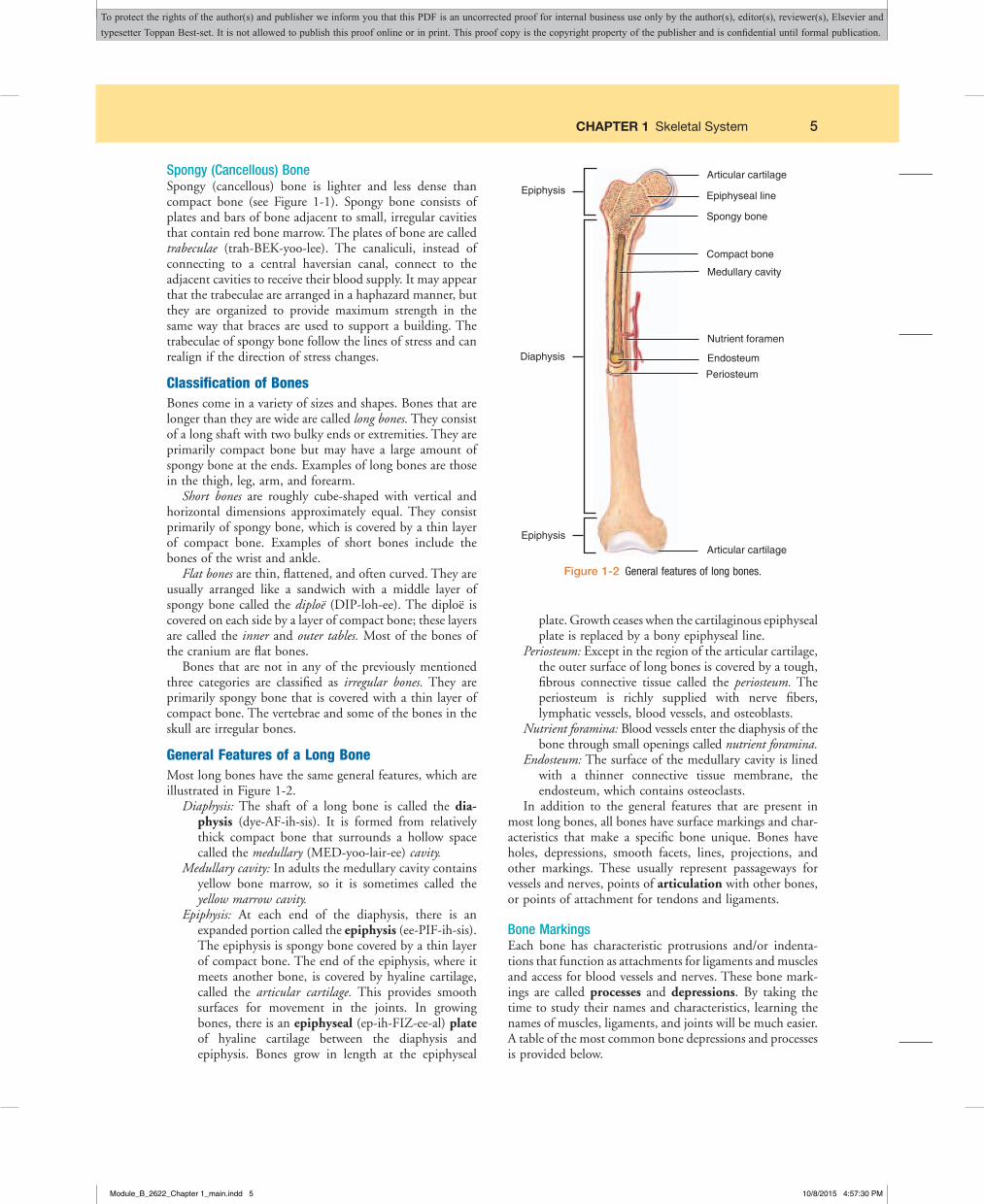
CHAPTER 1 Skeletal System 5
Spongy (Cancellous) BoneSpongy (cancellous) bone is lighter and less dense than compact bone (see Figure 1-1). Spongy bone consists of plates and bars of bone adjacent to small, irregular cavities that contain red bone marrow. The plates of bone are called trabeculae (trah-BEK-yoo-lee). The canaliculi, instead of connecting to a central haversian canal, connect to the adjacent cavities to receive their blood supply. It may appear that the trabeculae are arranged in a haphazard manner, but they are organized to provide maximum strength in the same way that braces are used to support a building. The trabeculae of spongy bone follow the lines of stress and can realign if the direction of stress changes.
Classification of BonesBones come in a variety of sizes and shapes. Bones that are longer than they are wide are called long bones. They consist of a long shaft with two bulky ends or extremities. They are primarily compact bone but may have a large amount of spongy bone at the ends. Examples of long bones are those in the thigh, leg, arm, and forearm.
Short bones are roughly cube-shaped with vertical and horizontal dimensions approximately equal. They consist primarily of spongy bone, which is covered by a thin layer of compact bone. Examples of short bones include the bones of the wrist and ankle.
Flat bones are thin, flattened, and often curved. They are usually arranged like a sandwich with a middle layer of spongy bone called the diploë (DIP-loh-ee). The diploë is covered on each side by a layer of compact bone; these layers are called the inner and outer tables. Most of the bones of the cranium are flat bones.
Bones that are not in any of the previously mentioned three categories are classified as irregular bones. They are primarily spongy bone that is covered with a thin layer of compact bone. The vertebrae and some of the bones in the skull are irregular bones.
General Features of a Long BoneMost long bones have the same general features, which are illustrated in Figure 1-2.
Diaphysis: The shaft of a long bone is called the diaphysis (dye-AF-ih-sis). It is formed from relatively thick compact bone that surrounds a hollow space called the medullary (MED-yoo-lair-ee) cavity.
Medullary cavity: In adults the medullary cavity contains yellow bone marrow, so it is sometimes called the yellow marrow cavity.
Epiphysis: At each end of the diaphysis, there is an expanded portion called the epiphysis (ee-PIF-ih-sis). The epiphysis is spongy bone covered by a thin layer of compact bone. The end of the epiphysis, where it meets another bone, is covered by hyaline cartilage, called the articular cartilage. This provides smooth surfaces for movement in the joints. In growing bones, there is an epiphyseal (ep-ih-FIZ-ee-al) plate of hyaline cartilage between the diaphysis and epiphysis. Bones grow in length at the epiphyseal
Epiphysis
Epiphysis
Diaphysis
Articular cartilage
Periosteum
Endosteum
Nutrient foramen
Medullary cavity
Compact bone
Spongy bone
Epiphyseal line
Articular cartilage
Figure1-2 General features of long bones.
plate. Growth ceases when the cartilaginous epiphyseal plate is replaced by a bony epiphyseal line.
Periosteum: Except in the region of the articular cartilage, the outer surface of long bones is covered by a tough, fibrous connective tissue called the periosteum. The periosteum is richly supplied with nerve fibers, lymphatic vessels, blood vessels, and osteoblasts.
Nutrient foramina: Blood vessels enter the diaphysis of the bone through small openings called nutrient foramina.
Endosteum: The surface of the medullary cavity is lined with a thinner connective tissue membrane, the endosteum, which contains osteoclasts.
In addition to the general features that are present in most long bones, all bones have surface markings and char-acteristics that make a specific bone unique. Bones have holes, depressions, smooth facets, lines, projections, and other markings. These usually represent passageways for vessels and nerves, points of articulation with other bones, or points of attachment for tendons and ligaments.
Bone Markings Each bone has characteristic protrusions and/or indenta-tions that function as attachments for ligaments and muscles and access for blood vessels and nerves. These bone mark-ings are called processes and depressions. By taking the time to study their names and characteristics, learning the names of muscles, ligaments, and joints will be much easier. A table of the most common bone depressions and processes is provided below.
Module_B_2622_Chapter 1_main.indd 5 10/8/2015 4:57:30 PM
To protect the rights of the author(s) and publisher we inform you that this PDF is an uncorrected proof for internal business use only by the author(s), editor(s), reviewer(s), Elsevier and typesetter Toppan Best-set. It is not allowed to publish this proof online or in print. This proof copy is the copyright property of the publisher and is confidential until formal publication.

6 CHAPTER 1 Skeletal System
Bone Depressions
Depression Combining Form Meaning/Function Example
fissure fissur/o Fairly deep cleft or groove Sphenoidal fissureforamen foramin/o Opening or hole Foramen magnum,
mental foraminafossa foss/o Hollow or depression, especially on the surface
of the end of a boneOlecranal fossa
fovea Small pit or depression Fovea capitis of humerus
sinus/antrum sinus/o, sin/o, antr/o Cavity or channel lined with a membrane Paranasal sinusessulcus sulc/o General term that refers to a groove or
depression in an anatomic structure, not as deep as a fissure
Intertubercular sulcus of humerus
Bone Processes
Process Combining Form Meaning/Function Example
condyle condyl/o Rounded projection at the end of a bone that anchors the ligaments and articulates with adjacent bones
Medial condyle of the femur
crest Narrow elongated elevation Iliac crestepicondyle epicondyl/o Projection on the surface of the bone
above the condyleLateral epicondyle of
the humerusfacet Small, smooth flat articular surface Vertebral facetshead(capitis) Rounded, usually proximal portion of
some long bonesFemoral head,
humeral headneck Narrowed area distal to a bone head Femoral neckramus Branchlike extension Mandibular ramusspine spin/o Thornlike projection Spinous process of
vertebratrochanter trochanter/o One of two bony projections on the
proximal ends of the femurs that serve as points of attachment for muscles
Greater trochanter
tubercle tubercul/o Nodule or small raised area Costal tubercletuberosity Elevation or protruberance; larger than a
tubercleIschial tuberosity
EXERCISE 1: Bone Basics
Match the bone word parts with their meanings.
____ 1. myel/o____ 2. -physis____ 3. peri-____ 4. condyl/o____ 5. spin/o____ 6. sin/o____ 7. foramin/o____ 8. -um
____ 9. -blast____ 10. epi-____ 11. foss/o____ 12. endo-____ 13. oste/o____ 14. -cyte____ 15. -clast
A. bone B. foramen, hole C. above, upon D. cell E. bone marrow F. surrounding, around G. embryonic H. spine
I. breaking down J. growth, nature K. hollow, depression L. condyle, knob M. within N. sinus, cavity O. structure
Fill in the blank.
16. Osteoblasts _______________ bone, whereas osteoclasts _______________ bone.
17. The shaft of a long bone is called the _______________; the ends of a long bone are called _______________ (plural!).
Module_B_2622_Chapter 1_main.indd 6 10/8/2015 4:57:30 PM
To protect the rights of the author(s) and publisher we inform you that this PDF is an uncorrected proof for internal business use only by the author(s), editor(s), reviewer(s), Elsevier and typesetter Toppan Best-set. It is not allowed to publish this proof online or in print. This proof copy is the copyright property of the publisher and is confidential until formal publication.

CHAPTER 1 Skeletal System 7
18. The outer covering of bone is the _______________, whereas the inner lining is the _______________.
19. A foramen, a sinus, and a fossa are examples of bone _______________. A condyle, a trochanter, and a tuberosity are examples of bone _______________.
20. A synonym for a sinus is a/an _______________.
B4.5 Proceduresperformedontendons,ligaments,bursae,andfasciasupportingajointarecodedtothebodypartintherespectivebodysystemthatisthefocusoftheprocedure.Proceduresperformedonjointstructuresthemselvesarecodedtothebodypartinthejointbodysystems.
Example:Repairoftheanteriorcruciateligamentofthekneeiscodedtothekneebursaeandliga-mentbodypartinthebursaeandligamentsbodysystem.KneearthroscopywithshavingofarticularcartilageiscodedtothekneejointbodypartintheLowerJointsbodysystem.
GUIDELINE ALERT
Bone Development and GrowthThe terms osteogenesis and ossification are often used synony-mously to indicate the process of bone formation. Parts of the skeleton form during the first few weeks after concep-tion. By the end of the eighth week after conception, the skeletal pattern is formed in cartilage and connective tissue membranes and ossification begins. Bone development con-tinues throughout adulthood. Even after adult stature is attained, bone development continues for repair of fractures and for remodeling to meet changing lifestyles. Three types of cells are involved in the development, growth, and remodeling of bones. Osteoblasts are bone-forming cells; osteocytes are mature bone cells; and osteoclasts break down and reabsorb bone.
Bone Growth in LengthBones grow in length at the epiphyseal plate located between the diaphysis and epiphysis of a long bone. The hyaline cartilage in the region of the epiphyseal plate next to the epiphysis continues to grow by mitosis. The chondrocytes in the region next to the diaphysis age and degenerate. Osteoblasts move in and ossify the matrix to form bone. This process continues throughout childhood and adoles-cence until the cartilage growth slows and finally stops. When cartilage growth ceases, usually in the early 20s, the epiphyseal plate completely ossifies so that only a thin epiphyseal line remains and the bones can no longer grow
B4.6. Ifaprocedureisperformedontheskin,subcutaneoustissue,orfasciaoverlyingajoint,theprocedureiscodedtothefollowingbodypart:• ShoulderiscodedtoUpperArm• ElbowiscodedtoLowerarm• WristiscodedtoLowerarm• HipiscodedtoUpperleg• KneeiscodedtoLowerLeg• AnkleiscodedtoFoot
GUIDELINE ALERT
in length. Bone growth occurs under the influence of growth hormone from the anterior pituitary gland and sex hormones from the ovaries and testes.
Even though bones stop growing in length in early adult-hood, they can continue to increase in thickness or diameter throughout life in response to stress from increased muscle activity or to weight gain. The increase in diameter is called appositional (ap-poh-ZISH-un-al) growth. Osteoblasts in the periosteum form compact bone around the external bone surface. At the same time, osteoclasts in the endos-teum break down bone on the internal bone surface, around the medullary cavity. These two processes together increase the diameter of the bone and at the same time keep the bone from becoming excessively heavy and bulky.
Divisions of the SkeletonThe typical adult human skeleton consists of 206 named bones. For convenience, the bones of the skeleton are grouped in two divisions, as illustrated in Figure 1-3. The 80 bones of the axial skeleton form the vertical axis of the body. They include the bones of the head, vertebral column, ribs, and breastbone or sternum. The appendicular skeleton consists of 126 bones and includes the free appendages and their attachments to the axial skeleton. The free append-ages are the upper and lower extremities, or limbs, and their attachments are called girdles. Table 1-1 lists the named bones of the body by category.
Module_B_2622_Chapter 1_main.indd 7 10/8/2015 4:57:31 PM
To protect the rights of the author(s) and publisher we inform you that this PDF is an uncorrected proof for internal business use only by the author(s), editor(s), reviewer(s), Elsevier and typesetter Toppan Best-set. It is not allowed to publish this proof online or in print. This proof copy is the copyright property of the publisher and is confidential until formal publication.

8 CHAPTER 1 Skeletal System
Skull
Scapula
Humerus
Radius
Ulna
Carpals
MetacarpalsPhalanges
Femur
Patella
Tibia
Fibula
Tarsals
Metatarsals
Phalanges
Vertebral columnCervical vertebrae
Thoracic vertebrae
Lumbar vertebrae
Sacrum
Coccyx
Clavicle
Sternum
Ribs
Os coxae
Figure1-3 Divisions of the skeleton with major bones identified. Yellow = axial skeleton. Blue = appendicular skeleton.
BONES OF THE AXIAL SKELETON
The axial skeleton, with 80 bones, is divided into the skull, hyoid, vertebral column, and rib cage.
SkullThe skull has 28 bones, as illustrated in Figures 1-4 and 1-5. The eight bones of the cranium are interlocked to enclose the brain. The anterior aspect of the skull, the face, consists of 14 bones. The remaining six bones are the audi-tory ossicles, tiny bones in the middle ear cavity. With the exception of the lower jaw, or mandible, and the auditory ossicles, the bones in the skull are tightly interlocked along irregular lines called sutures. Some of the bones in the skull contain sinuses, which are air-filled cavities lined with mucous membranes. The sinuses help to reduce the weight of the skull. The paranasal sinuses are arranged around the nasal cavity and drain into it.
CraniumFrontal BoneThe frontal bone forms the anterior portion of the skull above the eyes (forehead). The paranasal frontal sinuses are cavities in the frontal bone.
Parietal BonesThe two parietal (pah-RYE-eh-tal) bones form most of the superolateral aspect of the skull.
Occipital BoneThe single occipital (ahk-SIP-ih-tal) bone forms most of the posterior part of the skull. The foramen magnum is a large opening on the lower surface of the occipital bone. The spinal cord passes through this opening. Occipital condyles are rounded processes on each side of the foramen magnum. They articulate with the first cervical vertebra.
Module_B_2622_Chapter 1_main.indd 8 10/8/2015 4:57:31 PM
To protect the rights of the author(s) and publisher we inform you that this PDF is an uncorrected proof for internal business use only by the author(s), editor(s), reviewer(s), Elsevier and typesetter Toppan Best-set. It is not allowed to publish this proof online or in print. This proof copy is the copyright property of the publisher and is confidential until formal publication.

CHAPTER 1 Skeletal System 9
Temporal BonesThe two temporal bones, one on each side of the head, form parts of the sides and base of the cranium. Near the inferior margin of the temporal bone, there is an opening, the exter-nal auditory meatus, which is a canal that leads to the middle ear. Just anterior to the external auditory meatus, the tem-poral bone articulates with the mandible to form the tem-poromandibular joint (TMJ). Posterior and inferior to each external auditory meatus, there is a rough protuberance, the mastoid process. The mastoid process contains air cells that drain into the middle ear cavity.
Sphenoid BoneThe sphenoid (SFEE-noyd) bone is an irregularly shaped bone that spans the entire width of the cranial floor. It is wedged between other bones in the anterior portion of the cranium. The sphenoid bone contains paranasal sphenoid sinuses.
Ethmoid BoneThe ethmoid (ETH-moyd) bone is located anterior to the sphenoid bone and forms most of the bony area between the nasal cavity and the orbits. The ethmoid bone contains many small, paranasal ethmoidal sinuses.
Facial BonesThe 14 facial bones form the basic framework and shape of the face. They also provide attachments for the muscles that control facial expression and move the jaw for chewing. All facial bones except the vomer and mandible are paired. Facial bones are illustrated in Figures 1-4 and 1-5.
Maxillary BonesThe maxillary bones, or maxillae (maks-ILL-ee), form the upper jaw and the anterior part of the hard palate or roof of the mouth. Each maxilla has a large paranasal maxillary sinus. These are the largest of all the paranasal sinuses.
Palatine BonesThe palatine (PAL-ah-tyne) bones are behind, or posterior to, the maxillae and form the posterior portion of the hard palate.
Nasal BonesThe two nasal bones are small rectangular bones that form the bridge of the nose.
Lacrimal BonesThe small, thin lacrimal (LACK-rih-mal) bones are located in the medial walls of the orbits, between the ethmoid bone and the maxilla. Each one has a small lacrimal groove that is a pathway for a tube that carries tears from the eyes to the nasal cavity.
Zygomatic BonesThe zygomatic (zye-goh-MAT-ik) bones, also called malar bones, form the prominences of the cheeks.
Table 1-1 Names of Bones of the Body Listed by Category
Bones NumberAxial Skeleton (80 Bones)Skull(28bones)
CranialbonesParietal(2)Temporal(2)Frontal(1)Occipital(1)Ethmoid(1)Sphenoid(1)
8
FacialbonesMaxilla(2)Zygomatic(2)Mandible(1)Nasal(2)Palatine(2)Inferiornasalconcha(2)Lacrimal(2)Vomer(1)
14
AuditoryossiclesMalleus(2)Incus(2)Stapes(2)
6
Hyoid 1Vertebralcolumn
Cervicalvertebrae(7)Thoracicvertebrae(12)Lumbarvertebrae(5)Sacrum(1)Coccyx(1)
26
ThoraciccageSternum(1)Ribs(24)
25
Appendicular Skeleton (126 Bones)Pectoralgirdles
Clavicle(2)Scapula(2)
4
UpperextremityHumerus(2)Radius(2)Ulna(2)Carpals(16)Metacarpals(10)Phalanges(28)
60
PelvicgirdleCoxal,innominate,orhipbones(2)
2
LowerextremityFemur(2)Tibia(2)Fibula(2)Patella(2)Tarsals(14)Metatarsals(10)Phalanges(28)
60
FromApplegateE:The anatomy and physiology learning system,ed4,StLouis,2011,Saunders.
Module_B_2622_Chapter 1_main.indd 9 10/8/2015 4:57:32 PM
To protect the rights of the author(s) and publisher we inform you that this PDF is an uncorrected proof for internal business use only by the author(s), editor(s), reviewer(s), Elsevier and typesetter Toppan Best-set. It is not allowed to publish this proof online or in print. This proof copy is the copyright property of the publisher and is confidential until formal publication.

10 CHAPTER 1 Skeletal System
FRONTAL BONEPARIETAL BONE
SPHENOID BONE
NASAL BONEETHMOID BONELACRIMAL BONE
VOMER BONE
MANDIBLE
MAXILLA
TEMPORAL BONE
SPHENOID BONE
ZYGOMATIC BONE
INFERIOR NASALCONCHA
FRONTAL BONE
SPHENOID BONENASAL BONELACRIMAL BONE
ZYGOMATIC BONE
MANDIBLE
MAXILLA
TEMPORAL BONE
PARIETAL BONE
OCCIPITAL BONE
Externalauditory meatus
Mastoid process
Figure1-4 Skull, anterior view.
Figure1-5 Skull, lateral view.
Module_B_2622_Chapter 1_main.indd 10 10/8/2015 4:57:33 PM
To protect the rights of the author(s) and publisher we inform you that this PDF is an uncorrected proof for internal business use only by the author(s), editor(s), reviewer(s), Elsevier and typesetter Toppan Best-set. It is not allowed to publish this proof online or in print. This proof copy is the copyright property of the publisher and is confidential until formal publication.

CHAPTER 1 Skeletal System 11
Inferior Nasal ConchaeThe inferior nasal conchae (KONG-kee) are thin, curved bones that are attached to the lateral walls of the nasal cavity and project into the nasal cavity.
VomerThe thin, flat vomer (VOH-mer) is in the inferior portion of the midline in the nasal cavity. It forms part of the nasal septum.
MandibleThe mandible (MAN-dih-bul) is the lower jaw. It articulates with the temporal bone to form the temporomandibular (tem-por-oh-man-DIB-yoo-lar) joint.
Auditory OssiclesThree tiny bones form a chain in each middle ear cavity in the temporal bone. These are the malleus, incus, and stapes. These bones transmit sound waves from the tympanic membrane, or eardrum, to the inner ear, where the sound receptors are located.
Hyoid BoneThe hyoid bone is not really part of the skull, so it is listed separately. It is a U-shaped bone in the neck, sus-pended under the mandible. It is unique because it is the only bone in the body that does not articulate directly with another bone. It functions as a base for the tongue and as an attachment for several muscles associated with swallowing.
Vertebral ColumnThe vertebral column extends from the skull to the pelvis and contains 26 bones called vertebrae (singular, vertebra). The bones are separated by pads of fibrocartilage called intervertebral discs. The discs act as shock absorbers and allow the column to bend. Normally there are four curva-tures, illustrated in Figure 1-6, that increase the strength and resilience of the column. They are named according to the region in which they are located. The thoracic and sacral curvatures are concave anteriorly and are present at birth. The cervical curvature develops when an infant begins to hold his or her head erect. The lumbar curvature develops when an infant begins to stand and walk. Both the cervical and lumbar curvatures are convex anteriorly.
General Structure of VertebraeAll vertebrae have a common structural pattern, illustrated in Figure 1-7, although there are variations among them. The thick anterior, weight-bearing portion is the body or centrum. The posterior curved portion is the vertebral arch. The vertebral arch and body surround a central large opening, the vertebral foramen. When all the vertebrae are stacked together in a column, the vertebral foramina make a canal that contains the spinal cord. Transverse processes project laterally from the vertebral arch, and in the posterior
C1 Cervical vertebrae2
2
2
3
3
3
4
4
4
5
5
5
6
6
7
7
8
9
10
11
12
T1 Thoracic vertebrae
L1 Lumbar vertebrae
Coccygeal vertebrae
Sacrum
Sacral curve
Lumbar curve
Thoracic curve
Cervical curve
Figure 1-6 Curvatures of the vertebral column. The thoracic and sacral curvatures are concave anteriorly, and the cervical and lumbar curvatures are convex anteriorly.
Spinous process
Vertebral arch
Vertebral foramen
Transverse process
Body (centrum)
Figure1-7 General features of vertebrae, viewed from above.
Module_B_2622_Chapter 1_main.indd 11 10/8/2015 4:57:33 PM
To protect the rights of the author(s) and publisher we inform you that this PDF is an uncorrected proof for internal business use only by the author(s), editor(s), reviewer(s), Elsevier and typesetter Toppan Best-set. It is not allowed to publish this proof online or in print. This proof copy is the copyright property of the publisher and is confidential until formal publication.

12 CHAPTER 1 Skeletal System
midline there is a spinous process. These processes are places for muscle attachment. The spinous processes can be felt as bony projections along the midline of the back.
Composition of the Vertebral ColumnThe seven cervical vertebrae are designated C1 through C7. The 12 thoracic vertebrae are designated T1 through T12. Five lumbar vertebrae, designated L1 through L5, make up the part of the vertebral column in the small of the back. The lumbar vertebrae have large, heavy bodies because they support most of the body weight and have many back muscles attached to them.
The sacrum is a triangular bone just below the lumbar vertebrae. In the child there are five separate bones, but these fuse to form a single bone in the adult. The sacrum articulates with the pelvic girdle laterally, at the sacroiliac (say-kro-ILL-ee-ak) joint, and forms the posterior wall of the pelvic cavity.
The coccyx (KOK-siks), or tailbone, is the last part of the vertebral column (see Figure 1-8). A child has four (the number varies from three to five) separate small bones, but these fuse to form a single bone in the adult.
Thoracic CageThe thoracic cage, or bony thorax, protects the heart, lungs, and great vessels. It also supports the bones of the shoulder girdle and plays a role in breathing. The components of the thoracic cage are the thoracic vertebrae dorsally, the ribs laterally, and the sternum and costal cartilage anteriorly.
SternumThe sternum, or breastbone, is in the anterior midline (Figure 1-8). An important anatomic landmark, the jugular (suprasternal) notch is an easily palpable, central indentation
in the superior margin of the sternum. The superior portion of the sternum articulates with the clavicles and the first two pairs of ribs. The body of the sternum has notches along the sides where it attaches to the cartilage of the third through seventh ribs.
RibsTwelve pairs of ribs, illustrated in Figure 1-8, form the curved, lateral margins of the thoracic cage. One pair is attached to each of the 12 thoracic vertebrae. The upper seven pairs of ribs are called true, or vertebrosternal (ver-TEE-broh-stir-nal), ribs because they attach to the sternum directly by their individual costal cartilage. The lower five pairs of ribs are called false ribs because their costal cartilage does not reach the sternum directly. The first three pairs of false ribs reach the sternum indirectly by joining with the cartilage of the ribs above. These are called vertebrochondral (ver-TEE-broh-kahn-dral) ribs. The bottom two rib pairs have no anterior attachment and are called vertebral ribs or floating ribs.
BONES OF THE APPENDICULAR SKELETON
The 126 bones of the appendicular skeleton are suspended from two yokes or girdles that are anchored to the axial skeleton. They are additions or appendages to the axis of the body. The appendicular skeleton is designed for move-ment. If a portion is immobilized for a period of time, life without appendicular movement can be awkward.
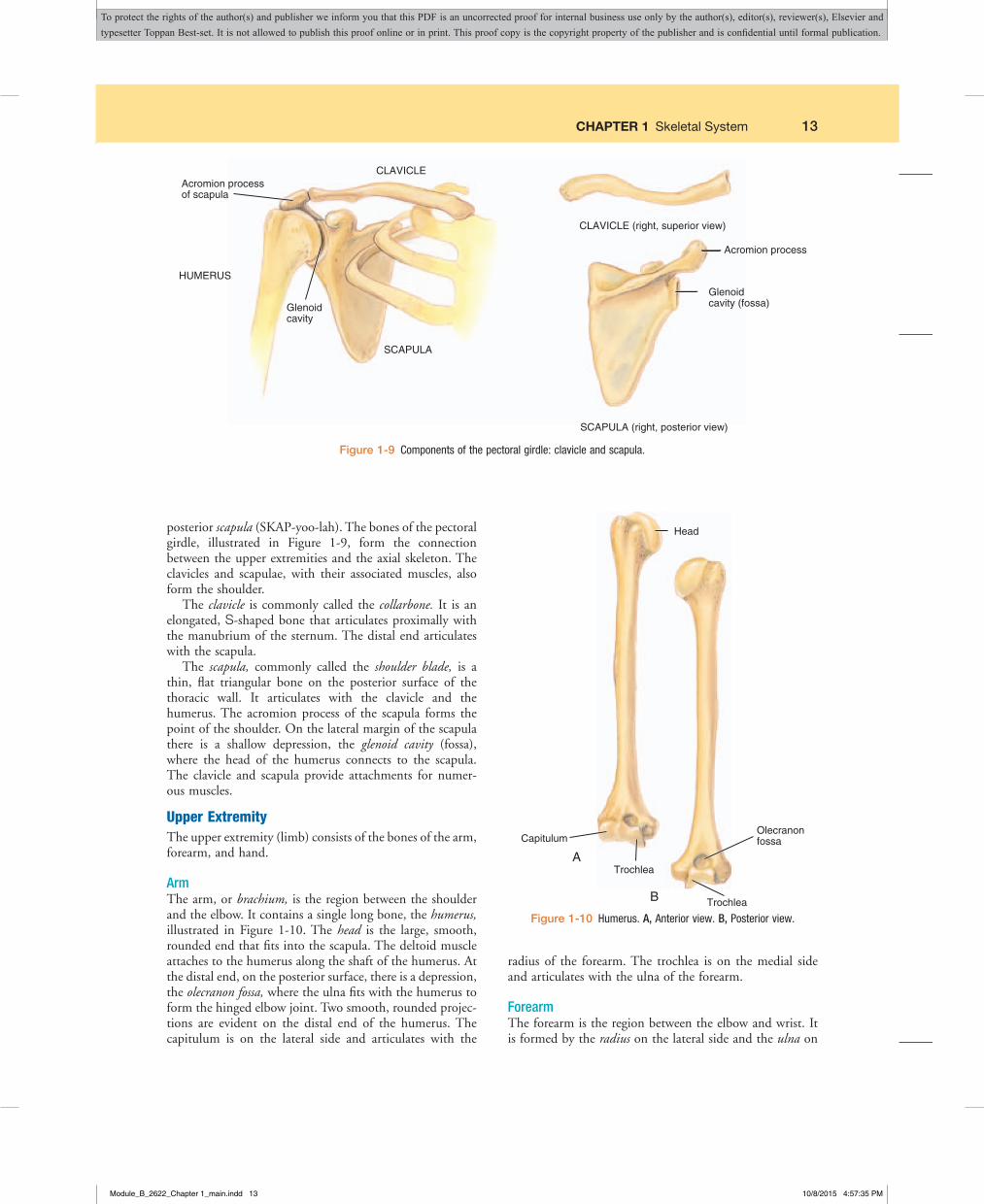
Pectoral GirdleEach half of the pectoral girdle, or shoulder girdle, consists of two bones: an anterior clavicle (KLAV-ih-kul) and a
1st thoracic vertebra (T1)
FALSERIBS
1
2
3
4
5
6
7
8
9
10
1112
TRUERIBS
Jugular notch
Costal cartilage
12th thoracic vertebra (T12)
STERNUMFigure1-8 Thoracic cage.
Module_B_2622_Chapter 1_main.indd 12 10/8/2015 4:57:34 PM
To protect the rights of the author(s) and publisher we inform you that this PDF is an uncorrected proof for internal business use only by the author(s), editor(s), reviewer(s), Elsevier and typesetter Toppan Best-set. It is not allowed to publish this proof online or in print. This proof copy is the copyright property of the publisher and is confidential until formal publication.
CHAPTER 1 Skeletal System 13
posterior scapula (SKAP-yoo-lah). The bones of the pectoral girdle, illustrated in Figure 1-9, form the connection between the upper extremities and the axial skeleton. The clavicles and scapulae, with their associated muscles, also form the shoulder.
The clavicle is commonly called the collarbone. It is an elongated, S-shaped bone that articulates proximally with the manubrium of the sternum. The distal end articulates with the scapula.
The scapula, commonly called the shoulder blade, is a thin, flat triangular bone on the posterior surface of the thoracic wall. It articulates with the clavicle and the humerus. The acromion process of the scapula forms the point of the shoulder. On the lateral margin of the scapula there is a shallow depression, the glenoid cavity (fossa), where the head of the humerus connects to the scapula. The clavicle and scapula provide attachments for numer-ous muscles.
Upper ExtremityThe upper extremity (limb) consists of the bones of the arm, forearm, and hand.
ArmThe arm, or brachium, is the region between the shoulder and the elbow. It contains a single long bone, the humerus, illustrated in Figure 1-10. The head is the large, smooth, rounded end that fits into the scapula. The deltoid muscle attaches to the humerus along the shaft of the humerus. At the distal end, on the posterior surface, there is a depression, the olecranon fossa, where the ulna fits with the humerus to form the hinged elbow joint. Two smooth, rounded projec-tions are evident on the distal end of the humerus. The capitulum is on the lateral side and articulates with the
radius of the forearm. The trochlea is on the medial side and articulates with the ulna of the forearm.
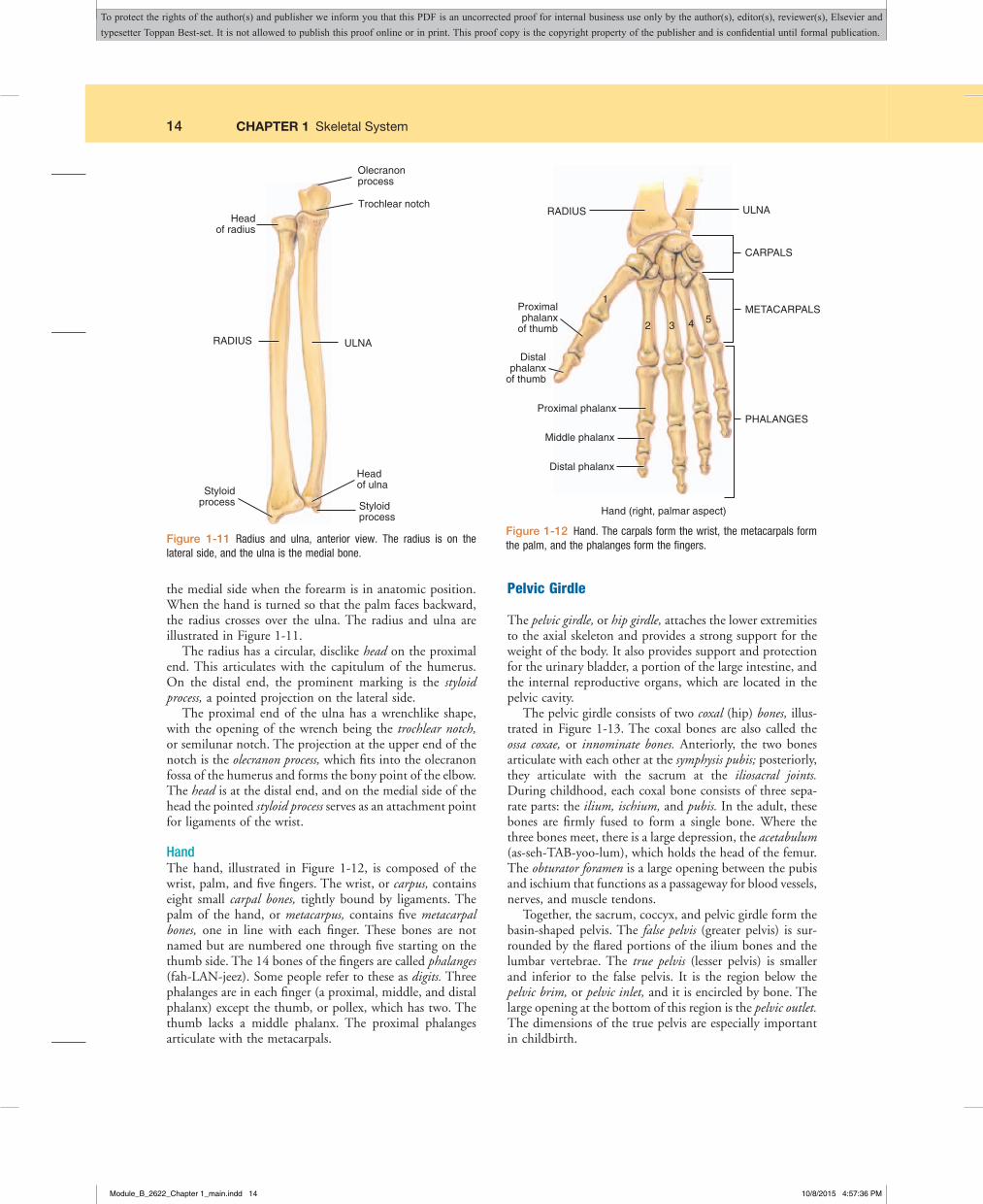
ForearmThe forearm is the region between the elbow and wrist. It is formed by the radius on the lateral side and the ulna on
Acromion processof scapula
CLAVICLE
Glenoidcavity
HUMERUS
SCAPULA
CLAVICLE (right, superior view)
Glenoidcavity (fossa)
SCAPULA (right, posterior view)
Acromion process
Figure1-9 Components of the pectoral girdle: clavicle and scapula.
Head
Capitulum
Trochlea
Trochlea
A
B
Olecranonfossa
Figure1-10 Humerus. A, Anterior view. B, Posterior view.
Module_B_2622_Chapter 1_main.indd 13 10/8/2015 4:57:35 PM
To protect the rights of the author(s) and publisher we inform you that this PDF is an uncorrected proof for internal business use only by the author(s), editor(s), reviewer(s), Elsevier and typesetter Toppan Best-set. It is not allowed to publish this proof online or in print. This proof copy is the copyright property of the publisher and is confidential until formal publication.
14 CHAPTER 1 Skeletal System
the medial side when the forearm is in anatomic position. When the hand is turned so that the palm faces backward, the radius crosses over the ulna. The radius and ulna are illustrated in Figure 1-11.
The radius has a circular, disclike head on the proximal end. This articulates with the capitulum of the humerus. On the distal end, the prominent marking is the styloid process, a pointed projection on the lateral side.
The proximal end of the ulna has a wrenchlike shape, with the opening of the wrench being the trochlear notch, or semilunar notch. The projection at the upper end of the notch is the olecranon process, which fits into the olecranon fossa of the humerus and forms the bony point of the elbow. The head is at the distal end, and on the medial side of the head the pointed styloid process serves as an attachment point for ligaments of the wrist.
HandThe hand, illustrated in Figure 1-12, is composed of the wrist, palm, and five fingers. The wrist, or carpus, contains eight small carpal bones, tightly bound by ligaments. The palm of the hand, or metacarpus, contains five metacarpal bones, one in line with each finger. These bones are not named but are numbered one through five starting on the thumb side. The 14 bones of the fingers are called phalanges (fah-LAN-jeez). Some people refer to these as digits. Three phalanges are in each finger (a proximal, middle, and distal phalanx) except the thumb, or pollex, which has two. The thumb lacks a middle phalanx. The proximal phalanges articulate with the metacarpals.
Pelvic Girdle
The pelvic girdle, or hip girdle, attaches the lower extremities to the axial skeleton and provides a strong support for the weight of the body. It also provides support and protection for the urinary bladder, a portion of the large intestine, and the internal reproductive organs, which are located in the pelvic cavity.
The pelvic girdle consists of two coxal (hip) bones, illus-trated in Figure 1-13. The coxal bones are also called the ossa coxae, or innominate bones. Anteriorly, the two bones articulate with each other at the symphysis pubis; posteriorly, they articulate with the sacrum at the iliosacral joints. During childhood, each coxal bone consists of three sepa-rate parts: the ilium, ischium, and pubis. In the adult, these bones are firmly fused to form a single bone. Where the three bones meet, there is a large depression, the acetabulum (as-seh-TAB-yoo-lum), which holds the head of the femur. The obturator foramen is a large opening between the pubis and ischium that functions as a passageway for blood vessels, nerves, and muscle tendons.
Together, the sacrum, coccyx, and pelvic girdle form the basin-shaped pelvis. The false pelvis (greater pelvis) is sur-rounded by the flared portions of the ilium bones and the lumbar vertebrae. The true pelvis (lesser pelvis) is smaller and inferior to the false pelvis. It is the region below the pelvic brim, or pelvic inlet, and it is encircled by bone. The large opening at the bottom of this region is the pelvic outlet. The dimensions of the true pelvis are especially important in childbirth.
Olecranonprocess
Trochlear notch
RADIUS ULNA
Headof radius
Styloidprocess Styloid
process
Head of ulna
RADIUS ULNA
CARPALS
1
2 3 4 5Proximalphalanx
of thumb
Proximal phalanx
Middle phalanx
Distal phalanx
Distalphalanx
of thumb
Hand (right, palmar aspect)
PHALANGES
METACARPALS
Figure 1-11 Radius and ulna, anterior view. The radius is on the lateral side, and the ulna is the medial bone.
Figure1-12 Hand. The carpals form the wrist, the metacarpals form the palm, and the phalanges form the fingers.
Module_B_2622_Chapter 1_main.indd 14 10/8/2015 4:57:36 PM
To protect the rights of the author(s) and publisher we inform you that this PDF is an uncorrected proof for internal business use only by the author(s), editor(s), reviewer(s), Elsevier and typesetter Toppan Best-set. It is not allowed to publish this proof online or in print. This proof copy is the copyright property of the publisher and is confidential until formal publication.

CHAPTER 1 Skeletal System 15
Lower ExtremityThe lower extremity (limb) consists of the bones of the thigh, leg, foot, and patella, or kneecap. The lower extremi-ties support the entire weight of the body when we are erect, and they are exposed to tremendous forces when we walk, run, and jump. With this in mind, it is not surprising that the bones of the lower extremity are larger and stronger than those in the upper extremity.
ThighThe thigh is the region from the hip to the knee. It contains a single long bone, the femur, illustrated in Figure 1-14. It is the largest, longest, and strongest bone in the body.
The large, smooth, ball-like head of the femur has a small depression called the fovea capitis. A ligament attaches here. Prominent projections at the proximal end, the greater and lesser trochanters, are major sites for muscle attachment. The neck is between the head and the trochanters. The distal end is marked by two large, rounded surfaces, the lateral and medial condyles. These form joints with the bones of the leg. The intercondylar notch is a depression between the condyles that contains ligaments associated with the knee joint. On the anterior surface, between the condyles, a smooth patellar surface marks the area for the kneecap.
LegThe leg is the region between the knee and the ankle. It is formed by the slender fibula (FIB-yoo-lah) on the lateral side and the larger, weight-bearing tibia (TIB-ee-ah), or shin bone, on the medial side. The tibia articulates with the femur to form the knee joint and with the talus (one of the foot bones) to allow flexion and extension at the ankle.
ISCHIUM
ILIUM
Acetabulum
Acetabulum
Obturator foramen
Pubic symphysis
OS COXAE (right, lateral view)PELVIC GIRDLE (anterior view)
PUBIS
Iliosacral articulation
Figure1-13 Bones of the pelvic girdle. The right and left ossa coxae form the pelvic girdle. Posteriorly, the two bones are separated by the sacrum. Anteriorly, they meet at the symphysis pubis.
Neck
Greatertrochanter
Lessertrochanter
FEMUR and PATELLA (right)
PATELLA
Head
Fovea capitis
Medial condyle
Patellarsurface
Intercondylarnotch
Lateral condyle
Figure1-14 Femur and patella (right). A, Anterior view. B, Posterior view.
Module_B_2622_Chapter 1_main.indd 15 10/8/2015 4:57:37 PM
To protect the rights of the author(s) and publisher we inform you that this PDF is an uncorrected proof for internal business use only by the author(s), editor(s), reviewer(s), Elsevier and typesetter Toppan Best-set. It is not allowed to publish this proof online or in print. This proof copy is the copyright property of the publisher and is confidential until formal publication.

16 CHAPTER 1 Skeletal System
The proximal end of the fibula is the head, and the pro-jection at the distal end is the lateral malleolus, which forms the lateral bulge of the ankle. The superior surface of the tibia is flattened and smooth, with two slightly concave regions called the lateral and medial condyles. The condyles of the femur fit into these regions. The anterior crest is a sharp ridge on the anterior surface and forms the shin. On the medial side of the distal end, the medial malleolus forms the medial bulge of the ankle. Figure 1-15 illustrates the tibia and fibula.
FootThe foot, illustrated in Figure 1-16, is composed of the ankle, instep, and five toes. The ankle, or tarsus, contains seven tarsal bones. These correspond to the carpals in the wrist. The largest tarsal bone is the calcaneus (kal-KAY-nee-us), or heel bone. The talus, another tarsal bone, rests on top of the calcaneus and articulates with the tibia. The instep of the foot, or metatarsus, contains five metatarsal bones, one in line with each toe. The distal ends of these bones form the ball of the foot. These bones are not named but are numbered one through five starting on the medial side. The tarsals and metatarsals, together with strong tendons and ligaments, form the arches of the foot. The 14 bones of the toes are called phalanges. Three phalanges are in each toe (a proximal, middle, and distal phalanx), except in the great (or big) toe, or hallux, which has only two. The great toe lacks a middle phalanx. The proximal phalanges articulate with the metatarsals.
PatellaThe patella, or kneecap, is a flat, triangular bone enclosed within the major tendon that anchors the anterior thigh
Anterior view (right)
Anterior crest
Medial condyle
Medial malleolus
TIBIA
Lateral condyle
Lateral malleolus
FIBULA
Head
Tarsals
Metatarsals
Phalanges
Calcaneus
ProximalMiddleDistal
12
34
5
Talus
Talus
Distalphalanx
Proximalphalanx First
metatarsal CalcaneusA B
Figure1-15 Tibia and fibula, anterior view (right). The fibula is on the lateral side of the leg, and the tibia is on the medial side.
Figure1-16 Bones of the foot. A, Superior view. B, Lateral view.
muscle to the tibia. It provides a smooth surface for the tendon as it turns the corner between the thigh and leg when the knee is flexed. It also protects the knee joint anteriorly.
Module_B_2622_Chapter 1_main.indd 16 10/8/2015 4:57:37 PM
To protect the rights of the author(s) and publisher we inform you that this PDF is an uncorrected proof for internal business use only by the author(s), editor(s), reviewer(s), Elsevier and typesetter Toppan Best-set. It is not allowed to publish this proof online or in print. This proof copy is the copyright property of the publisher and is confidential until formal publication.

CHAPTER 1 Skeletal System 17
LIGAMENTS AND BURSAE
Ligaments are strong bands of white fibrous connective tissue that connect one bone to another at the joints.
Bursae are the sacs that appear in some synovial joints with the function of providing additional cushioning. The sacs are lined with a synovial membrane and filled with synovial fluid, a clear, viscous lubricating liquid. The term synovial is derived from its similar appear-ance to egg white.
The names of the bursae and ligaments echo the names of their associated bones and joints. If you have been working in sequence, the combining forms should help you recognize the location of many of these structures.
ligament = ligament/o, syndesm/o
bursa = burs/osynovial = synovi/o
B4.3 Bilateral body part values are available for a limited number of body parts. If the identicalprocedureisperformedonthecontralateralbodyparts,andabilateralbodypartvalueexistsforthatbodypart,asingleprocedure iscodedusing thebilateralbodypart value. Ifnobilateralbodypartvalueexists,codeeachprocedureseparatelyusingtheappropriatebodypartvalue.
GUIDELINE ALERT
DiarthrosesMost joints in the adult body are diarthroses (dye-ahr-THROH-seez) or freely movable joints. The singular form is diarthrosis. In this type of joint, the ends of the opposing bones are covered with hyaline cartilage, the articular cartilage, and they are separated by a space called the joint cavity. The components of the joints are enclosed in a dense fibrous joint capsule (Figure 1-17).
The outer layer of the capsule consists of the ligaments that hold the bones together. The inner layer is the synovial membrane, which secretes synovial fluid into the joint cavity for lubrication. Because all of these joints have a synovial membrane, they are sometimes called synovial joints.
Whilemostligamentsconnectbonesaroundajoint,thereareotherligamentsthatarenottheexpectedelasticbandsofconnectivetissue.Instead,theseligamentsarefoldsofperitonealtissue(theliningoftheabdominopelviccavity)thatholdtheabdominopelviccavityviscera(organs)inplace.Thesearenotconsideredorgansofthemusculoskeletalsystem,butarereferencedinthedigestiveandurogenitalsystems.
NOTE
Synovialmembrane
Articularcartilage
Joint cavityfilled withsynovial fluid
Ligaments
Fibrous joint capsule
Figure1-17 Generalized structure of a synovial joint.
Module_B_2622_Chapter 1_main.indd 17 10/8/2015 4:57:38 PM
To protect the rights of the author(s) and publisher we inform you that this PDF is an uncorrected proof for internal business use only by the author(s), editor(s), reviewer(s), Elsevier and typesetter Toppan Best-set. It is not allowed to publish this proof online or in print. This proof copy is the copyright property of the publisher and is confidential until formal publication.
18 CHAPTER 1 Skeletal System
Hinge jointConvex projection
of one bone fits intoconcave depressionin another. Permits
flexion and extensiononly. Examples are theelbow and knee joints.
Gliding joint Flat or slightly curved surfaces moving against each other. Permits sliding or twisting without circular movement. Examples are joints between carpals in wrist and between tarsals in ankle.
Pivot jointRounded or conicalsurface of one bonefits into ring of bone
or tendon. Permitsrotation. Examples
are the joint betweenatlas and axis and
the proximalradioulnar joint.
Ball and socket joint Ball shaped end of one bone fits into cup- shaped socket of another. Permits the widest range of movement in all planes, including rotation. Examples are shoulder and hip.
Saddle joint Articulating surfaces of both bones have concave and convex regions; shapes of two bones complement each other. Permits wide range of movement. The carpo- metacarpal joint of thumb is the only saddle joint in the body.
Condyloid (ellipsoidal) joint Oval-shaped condyle fits into elliptical cavity of another. Permits angular motion but not rotation. Examples are the occipital condyles with atlas; metacarpals and metatarsals with phalanges.
Figure1-18 Types of freely movable joints.
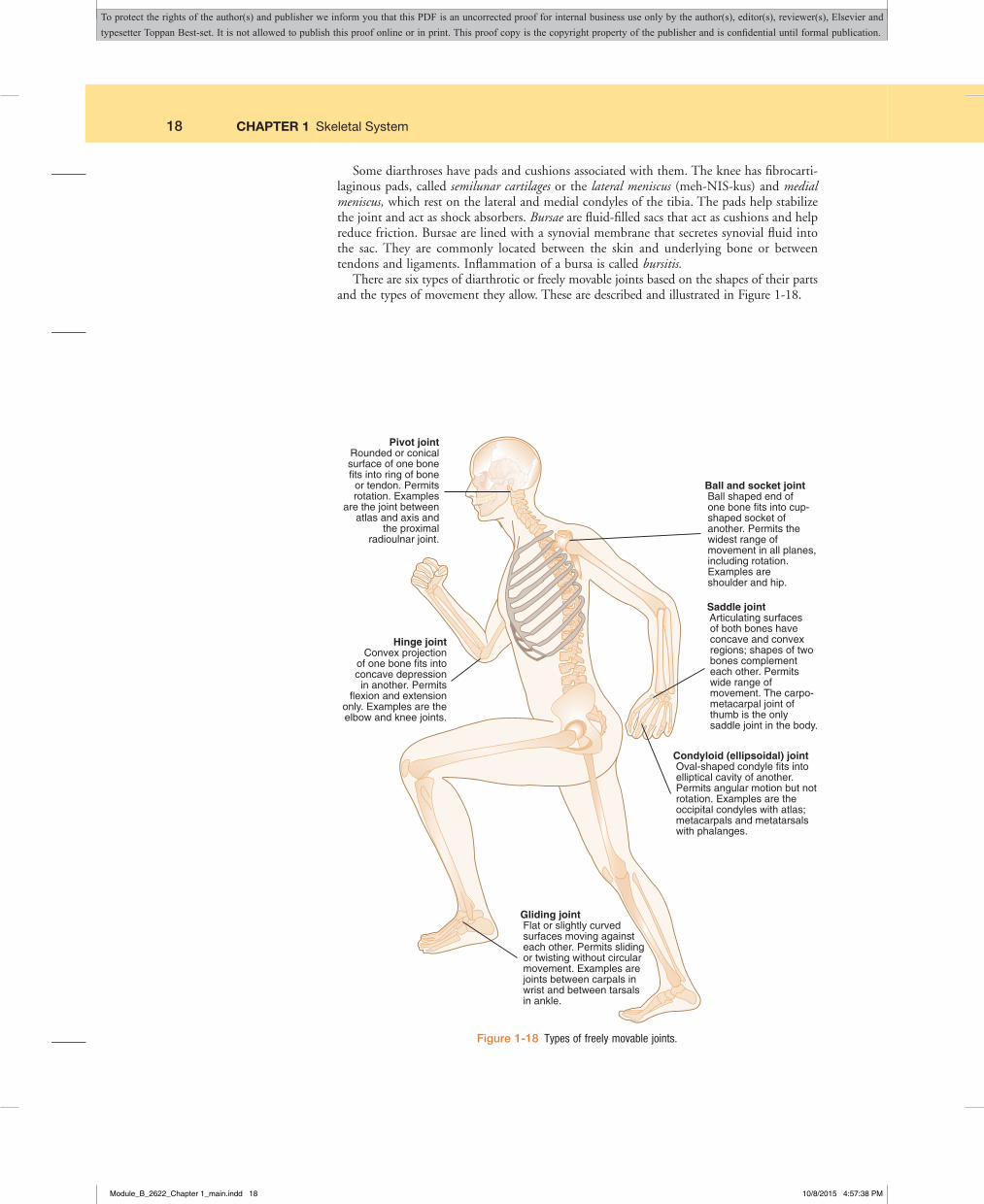
Some diarthroses have pads and cushions associated with them. The knee has fibrocarti-laginous pads, called semilunar cartilages or the lateral meniscus (meh-NIS-kus) and medial meniscus, which rest on the lateral and medial condyles of the tibia. The pads help stabilize the joint and act as shock absorbers. Bursae are fluid-filled sacs that act as cushions and help reduce friction. Bursae are lined with a synovial membrane that secretes synovial fluid into the sac. They are commonly located between the skin and underlying bone or between tendons and ligaments. Inflammation of a bursa is called bursitis.
There are six types of diarthrotic or freely movable joints based on the shapes of their parts and the types of movement they allow. These are described and illustrated in Figure 1-18.
Module_B_2622_Chapter 1_main.indd 18 10/8/2015 4:57:38 PM
To protect the rights of the author(s) and publisher we inform you that this PDF is an uncorrected proof for internal business use only by the author(s), editor(s), reviewer(s), Elsevier and typesetter Toppan Best-set. It is not allowed to publish this proof online or in print. This proof copy is the copyright property of the publisher and is confidential until formal publication.

CHAPTER 1 Skeletal System 19
AGING OF THE SKELETAL SYSTEM
The major age-related change in the skeletal system is the loss of calcium from the bones. Calcium loss occurs in both men and women, but it starts at an earlier age and is more severe in women. The exact reasons for the loss are unknown and possibly involve a combination of several factors. These may include an imbalance between osteoblast and osteoclast activity, imbalance between cal-citonin and parathormone levels, reduced absorption of calcium and/or vitamin D from the digestive tract, poor diet, and lack of exercise. Whatever the cause, there is no sure way of preventing the loss, but adequate calcium and vitamin D in the diet may help reduce the effects.
Another change with age is a decrease in the rate of col-lagen synthesis. This means that the bones have less strength
and are more brittle. Bones fracture more readily in elderly individuals, and the healing process may be slow or incom-plete. Tendons and ligaments become less flexible because of the changes in collagen.
The articular cartilage at the ends of bones tends to become thinner and deteriorates with age. This causes joint disorders that are commonly found in older individuals. People also appear to get shorter as they get older. This is caused partially by loss of bone mass and partially by com-pression of the intervertebral discs.
Age-related changes in the skeletal system cannot be prevented. An active and healthy lifestyle with appropriate exercise and an adequate diet help reduce the effect of the changes in the skeletal system.
Ankylosing spondylitis (ANG-kih-loh-sing spahn-dih-LYE-tis)Inflammationofthespinethatischaracterizedbystiffeningofthe spinal joints and ligaments so that movement becomesincreasingly painful and difficult; also called rheumatoid spondylitis
Arthritis(ahr-THRYE-tis)InflammationofajointBunion(BUN-yun)Abnormalswellingofthejointbetweenthebig
toeand thefirstmetatarsalbone, resulting fromabuildupofsofttissuesandbonecausedbychronicirritationfromill-fittingshoes
Carpal tunnel syndrome(KAHR-pullTUH-nulSIN-drohm)Condi-tioncharacterizedbypainandburningsensationsinthefingersandhand, causedbycompressionof themediannerveas itpassesbetweenawrist ligamentandthebonesandtendonsofthewrist
Dislocation (dis-loh-KAY-shun)Displacementofabonefromitsjointwithtearingofligaments,tendons,andarticularcapsule;alsocalledluxation
Gout(GOWT)Aformofacutearthritisinwhichuricacidcrystalsdevelopwithina jointand irritatethecartilage,causingacuteinflammation, swelling, and pain; most commonly occurs inmiddle-agedandoldermen
Lyme disease (LYMEdih-ZEEZ)Abacterialdisease transmittedtohumansbydeerticks;characterizedbyjointstiffness,head-ache, fever andchills, nausea, andbackpain; complicationsincludeseverearthritisandcardiacproblems;earlystagesofthediseaserespondwelltoantibiotics
Osteoarthritis (ahs-tee-oh-ahr-THRYE-tis) A noninflammatorydiseaseof the joints that is characterizedbydegenerationofthearticularcartilageandchangesinthesynovialmembrane;alsocalleddegenerative joint disease(DJD)
Osteomalacia(ahs-tee-oh-mah-LAY-shee-ah)Softeningofbonebecause of inadequate amounts of calciumand phosphorus;bonesbendeasilyandbecomedeformed;inchildhoodthisiscalledrickets
Osteomyelitis (ahs-tee-oh-my-eh-LYE-tis) Inflammation of thebonemarrowcausedbybacteria
Osteoporosis(ahs-tee-oh-por-OH-sis)Decreaseinbonedensityandmass;commonlyoccurs inpostmenopausalwomenasaresult of increased osteoclast activity caused by diminishedestrogenlevels;bonesfractureeasily
Osteosarcoma (ahs-tee-oh-sahr-KOH-mah) Malignant tumorderivedfrombone;alsocalledosteogenic sarcoma;osteoblastsmultiplywithoutcontrolandformlargetumorsinbone
Rheumatoid arthritis (ROO-mah-toydahr-THRYE-tis)Achronicsystemic disease with changes occurring in the connectivetissuesofthebody,especiallythejoints;incontrasttoosteo-arthritis, the symptoms are usually more generalized andsevere; evidence indicates it may be an autoimmunedisease
Spina bifida(SPY-nahBIFF-ih-dah)Adevelopmentalanomalyinwhich the vertebral laminae do not close around the spinalcord,leavinganopeningthroughwhichthecordandmeningesmayormaynotprotrude
Sprain(SPRAYN)Twistingofajointwithpain,swelling,andinjuryto ligaments, tendons, muscles, blood vessels, and nerves;most often occurs in the ankle; more serious than a strain,which is theoverstretchingof themusclesassociatedwithajoint
Talipes (TAL-ih-peez) Congenital deformity of the foot in whichthepatient cannot standwith the soleof the foot flat on theground;alsocalledclubfoot n
Highlight on Conditions Affecting the Skeletal System
Module_B_2622_Chapter 1_main.indd 19 10/8/2015 4:57:38 PM
To protect the rights of the author(s) and publisher we inform you that this PDF is an uncorrected proof for internal business use only by the author(s), editor(s), reviewer(s), Elsevier and typesetter Toppan Best-set. It is not allowed to publish this proof online or in print. This proof copy is the copyright property of the publisher and is confidential until formal publication.

20 CHAPTER 1 Skeletal System
Prefixes for the Anatomy of the Musculoskeletal SystemPrefix Meaning
amphi- bothdia- through, completeendo-, end- withinepi- above, upon
inter- betweenintra- withinperi- surrounding, aroundsyn- together, joined
Prefix Meaning
Suffixes for the Anatomy of the Musculoskeletal SystemSuffixes Meaning
-ar, -al, -ic, -ous, -eal pertaining to-blast embryonic-clast breaking down-cyte cell-genesis production, origin
Suffixes Meaning
-oid resembling, like-physis growth-poiesis formation-sis condition-um structure
Combining Forms for the Anatomy of the Musculoskeletal System
Meaning Combining Formacromion acromi/obone marrow myel/obone oste/o, osse/o, oss/ibursa burs/ocalcaneus (heel bone) calcane/ocarpus (wrist) carp/ocartilage chondr/o, cartilag/ochin ment/o, geni/oclavicle (collarbone) clavicul/o, cleid/ococcyx (tailbone) coccyg/ocondyle condyl/oelbow (olecranon) olecran/oepicondyle epicondyl/oethmoid ethmoid/ofemur (thigh bone) femor/ofibula (lower lateral leg
bone)fibul/o, perone/o
finger, toe (whole), digitus dactyl/o, digit/oforamen foramin/ofrontal bone front/oglenoid glen/ohallux halluc/ohumerus (upper arm
bone)humer/o
ilium ili/oischium ischi/ojaw (entire) gnath/ojoint (articulation) arthr/o, articul/olacrima lacrim/olamina lamin/oligament ligament/o, syndesm/olower back lumb/omandible (lower jaw bone) mandibul/omaxilla (upper jaw bone) maxill/omeniscus menisc/o
Meaning Combining Form
metacarpus (hand bone) metacarp/ometatarsus (foot bone) metatars/omuscle my/o, myos/o, muscul/oneck cervic/oocciput occipit/oolecranon olecran/opalatine bone palat/oparietal bone pariet/opatella (kneecap) patell/o, patell/apelvis pelv/i, pelv/ophalanx (one of the bones
of the fingers or toes)phalang/o
pubis (pubic bone) pub/oradius (lower lateral arm
bone)radi/o
rib (costa) cost/osacrum sacr/oscapula (shoulder blade) scapul/osinus sin/o, sinus/o, antr/oskeleton skelet/oskull (cranium) crani/osphenoid sphenoid/ospinal column, spine spin/o, rachi/o, vertebr/osternum, breastbone stern/otarsus (anklebone) tars/otemporal bone tempor/otendon tendin/o, tendon/o, ten/o,
tend/othorax (chest) thorac/otibia (shinbone) tibi/oulna uln/overtebra (backbone) vertebr/o, spondyl/ovomer vomer/oxiphoid process xiph/ozygoma (cheekbone) zygomat/o
Module_B_2622_Chapter 1_main.indd 20 10/8/2015 4:57:39 PM
To protect the rights of the author(s) and publisher we inform you that this PDF is an uncorrected proof for internal business use only by the author(s), editor(s), reviewer(s), Elsevier and typesetter Toppan Best-set. It is not allowed to publish this proof online or in print. This proof copy is the copyright property of the publisher and is confidential until formal publication.

CHAPTER 1 Skeletal System 21
EXERCISE 2: Emergency Room Report
Use the emergency room record above to answer the following questions. 1. Dylan’s injury is to the fleshy area of the palm near the thumb, the “thenar eminence.” Explain the difference between a DIP
(distal interphalangeal joint) and a PIP (proximal interphalangeal) joint.
2. Not being able to “flex” a body part means one is unable to .
3. “First digit, R hand” means .
4. The first metacarpal is a bone of the .
…
KOLDMANN, DYLAN M. - 507940 Opened by Bradley Oppenheimer, MDTask Edit View
Flowsheet: Level:ED ED Record Table Group List
Time Scale Options Help
KOLDMANN, DYLAN M. Age: 7 yearsDOB: 1/27/2004
Sex: MaleMRN: 507940
Loc: ARHFIN: 3506004
Navigator
PROD MAHAFC
As Of 16:10
26 March 2011 16:10
ED Record
Reference Text Browser
Orders ED Lab Surgery Pt. Info Pt. Schedule Task List I & O MARClinical NotesAssessmentsRadiologyLast 48 Hours
Form Browser Medication Profile
7-year-old sustained injury to right hand when fell off bike. Pain over thenar eminence. Able to bend at his wrist and flex at his DIP and PIP joints and every digit of the hand with exception of first digit.
Exam of right hand is significant for mild protrusion but no ecchymosis and minimal edema overlying thenar eminence of the right hand. Good wrist mobility. X-ray significant for what appears to be a Salter-Harris fracture of the first metacarpal. Immobile and follow-up tomorrow with Ortho for possible cast.
inter- betweenintra- withinperi- surrounding, aroundsyn- together, joined
-oid resembling, like-physis growth-poiesis formation-sis condition-um structure
metacarpus (hand bone) metacarp/ometatarsus (foot bone) metatars/omuscle my/o, myos/o, muscul/oneck cervic/oocciput occipit/oolecranon olecran/opalatine bone palat/oparietal bone pariet/opatella (kneecap) patell/o, patell/apelvis pelv/i, pelv/ophalanx (one of the bones
of the fingers or toes)phalang/o
pubis (pubic bone) pub/oradius (lower lateral arm
bone)radi/o
rib (costa) cost/osacrum sacr/oscapula (shoulder blade) scapul/osinus sin/o, sinus/o, antr/oskeleton skelet/oskull (cranium) crani/osphenoid sphenoid/ospinal column, spine spin/o, rachi/o, vertebr/osternum, breastbone stern/otarsus (anklebone) tars/otemporal bone tempor/otendon tendin/o, tendon/o, ten/o,
tend/othorax (chest) thorac/otibia (shinbone) tibi/oulna uln/overtebra (backbone) vertebr/o, spondyl/ovomer vomer/oxiphoid process xiph/ozygoma (cheekbone) zygomat/o
Module_B_2622_Chapter 1_main.indd 21 10/8/2015 4:57:39 PM
To protect the rights of the author(s) and publisher we inform you that this PDF is an uncorrected proof for internal business use only by the author(s), editor(s), reviewer(s), Elsevier and typesetter Toppan Best-set. It is not allowed to publish this proof online or in print. This proof copy is the copyright property of the publisher and is confidential until formal publication.

22 CHAPTER 1 Skeletal System
EXERCISE 3: Congenital Disorders
Build the terms.
1. Process of joined fingers/toes
2. Condition of formation without cartilage
3. Process of many fingers/toes
PATHOLOGY
Figure1-19 Polydactyly.
Figure1-20 Syndactyly.
TERMINOLOGY REVIEW
Term Word Origin Definitionachondroplasia a- no, not, without
chondr/o cartilage-plasia condition of formation
Disorder of the development of cartilage at the epiphyses of the long bones and skull, resulting in dwarfism.
polydactyly poly- many, muchdactyl/o fingers, toes-y process of
Condition of more than five fingers or toes on each hand or foot (Figure 1-19).
spina bifida occulta spin/o spinebi- two-fida to splitocculta hidden
Congenital malformation of the bony spinal canal without involvement of the spinal cord.
syndactyly syn- joined, togetherdactyl/o fingers, toes-y process of
Condition of the joining of the fingers or toes, giving them a webbed appearance (Figure 1-20).
Terms Related to Congenital Conditions (QØØ-Q99)
Module_B_2622_Chapter 1_main.indd 22 10/8/2015 4:57:40 PM
To protect the rights of the author(s) and publisher we inform you that this PDF is an uncorrected proof for internal business use only by the author(s), editor(s), reviewer(s), Elsevier and typesetter Toppan Best-set. It is not allowed to publish this proof online or in print. This proof copy is the copyright property of the publisher and is confidential until formal publication.

CHAPTER 1 Skeletal System 23
TERMINOLOGY REVIEW
Term Word Origin Definitionarthrosis arthr/o joint
-osis abnormal conditionAbnormal condition of a joint; may be hemarthrosis, hydrarthrosis, or
pyarthrosis (blood, fluid, or pus respectively, in a joint cavity).bunion bunion/o bunion Fairly common, painful enlargement and inflammation of the first
metatarsophalangeal joint (the base of the great toe). Also called hallux valgus.
contracture con- togethertract/o pulling-ure condition
Chronic fixation of a joint in flexion (such as a finger) caused by atrophy and shortening of muscle fibers after a long period of disuse.
crepitus crepit/o crackling-us thing
Crackling sound heard in joints.
gout Type of arthritis due to excessive uric acid that causes crystals to form. The joints then become swollen and inflamed.
osteoarthritis (OA) oste/o bonearthr/o joint-itis inflammation
Joint disease characterized by degenerative articular cartilage and a wearing down of the bones’ edges at a joint; considered a “wear and tear” disorder. Also called degenerative joint disease (DJD) (Figure 1-21).
osteophytosis oste/o bonephyt/o growth, nature-osis abnormal condition
Abnormal bone growth in a joint. Bouchard nodes are osteophytes of the proximal interphalangeal joints in rheumatoid arthritis (Figure 1-22).
rheumatoid arthritis (RA) rheumat/o watery flow-oid resembling, likearthr/o joint-itis inflammation
Inflammatory joint disease believed to be autoimmune in nature; occurs in a much younger population (ages 20 to 45) than OA (see Figure 1-22). Diagnosed with a rheumatoid factor test.
temporomandibular joint disorder (TMJ)
tempor/o temporal bonemandibul/o lower jaw-ar pertaining to
Dysfunctional temporomandibular joint, accompanied by gnathalgia (jaw pain).
Cervical vertebrae
Hip
First carpometacarpal
Distal interphalangeal
Knee
First metatarsophalangeal
Lower lumbar vertebrae
Terms Related to Arthropathies (MØØ-M25) and Dentofacial Anomalies (M26-M27)
A B
Figure 1-21 Joints most frequently involved in osteoarthritis.
Figure1-22 A, Bouchard’s nodes seen in rheumatoid arthritis of the hands. Moder-ate involvement. B, Arthrogram of wrist showing RA and resultant osteophytosis.
Module_B_2622_Chapter 1_main.indd 23 10/8/2015 4:57:42 PM
To protect the rights of the author(s) and publisher we inform you that this PDF is an uncorrected proof for internal business use only by the author(s), editor(s), reviewer(s), Elsevier and typesetter Toppan Best-set. It is not allowed to publish this proof online or in print. This proof copy is the copyright property of the publisher and is confidential until formal publication.

24 CHAPTER 1 Skeletal System
TERMINOLOGY REVIEW
Term Word Origin Definitionankylosing spondylitis ankyl/o stiffening
spondyl/o vertebra-itis inflammation
Chronic inflammatory disease of idiopathic origin, which causes a fusion of the spine.
herniated intervertebral disk inter- betweenvertebr/o vertebra-al pertaining to
Protrusion of the central part of the disk that lies between the vertebrae, resulting in compression of the nerve root and pain.
kyphosis kyph/o round back-osis abnormal condition
Extreme posterior curvature of the thoracic area of the spine (Figure 1-23, A ).
lordosis lord/o swayback-osis abnormal condition
Swayback; exaggerated anterior curve of the lumbar vertebrae (lower back) (Figure 1-23, B ).
sciatica Inflammation of the sciatic nerve. Symptoms include pain and tenderness along the path of the nerve through the thigh and leg.
scoliosis scoli/o curvature-osis abnormal condition
Lateral S curve of the spine that can cause an individual to lose inches in height (Figure 1-23, C ).
spinal stenosis spin/o spine-al pertaining tostenosis abnormal condition of narrowing
Abnormal condition of narrowing of the spinal canal with attendant pain, sometimes caused by osteoarthritis or spondylolisthesis (Figure 1-24).
spondylolisthesis spondyl/o vertebra-listhesis slipping
Condition resulting from the partial forward dislocation of one vertebra over the one beneath it.
spondylosis spondyl/o vertebra-osis abnormal condition
An abnormal condition characterized by stiffening of the vertebral joints.
Terms Related to Systemic Connective Tissue Disorders (M3Ø-M36) and Deforming Dorsopathies (M4Ø-M54)
Lordosis
B CA
Kyphosis ScoliosisLaminaSpinous process
Vertebral body
Spinal nervesTransverseprocessSpinal cord
Figure1-23 A, Kyphosis, B, Lordosis, C, Scoliosis. Figure 1-24 Spinal stenosis. Bony overgrowth has narrowed the spinal canal and pinched the spinal nerves.
Module_B_2622_Chapter 1_main.indd 24 10/8/2015 4:57:43 PM
To protect the rights of the author(s) and publisher we inform you that this PDF is an uncorrected proof for internal business use only by the author(s), editor(s), reviewer(s), Elsevier and typesetter Toppan Best-set. It is not allowed to publish this proof online or in print. This proof copy is the copyright property of the publisher and is confidential until formal publication.
CHAPTER 1 Skeletal System 25
EXERCISE 4: Arthropathies and Dentofacial Anomalies; Systemic Connective Tissue Disorders and Dorsopathies
Fill in the blanks using the terms from the list below.
rheumatoid arthritis, crepitus, bunion, TMJ, osteophytosis, ankylosing spondylitis, spinal stenosis, herniated intervertebral disk, contracture, lordosis, arthrosis, gout
1. An abnormal condition of a joint is called .
2. A painful enlargement and inflammation of the great toe is .
3. Chronic fixation of a joint in flexion is called .
4. _______________ is a crackling sound heard in joints.
5. A type of arthritis due to excessive uric acid that causes crystals to form is .
6. Abnormal bone growth in a joint is called .
7. _______________ is an autoimmune inflammatory joint disease.
8. _______________ is a dysfunctional temporomandibular joint, accompanied by gnathalgia.
9. A chronic inflammatory disease that causes fusion of the spine .
10. A protrusion of the central part of the disk that lies between the vertebrae is called .
11. Another word for swayback is .
12. An abnormal, painful narrowing of the spinal canal is called .
Build the terms.
13. inflammation of a bone and joint
14. condition of slipping of the vertebrae
15. abnormal condition of vertebra
16. abnormal condition of curvature
17. abnormal condition of round back
TERMINOLOGY REVIEW
Term Word Origin Definitionbursitis burs/o bursa
-itis inflammationInflammation of a bursa.
Terms Related to Soft Tissue Disorders (M6Ø-M79)
Module_B_2622_Chapter 1_main.indd 25 10/8/2015 4:57:43 PM
To protect the rights of the author(s) and publisher we inform you that this PDF is an uncorrected proof for internal business use only by the author(s), editor(s), reviewer(s), Elsevier and typesetter Toppan Best-set. It is not allowed to publish this proof online or in print. This proof copy is the copyright property of the publisher and is confidential until formal publication.

26 CHAPTER 1 Skeletal System
TERMINOLOGY REVIEW
Term Word Origin Definitionchondromalacia chondr/o cartilage
-malacia softeningSoftening of the cartilage.
costochondritis cost/o ribchondr/o cartilage-itis inflammation
Inflammation of the cartilage of the ribs.
osteitis deformans oste/o bone-itis inflammationdeformans misshapen
Misshaped bone resulting from inflammation. Also known as Paget’s disease of the bone.
osteomalacia oste/o bone-malacia softening
Softening of bone caused by loss of minerals from the bony matrix as a result of vitamin D deficiency. When osteomalacia occurs in childhood, it is called rickets.
osteomyelitis oste/o bonemyel/o bone marrow-itis inflammation
Inflammation of the bone and bone marrow.
osteoporosis oste/o bonepor/o passage-osis abnormal condition
Loss of bone mass, which results in the bones being fragile and at risk for fractures (Figure 1-25). Osteopenia refers to a less severe bone mass loss.
Terms Related to Osteopathies and Chondropathies (M8Ø-M94)
AB
Figure1-25 A, The hallmark of osteoporosis: the dowager hump. Affected persons lose height, have a bent spine, and appear to sink into their hips. B, X-ray demonstrating a compression fracture of T12 and L1 subsequent to osteoporosis.
EXERCISE 5: Soft Tissue Disorders; Osteopathies and Chondropathies
Fill in the blanks using the terms from the list below.
bursitis, costochondritis, osteitis deformans, osteomalacia, osteomyelitis
1. Inflammation of the cartilage of the ribs is called .
2. Inflammation of a bursa is called .
3. Paget’s disease of the bone is also known as .
Module_B_2622_Chapter 1_main.indd 26 10/8/2015 4:57:43 PM
To protect the rights of the author(s) and publisher we inform you that this PDF is an uncorrected proof for internal business use only by the author(s), editor(s), reviewer(s), Elsevier and typesetter Toppan Best-set. It is not allowed to publish this proof online or in print. This proof copy is the copyright property of the publisher and is confidential until formal publication.

CHAPTER 1 Skeletal System 27
4. Softening of the bone is called .
5. Inflammation of the bone and bone marrow is called .
Decode the terms.
6. chondromalacia
7. osteoporosis
Trauma
FracturesPut simply, a fracture (fx, #) is a broken bone. However, there are a number of types of breaks, each with its own name. Most fractures occur as a result of trauma, but some can result from an underlying disease, such as osteoporosis or cancer; these pathologic fractures are also sometimes called spontaneous fractures. All fractures may be classified into simple (closed) or compound (open) fractures. Fractures are additionally characterized as nondisplaced fractures, meaning that the broken bones are still in alignment, or displaced fractures, meaning that the ends of the fractured bones are not in alignment (Figure 1-26). The break in a simple fracture does not rupture the skin, but a compound fracture splits open the skin, which allows more opportunity for infection to take place. See the table in Termi-nology Review for different types of fractures.
Sprain/Strain and Dislocation/SubluxationA sprain is a traumatic injury to a joint involving the ligaments. Swelling, pain, and discol-oration of the skin may be present. The severity of the injury is measured in grades.
A bone that is completely out of its place in a joint is called a dislocation. If the bone is partially out of the joint, it is considered to be a subluxation. This can be a congenital or an acquired condition.
19a. Code ExtensionsMostcategories inChapter19have7thcharacterextensions thatare required foreachapplicablecode.Mostcategoriesinthischapterhavethreeextensions(withtheexceptionoffractures):A,initialencounter;D,subsequentencounter;andS,sequela.
Extension“A”,initialencounterisusedwhilethepatientisreceivingactivetreatmentfortheinjury.Examplesofactivetreatmentare:surgicaltreatment,emergencydepartmentencounter,andevalua-tionandtreatmentbyanewphysician.
Extension“D”subsequentencounterisusedforencountersafterthepatienthasreceivedactivetreatmentoftheinjuryandisreceivingroutinecarefortheinjuryduringthehealingorrecoveryphase.Examples of subsequent care are: cast change or removal, removal of externalor internal fixationdevice,medicationadjustment,otheraftercareandfollowupvisitsfollowinginjurytreatment.
TheaftercareZcodesshouldnotbeusedforaftercareforinjuries.Foraftercareofaninjury,assigntheacuteinjurycodewiththe7thcharacter“D”(subsequentencounter).
Extension“S”,sequela,isforuseforcomplicationsorconditionsthatariseasadirectresultofaninjury,suchasscarformationafteraburn.Thescarsaresequelaeoftheburn.Whenusingextension“S”, it is necessary touseboth the injury code that precipitated the sequela and the code for thesequela itself. The “S” is added only to the injury code, not the sequela code. The “S” extensionidentifiestheinjuryresponsibleforthesequela.Thespecifictypeofsequela(e.g.scar)issequencedfirst,followedbytheinjurycode.
GUIDELINE ALERT
Displacedfracture
Non-displacedfracture
Figure 1-26 Nondisplaced and displaced fractures.
Module_B_2622_Chapter 1_main.indd 27 10/8/2015 4:57:44 PM
To protect the rights of the author(s) and publisher we inform you that this PDF is an uncorrected proof for internal business use only by the author(s), editor(s), reviewer(s), Elsevier and typesetter Toppan Best-set. It is not allowed to publish this proof online or in print. This proof copy is the copyright property of the publisher and is confidential until formal publication.

28 CHAPTER 1 Skeletal System
19.c. Coding of Traumatic FracturesTheprinciplesofmultiplecodingofinjuriesshouldbefollowedincodingfractures.Fracturesofspeci-fiedsitesarecodedindividuallybysiteinaccordancewithboththeprovisionswithinspecificcategoriesandthelevelofdetailfurnishedbymedicalrecordcontent.
A fracturenot indicatedas openor closed shouldbe coded to closed.A fracturenot indicatedwhetherdisplacedornotdisplacedshouldbecodedtodisplaced.
GUIDELINE ALERT
TheGastiloclassificationforopenfracturesisusedtogivemoreinformationaboutthetypeofwoundcausedbythebreak.Itincludesinformationaboutthesizeofthewound,presenceorabsenceofcontamination,andadditionalcomplications.NO
TE
TERMINOLOGY REVIEW
Type DefinitionFRACTURES (Figure 1-27)
avulsion Part of the bone is pulled away by a tendon or ligament.Colles’ Fracture at distal end of the radius at the epiphysis. Often occurs when
patient has attempted to break his/her fall.comminuted Bone is crushed and/or shattered into multiple pieces.complicated Bone is broken and pierces an internal organ.compression Fractured area of bone collapses on itself.greenstick Partially bent and partially broken. Relatively common in children.hairline Minor fracture appearing as a thin line on x-ray. May not extend through
bone.impacted Broken bones with ends driven into each other.Salter-Harris Fracture of epiphyseal plate in children.torus Compression injury in children in which the bone does not break, but
buckles because of its pliable nature. Also called a buckle fracture.OTHER TRAUMA (Figures 1-28 and 1-29)
dislocation Bone that is completely out of its joint socket.subluxation Partial dislocation.sprain Traumatic injury to ligaments of a joint, including tearing of a ligament.
Normal Anteriordislocation
PosteriordislocationA B C
Sprain (ligament)
Terms Related To Injury, Poisoning, and Certain Other Consequences of External Causes (SØØ-T88)
Figure1-28 Normal shoulder (A). Dislocated shoulder, anterior (B) and posterior (C). Figure1-29 Sprain.
Comminuted Compression
Colles’ Complicated
Impacted Hairline
Greenstick Salter-Harris
Avulsion fracture
Figure1-27 Fractures.
Module_B_2622_Chapter 1_main.indd 28 10/8/2015 4:57:45 PM
To protect the rights of the author(s) and publisher we inform you that this PDF is an uncorrected proof for internal business use only by the author(s), editor(s), reviewer(s), Elsevier and typesetter Toppan Best-set. It is not allowed to publish this proof online or in print. This proof copy is the copyright property of the publisher and is confidential until formal publication.

CHAPTER 1 Skeletal System 29
EXERCISE 6: Fractures
Match the fractures with their definitions.
____ 1. complicated____ 2. greenstick____ 3. Colles’____ 4. impacted____ 5. comminuted____ 6. simple/closed____ 7. compound/open____ 8. hairline____ 9. pathologic
A. broken bone pierces internal organ. B. broken bone pierces skin. C. spontaneous fracture as a result of disease. D. bone is partially bent and partially broken. E. bone is broken, skin is closed. F. distal end of radius is broken.G. ends of broken bone are driven into each other. H. fracture appears as a line on the bone and fracture may not
be completely through bone. I. bone is crushed.
EXERCISE 7: Other Trauma
1. A partial displacement of a bone at a joint is a _______________________________________________; full displacement
is a _______________________________________________________________________________________________________.
2. An injury that can be described in grades and involves the soft tissue of a joint is a
___________________________________________________________________________________________________________.
TERMINOLOGY REVIEW
Term Word Origin Definitionchondroma chondr/o cartilage
-oma tumorBenign tumor of the cartilage, usually occurring in children and
adolescents.exostosis ex- out
oste/o bone-osis abnormal condition
Abnormal condition of bony growth. Also called hyperostosis and osteochondroma.
osteoma oste/o bone-oma tumor
Benign bone tumor, usually of compact bone.
Terms Related to Benign Neoplasms (D1Ø-D36)
TERMINOLOGY REVIEW
Term Word Origin Definitionchondrosarcoma chondr/o cartilage
-sarcoma connective tissue cancerMalignant tumor of the cartilage. Occurs most
frequently in adults (Figure 1-30).osteosarcoma oste/o bone
-sarcoma connective tissue cancerMalignant tumor of bone. Also called Ewing’s
sarcoma. Most common children’s bone cancer.
Terms Related to Malignant Neoplasms (CØØ-C96)
A B
Figure 1-30 A, Chondrosarcoma of femur. B, X-ray showing prominent dense calcification in a large neoplas-tic mass.
Module_B_2622_Chapter 1_main.indd 29 10/8/2015 4:57:45 PM
To protect the rights of the author(s) and publisher we inform you that this PDF is an uncorrected proof for internal business use only by the author(s), editor(s), reviewer(s), Elsevier and typesetter Toppan Best-set. It is not allowed to publish this proof online or in print. This proof copy is the copyright property of the publisher and is confidential until formal publication.

30 CHAPTER 1 Skeletal System
EXERCISE 8: Neoplasms
Match the neoplasms with their definitions.
____ 1. osteosarcoma____ 2. chondrosarcoma
A. connective tissue cancer of bone B. connective tissue cancer of cartilage
…
Herold, Jean F - 12438 Opened by Landrey, Melissa, PATask Edit View
Flowsheet: Level:Assessments History & Physical Table Group List
Time Scale Options Help
Herold, Jean F Age: 54 yearsDOB: 7/23/1957
Sex: FemaleMRN: 12438
Loc: AR-OCFIN: 8425633
Navigator
PROD MAHAFC
As Of 09:19
22 Jan 2012 09:19
History & Physical
Reference Text Browser
Orders ED Lab Surgery Pt. Info Pt. Schedule Task List I & O MARClinical NotesAssessmentsRadiologyLast 48 Hours
Form Browser Medication Profile
CHIEF COMPLAINT: Right shoulder pain/fracture
HISTORY OF PRESENT ILLNESS: Patient is a 54-year-old female who works as a health-care worker. While out exercising last night, she fell on her right shoulder. She has a comminuted fracture of the proximal humerus involving the humeral head, extending into the joint space. Admitted for observation and analgesia. CT of shoulder reveals the need for a humeral prosthesis. Some discomfort with deep inspiration. Unclear whether this is in the shoulder or possible right chest wall.
PAST MEDICAL HISTORY: Cholecystectomy 1986. ORIF left forearm, fracture same forearm, age 9. Has some dependent edema and takes Lasix 80 mg daily for it. Does not wear compression stockings as they make her feet feel cold. Also diagnosis of fibromyalgia in 2008.
FAMILY HISTORY: Mother died age 64 post surgical pulmonary embolus.
REVIEW OF SYSTEMS: Negative.
PHYSICAL EXAM: Pleasant, uncomfortable, overweight female appearing her stated age and in no distress. HEENT normal, neck supple, thyroid normal. No JVD, carotids normal. Lungs decreased breath sounds at bases. Heart regular rate and rhythm. Extremities: normal range of motion of lower extremities. Motor sensory deep tendon reflexes are normal in arm. Trace pretibial edema bilaterally without venostasis changes. Excellent peripheral pulses. Cannot adduct her arm and shoulder without pain. X-ray of shoulder and CT show comminuted fracture.
ASSESSMENT: Comminuted right proximal humeral fracture involving humeral head.
PLAN: Admit for analgesia, IV fluids. Has a little nausea probably from analgesics. Won’t have surgery until tomorrow. Preoperative labs, EKG, and chest x-ray will be obtained before that time.
Module_B_2622_Chapter 1_main.indd 30 10/8/2015 4:57:46 PM
To protect the rights of the author(s) and publisher we inform you that this PDF is an uncorrected proof for internal business use only by the author(s), editor(s), reviewer(s), Elsevier and typesetter Toppan Best-set. It is not allowed to publish this proof online or in print. This proof copy is the copyright property of the publisher and is confidential until formal publication.

CHAPTER 1 Skeletal System 31
Build the term.
3. Benign bone tumor
4. Benign tumor of cartilage
5. An abnormal condition of out(growth) of bone
EXERCISE 9: Admission Record
Using the admission record above, answer the following questions:
1. Which bone did she break while exercising? Give the medical and English names.
2. Did she fracture the area closest to her shoulder or farther from her shoulder? Circle one.
3. Describe the type of fracture sustained.
4. What does “cannot adduct her arm and shoulder without pain” mean?
Beawarethatmanyofthemusculoskeletalproceduralcodesaredependentonthecorrectidentificationofaleftorrightanatomicalterm.Alittlecommonsense(certainlythereareleftandrightkneecaps,butonlyonesacrum)andknowledgeoftheanatomyofthatparticularsystemwillhelpwhencheckingone’swork.
NOTE
PROCEDURES
B2.1b Bodysystemsdesignatedasupperor lowercontainbodyparts locatedaboveorbelow thediaphragmrespectively.
Example:Jointsarecategorizedaseitherupperorlowerjoints.Thedividinglineistheendofthethoracicvertebrae(upper)andthebeginningofthelumbarvertebrae(lower).Thethoracolumbarjointanddiscarepartoftheupperbodydesignation.
GUIDELINE ALERT
This is where you reap the benefits of learning the bone processes, as they are used within the procedural part of coding to address specific parts of bones and name many muscles, ligaments, and tendons. With the general terms for the bones, you’ll need to decide if it’s a procedure on the head and facial bones, upper bones, or lower bones. With the more specific terms, you’ll need to think back to where the bone is in the skeleton and how it would be categorized.
B4.1b If theprefixperi- isusedwithabodypart to identify theprocedure, thebodypart value isdefinedasthatbodypart.
Example:peripatellaplastycodedasapatellaplasty.
GUIDELINE ALERT
BE CAREFUL! Geni/omeans chin, while-genmeans producing andgen/umeans knee.
BE CAREFUL! Ment/omeans both chin and mind.
Module_B_2622_Chapter 1_main.indd 31 10/8/2015 4:57:46 PM
To protect the rights of the author(s) and publisher we inform you that this PDF is an uncorrected proof for internal business use only by the author(s), editor(s), reviewer(s), Elsevier and typesetter Toppan Best-set. It is not allowed to publish this proof online or in print. This proof copy is the copyright property of the publisher and is confidential until formal publication.

32 CHAPTER 1 Skeletal System
TERMINOLOGY REVIEW
Term Word Origin Definitionacetabuloplasty acetabul/o acetabulum
-plasty surgically formingReconstruction of the acetabulum to correct hip dysplasia,
remove metastatic lesions, or as part of hip replacement surgery.
alveoloplasty alveol/o small cavity-plasty surgically forming
Surgically shaping the alveolus of the jaw in preparation for dentures.
amputation Removal of a limb when there are no feasible options to save it.bunionectomy bunion/o bunion
-ectomy cutting outCutting off a bunion (Figure 1-31).
carpectomy carp/o carpus, wrist bone-ectomy cutting out
Cutting off part or all of a wrist bone. A proximal row carpectomy is done to reduce pain but maintain ROM.
chondrectomy chondr/o cartilage-ectomy cutting out
Surgically cutting off part or all of a damaged cartilage.
claviculectomy clavicul/o collarbone, clavicle-ectomy cutting out
Cutting off part or all of a collarbone. A partial (or distal) claviculectomy is performed to reduce pain in the acromioclavicular (AC) joint due to osteoarthritis.
condylotomy condyl/o condyle-tomy cutting
Cutting a condyle, e.g., to treat TMJ.
costectomy cost/o rib-ectomy cutting out
Cutting out part or all of a rib. May be done as part of a procedure to treat scoliosis.
densitometry densit/o density-metry measuring
Process of measuring bone density. An example is dual energy x-ray absorptiometry (DEXA) (Figure 1-32).
discography disc/o disc-graphy recording
X-ray recording of an intervertebral disc using a contrast medium to establish a diagnosis for herniated discs.
genioplasty geni/o chin-plasty surgically forming
Surgical formation to augment or reduce the size of the chin. Also called mentoplasty.
kyphoplasty kyph/o round back-plasty surgically forming
Minimally invasive procedure designed to address the pain of fractured vertebrae resulting from osteoporosis or cancer. A balloon is used to inflate the area of fracture before a cementlike substance is injected. The substance hardens rapidly, and pain relief is immediate in most patients (Figure 1-33).
Terms Related to Bone Procedures
Figure1-31 Bunionectomy.
Medial eminenceof metatarsal bone
is removed
Module_B_2622_Chapter 1_main.indd 32 10/8/2015 4:57:46 PM
To protect the rights of the author(s) and publisher we inform you that this PDF is an uncorrected proof for internal business use only by the author(s), editor(s), reviewer(s), Elsevier and typesetter Toppan Best-set. It is not allowed to publish this proof online or in print. This proof copy is the copyright property of the publisher and is confidential until formal publication.

CHAPTER 1 Skeletal System 33
X-ray fanbeam
Linearscan path
AB
TERMINOLOGY REVIEW—cont’d
Terms Related to Bone Procedures
Term Word Origin Definitionmandibulectomy mandibul/o mandible, lower jaw
-ectomy cutting outCutting off part or all of the lower jaw. May be done to treat
cancer of the jaw.metatarsectomy metatars/o metatarsus, foot
bone-ectomy cutting out
Cutting off part or all of a foot bone to surgically treat intractable metatarsal pain.
osteoclasis oste/o bone-clasis breaking
Surgical fracture of a bone to correct a malformation.
patellapexy patell/a kneecap, patella-pexy fixation
Fixing the kneecap to the femur to stabilize the joint.
sacrectomy sacr/o sacrum-ectomy cutting out
Cutting off part or all of the sacrum usually to remove an attached tumor.
scapulopexy scapul/o shoulder blade, scapula
-pexy fixation
Fixing the shoulder blade in place to treat a protruding shoulder blade.
sequestrectomy sequestr/o sequestrum-ectomy cutting out
Cutting out a necrosed (dead) bone fragment to prevent or correct possible complications.
spondylosyndesis spondyl/o vertebrasyn- joined, together-desis binding
Binding the vertebrae together to stabilize the spine. Also called spinal fusion and spondylodesis.
sternotomy stern/o breastbone, sternum-tomy cutting
Cutting the sternum to allow access to the heart and thoracic cavity.
tarsectomy tars/o tarsus, ankle bone-ectomy cutting out
Cutting out one of the tarsal bones. May be done to correct an abnormally high arch of the foot.
Figure 1-32 Dual energy x-ray absorptiometry (DEXA). A, DEXA system. B, Scan of lumbar vertebrae.
Figure1-33 Kyphoplasty.
Module_B_2622_Chapter 1_main.indd 33 10/8/2015 4:57:47 PM
To protect the rights of the author(s) and publisher we inform you that this PDF is an uncorrected proof for internal business use only by the author(s), editor(s), reviewer(s), Elsevier and typesetter Toppan Best-set. It is not allowed to publish this proof online or in print. This proof copy is the copyright property of the publisher and is confidential until formal publication.
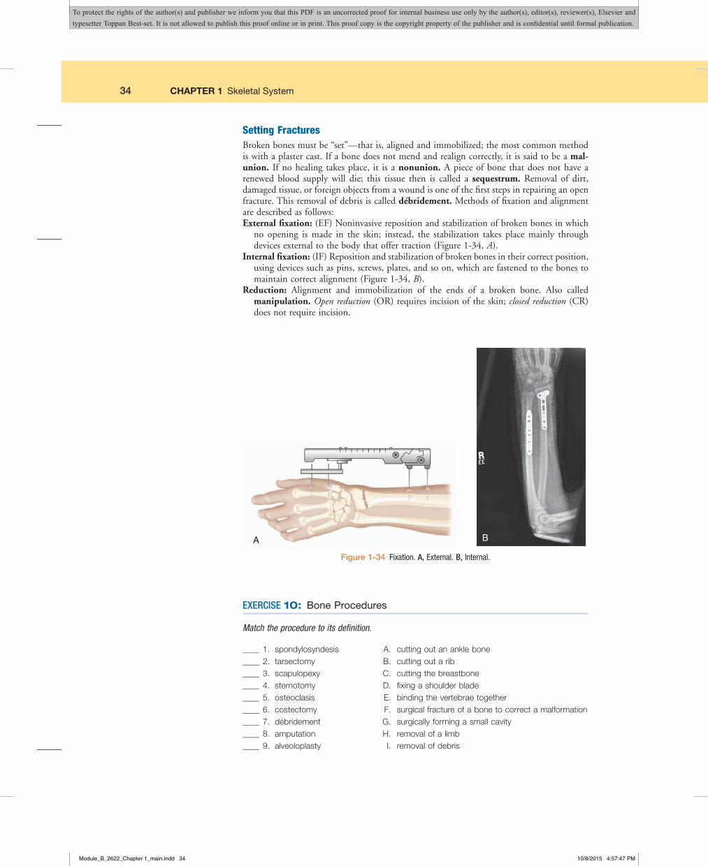
34 CHAPTER 1 Skeletal System
Setting FracturesBroken bones must be “set”—that is, aligned and immobilized; the most common method is with a plaster cast. If a bone does not mend and realign correctly, it is said to be a malunion. If no healing takes place, it is a nonunion. A piece of bone that does not have a renewed blood supply will die; this tissue then is called a sequestrum. Removal of dirt, damaged tissue, or foreign objects from a wound is one of the first steps in repairing an open fracture. This removal of debris is called débridement. Methods of fixation and alignment are described as follows:External fixation: (EF) Noninvasive reposition and stabilization of broken bones in which
no opening is made in the skin; instead, the stabilization takes place mainly through devices external to the body that offer traction (Figure 1-34, A).
Internal fixation: (IF) Reposition and stabilization of broken bones in their correct position, using devices such as pins, screws, plates, and so on, which are fastened to the bones to maintain correct alignment (Figure 1-34, B).
Reduction: Alignment and immobilization of the ends of a broken bone. Also called manipulation. Open reduction (OR) requires incision of the skin; closed reduction (CR) does not require incision.
A B
Figure1-34 Fixation. A, External. B, Internal.
EXERCISE 10: Bone Procedures
Match the procedure to its definition.
____ 1. spondylosyndesis____ 2. tarsectomy____ 3. scapulopexy____ 4. sternotomy____ 5. osteoclasis____ 6. costectomy____ 7. débridement____ 8. amputation____ 9. alveoloplasty
A. cutting out an ankle bone B. cutting out a rib C. cutting the breastbone D. fixing a shoulder blade E. binding the vertebrae together F. surgical fracture of a bone to correct a malformation G. surgically forming a small cavity H. removal of a limb I. removal of debris
Module_B_2622_Chapter 1_main.indd 34 10/8/2015 4:57:47 PM
To protect the rights of the author(s) and publisher we inform you that this PDF is an uncorrected proof for internal business use only by the author(s), editor(s), reviewer(s), Elsevier and typesetter Toppan Best-set. It is not allowed to publish this proof online or in print. This proof copy is the copyright property of the publisher and is confidential until formal publication.

CHAPTER 1 Skeletal System 35
Build the terms.
10. cutting out dead bone
11. fixation of a kneecap
12. measuring density
13. cutting out cartilage
14. surgically forming a chin
15. breaking a bone
16. recording a disc
TERMINOLOGY REVIEW
Term Word Origin Definitionacromioplasty acromi/o acromion
-plasty surgically formingForming the acromion process to correct a defect as in rotator cuff surgery.
arthrocentesis arthr/o joint-centesis surgical puncture
Surgical puncture of a joint to remove fluid, pus, or blood.
arthrodesis arthr/o joint-desis binding
Binding a joint in order to stabilize it.
arthrography arthr/o joint-graphy recording
X-ray recording of a joint.
arthroplasty arthr/o joint-plasty surgically forming
Surgically forming a joint. Examples include total knee replacement (TKR) and total hip replacement (THR) (Figure 1-35).
arthroscopy arthr/o joint-scopy viewing
Internal viewing of a joint, especially the shoulder and knees to aid in the diagnosis of ligament tears and injuries (Figure 1-36).
arthrotomy arthr/o joint-tomy cutting
An incision of a joint, usually as a means of access for a surgical procedure, such as a joint replacement. It may also be performed to provide drainage for infection.
Terms Related To Joint Procedures
Module_B_2622_Chapter 1_main.indd 35 10/8/2015 4:57:47 PM
To protect the rights of the author(s) and publisher we inform you that this PDF is an uncorrected proof for internal business use only by the author(s), editor(s), reviewer(s), Elsevier and typesetter Toppan Best-set. It is not allowed to publish this proof online or in print. This proof copy is the copyright property of the publisher and is confidential until formal publication.

36 CHAPTER 1 Skeletal System
Term Word Origin Definitionbursectomy burs/o bursa
-ectomy cutting outCutting out part or all of a bursa.
bursocentesis burs/o bursa-centesis surgical puncture
Surgical puncture of a bursa to remove excess fluid.
discectomy disc/o disc-ectomy cutting out
Removal of an intervertebral disc. Also called diskectomy.
laminectomy lamin/o lamina, thin plate-ectomy cutting out
Cutting out one of the vertebral laminae to treat spinal stenosis (Figure 1-37).
meniscectomy menisc/o meniscus-ectomy cutting out
Cutting out a meniscus to treat a tear of the meniscus.
synovectomy synov/o synovial membrane-ectomy cutting out
Cutting out a synovial membrane to treat severe rheumatoid arthritis or hemarthrosis.
TERMINOLOGY REVIEW—cont’d
Terms Related To Joint Procedures
Femur
Pelvis
A
Femur
Tibia Fibula
Screws
Metal surface
Plastic bearing
B
Figure1-35 A, Total hip replacement (THR) B, Total knee replacement (TKR).
Module_B_2622_Chapter 1_main.indd 36 10/8/2015 4:57:48 PM
To protect the rights of the author(s) and publisher we inform you that this PDF is an uncorrected proof for internal business use only by the author(s), editor(s), reviewer(s), Elsevier and typesetter Toppan Best-set. It is not allowed to publish this proof online or in print. This proof copy is the copyright property of the publisher and is confidential until formal publication.

CHAPTER 1 Skeletal System 37
Spinal cord
Vertebral disk
Lamina
Spinous process
ACL
PCL
Patella
Trochlea Medialcondyle
Medialgutter
Medialcondyle
Lateralcondyle
Lateralcondyle
Medial meniscus
Tibial plateau
Tibialplateau
Lateralmeniscus
Lateralmeniscus
Popliteustendon
BA
Figure1-36 A, Arthroscopy of the knee. B, Knee structures that can be seen during arthroscopy at 6 different points (circles).
Figure1-37 Laminectomy.
EXERCISE 11: Joint, Muscle and Fascia Procedures
Match the procedure to its definition.
____ 1. arthroscopy____ 2. acromioplasty____ 3. bursectomy____ 4. bursocentesis____ 5. arthrography____ 6. laminectomy
A. recording a jointB. surgically forming the acromion processC. surgical puncture of a bursaD. cutting out a vertebral laminaE. cutting out a bursaF. viewing a joint
Decode the term.
7. arthrocentesis
8. arthrodesis
Module_B_2622_Chapter 1_main.indd 37 10/8/2015 4:57:50 PM
To protect the rights of the author(s) and publisher we inform you that this PDF is an uncorrected proof for internal business use only by the author(s), editor(s), reviewer(s), Elsevier and typesetter Toppan Best-set. It is not allowed to publish this proof online or in print. This proof copy is the copyright property of the publisher and is confidential until formal publication.

38 CHAPTER 1 Skeletal System
EXERCISE 12: Pharmacology
1. What class of drugs may prevent osteoporosis? .
2. Rheumatoid arthritis progression may be treated with .
3. NSAIDs are used to treat what kinds of symptoms?
4. are used to treat muscle spasms.
Suffixes and Root Operations for the Musculoskeletal SystemSuffix Root Operation
-clasis Division-desis Fusion-ectomy Excision, resection, extirpation-pexy Repair, reposition-plasty Repair, replacement, supplement, alteration-tomy Release, division, drainage
PHARMACOLOGY
analgesics: Reduce pain. Examples include morphine (MS Contin), hydrocodone (Vicodin or Lortab, in combination with acetaminophen), acetaminophen (Tylenol), and NSAIDs such as naproxen (Anaprox).
antiinflammatories: Used to reduce inflammation and pain. Examples include steroidal and nonsteroidal antiinflammatory drugs (NSAIDs). Prednisolone (Delta-Cortef ) is an example of a steroid; ibuprofen (Advil, Motrin) and celecoxib (Celebrex) are examples of NSAIDs.
antirheumatics: Manage symptoms of rheumatoid arthritis. Methotrexate, hydroxychloro-quine (Plaquenil), and gold sodium thiomalate (Myochrysine) are common examples.
bisphosphonates: Prevent and sometimes reverse bone loss to treat diseases such as osteo-porosis, Paget disease, or bone cancer. Examples include alendronate (Fosamax) and zoledronic acid (Zometa).
diseasemodifying antirheumatic drugs (DMARDs): Slow progression of rheumatoid arthritis while also reducing signs and symptoms. Examples include leflunomide (Arava), etanercept (Enbrel), and infliximab (Remicade).
muscle relaxants: Relieve pain caused by muscle spasms by relaxing the skeletal muscles. Examples include cyclobenzaprine (Flexeril) and metaxalone (Skelaxin).
RECOGNIZING SUFFIXES FOR PCS
Now that you’ve finished reading about the procedures for the musculoskeletal system, take a look at this review of the suffixes used in their terminology. Each of these suffixes is associ-ated with one or more root operations in the medical surgical section or one of the other categories in PCS.
Module_B_2622_Chapter 1_main.indd 38 10/8/2015 4:57:50 PM
To protect the rights of the author(s) and publisher we inform you that this PDF is an uncorrected proof for internal business use only by the author(s), editor(s), reviewer(s), Elsevier and typesetter Toppan Best-set. It is not allowed to publish this proof online or in print. This proof copy is the copyright property of the publisher and is confidential until formal publication.
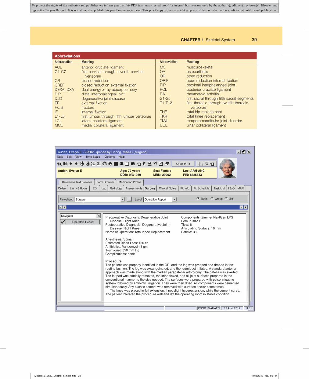
CHAPTER 1 Skeletal System 39
AbbreviationsAbbreviation Meaning
ACL anterior cruciate ligamentC1-C7 first cervical through seventh cervical
vertebraeCR closed reductionCREF closed reduction external fixationDEXA, DXA dual energy x-ray absorptiometryDIP distal interphalangeal jointDJD degenerative joint diseaseEF external fixationFx, # fractureIF internal fixationL1-L5 first lumbar through fifth lumbar vertebraeLCL lateral collateral ligamentMCL medial collateral ligament
MS musculoskeletalOA osteoarthritisOR open reductionORIF open reduction internal fixationPIP proximal interphalangeal jointPCL posterior cruciate ligamentRA rheumatoid arthritisS1-S5 first sacral through fifth sacral segmentsT1-T12 first thoracic through twelfth thoracic
vertebraeTHR total hip replacementTKR total knee replacementTMJ temporomandibular joint disorderUCL ulnar collateral ligament
Abbreviation Meaning
…
Auden, Evelyn E - 29202 Opened by Chong, Mae-Li (surgeon)Task Edit View
Flowsheet: Level:Surgery Operative Report Table Group List
Time Scale Options Help
Auden, Evelyn E Age: 72 yearsDOB: 9/2/1939
Sex: FemaleMRN: 29202
Loc: ARH-ANCFIN: 8425633
Navigator
PROD MAHAFC
As Of 11:11
12 April 2012 11:11
Operative Report
Reference Text Browser
Orders ED Lab Surgery Pt. Info Pt. Schedule Task List I & O MARClinical NotesAssessmentsRadiologyLast 48 Hours
Form Browser Medication Profile
Preoperative Diagnosis: Degenerative Joint Disease, Right KneePostoperative Diagnosis: Degenerative Joint Disease, Right KneeName of Operation: Total Knee Replacement
Anesthesia: SpinalEstimated Blood Loss: 150 ccAntibiotics: Vancomycin 1 gmTourniquet: 350 mm HgComplications: none
ProcedureThe patient was properly identified in the OR, and the leg was prepped and draped in the routine fashion. The leg was exsanguinated, and the tourniquet inflated. A standard anterior approach was made along with the median parapatellar arthrotomy. The patella was everted. The fat pad was partially removed, the knee flexed, and all joint surfaces prepared in the conventional manner to the size needed. The surfaces were prepared with pulse irrigating system followed by antibiotic irrigation. They were then dried. All components were cemented simultaneously. Any excess cement was removed with curettes and/or osteotomes. The knee was placed in full extension, if not slight hyperextension, while the cement cured. The patient tolerated the procedure well and left the operating room in stable condition.
Components: Zimmer NextGen LPSFemur: size GTibia: 6Articulating Surface: 10 mmPatella: 38
Module_B_2622_Chapter 1_main.indd 39 10/8/2015 4:57:50 PM
To protect the rights of the author(s) and publisher we inform you that this PDF is an uncorrected proof for internal business use only by the author(s), editor(s), reviewer(s), Elsevier and typesetter Toppan Best-set. It is not allowed to publish this proof online or in print. This proof copy is the copyright property of the publisher and is confidential until formal publication.

40 CHAPTER 1 Skeletal System
EXERCISE 13: Healthcare Report
Using the operative report on the previous page, answer the following questions.
1. A synonym for the preoperative diagnosis of degenerative joint disease is .
2. An “anterior approach” to the knee would be through which part of the knee?
3. What is the patella?
4. To what does the term parapatellar refer?
5. What is an arthrotomy?
6. If the patella was everted, how would it be placed?
7. What is an osteotome?
Module_B_2622_Chapter 1_main.indd 40 10/8/2015 4:57:50 PM
To protect the rights of the author(s) and publisher we inform you that this PDF is an uncorrected proof for internal business use only by the author(s), editor(s), reviewer(s), Elsevier and typesetter Toppan Best-set. It is not allowed to publish this proof online or in print. This proof copy is the copyright property of the publisher and is confidential until formal publication.
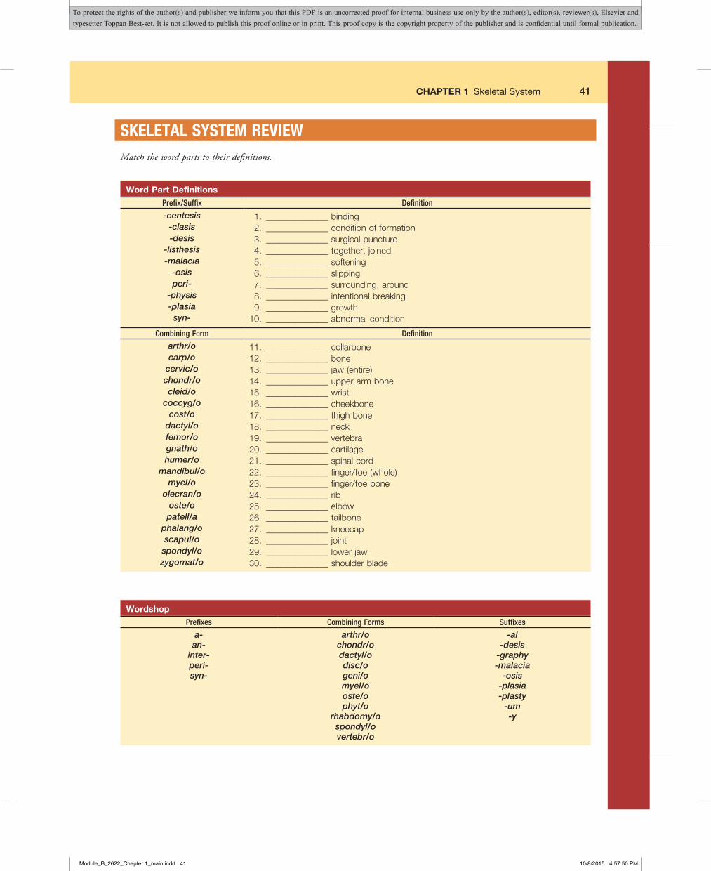
CHAPTER 1 Skeletal System 41
SKELETAL SYSTEM REVIEWMatch the word parts to their definitions.
Word Part DefinitionsPrefix/Suffix Definition
-centesis 1. ______________ binding-clasis 2. ______________ condition of formation-desis 3. ______________ surgical puncture
-listhesis 4. ______________ together, joined-malacia 5. ______________ softening
-osis 6. ______________ slippingperi- 7. ______________ surrounding, around
-physis 8. ______________ intentional breaking-plasia 9. ______________ growth
syn- 10. ______________ abnormal condition
Combining Form Definition
arthr/o 11. ______________ collarbonecarp/o 12. ______________ bone
cervic/o 13. ______________ jaw (entire)chondr/o 14. ______________ upper arm bonecleid/o 15. ______________ wrist
coccyg/o 16. ______________ cheekbonecost/o 17. ______________ thigh bone
dactyl/o 18. ______________ neckfemor/o 19. ______________ vertebragnath/o 20. ______________ cartilagehumer/o 21. ______________ spinal cord
mandibul/o 22. ______________ finger/toe (whole)myel/o 23. ______________ finger/toe bone
olecran/o 24. ______________ riboste/o 25. ______________ elbowpatell/a 26. ______________ tailbone
phalang/o 27. ______________ kneecapscapul/o 28. ______________ joint
spondyl/o 29. ______________ lower jawzygomat/o 30. ______________ shoulder blade
WordshopPrefixes Combining Forms Suffixes
a- arthr/o -alan- chondr/o -desis
inter- dactyl/o -graphyperi- disc/o -malaciasyn- geni/o -osis
myel/o -plasiaoste/o -plastyphyt/o -um
rhabdomy/o -yspondyl/overtebr/o
Module_B_2622_Chapter 1_main.indd 41 10/8/2015 4:57:50 PM
To protect the rights of the author(s) and publisher we inform you that this PDF is an uncorrected proof for internal business use only by the author(s), editor(s), reviewer(s), Elsevier and typesetter Toppan Best-set. It is not allowed to publish this proof online or in print. This proof copy is the copyright property of the publisher and is confidential until formal publication.

42 CHAPTER 1 Skeletal System
Build musculoskeletal terms by combining the word parts on previous page. Some word parts may be used more than once. Some may not be used at all. The number in parentheses indicates the number of word parts needed.
Definition Term
1. structure surrounding bone (3) 2. pertaining to between vertebrae (3) 3. softening of cartilage (2) 4. process of many fingers or toes (3) 5. abnormal condition of bone growth (3) 6. binding of a joint (2) 7. process of joined fingers or toes (3) 8. recording a disc (2) 9. binding together of vertebrae (3) 10. surgical puncture of a bursa (2) 11. condition of formation of no cartilage (3) 12. surgical repair of the chin (2)
Sort the terms into the correct categories.
Term SortingAnatomy and Physiology Pathology Procedures
achondroplasia diaphysis osteosarcomaamputation digitus patellapexy
arthrocentesis endosteum perichondriumarthrodesis genioplasty radius
arthrography humerus sacrectomyarthrosis lamellae scapula
arthrotomy laminectomy scapulopexyarticulation ligament spondylolisthesis
bunion meniscectomy sternumbursitis osteoclasis syndactyly
carpectomy osteogenesis TMJcartilage osteomyelitis ulna
contracture osteoporosiscosta
densitometry
Module_B_2622_Chapter 1_main.indd 42 10/8/2015 4:57:50 PM
To protect the rights of the author(s) and publisher we inform you that this PDF is an uncorrected proof for internal business use only by the author(s), editor(s), reviewer(s), Elsevier and typesetter Toppan Best-set. It is not allowed to publish this proof online or in print. This proof copy is the copyright property of the publisher and is confidential until formal publication.
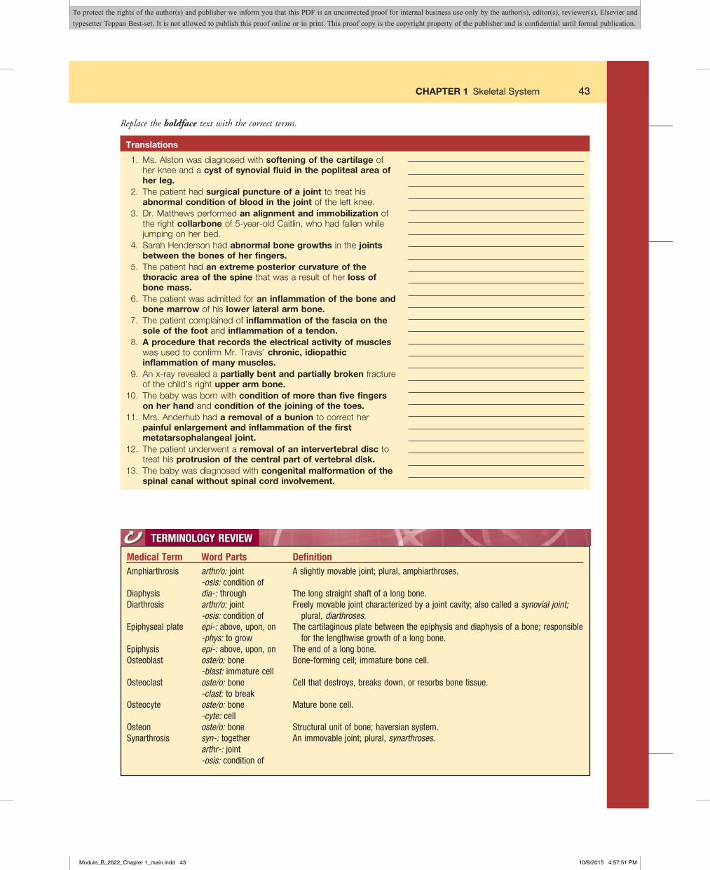
CHAPTER 1 Skeletal System 43
Replace the boldface text with the correct terms.
Translations
1. Ms. Alston was diagnosed with softening of the cartilage of her knee and a cyst of synovial fluid in the popliteal area of her leg.
2. The patient had surgical puncture of a joint to treat his abnormal condition of blood in the joint of the left knee.
3. Dr. Matthews performed an alignment and immobilization of the right collarbone of 5-year-old Caitlin, who had fallen while jumping on her bed.
4. Sarah Henderson had abnormal bone growths in the joints between the bones of her fingers.
5. The patient had an extreme posterior curvature of the thoracic area of the spine that was a result of her loss of bone mass.
6. The patient was admitted for an inflammation of the bone and bone marrow of his lower lateral arm bone.
7. The patient complained of inflammation of the fascia on the sole of the foot and inflammation of a tendon.
8. A procedure that records the electrical activity of muscles was used to confirm Mr. Travis’ chronic, idiopathic inflammation of many muscles.
9. An x-ray revealed a partially bent and partially broken fracture of the child’s right upper arm bone.
10. The baby was born with condition of more than five fingers on her hand and condition of the joining of the toes.
11. Mrs. Anderhub had a removal of a bunion to correct her painful enlargement and inflammation of the first metatarsophalangeal joint.
12. The patient underwent a removal of an intervertebral disc to treat his protrusion of the central part of vertebral disk.
13. The baby was diagnosed with congenital malformation of the spinal canal without spinal cord involvement.
TERMINOLOGY REVIEW
Medical Term Word Parts DefinitionAmphiarthrosis arthr/o: joint
-osis: condition ofA slightly movable joint; plural, amphiarthroses.
Diaphysis dia-: through The long straight shaft of a long bone.Diarthrosis arthr/o: joint
-osis: condition ofFreely movable joint characterized by a joint cavity; also called a synovial joint;
plural, diarthroses.Epiphyseal plate epi-: above, upon, on
-phys: to growThe cartilaginous plate between the epiphysis and diaphysis of a bone; responsible
for the lengthwise growth of a long bone.Epiphysis epi-: above, upon, on The end of a long bone.Osteoblast oste/o: bone
-blast: immature cellBone-forming cell; immature bone cell.
Osteoclast oste/o: bone-clast: to break
Cell that destroys, breaks down, or resorbs bone tissue.
Osteocyte oste/o: bone-cyte: cell
Mature bone cell.
Osteon oste/o: bone Structural unit of bone; haversian system.Synarthrosis syn-: together
arthr-: joint-osis: condition of
An immovable joint; plural, synarthroses.
Module_B_2622_Chapter 1_main.indd 43 10/8/2015 4:57:51 PM
To protect the rights of the author(s) and publisher we inform you that this PDF is an uncorrected proof for internal business use only by the author(s), editor(s), reviewer(s), Elsevier and typesetter Toppan Best-set. It is not allowed to publish this proof online or in print. This proof copy is the copyright property of the publisher and is confidential until formal publication.

44 CHAPTER 1 Skeletal System
For information on the skeletal system:
Loyola University Medical Education Network: Pick a Bone: www.meddean.luc.edu/lumen/MedEd/GrossAnatomy/learnem/bones/main_bone.htm
Merck Manual: Bone, Joint, and Muscle Disorders: www.merck.com/mmhe (Click on “Bone, Joint, and Muscle Disorders”)
National Osteoporosis Foundation: www.nof.org
ON THE WEB
Module_B_2622_Chapter 1_main.indd 44 10/8/2015 4:57:51 PM
To protect the rights of the author(s) and publisher we inform you that this PDF is an uncorrected proof for internal business use only by the author(s), editor(s), reviewer(s), Elsevier and typesetter Toppan Best-set. It is not allowed to publish this proof online or in print. This proof copy is the copyright property of the publisher and is confidential until formal publication.

CHAPTER 1 Skeletal System 45
CHAPTER REVIEW
Name ____________________________________________________________ Date __________________________________
PRETEST
True or False
_____ 1. The skeletal system provides a rigid framework for the body._____ 2. Blood cell formation takes place in the spleen._____ 3. Vertebrae are made up of flat bones._____ 4. Articular cartilage covers the ends of long bones._____ 5. Long bones grow in length at the epiphyseal line._____ 6. The maxillary bones form the upper jaw._____ 7. The sacrum makes up the small of the back._____ 8. The shoulder is an example of a hinge joint._____ 9. The patella is the kneecap._____ 10. The humerus makes up the thigh.
Module_B_2622_Chapter 1_main.indd 45 10/8/2015 4:57:51 PM
To protect the rights of the author(s) and publisher we inform you that this PDF is an uncorrected proof for internal business use only by the author(s), editor(s), reviewer(s), Elsevier and typesetter Toppan Best-set. It is not allowed to publish this proof online or in print. This proof copy is the copyright property of the publisher and is confidential until formal publication.

46 CHAPTER 1 Skeletal System
Notes
Module_B_2622_Chapter 1_main.indd 46 10/8/2015 4:57:51 PM
To protect the rights of the author(s) and publisher we inform you that this PDF is an uncorrected proof for internal business use only by the author(s), editor(s), reviewer(s), Elsevier and typesetter Toppan Best-set. It is not allowed to publish this proof online or in print. This proof copy is the copyright property of the publisher and is confidential until formal publication.

CHAPTER 1 Skeletal System 47
KEY TERM ASSESSMENT
A. Definitions
Directions: Match each medical term with its definition.
_____ 1. Amphiarthrosis_____ 2. Diaphysis_____ 3. Diarthrosis_____ 4. Epiphyseal plate_____ 5. Epiphysis_____ 6. Osteoblast_____ 7. Osteoclast_____ 8. Osteocyte_____ 9. Osteon_____ 10. Synarthrosis
A. A slightly movable joint B. An immovable joint C. Bone-forming cell D. Cell that destroys or resorbs bone tissue E. Freely movable joint characterized by a joint cavity; also called a synovial
joint F. Mature bone cell G. Structural unit of bone; haversian system H. The cartilaginous plate between the epiphysis and diaphysis of a bone;
responsible for the lengthwise growth of a long bone I. The end of a long bone J. The long, straight shaft of a long bone
B. Word Parts
Directions: Indicate the meaning of each word part in the space provided. List as many medical terms as possible that incorporate the word part in the space provided.
Word Part Meaning of Word Part Medical Terms That Incorporate Word Part
1. arthr/o
2. -osis
3. dia-
4. epi-
5. -physis
6. oste/o
7. -blast
8. -clast
9. -cyte
10. syn-
Module_B_2622_Chapter 1_main.indd 47 10/8/2015 4:57:51 PM
To protect the rights of the author(s) and publisher we inform you that this PDF is an uncorrected proof for internal business use only by the author(s), editor(s), reviewer(s), Elsevier and typesetter Toppan Best-set. It is not allowed to publish this proof online or in print. This proof copy is the copyright property of the publisher and is confidential until formal publication.

48 CHAPTER 1 Skeletal System
EVALUATION OF LEARNING
Directions: Fill in each blank with the correct answer.
1. What structures make up the skeletal system?
2. What are the five functions of the skeletal system?
3. How is the blood calcium level maintained in the body?
4. What is the function of red bone marrow?
5. Where is red bone marrow found in the adult?
6. What is an osteon?
7. Describe the following structures that make up an osteon:
a. Osteonic canal (haversian canal):
b. Lamella:
c. Osteocytes:
d. Lacunae:
e. Canaliculi:
Module_B_2622_Chapter 1_main.indd 48 10/8/2015 4:57:51 PM
To protect the rights of the author(s) and publisher we inform you that this PDF is an uncorrected proof for internal business use only by the author(s), editor(s), reviewer(s), Elsevier and typesetter Toppan Best-set. It is not allowed to publish this proof online or in print. This proof copy is the copyright property of the publisher and is confidential until formal publication.

CHAPTER 1 Skeletal System 49
8. What is the difference between spongy bone and compact bone?
9. List examples of each of the following classifications of bone:
a. Long bones:
b. Short bones:
c. Flat bones:
d. Irregular bones:
10. Describe each of the following structures that make up a long bone:
a. Diaphysis:
b. Medullary cavity:
c. Epiphysis:
d. Articular cartilage:
e. Periosteum:
f. Nutrient foramina:
g. Endosteum:
11. What is ossification?
12. What is the function of each of the following types of bone cells?
a. Osteoblast:
b. Osteocyte:
c. Osteoclast:
13. Where is the epiphyseal plate located in a long bone?
14. What type of cartilage is found in the epiphyseal plate?
15. How do long bones grow in length?
Module_B_2622_Chapter 1_main.indd 49 10/8/2015 4:57:51 PM
To protect the rights of the author(s) and publisher we inform you that this PDF is an uncorrected proof for internal business use only by the author(s), editor(s), reviewer(s), Elsevier and typesetter Toppan Best-set. It is not allowed to publish this proof online or in print. This proof copy is the copyright property of the publisher and is confidential until formal publication.

50 CHAPTER 1 Skeletal System
16. When does an individual stop growing in length?
17. What happens to the epiphyseal plate when long bones stop growing?
18. What influences bone growth in the body?
19. How many bones make up the skeleton of an adult?
20. What are the two divisions of the skeleton? What structures are included in each division?
21. How many bones make up the skull?
22. What is the function of the cranium?
23. What are sinuses and what is their function?
24. What bones make up the cranium?
25. What is the function of the facial bones?
26. What are the names of the three small bones located in the middle ear?
27. What is the function of the hyoid bone?
Module_B_2622_Chapter 1_main.indd 50 10/8/2015 4:57:51 PM
To protect the rights of the author(s) and publisher we inform you that this PDF is an uncorrected proof for internal business use only by the author(s), editor(s), reviewer(s), Elsevier and typesetter Toppan Best-set. It is not allowed to publish this proof online or in print. This proof copy is the copyright property of the publisher and is confidential until formal publication.

CHAPTER 1 Skeletal System 51
28. How many vertebrae make up the vertebral column?
29. What are the functions of the intervertebral disks?
30. What structures make up the vertebrae?
31. How many vertebrae are included in each of the following divisions of the vertebral column?
a. Cervical:
b. Thoracic:
c. Lumbar:
32. Describe the following:
a. Sacrum:
b. Coccyx:
33. What are the functions of the thoracic cage?
34. What is the name of the central indentation in the superior margin of the sternum?
35. How many pairs of ribs are present in the human skeleton?
36. What is the difference between true ribs and false ribs?
37. What are floating ribs?
Module_B_2622_Chapter 1_main.indd 51 10/8/2015 4:57:51 PM
To protect the rights of the author(s) and publisher we inform you that this PDF is an uncorrected proof for internal business use only by the author(s), editor(s), reviewer(s), Elsevier and typesetter Toppan Best-set. It is not allowed to publish this proof online or in print. This proof copy is the copyright property of the publisher and is confidential until formal publication.

52 CHAPTER 1 Skeletal System
38. What is the function of the appendicular skeleton?
39. What two bones make up the pectoral girdle?
40. What is another name for the clavicle?
41. What is the name of the shallow depression on the scapula where the head of the humerus connects to the scapula?
42. What bone is located in the upper arm?
43. What bones are located in the forearm?
44. State the location of the following bones making up the hand:
a. Carpal bones:
b. Metacarpal bones:
c. Phalanges:
45. What are the functions of the pelvic girdle?
46. What three bones fuse to form a coxal bone?
47. What is the symphysis pubis?
48. What bone is located in the thigh?
49. What is the patella?
50. What is the function of the patella?
Module_B_2622_Chapter 1_main.indd 52 10/8/2015 4:57:51 PM
To protect the rights of the author(s) and publisher we inform you that this PDF is an uncorrected proof for internal business use only by the author(s), editor(s), reviewer(s), Elsevier and typesetter Toppan Best-set. It is not allowed to publish this proof online or in print. This proof copy is the copyright property of the publisher and is confidential until formal publication.

CHAPTER 1 Skeletal System 53
51. What bones are located in the leg?
52. What does the lateral malleolus do?
53. Describe the location of the following bones making up the foot:
a. Tarsal bones:
b. Calcaneus bone:
c. Metatarsal bones:
d. Phalanges:
54. What is an articulation?
55. What is a synarthrosis?
56. What is an example of a synarthrosis?
57. What is an amphiarthrosis?
58. What is an example of an amphiarthrosis?
59. What is a diarthrosis?
60. Describe the following parts of a diarthrosis:
a. Articular cartilage:
b. Joint cavity:
c. Joint capsule:
d. Synovial membrane:
e. Synovial fluid:
61. What is the function of fibrocartilaginous pads located in the knee?
Module_B_2622_Chapter 1_main.indd 53 10/8/2015 4:57:51 PM
To protect the rights of the author(s) and publisher we inform you that this PDF is an uncorrected proof for internal business use only by the author(s), editor(s), reviewer(s), Elsevier and typesetter Toppan Best-set. It is not allowed to publish this proof online or in print. This proof copy is the copyright property of the publisher and is confidential until formal publication.
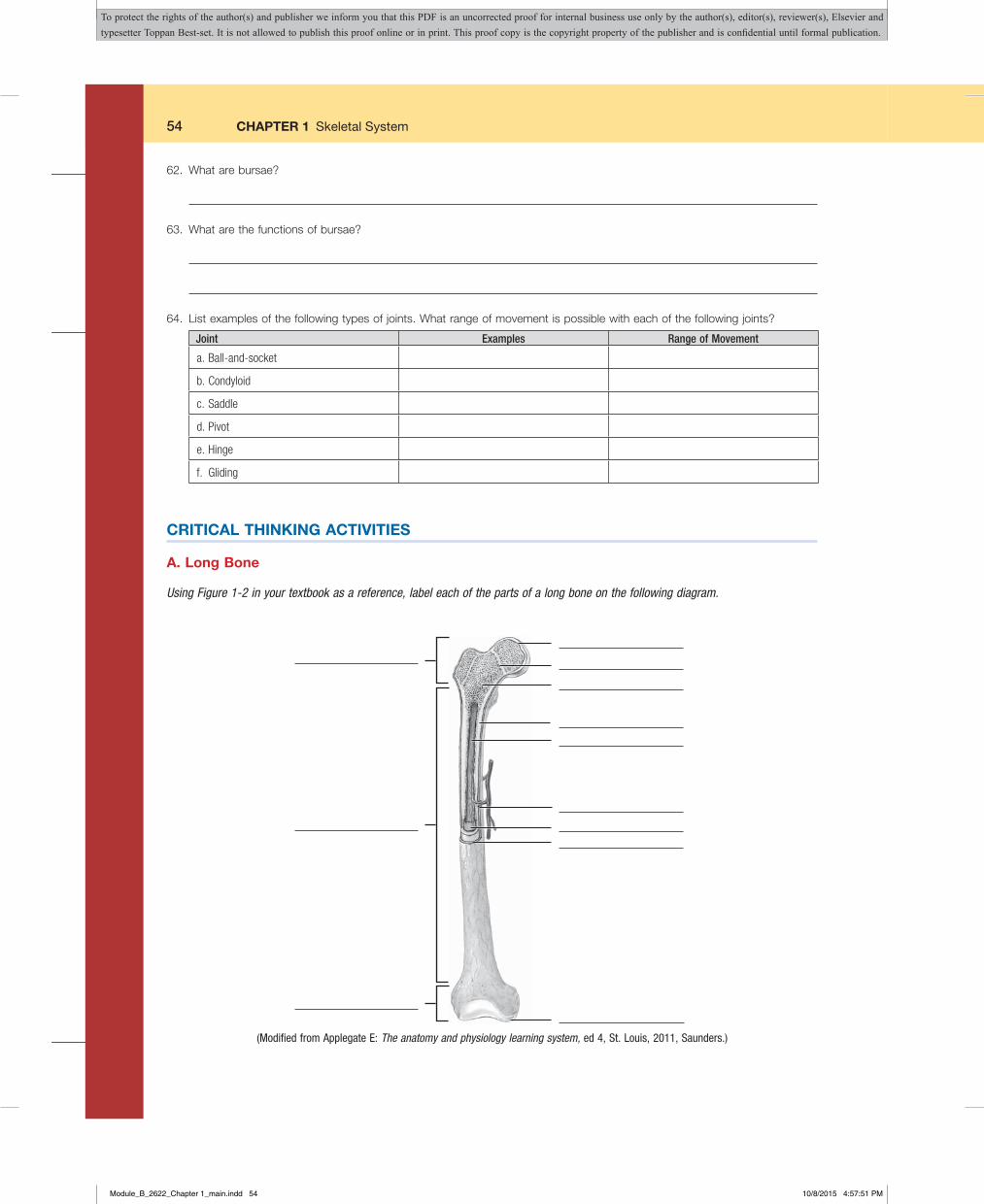
54 CHAPTER 1 Skeletal System
62. What are bursae?
63. What are the functions of bursae?
64. List examples of the following types of joints. What range of movement is possible with each of the following joints?
Joint Examples Range of Movement
a.Ball-and-socket
b.Condyloid
c.Saddle
d.Pivot
e.Hinge
f. Gliding
CRITICAL THINKING ACTIVITIES
A. Long Bone
Using Figure 1-2 in your textbook as a reference, label each of the parts of a long bone on the following diagram.
(Modified from Applegate E: The anatomy and physiology learning system, ed 4, St. Louis, 2011, Saunders.)
Module_B_2622_Chapter 1_main.indd 54 10/8/2015 4:57:51 PM
To protect the rights of the author(s) and publisher we inform you that this PDF is an uncorrected proof for internal business use only by the author(s), editor(s), reviewer(s), Elsevier and typesetter Toppan Best-set. It is not allowed to publish this proof online or in print. This proof copy is the copyright property of the publisher and is confidential until formal publication.

CHAPTER 1 Skeletal System 55
B. Synovial Joint
Using Figure 1-17 in your textbook as a reference, label each of the parts of a synovial joint on the following diagram.
(Modified from Applegate E: The anatomy and physiology learning system, ed 4, St. Louis, 2011, Saunders.)
C. Inquiring Patients Want to Know
You are working in a general practice medical office. Your patients ask you the following questions. In the space provided, indicate how you would respond to each question in terms the patient would understand. Use your textbook and Internet resources to develop your responses.
1. What causes osteoporosis?
2. What causes my sinus headaches?
3. How does a baby get through the bones of the pelvis during childbirth?
Module_B_2622_Chapter 1_main.indd 55 10/8/2015 4:57:52 PM
To protect the rights of the author(s) and publisher we inform you that this PDF is an uncorrected proof for internal business use only by the author(s), editor(s), reviewer(s), Elsevier and typesetter Toppan Best-set. It is not allowed to publish this proof online or in print. This proof copy is the copyright property of the publisher and is confidential until formal publication.

56 CHAPTER 1 Skeletal System
4. What causes older people to break their hips?
5. What causes gout?
6. Why do people get shorter when they get older?
7. Why do babies have “soft spots” on their heads?
8. What is the difference between osteoarthritis and rheumatoid arthritis?
9. What happens when someone cracks his or her knuckles?
10. What is the difference between a strain and a sprain?
Module_B_2622_Chapter 1_main.indd 56 10/8/2015 4:57:52 PM
To protect the rights of the author(s) and publisher we inform you that this PDF is an uncorrected proof for internal business use only by the author(s), editor(s), reviewer(s), Elsevier and typesetter Toppan Best-set. It is not allowed to publish this proof online or in print. This proof copy is the copyright property of the publisher and is confidential until formal publication.

CHAPTER 1 Skeletal System 57
1
2
3
4 5
6
7 8
9 10
11
12
13 14
15
16
17
18
19
20
D. Crossword Puzzle: Skeletal System
Directions: Complete the crossword puzzle using the clues provided.
Across Down
1 Abnormal side-to-side spinal curvature 2 Bones that protect brain 4 Freely movable joint 6 Upper arm bone 7 Shaft of a long bone 9 Softening of bones11 Collarbone14 Bone cell15 Air-filled cavity in the skull17 Shoulder blade18 Spinal cord passes through this part of the skull19 Bones of the upper jaw20 Lower jaw bones
1 Immovable joint 3 Tailbone 5 Ankle 8 Finger bones10 Where bones grow in length12 Breastbone13 Outer surface of a long bone14 Process of bone formation16 Kneecap18 Thigh bone
Module_B_2622_Chapter 1_main.indd 57 10/8/2015 4:57:52 PM
To protect the rights of the author(s) and publisher we inform you that this PDF is an uncorrected proof for internal business use only by the author(s), editor(s), reviewer(s), Elsevier and typesetter Toppan Best-set. It is not allowed to publish this proof online or in print. This proof copy is the copyright property of the publisher and is confidential until formal publication.

58 CHAPTER 1 Skeletal System
Notes
Module_B_2622_Chapter 1_main.indd 58 10/8/2015 4:57:52 PM
To protect the rights of the author(s) and publisher we inform you that this PDF is an uncorrected proof for internal business use only by the author(s), editor(s), reviewer(s), Elsevier and typesetter Toppan Best-set. It is not allowed to publish this proof online or in print. This proof copy is the copyright property of the publisher and is confidential until formal publication.
CHAPTER 1 Skeletal System 59
Name ____________________________________________________________ Date __________________________________
POSTTEST
True or False
_____ 1. The formation of blood cells is known as hemogenesis._____ 2. Osteons are the microscopic units of compact bone._____ 3. Calcium is located in an osteonic canal._____ 4. The shaft of a long bone is the diaphysis._____ 5. The endosteum is the tough fibrous connective tissue that covers a long bone._____ 6. A mature bone cell is an osteoblast._____ 7. The central indentation of the sternum is the jugular (suprasternal) notch._____ 8. The clavicle and scapula make up the pelvic girdle._____ 9. The ileum, ischium, and pubis make up the coxal bones._____ 10. The ulna is located on the lateral side of the forearm.
Module_B_2622_Chapter 1_main.indd 59 10/8/2015 4:57:53 PM
To protect the rights of the author(s) and publisher we inform you that this PDF is an uncorrected proof for internal business use only by the author(s), editor(s), reviewer(s), Elsevier and typesetter Toppan Best-set. It is not allowed to publish this proof online or in print. This proof copy is the copyright property of the publisher and is confidential until formal publication.

60 CHAPTER 1 Skeletal System
Notes
Module_B_2622_Chapter 1_main.indd 60 10/8/2015 4:57:53 PM
To protect the rights of the author(s) and publisher we inform you that this PDF is an uncorrected proof for internal business use only by the author(s), editor(s), reviewer(s), Elsevier and typesetter Toppan Best-set. It is not allowed to publish this proof online or in print. This proof copy is the copyright property of the publisher and is confidential until formal publication.