Embed Size (px)
Citation preview
SKELETAL SYSTEM
radtech-board.ohd.hr.state.or.us
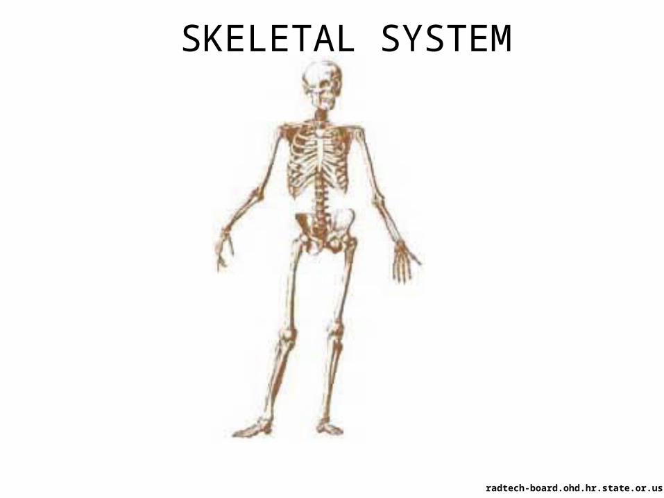
Functions of the Skeletal System
•Provide points for attachment for muscles, assistance in movement
•Protect and support softer tissues•Blood cell production
•Stores minerals such as calcium and phosphorus.
and releases into or absorbs minerals from
bloodstream•Form passageways for blood vessels and nerves
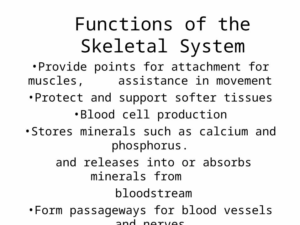
Bones classified according to shape
1. Long bones– long with expanded ends– thigh bones (femur) and forearm
2. Short bones– cube-like – wrists and ankles
3. Flat bones– plate-like structures with broad surfaces
– Ribs, scapula and some bones of skull
4. Irregular bones– variety of shapes– Vertebrae and many facial bones
5. Sesamoid bones– small and nodular– Kneecap (patella)
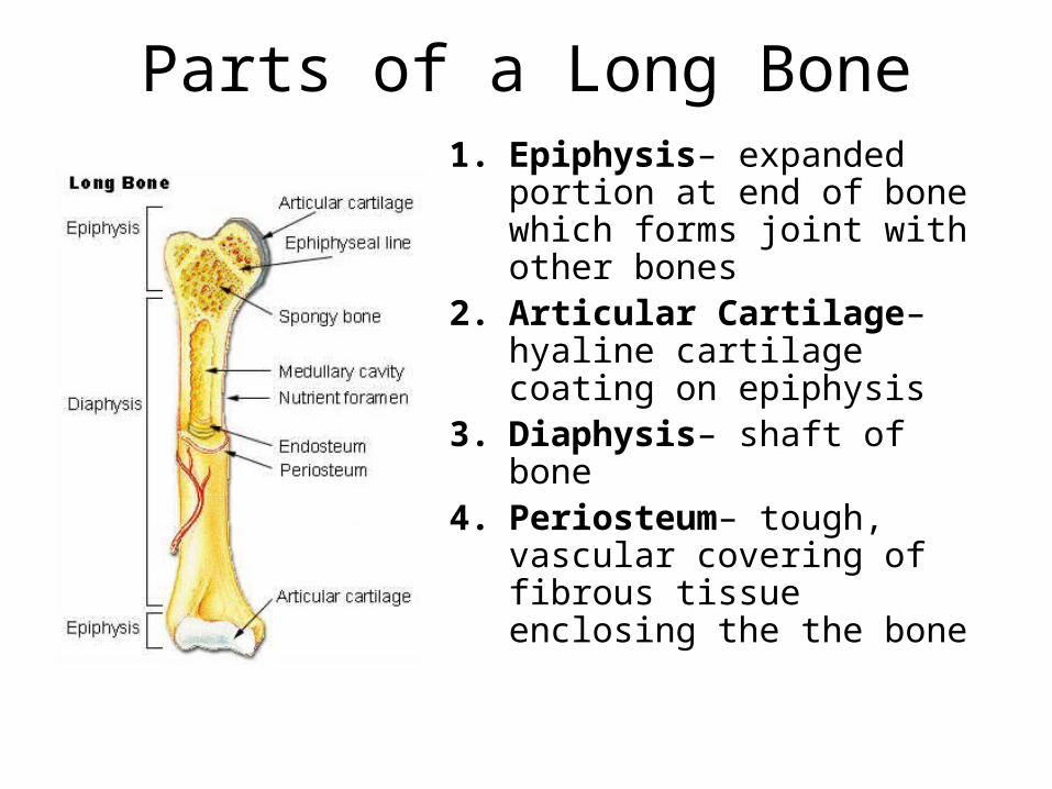
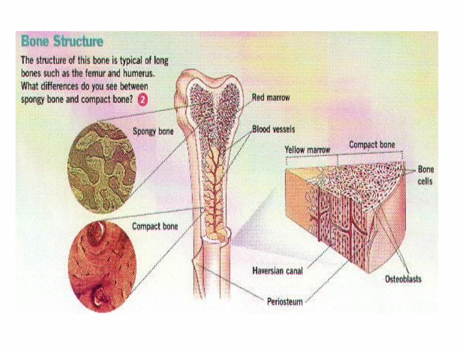
Parts of a Long Bone1. Epiphysis– expanded
portion at end of bone which forms joint with other bones
2. Articular Cartilage– hyaline cartilage coating on epiphysis
3. Diaphysis– shaft of bone4. Periosteum– tough,
vascular covering of fibrous tissue enclosing the the bone
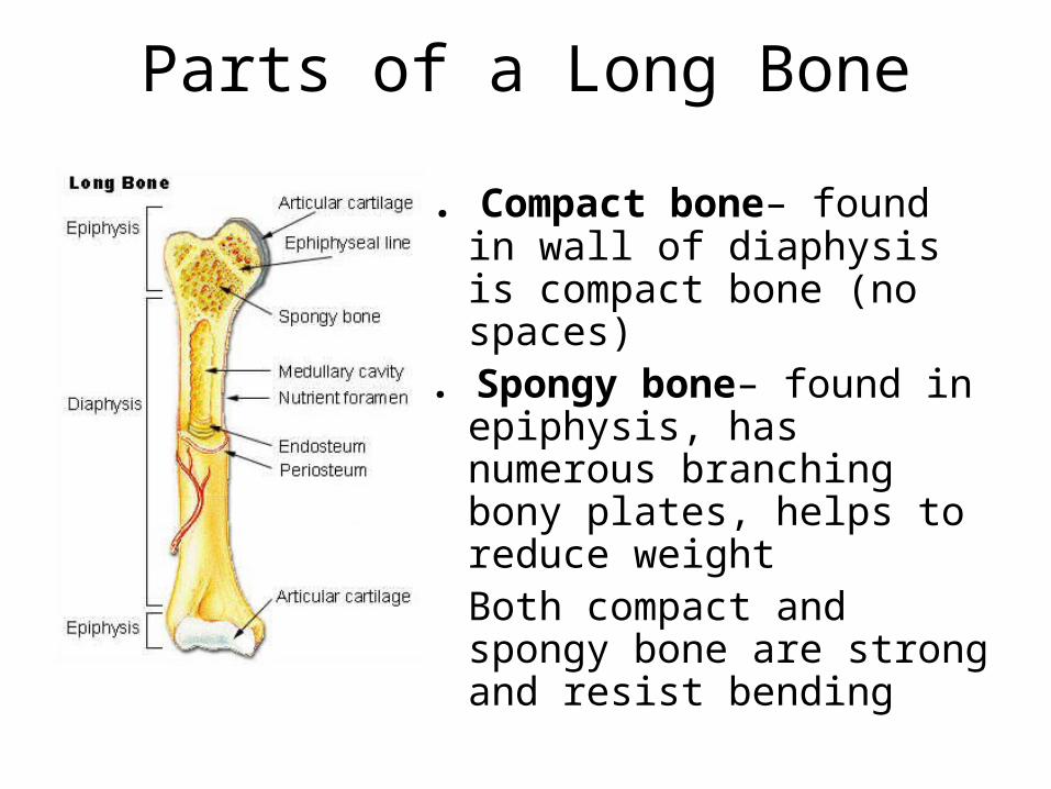
Parts of a Long Bone
5. Compact bone– found in wall of diaphysis is compact bone (no spaces)
6. Spongy bone– found in epiphysis, has numerous branching bony plates, helps to reduce weight
• Both compact and spongy bone are strong and resist bending
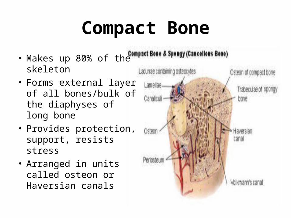
Compact Bone
• Makes up 80% of the skeleton
• Forms external layer of all bones/bulk of the diaphyses of long bone
• Provides protection, support, resists stress
• Arranged in units called osteon or Haversian canals
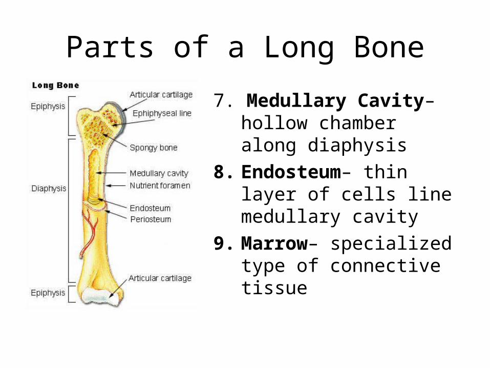
Parts of a Long Bone
7. Medullary Cavity– hollow chamber along diaphysis
8. Endosteum– thin layer of cells line medullary cavity
9. Marrow– specialized type of connective tissue

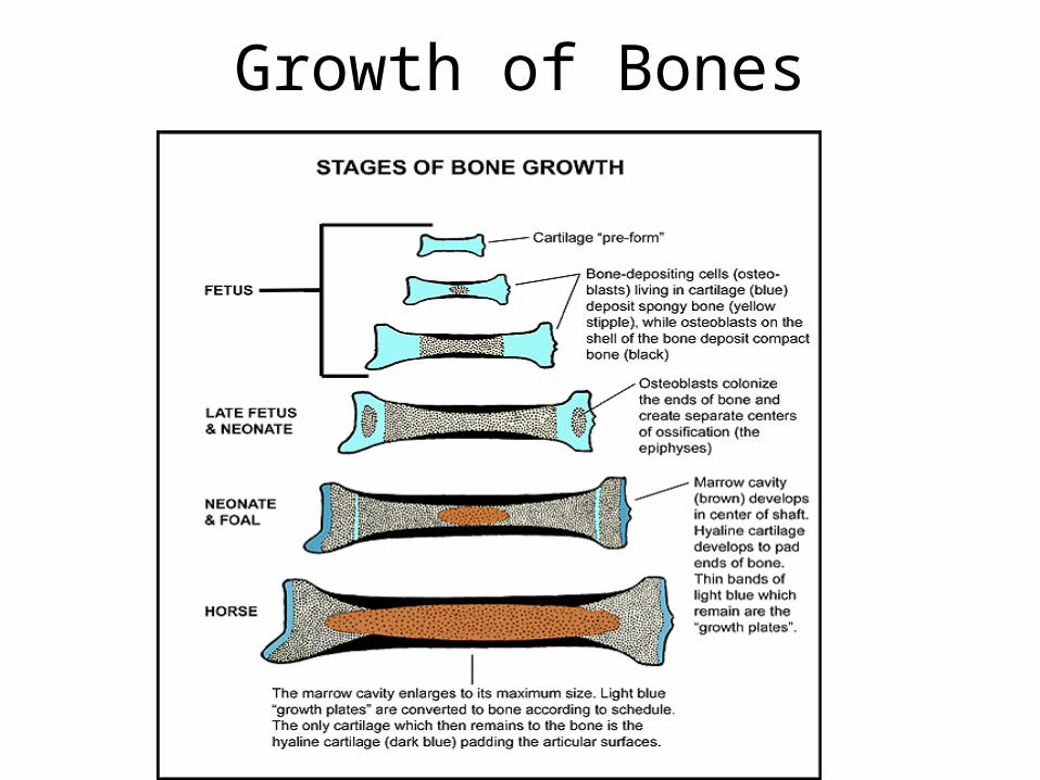
Endochondral Bones(and bone growth)
• Most of the bones in the skeleton
• Develop in the fetus from masses of hyaline cartilage shaped like future bony structures
• Ossification– formation of bones
• Osteoblasts– bone-forming cells
• Ossification (bone growth) begins in the center of the diaphysis
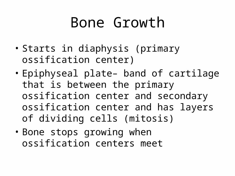
Bone Growth
• Starts in diaphysis (primary ossification center)
• Epiphyseal plate– band of cartilage that is between the primary ossification center and secondary ossification center and has layers of dividing cells (mitosis)
• Bone stops growing when ossification centers meet
Growth of Bones
Bone Growth
• Osteoblasts deposit bony matrix around them in the connective tissue
• When they are completely surrounded by the bony matrix, they are osteocytes
• Osteocytes are bone cells.
• Osteoclasts– secrete an acid and clean up old bone cells so new bone can be produced.
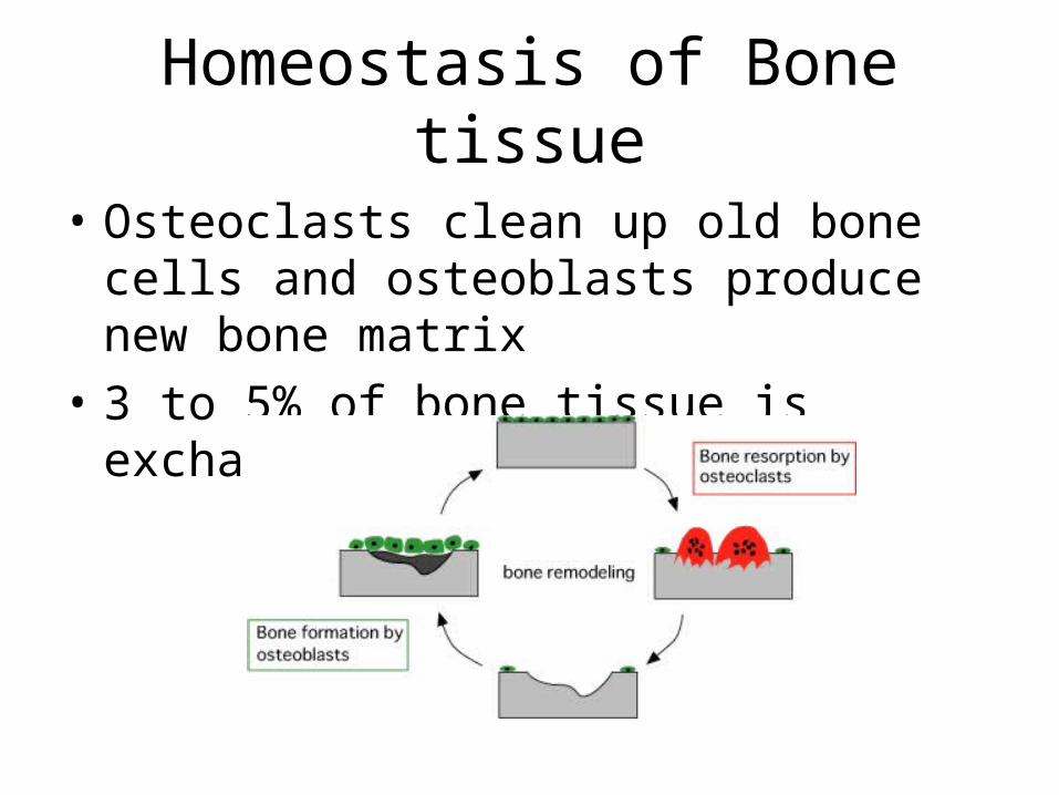
Homeostasis of Bone tissue
• Osteoclasts clean up old bone cells and osteoblasts produce new bone matrix
• 3 to 5% of bone tissue is exchanged each year
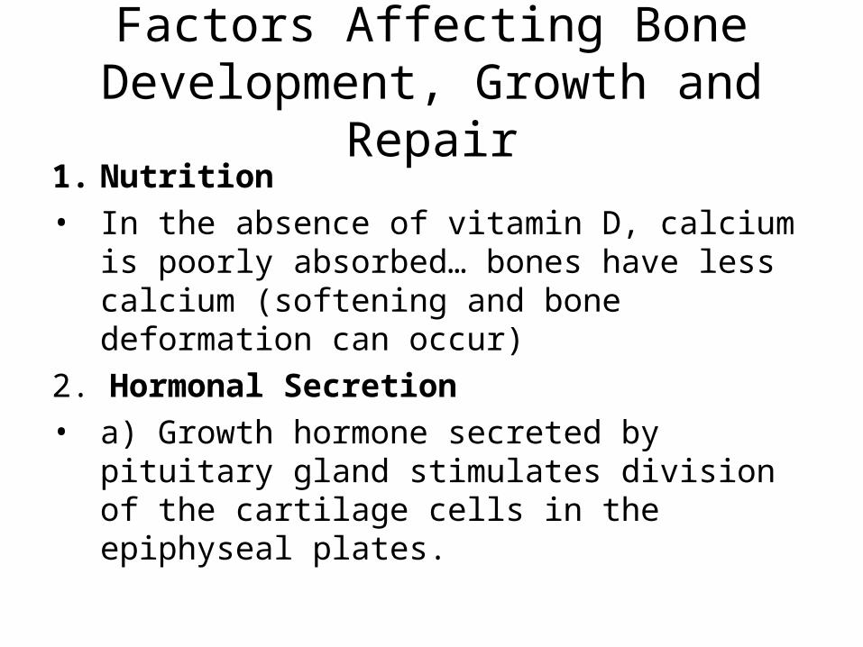
Factors Affecting Bone Development, Growth and Repair
1. Nutrition• In the absence of vitamin D, calcium is
poorly absorbed… bones have less calcium (softening and bone deformation can occur)
2. Hormonal Secretion• a) Growth hormone secreted by pituitary
gland stimulates division of the cartilage cells in the epiphyseal plates.
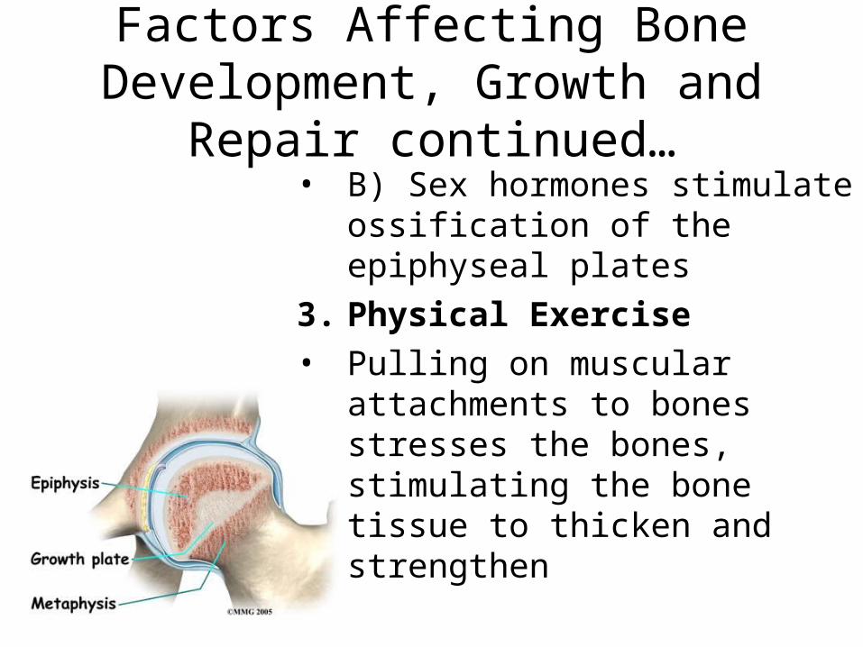
Factors Affecting Bone Development, Growth and Repair
continued…• B) Sex hormones stimulate
ossification of the epiphyseal plates
3. Physical Exercise
• Pulling on muscular attachments to bones stresses the bones, stimulating the bone tissue to thicken and strengthen
Bone Fractures
Use the info on page 137
• Define open fracture (compound fracture)
• Complete the chart on Bone Fracture types.
Blood Cell Formation• Hematopoiesis– the process of blood
cell formation• As a fetus, hematopoeisis occurs in the
yolk sac, then later in the liver and spleen, then in the bone marrow.
2 types of marrow:1. Red marrow– in an infant, it is found in
the cavities of most bones• As you age, yellow marrow replaces it• And red marrow is found in the spongy
bones of the skull, ribs, sternum, clavicles, vertebrae and hip
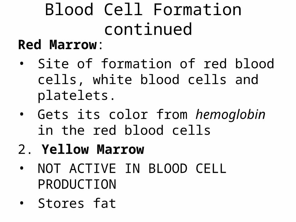
Blood Cell Formation continued
Red Marrow:
• Site of formation of red blood cells, white blood cells and platelets.
• Gets its color from hemoglobin in the red blood cells
2. Yellow Marrow
• NOT ACTIVE IN BLOOD CELL PRODUCTION
• Stores fat
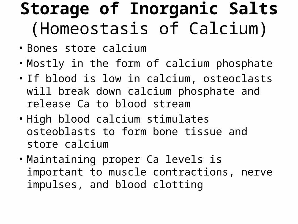
Storage of Inorganic Salts (Homeostasis of Calcium)
• Bones store calcium
• Mostly in the form of calcium phosphate
• If blood is low in calcium, osteoclasts will break down calcium phosphate and release Ca to blood stream
• High blood calcium stimulates osteoblasts to form bone tissue and store calcium
• Maintaining proper Ca levels is important to muscle contractions, nerve impulses, and blood clotting

Osteoporosislow bone density

2 Main Parts of Skeleton
1. Axial Skeleton– supports the head, neck and trunk
• Skull– cranium (brain case) and facial bones
• Hyoid– between lower jaw and larynx supports the tongue and attachments
• Vertebral Column-- backbone
• Thoracic cage– protects organs of the thoracic cavity
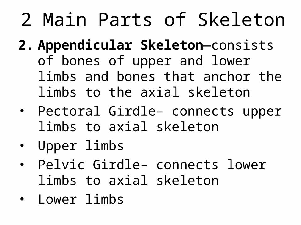
2 Main Parts of Skeleton2. Appendicular Skeleton—consists of
bones of upper and lower limbs and bones that anchor the limbs to the axial skeleton
• Pectoral Girdle– connects upper limbs to axial skeleton
• Upper limbs
• Pelvic Girdle– connects lower limbs to axial skeleton
• Lower limbs
Infantile Skull• Skull is incompletely developed with fibrous
membranes connecting the cranial bones• Fontanels– incomplete intramembranous
ossification or “soft spots”
• Allows the skull to be compressible and change shapes to pass through birth canal.
• Fontanels will close after birth
Skeleton Parts
• Complete skeleton chart
• Define words and be able to label bones
Function of Joints
Joint– functional junctions between bones
• They bind parts of the skeletal system
• Make possible bone growth
• Permit parts of the skeleton to change shape during childbirth
• Enable the body to move in response to skeletal muscle contractions

Types of Joints
• Classified by type of tissue (most commonly used type of classification)
1. Fibrous joint– lie between bones that closely contact one another
• A thin layer of dense connective tissue joins the bones at such joints
• No appreciable movement
• Called a SUTURE between skull bones
Types of Joints
2. Cartilaginous Joints
• Hyaline or fibrocartilage connects the bones at these joints
• Slight flexibility at the discs allows for limited movement
• Examples: intervertebral discs, symphysis pubis and 1st rib with the sternum
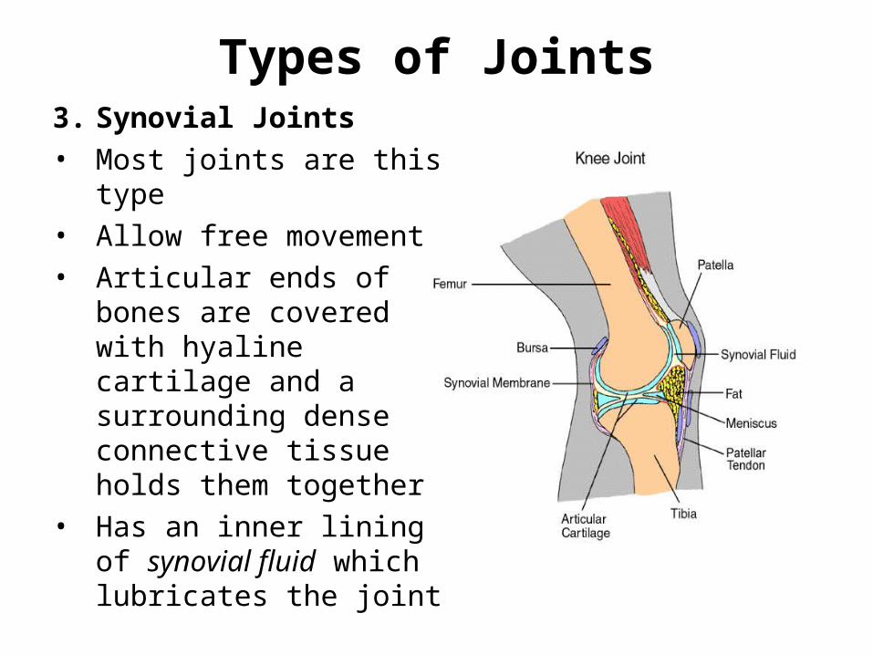
Types of Joints3. Synovial Joints• Most joints are this type• Allow free movement• Articular ends of bones
are covered with hyaline cartilage and a surrounding dense connective tissue holds them together
• Has an inner lining of synovial fluid which lubricates the joint
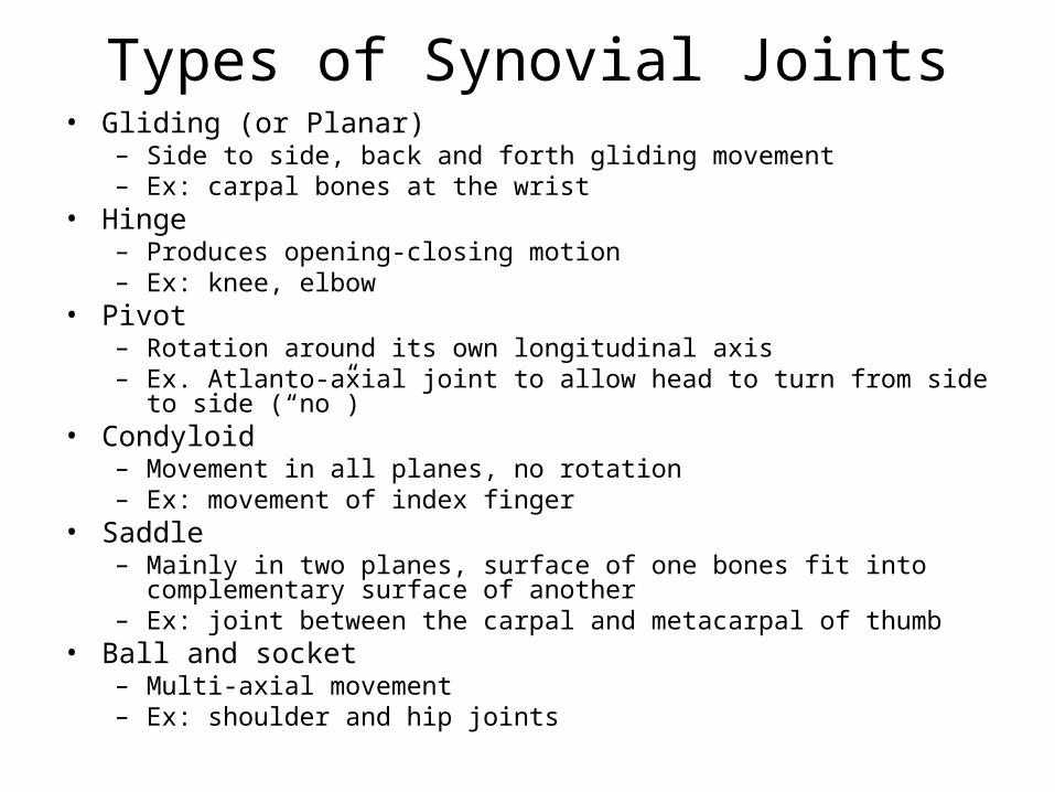
Types of Synovial Joints• Gliding (or Planar)
– Side to side, back and forth gliding movement– Ex: carpal bones at the wrist
• Hinge– Produces opening-closing motion– Ex: knee, elbow
• Pivot– Rotation around its own longitudinal axis– Ex. Atlanto-axial joint to allow head to turn from side to side (“no”)
• Condyloid– Movement in all planes, no rotation– Ex: movement of index finger
• Saddle– Mainly in two planes, surface of one bones fit into complementary surface
of another– Ex: joint between the carpal and metacarpal of thumb
• Ball and socket– Multi-axial movement – Ex: shoulder and hip joints
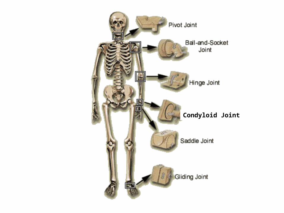
Condyloid Joint
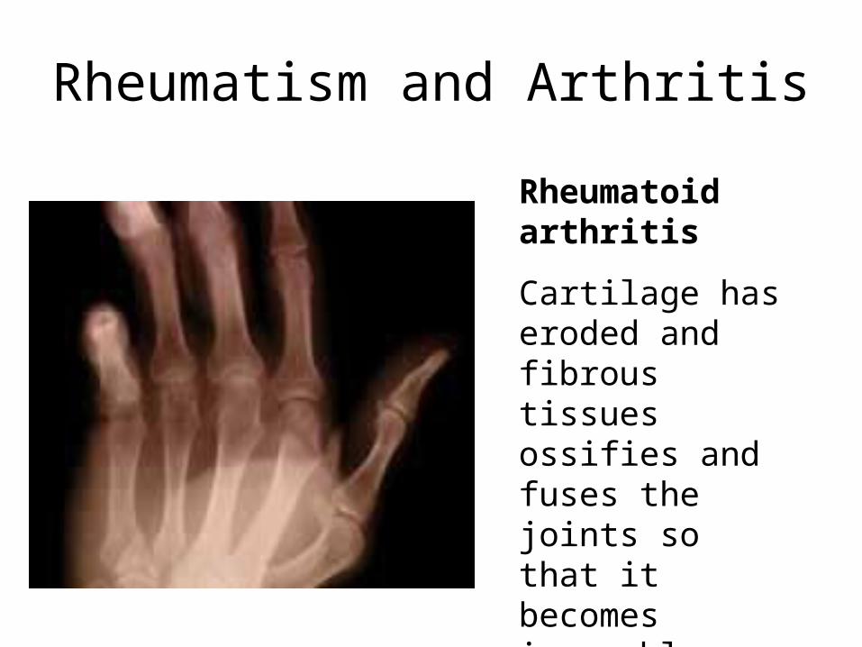
Rheumatism and Arthritis
Rheumatoid arthritis
Cartilage has eroded and fibrous tissues ossifies and fuses the joints so that it becomes immovable
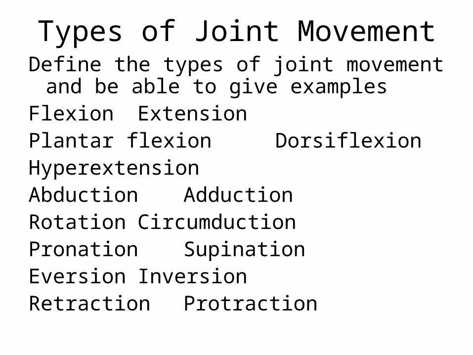
Types of Joint MovementDefine the types of joint movement and be
able to give examplesFlexion ExtensionPlantar flexion DorsiflexionHyperextensionAbduction AdductionRotation CircumductionPronation SupinationEversion InversionRetraction Protraction
Skeletal System Interaction (with other systems)
Complete skeletal system interaction chart.