Embed Size (px)
Citation preview

Skin cancerInformation for patients
Our pathologists are specialists, and their experience and commitment to professional development results in a broad range of expertise that can assist in the diagnosis and management of your skin condition.
Many disciplines of pathology contribute to the diagnosis of disorders of the skin and it is this collaboration that ensures doctors and their patients receive accurate and reliable results.
Our specialist pathologists1 PreventionThe key to reducing your risk of developing a skin cancer is to be cautious about how much sunlight your body is exposed to and to understand ways of preventing damage to your skin.
� Stay out of the sun during peak UV times (usually between 10.00 am and 2.00 pm).
� Wear protective clothing when out in the sun. � Apply sunscreen liberally to exposed skin areas, and
always reapply after swimming or heavy exercise. � Try to utilise natural shade areas where possible. � Regularly check your skin for any new spots, or
changes in colour and appearance in existing spots. � Have a doctor examine any suspicious spot as soon as
possible.
2 Early detectionIt is important to visit the doctor regularly to have your skin checked. This includes areas routinely exposed to the sun and those areas which only have intermittent sun exposure. It is widely agreed that early detection of skin cancer can lead to a cure in 99% of cases, and the rise in survival rate is thought to be due to treatment at an earlier stage.
3 Skin changesSkin cancers can look harmless and are often not painful. New spots, existing sores or lumps which don’t heal within a month, or any mole or freckle that grows or alters, are all important changes that should be reported to your doctor as soon as possible.
4 TreatmentIf a skin cancer is found, the treatment will vary, depending on the location, the type of skin cancer that was diagnosed and the thickness of the lesion. Taking these factors into account, your own doctor is best able to advise you on the most appropriate treatment.
The pathologist will carefully review the slides and produce a report that is sent back to your doctor, containing the diagnosis of the lesion, along with any other important information that will assist your doctor if further treatment is necessary.
How can I reduce the likelihood of a
skin cancer forming?
DOUGLASS HANLY MOIR PATHOLOGY
14 GIFFNOCK AVENUE • MACQUARIE PARK • NSW 2113 • AUSTRALIA P (02) 98 555 222 • F (02) 9878 5077MAIL ADDRESS • LOCKED BAG 145 • NORTH RYDE • NSW 1670 • AUSTRALIA
www.dhm.com.au SS
DX
-DH
M-P
t-V2
-20
18
-D0
00
71
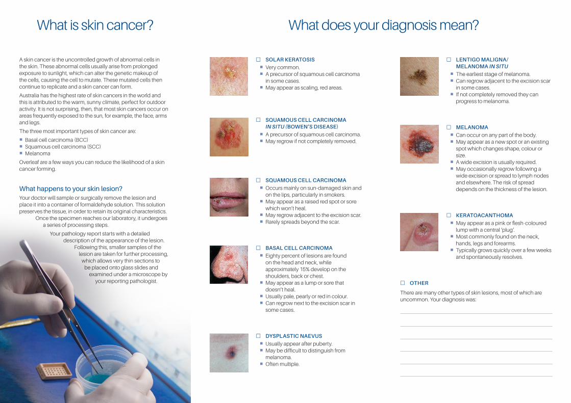
� SOLAR KERATOSIS
� Very common. � A precursor of squamous cell carcinoma
in some cases. � May appear as scaling, red areas.
� SQUAMOUS CELL CARCINOMA IN SITU (BOWEN’S DISEASE)
� A precursor of squamous cell carcinoma. � May regrow if not completely removed.
� SQUAMOUS CELL CARCINOMA
� Occurs mainly on sun-damaged skin and on the lips, particularly in smokers.
� May appear as a raised red spot or sore which won’t heal.
� May regrow adjacent to the excision scar. � Rarely spreads beyond the scar.
� BASAL CELL CARCINOMA
� Eighty percent of lesions are found on the head and neck, while approximately 15% develop on the shoulders, back or chest.
� May appear as a lump or sore that doesn’t heal.
� Usually pale, pearly or red in colour. � Can regrow next to the excision scar in
some cases.
� DYSPLASTIC NAEVUS
� Usually appear after puberty. � May be difficult to distinguish from
melanoma. � Often multiple.
� LENTIGO MALIGNA/ MELANOMA IN SITU
� The earliest stage of melanoma. � Can regrow adjacent to the excision scar
in some cases. � If not completely removed they can
progress to melanoma.
� MELANOMA
� Can occur on any part of the body. � May appear as a new spot or an existing
spot which changes shape, colour or size.
� A wide excision is usually required. � May occasionally regrow following a
wide excision or spread to lymph nodes and elsewhere. The risk of spread depends on the thickness of the lesion.
� KERATOACANTHOMA
� May appear as a pink or flesh-coloured lump with a central ‘plug’.
� Most commonly found on the neck, hands, legs and forearms.
� Typically grows quickly over a few weeks and spontaneously resolves.
� OTHER
There are many other types of skin lesions, most of which are uncommon. Your diagnosis was:
What does your diagnosis mean?What is skin cancer?
A skin cancer is the uncontrolled growth of abnormal cells in the skin. These abnormal cells usually arise from prolonged exposure to sunlight, which can alter the genetic makeup of the cells, causing the cell to mutate. These mutated cells then continue to replicate and a skin cancer can form.
Australia has the highest rate of skin cancers in the world and this is attributed to the warm, sunny climate, perfect for outdoor activity. It is not surprising, then, that most skin cancers occur on areas frequently exposed to the sun, for example, the face, arms and legs.
The three most important types of skin cancer are:
� Basal cell carcinoma (BCC) � Squamous cell carcinoma (SCC) � Melanoma
Overleaf are a few ways you can reduce the likelihood of a skin cancer forming.
What happens to your skin lesion?Your doctor will sample or surgically remove the lesion and place it into a container of formaldehyde solution. This solution preserves the tissue, in order to retain its original characteristics.
Once the specimen reaches our laboratory, it undergoes a series of processing steps.
Your pathology report starts with a detailed description of the appearance of the lesion.
Following this, smaller samples of the lesion are taken for further processing,
which allows very thin sections to be placed onto glass slides and
examined under a microscope by your reporting pathologist.