Embed Size (px)
Citation preview

Slaughtered Hogs with Discoloured Bones and the Relationship with Tetracycline Medication in the Grower-Finisher Stage
by
Norma Patricia Varela Cruz
A Thesis presented to
The University of Guelph
In partial fulfilment of requirements for the degree of
Doctor of Philosophy in
Population Medicine
Guelph, Ontario, Canada
© Norma Patricia Varela Cruz, May, 2012

ABSTRACT
PREVALENCE OF DISCOLOURED BONES IN HOGS AT SLAUGHTER AND ITS RELATIONSHIP WITH TETRACYCLINE-MEDICATED FEEDING
PROGRAMS FOR GROWER-FINISHER PIGS
Norma Patricia Varela Cruz Advisors: University of Guelph, 2012 R. Friendship, C. Dewey
Bone discolouration of pig carcasses is a quality concern that has been
observed in Ontario slaughter plants. The objectives of this study were to
establish the prevalence of pig carcasses showing bone discolouration, its
relationship with residues of tetracyclines in bones, and to investigate the use of
tetracyclines in feeding programs for grower-finisher pigs as the main risk factor
for discolouration.
Abattoir data were examined to determine the extent of the problem and the
prevalence of bone discolouration during 2006, 2008, 2009, and 2010 was found
to be 0.13%, 0.22%, 0.26%, and 0.28%, respectively, indicating that the issue of
bone discolouration was present at low levels over the entire period of the study.
A controlled trial using feed, water, and injectable tetracycline products to
investigate the effect of tetracyclines on residue and bone colour was conducted.
Bones were assessed visually for signs of discolouration, and high performance
liquid chromatography (HPLC) was used to measure the levels of tetracycline
residues in the bones. Results from this trial demonstrated that discolouration
could be produced with 660ppm of chlortetracycline (CTC) in feed for 12 weeks

even when 33 days of withdrawal time was observed. It was also found that
residues of tetracyclines can be present in bones in the absence of
discolouration.
A retrospective study was conducted to investigate tetracycline use in herds
identified as having discoloured bones at slaughter. Positive shipments were
associated with dosage and duration of CTC use as well as with length of
withdrawal.
In conclusion, discoloured bones of pig carcasses were identified at low
levels in one large Ontario abattoir; however, further investigation is needed in
order to determine the impact it may have on the swine industry.

iv
DEDICATION
To the most important people in my life:
My mother Cecilia Cruz
My brother Michel Varela
My Husband Cesar Caballero
My lovely kids David and Matthew.

v
ACKNOWLEDGEMENTS
I would like to acknowledge and express my gratitude to all the people who made
this dissertation possible.
First and foremost, I would like to thank my graduate advisory committee: Dr.
Robert M. Friendship, Dr. Cate Dewey, and Dr. Jerome R. E. del Castillo. I thank
them for their support and mentorship throughout my PhD program.
Dr Friendship, I would like to offer my deepest thanks for your time, advice, and
invaluable and unconditional support, always available to me.
This project was possible due to the financial support of Ontario Pork and the
University of Guelph- Ontario Ministry of Agriculture (OMAFRA) Sustainable
Production Systems Program.
I would like to thank the Department of Population Medicine at the Ontario
Veterinary College for giving me the opportunity to perform my graduate studies.
A special thanks goes to the faculty members, fellow graduate students,
secretaries, and friends who created a stimulating environment making this a
positive experience for me.

vi
I would like to thank all members of the Laboratory Services Division – University
of Guelph, particularly Louise Spilsbury, for her technical assistance with the
High Performance Liquid Chromatography (HPLC).
I would like to recognize William Sears for his statistical support.
I would like to thank Eric Brandt for his unconditional support and friendship since
Cesar and I arrived to this beautiful country “Canada”.
In particular, I would like to thank those special people who always supported
and encouraged me to achieve my dreams. Thanks to my mother, Cecilia Cruz,
my brother, Michel Varela, my husband, Cesar Caballero, and my sons, David
and Matthew.
To my mother, thank you mom for always being there believing in me, your
support, love and guidance have meant so much to me. I feel blessed to have a
mom like you. “I love you mom”.
To my brother, thank you Michel, I know you are with God, but somehow you are
also with us.
To my husband Cesar, your love, friendship, and support means more to me
than you could ever imagine. I feel so lucky to have you in my life. “I love you”.

vii
To my kids, David and Matthew, I have no word to express how much you guys
mean to me. “I love you”.

viii
TABLE OF CONTENTS
CHAPTER 1
General Introduction and Literature Review
General Introduction..............................................................................
Meat Inspection and Xenobiotic Residues.............................................
Issues of Carcass Quality...........................................................
Bone Growth and Physiology................................................................
Structure......................................................................................
Bone Formation...........................................................................
Organic Matrix Synthesis (Osteoid)..................................
Intramembranous Bone Formation........................
Endochondral Bone Formation (Bone Modeling)..
Mineralization of Organic Matrix to form Bone.................
Bone Resorption...............................................................
Bone Remodelling............................................................
Tetracyclines..........................................................................................
The Origin of Tetracyclines........................................................
Chemical Structure......................................................................
Mechanism of Action...................................................................
Pharmacokinetics of Tetracyclines..............................................
Absorption........................................................................
Distribution.......................................................................
Metabolism and Excretion................................................
Properties of Tetracyclines..........................................................
Antimicrobial.....................................................................
Non-Antimicrobial.............................................................
Metal Chelation................................................................
Use of Tetracyclines in Swine Production...................................
1
3
6
7
8
10
10
11
12
13
14
15
16
16
17
18
19
19
20
23
24
24
24
25
26

ix
Bone Discolouration in Animals Intended for Human
Consumption...............................................................................
Liquid Chromatography Method to Detect and Measure
Tetracycline Residues in Bone Tissue of Food Producing
Animals.......................................................................................
Summary of the Problem.......................................................................
Specific Research Objectives................................................................
References.............................................................................................
CHAPTER 2
The Prevalence of Carcasses with Discoloured Bones at an Ontario
Abattoir
Abstract..................................................................................................
Introduction............................................................................................
Materials and Methods...........................................................................
Data Handling and Analysis...................................................................
Results...................................................................................................
Discussion..............................................................................................
References.............................................................................................
CHAPTER 3
An Investigation of Residues of Tetracyclines in Pig Bones
Abstract..................................................................................................
Introduction............................................................................................
Materials and Methods...........................................................................
Part 1 – Controlled Trial to Compare Tetracycline Exposure
With Bone Discolouration and Fluorescence at laughter..........
29
31
34
36
37
47
48
49
50
51
52
55
58
59
60
61
x
Part 2 – Tetracycline Residues in Edible Tissue and Bones as
Determined by HPLC .................................................................
Data Handling and Analysis...................................................................
Results...................................................................................................
Discussion..............................................................................................
Conclusion.............................................................................................
References.............................................................................................
CHAPTER 4
The Relationship between Discoloured Bones in Hog Carcasses and
Medicated Feeding Programs for Grower-Finisher Pigs
Abstract..................................................................................................
Introduction............................................................................................
Materials and Methods...........................................................................
Source of Data............................................................................
Descriptive Statistics...................................................................
Logistic Regression Analysis......................................................
Results...................................................................................................
Descriptive Statistics...................................................................
Logistic Regression Analysis......................................................
Discussion..............................................................................................
References.............................................................................................
CHAPTER 5
General Discussion.............................................................................
References............................................................................................
64
65
66
67
71
73
76
77
78
78
81
82
83
83
85
86
92
100
105
xi
APPENDICES
A.1.1. Letter from Animal Health Laboratory (AHL) – University of
Guelph, Reporting an Outbreak of Disease in Fall 2005......................
A.1.2. Meat Hygiene Directive – January 4, 2006: Disposition of
Carcasses with Bones Discoloured by Tetracyclines...........................
A.1.3. Meat Hygiene Directive – March 10, 2006: Disposition of
Carcasses with Bones Discoloured by Tetracyclines...........................
107
108
110

xii
LIST OF TABLES
Table 1.1. Approved Brands, Claims, Levels, Directions and Withdrawal
Times for Chlortetracycline Hydrochloride as Listed in the
Compendium of Medicating Ingredient Brochures..........................
Table 1.2. Approved Brands, Claims, Levels, Directions and Withdrawal
Times for Oxytetracycline Hydrochloride as Listed in the
Compendium of Medicating Ingredient Brochures.........................
Table 3.1. Levels of Tetracycline HCL, Chlortetracycline, and
Oxytetracycline (ppm) in Bone as Measured by High
Performance Liquid Chromatography.............................................
Table 4.1. Significant Independent Variables Included in the Final Model of
Discoloured Bones of Hogs Marketed by One Commercial Swine
Company in Ontario – 2006............................................................
44
45
75
94

xiii
LIST OF FIGURES
Figure 1.1. Basic Molecular Structure of the Main Tetracycline Antibiotics....
Figure 2.1. Monthly Prevalence of Pig Carcasses Detected with Discoloured
Bones at One Ontario Abattoir from 2006 to 2010........................
Figure 4.1. Proportion of Pig Carcasses with Discoloured Bones at
Slaughter, Based on the Estimated Cumulative Chlortetracycline
Consumption of Hogs Marketed by a Commercial Swine
Company in Ontario - 2006...........................................................
Figure 4.2. Proportion of Pig Carcasses with Discoloured Bones at
Slaughter, Based on Duration of Chlortetracycline
Treatment......................................................................................
Figure 4.3. Proportion of Pig Carcasses with Discoloured Bones Based on
Withdrawal Times of Hogs Marketed by a Commercial Swine
Company in Ontario – 2006...........................................................
Figure 4.4. Proportion of Pig Carcasses with Discoloured Bones at
Slaughter, Based on Shipment from the Grower-Finisher
Barn...............................................................................................
Figure 4.5. Probability of Pig Carcasses Showing Bone Discolouration
According to Chlortetracycline (CTC) and Withdrawal Time
Interaction......................................................................................
46
57
95
96
97
98
99

1
CHAPTER 1: General Introduction and Literature Review
1.1. GENERAL INTRODUCTION
In October 2005, a shipment of pigs from one producer in Ontario caused
alarm among meat inspectors because the bones of most of the pigs were
strikingly yellow. This one incident triggered discussion and awareness among
Canadian Food Inspection Agency (CFIA) inspectors in Ontario and possibly as a
result, about 500 pig carcasses were identified with bone discolouration over the
next two months. The original shipment resulted in a farm investigation and it
was revealed that tetracyclines had been fed to these pigs at relatively high
levels continuously for at least 12 weeks in the grower-finisher barn.
Tetracyclines have been used for more than 60 years in food animal
production; with tetracycline, chlortetracycline and oxytetracycline being among
the most widely used antimicrobial drugs in the Canadian swine industry (Health
Canada, 2002). Tetracyclines are commonly used in feed, water, or as injectable
products to improve growth rate and to prevent, treat or control disease (Riviere
and Spoo, 1995). It has long been known that tetracyclines bind to calcium and
can lead to bone discolouration (Albert and Rees, 1956; Benitz et al, 1967;
Buyske et al, 1960; Weinberg, 1957). Bound tetracyclines in bone is an important
issue in the swine industry regarding the safety and acceptability of pork products
since residues of tetracyclines not only decrease the quality of pork carcasses,
but also may affect food safety through contamination of both mechanically
deboned meat and bone meal (Kühne and Körner, 2001; Kühne et al, 2000).
Meat obtained from mechanically deboned carcasses may contain bone splinters

2
and thus be contaminated with tetracyclines; likewise bone meal from pigs fed
high levels of tetracyclines used for preparation of animal feeds could be a
concern given that complete destruction of tetracycline and chlortetracycline has
not been demonstrated after treatment at 133ºC for up to 45 min (Kühne et al.,
2001). Despite widespread use of tetracyclines in the pig industry worldwide,
there are few reports of problems with discoloured bones at slaughter. It is
possible that because the Ontario pig industry was dealing with a disease
outbreak in the Fall of 2005 (See Appendix A.1.1) that higher levels of antibiotics
were being used on pig farms during this period.
Although muscle and organ tissue from pigs with discoloured bones in the
October 2005 incident were tested and found to be acceptable for human
consumption with residue levels of tetracyclines well below the targets set by
regulatory agencies, the CFIA released a Meat Hygiene Directive ordering
positive carcasses deboned (See Appendix A.1.2). Subsequently a second Meat
Hygiene Directive was issued that described the problem as one of cosmetic
concern rather than a food safety issue and gave the packers the responsibility of
dealing with the problem (See Appendix A.1.3).
This remains an important issue in the Canadian pig industry because
consumer confidence is essential to maintain good sales both domestically and
internationally.

3
1.2. MEAT INSPECTION AND XENOBIOTIC RESIDUES
Meat inspection is a sanitary control process of slaughter animals and meat
intended for human consumption. The main purpose of inspection is to assist in
preventing the spread of food borne disease, or undesirable meat from entering
the food supply and, thus ensuring safe and wholesome meat and meat
products. This activity, which involves examination of the living animals awaiting
slaughter and examination after slaughter of head, carcass, and viscera, is
carried out by public health authorities represented by veterinary meat inspectors
at the abattoir stage. In conducting an inspection, an inspector must apply the
standards related to food safety and animal health under the Food and Drugs Act
(Canada) and the Meat Inspection Act (Canada).
The presence of chemical residues in food accounts for a high level of
concern among consumers because toxicity and allergic reactions may occur.
Furthermore, exposure to low levels of antibiotics could possibly result in the
development of antibiotic resistant strains of bacteria (Nisha, 2008).
Antibiotics are widely used in livestock production and therefore there is
always a risk that residues may be present at slaughter. Residue testing of edible
tissue is often carried out if inspectors suspect an animal has been treated with
medication. Antibiotics used to treat livestock undergo an intensive approval
process. In Canada, prior to being offered for sale, veterinary drugs must be
tested and approved by Health Canada’s Health Products and Food Branch,
Veterinary Drugs Directorate. Thus, when used according to label directions,

4
levels of drug or antibiotic residue will not violate the allowable limits established
for the drug or antibiotic.
Veterinarians have the responsibility to extend withdrawal periods if
necessary when a drug is prescribed in an extra-label manner in order to avoid
drug residues. When tetracyclines or other drug residues are found in pork and
pork products above allowable limits, there is a potential risk to human health
and it is a violation of the Canada Food and Drugs Act and Regulations (FDAR).
The FDAR lists the permitted feed additives and how they may be used; its
implementation and enforcement with respect to foods is the responsibility of the
Canadian Food Inspection Agency (CFIA). In swine, the tolerance levels for
chlortetracycline, oxytetracycline and tetracycline residues are 0.2 ppm, 0.6 ppm,
and 1.2 ppm in muscle, liver, and kidney tissue, respectively (Health Canada
2011). Maximum Residue Limits (MRLs), a residue considered to pose no
adverse health effect if ingested daily by humans over a lifetime, are established
for edible tissues such as skeletal muscle, liver, and kidney and not for non-
edible tissues (bile, blood, bone, brain, diaphragm, gastrointestinal contents,
gastrointestinal tract, heart, lung, skin, spleen, and thyroid gland). The World
Health Organization (WHO) and the Food and Agriculture Organization (FAO)
have set standards for acceptable daily intake (ADI) and maximum residue limits
(MRLs) in food of animal origin. The acceptable MRLs for chlortetracycline,
oxytetracycline, and tetracycline as recommended by the Joint FAO/WHO Expert
Committee on Food Additives is 200, 600, and 1200 µg/kg for muscle, liver, and

5
kidney, respectively (World Health Organization: Join FAO/WHO Expert
Committee on Food Additives, 1998)
In most cases, antimicrobial residues in pork are caused by farmers’ failure
to observe appropriate withdrawal times. In addition, on-farm feed preparation
practices that allow medicated feed to mix with non-medicated feed, human
errors in feeding or in the use of inaccurate scales, can all lead to residues in
meat (Friendship, 2006).
When conducting a carcass inspection, meat inspectors should be able to
identify any abnormality that may compromise the food safety and the
wholesome image of Canadian pork products; this includes the identification of
carcasses from animals that had been treated with antibiotics or any other
chemical agent (Herenda et al., 1994). According to the Canadian Food
Inspection Agency (Canadian Food Inspection Agency, 2010), antibiotic use
should be suspected in any animal in which an injection site is found, and any
animal affected with a septic condition, which might have been treated with
antibiotics (Canadian Food Inspection Agency, 2010). It is important to consider,
however, that the extent and severity of any condition differs among inspectors
because judgement is based on subjective evaluation and then observational
bias may happen. Variability in condemnation rates among slaughter plants with
similar condemnation criteria has been documented (Tuovinen et al., 1994,
Jackowiak et al., 2006, Elbers et al., 1992). Carcasses from animals which an
inspector believes may have been exposed to a drug should be held until
appropriate testing is conducted and results received, If a residue of a medication

6
normally administered to a group of animals is detected in one animal, all the
animals in the same lot are suspected of the residue (Canadian Food Inspection
Agency, 2010). Appropriate testing and regulations have been established by the
legal authorities depending on the particular residue suspected and type of
exposure (individual or lot), respectively. Overall, individual exposure testing is
dependent on the particular compound suspected, and the held product can be
treated as condemned material keeping samples for additional testing. On the
other hand, if exposure of a lot is suspected, initial screening of 6 carcasses from
the suspected lot is conducted using the Swab Test On Premises (STOP). If the
test result is negative the carcasses are released; otherwise, the carcass
remains under detention and will be condemned should the muscle sample
contain violative levels (Canadian Food Inspection Agency, 2010).
To improve the quality and safety of pork, different programs have been
instituted in different countries with the main objective to increase farmers’
awareness of antibiotic use and improve adherence to withdrawal times. In
Canada, the Canadian Quality Assurance Program, CQA was developed for
producers to encourage them to adhere to withdrawal times by keeping a
complete drug record under a veterinarian’s supervision. The use of on-farm
mixing medication is also examined under the CQA program.
1.2.1. Issues of Carcass Quality
Being recognized as one of the major world pork exporters, the Canadian
pork industry depends on a reputation for a safe and high quality product. One

7
area associated with pork quality is colour. The colour of meat is a primary
indicator of quality and has a huge impact on export sales, especially to Japan.
Although generally darker pork is preferable, it may vary from bright reddish-pink
to slightly darker; both pale and dark colour are unattractive pork colour to
consumers and indicate possible issues of quality (Lammers et al., 2007).
Besides muscle colour, there are other characteristics that are important when
assessing cuts of meat such as bone content, fat trim, and marbling or
intramuscular fat (Sebranek and Judge, 1990). Fat should be firm, greasy and
white; fat and lean separation are undesiable (Wilson, 2005).Various feed
ingredients can affect the appearance of fat and impact consumer acceptance;
diets containing high saturated fatty acids such as soybean oil and poultry fat are
known to produce soft fat, which results in difficult processing and pork cuts that
are less firm and unattractive to the customer (Goodband et al., 2006).
Pork is often sold as cuts of meat with the bone included which is expected
by consumers to be shiny and white; thus discolouration of the bones is a quality
defect. Cuts of meat showing discoloured bones results in consumer rejection
because of bone colour and for this reason the value of the carcasses with
discoloured bones is reduced.
1.3. BONE GROWTH AND PHYSIOLOGY
Bone is a dynamic and rigid tissue that forms part of the endoskeleton of
vertebrates. Its main function involves: mechanical support and protection,
storage for body calcium, and a site for adult haematopoiesis (Salo et al., 1996).

8
1.3.1. Structure
Bone is composed of two types of osseous tissue mainly classified on the
basis of porosity as: compact or cortical bone and trabecular bone also known as
cancellous or spongy bone (Garner et al., 1996). Compact or cortical bone
makes up to 80% of the total bone mass and is mainly responsible for the
support function; it is a denser tissue with a high mineral content, which gives
bones their smooth, white, and solid appearance. Trabecular bone is much more
porous and accounts for the remaining 20% of the total bone mass; it is
composed of bone plates filled with blood vessels and marrow, which makes
trabecular tissue an important component for bone remodelling.
Regarding its cellular structure, skeletal tissue is composed of various types
of cells grouped into two general types, bone-forming cells and bone-resorbing
cells. Bone-forming cells or osteoblasts originate from the mesenchymal stem
cells by a linear sequence that progresses from progenitor cells, restricted to
osteoblast development and bone formation known as osteoprogenitor, to
preosteoblasts, osteoblasts and then lining cells or osteocytes (Aubin, 1998;
Aubin, 2001). Preosteoblasts are elongated cells with the unique ability to divide,
and are found in the tissue layer near bone-forming surfaces. Osteoblasts
secrete collagen and lay down new extracellular bone matrix known as osteoid,
which mineralizes to become bone (Cool and Nurcombe, 2005). During the
process of extracellular matrix formation and in the presence of tetracyclines in
serum, some tetracycline may become incorporated with the matrix. Calcium ions
at the site of mineralization may be chelated by the extracellular compounds

9
such as tetracycline, following its incorporation into any hydroxyapatite forming
crystal or into the collagenous matrix (Skinner and Nalbandian, 1975).
Tetracyclines or any other compound previously incorporated into bone tissue
can be released by dissolution of the bone during osteoclastic activity (bone
resorption), and thus bone remodelling plays an important role in the
permanence of tetracyclines in bone tissue (Skinner and Nalbandian, 1975).
Osteoblasts give rise to two cell types within bone: osteocytes and bone-
lining cells which maintain some osteoblastic characteristics but no longer
synthesize collagen for the formation of new bone (Garner et al., 1996). As
extracellular matrix is secreted and calcified around osteoblasts, osteoblasts
become trapped and differentiate further into osteocytes, which are in charge of
homeostasis, exchanging wastes for nutrients from the blood, and regulating
calcium release back into the blood stream (Horton, 1995). Bone-lining cells, also
known as resting osteoblasts or surface osteocytes, are inactive osteoblasts
which are essential for the maintenance of blood calcium levels by pumping Ca+
ions from the bone fluid compartment to the extracellular fluid compartment
(Garner et al., 1996).
Different compounds present in the blood stream may diffuse from the
extracellular fluid to the hydroxyapatite crystals of the bone mineral and be
incorporated into new or remodelled bone.
Bone-resorbing cells also known as osteoclasts are derived from
hematopoietic stem cells and are responsible for bone resorption by dissolving
crystalline hydroxyapatite and degrading organic bone matrix rich in collagen

10
fibres (Vaananen et al., 2000). Its promyeloid precursor can differentiate into
either an osteoclast, a macrophage or a dentritic cell if it is exposed to osteoclast
differentiation factor (ODF), macrophage colony-stimulating factor (M-CSF), or
granulocyte-macrophage colony-stimulating factor (GM-CSF), respectively (Suda
et al., 1999).
At the molecular level, bone consists of two fundamentally different
components: the organic and inorganic. The organic or intercellular component
is formed by approximately 90% of collagen, synthesized and secreted by
osteoblasts, and 10% of a mixture of proteins that are either synthesized by
osteoblasts or are serum-derived (Garner et al., 1996). The inorganic component
is formed mainly by mineral salts and calcium present in the form of
hydroxyapatite crystals deposited within an organic phase of cross-linked
collagen fibres. The process of calcification occurs when these crystals are laid
down between the collagen fibres of the bone matrix (Ham, 1974).
1.3.2. Bone Formation
The process of bone formation or osteogenesis involves four main steps:
organic matrix synthesis by osteoblasts, mineralization of the matrix to form
bone, bone resorption and bone remodelling.
1.3.2.1. Organic Matrix Synthesis (Osteoid)
The prenatal development of bone occurs by deposition of hydroxyapatite
crystals within the collagen matrix; however, the process by which the collagen

11
matrix is laid down differs according to the two major types of bone formation:
intramembranous and endochondral. These terms refer to the sites in which
ossification occurs: endochondral means “in cartilage” and intramembranous
means “within membrane”; in both cases, bone forms because osteoblasts
evolve and secrete the organic intracellular substance of the bone matrix (Garner
et al., 1996; Ham, 1974).
1.3.2.1.1. Intramembranous Bone Formation
Intramembranous ossification, in which cartilage is not present, mainly
occurs during formation of most of the bones of the skull, the jaw, and the ribs
(Garner et al., 1996; Ham, 1974). During this process, embryologic mesenchymal
cells differentiate directly into preosteoblasts and then into osteoblasts in areas of
highly vascular embryonic connective tissue to begin the process of ossification
(Garner et al., 1996). Osteoblasts begin to secrete the organic matrix of the
bone, known as “osteoid tissue” or uncalcified bone and those that completely
surround themselves with osteoid tissue differentiate into osteocytes (Ham,
1974).
Cells that do not differentiate and osteoblasts, remain at the margin of the
bone already formed, with some of the osteogenic cells continuing to proliferate
and others differentiating and secreting intracellular substance around
themselves to become osteocytes to form a beam of bone called a spicule (Ham,
1974). As spicules continue to grow, they join together to form the trabeculae.
Osteoblasts continue to line up on the surface and as growth continues,
12
trabeculae become interconnected and immature or woven bone is formed (Ham,
1974).Differentiating mesenchymal cells surrounding the trabeculae will produce
the periosteum richly supplied with blood vessels involved in the nutrition of the
bone, and osteoblasts from the periosteum on the bone matrix will replace woven
bone by lamellar or compact bone.
1.3.2.1.2. Endochondral Bone Formation (Bone Modeling)
Endochondral ossification, in which bone is formed by calcification of a
cartilaginous structure that serves as a model for the new bone, occurs in long
bones such as those of the limbs; and it is also an essential process during the
growth of the length of bones and the natural healing of the bone fractures.
Young cartilage can grow by two different methods: interstitial and
appositional growth.
The word interstitial refers to the cells in the lacunae known as chondrocytes.
Cartilage is formed by chondroblast cells, which are derived from
prechondroblasts arising from the same mesenchymal cells that form osteoblasts
(Garner et al., 1996). Chondroblasts secrete intercellular collagen matrix,
eventually enclosing themselves, to differentiate into chondrocytes characterized
by their ability to divide until they become mature. Chondrocytes can continue to
enlarge and divide expanding the lacunar spaces, and this continuous cell
division with their subsequent formation of intercellular collagen matrix causes
the cartilage to grow in length (Garner et al., 1996; Ham, 1974).

13
Appositional growth implies a mechanism whereby new layers of cartilage
are apposed to one of its surfaces. During this process the deeper cells of the
connective tissue that surrounds the cartilage of the developing bone, known as
perichondrium, divide to increase their numbers. These cells differentiate into
chondroblasts and then into chondrocytes surrounding themselves with
intercellular substance which is applied to the surface of the cartilage. By this
mechanism a new layer of cartilage is laid down under the perichondrium on the
surface of the cartilage causing the cartilage to grow in width (Ham, 1974).
1.3.2.2. Mineralization of Organic Matrix to Form Bone
Since cartilage is an avascular tissue, cells receive their nutrients by diffusion
through the fluid surrounding the collagen fibers; when the cartilage is invaded by
blood vessels, the process of mineralization of the organic matrix begins (Garner
et al., 1996). In the earliest bone formation either in endochondral formation, or in
woven bone, mineralization is preceded by vesicle-driven-mineralization.
Osteoclasts secrete matrix vesicles that provide a site for the hydroxyapatite
crystal formation; these vesicles act as the foci for calcium and phosphate
deposition and when crystallization produces a crystal larger than the original
vesicle, the vesicle is destroyed and acts as a centre for crystals to grow on
(Garner et al., 1996).
In lamellar bone formed during remodelling, mineralization occurs directly
within the collagen matrix where mineral is deposited in association with the

14
tightly spaced collagen fibrils and associated proteins; vesicle formation is not
involved in this process (Garner et al., 1996).
1.3.2.3. Bone Resorption
Bone resorption is defined as the removal of both the mineral and organic
matrix to produce a cavity in the bone structure. When dead bone, calcified bone
in which the lacunae are empty because the osteocytes they formerly contained
have died and dissolved, is exposed to the contents of a Haversian canal
(capillaries and osteogenic cells) cells of the monocyte-macrophage cell line
differentiate into osteoclasts and the dead bone is resorbed (Chambers, 2010).
During this process minerals are released and transfer of calcium from bone fluid
to blood occurs (Garner et al., 1996; Ham, 1974).
In general, the process of bone resorption involves migration of osteoclasts
to the resorption site, its attachment to bone matrix, polarization and formation of
new membrane domains, dissolution of hydroxyapatite crystals, degradation of
organic matrix, removal of degradation products from the resorption lacuna, and
either apoptosis of the osteoclasts or their return to the non-resorbing stage
(Vaananen et al., 2000).
When bone resorption initiates, osteoclasts migrate to a resorption site, and
three distinct membrane domains are formed: the sealing zone which attach the
osteoclastic plasmatic membrane to the bone matrix from its surroundings, the
ruffled border with finger-like projections formed by fusion of intracellular acidic
vesicles that penetrate the bone matrix, and the functional secretory domain

15
used to transfer degradation products from the resorption lacuna to the
extracellular space (Vaananen et al., 2000). Osteoclasts attach to bone matrix
through the sealing zone and the ruffled border and dissolution of
minerals/hydroxyapatite crystals occurs by targeted secretion of HCl through the
ruffled border into the resorption lacuna, an extracellular space between the
ruffled border and the bone matrix (Vaananen et al., 1990; Vaananen et al.,
2000). After dissolution of minerals occurs, several proteolytic enzymes degrade
the organic bone matrix which is rich in collagen, and degradation products are
transferred from the resorption lacuna from the ruffled border to the functional
secretory domain to be further liberated into the extracellular space (Vaananen et
al., 2000).
1.3.2.4. Bone Remodelling
The remodelling of bone can only be achieved by bone being absorbed from
a surface and added to other surfaces; it is an important process that occurs in
healthy bones to maintain their optimal shape for support (Ham, 1974).
After bone resorption has occurred, the osteogenic cells line up around the
inner surface of the cavity and differentiate into osteoblasts; osteoblasts which
will line the surface of the cavity and lay down new bone until they have filled the
excavation leaving space for blood vessels and nerves (Garner et al., 1996;
Ham, 1974). In growing bone, remodeling (resorption and formation) takes place
in separate regions of the bone with formation exceeding resorption; while in
mature bone, osteoblastic bone formation occurs only at sites of osteoclastic

16
activity, resulting in replacement of the exact quantity of bone removed by
resorption (Garner et al., 1996).
1.4. TETRACYCLINES
Tetracyclines compose a large family of antibiotics but only chlortetracycline,
oxytetracycline and tetracycline are used in pig production in Canada and
therefore the following discussion will primarily focus on these three forms of
tetracyclines.
1.4.1. The Origin of Tetracyclines
Tetracyclines are a large family of antibiotics first described in 1948 as the
fermentation product of a soil bacterium: Streptomyces spp (Wainwright, 1990).
Streptomyces spp. produces metabolites known as polyketides, the key
tetracycline precursors, which are used as defensive toxins. Chemical isolation
and purification of Streptomyces aureofaciens polyketides produced the
compound chlortetracycline, an antibiotic with many advantages over previously
discovered penicillin, streptomycin and chloramphenicol, such as an increased
spectrum of activity especially against Gram-negative organisms (Nelson, 1998).
Examination of other strains of Streptomyces led to the production of other
natural compounds. Oxytetracycline is obtained as a metabolic product of
Streptomyces rimosus (Chambers, 2006). Tetracycline was first produced as a
semi-synthetic derivate from chlortetracycline (Nelson, 1998). Nowadays,

17
tetracycline is produced by a mutant strain of Streptomyces aureofaciens (del
Castillo, 2001).
1.4.2. Chemical Structure
Tetracyclines are a group of four-ringed amphoteric substances slightly
soluble in water at pH 7.0 (Prescott, 2000). They are available for use, mainly as
hydrochloride (acid solutions) as tetracyclines are stable in this form; however,
they rapidly lose their activity in solution (Bryskier, 2005).
In spite of diverse pharmacokinetic properties of tetracyclines, they have the
same basic molecular structure required for bioactivity: the tetracyclic
naphthacene carboxamide system conformed by two rings: A and DCB ring
(Figure 1.1) (Nelson, 1998; Prescott, 2000). The DCBA ring structure is
composed of two distinct regions, the upper and lower peripheral regions which
contain different chemical functional groups and substituents. Tetracyclines with
antibiotic activity must possess a functional group along the upper peripheral
region responsible for the antibiotic properties: the dimethylamine group at
carbon 4 (C4) in Ring A (Sapadin and Fleischmajer, 2006). Similarly, along the
lower peripheral region bioactive tetracyclines must possess the following
functional groups: a diketo-enol system across carbons C11 and C12 between
rings C and B, a tertiary hydroxyl group at the lower bond between rings B and A,
and the tricarbonyl-methane system spread across carbon C1, C2 and C3
(Nelson, 1998; del Castillo, 2001). Structural variations from the reference a a a
molecule tetracycline which has the empirical formula of C22H24N2O8 (Figure 1.1),

18
constitute the specific sequence of other tetracyclines. Chlortetracycline has a
chlorine atom substituent at position C7 and thus its empirical formula is
C22H23N2O8 Cl. Oxytetracycline has a hydroxyl substituent at position C5 and its
empirical formula is C22H24N2O9 (del Castillo, 2001; Bryskier, 2005).
Removal of the dimethylamine group at C4 in Ring A enhances the activity of
this compound against non-antibiotic targets but abolishes antibiotic potency
(Nelson, 1998; Sapadin and Fleischmajer, 2006).
1.4.3. Mechanism of Action
Tetracyclines inhibit bacterial protein synthesis in susceptible
microorganisms by binding to the 30S subunit of the ribosome. In Gram-negative
bacteria, the more lipophilic molecules such as doxycycline and minocycline
penetrate the outer membrane partly by passive diffusion, while the more
hydrophilic molecules such as oxytetracycline and tetracycline cross the
membrane by active transport through water-lined transmembrane protein porin
routs as positive charged cations (Foye et al., 2008; Bryskier, 2005).
Once inside the cell, tetracyclines form a complex with Mg2+ and bind
reversibly to the 30S and 16S subunits of the bacterial ribosome. Binding of the
molecule to the 30S ribosomal subunit ensure inhibition of protein synthesis by
interfering with the binding of aminoacyl-tRNA to the acceptor (A) site on the
messenger RNA molecule/ribosome complex, needed for protein synthesis in
growing organisms (Riviere and Spoo, 1995; Chambers, 2006; Prescott, 2000;
Giguère, 2006). Although tetracyclines have less affinity for mammalian

19
ribosomes, interaction with the16S rRNA occurs indirectly through primarily
distortion of the ribosome (Bryskier, 2005). The binding of tetracyclines to their
target is reversible, which would explain their purely bacteriostatic nature in
prokaryotic cells, due to their capacity to accumulate in the cytoplasm at greater
internal concentrations than the external ones (Bryskier, 2005).
1.4.4. Pharmacokinetics of Tetracyclines
1.4.4.1. Absorption
Unless given IV, a drug must cross several semi-permeable cell membranes
or biologic barriers before it reaches the systemic circulation. These membranes
are mainly composed of biomolecular lipid matrix which determines its
permeability characteristic (Kahn, 2005). Efficient assimilation and enhanced
distribution of oral administrated tetracyclines in the body relies on their lipid
solubility (Aronson, 1980). Tetracyclines are rapidly absorbed from the intestinal
lumen into the blood stream and a maximum plasma concentration is reached
within 2 – 4 hours in carnivores (Agwuh and MacGowan, 2006; del Castillo,
2001). In pigs, the level of absorption of chlortetracycline (28%) has been found
to be significantly greater than that of oxytetracycline (5%) when similar levels of
the drug are fed (del Castillo et al., 2000). The low bioavailability of orally
administered tetracyclines in pigs is probably due to the expression of P-
glycoprotein, an energy-dependent efflux transporter found in the gastrointestinal
tract, responsible for intestinal clearance of lipophilic drugs such as tetracyclines
(del Castillo et al., 2000). As tetracyclines diffuse through the epithelial intestinal

20
cells known as enterocytes, the transporter protein “P-glycoprotein” dump back
the drug molecule into the intestinal lumen preventing them from reaching the
systemic circulation and therefore limiting its bioavailability (Horn et al, 2004).
Furthermore, the oral bioavailability of tetracyclines may be impaired by different
factors such as food particles and divalent cations found in dairy products,
aluminum hydroxide gels, and bismuth subsalicylate (Chambers, 2006). Swiss
researchers documented reduced chlortetracycline bioavailability, a parameter
used to measure absorption, when high dietary calcium was used (Wanner et al.,
1991). Binding of tetracyclines to organic macromolecules occurs in the presence
of Ca++ and other cations present in the diet. The tetracyclines precipitate and
become unable to cross the biological membranes, which in turn decreases their
absorption markedly (Nelson, 1998; Agwuh and MacGowan, 2006; Poiger and
Schlatter, 1979; White and Pierce, 1982). In contrast, dietary citric acid positively
affects the bioavailability and absorption of orally administered tetracyclines,
especially chlortetracycline. The bioavailability of orally administered
oxytetracycline and chlortetracycline in piglets was reported to be significantly
increased by the citric acid content in feed, even when a basal calcium diet was
administered (Wanner et al., 1991; Pollet et al., 1983; Wanner et al., 1990).
1.4.4.2. Distribution
Once tetracyclines are absorbed into the bloodstream, they bind to plasma
proteins (Riviere and Spoo, 1995). When plasma-protein binding of
antimicrobials is greater than 85%, it negatively affects drug distribution because

21
only free molecules are microbially active and able to diffuse into the target tissue
(Craig and Suh, 1978). Plasma-protein binding of more lipid-soluble
chlortetracycline and less lipid-soluble oxytetracycline are 70% and 20%,
respectively (del Castillo et al., 1998). High lipid soluble tetracyclines have the
advantage of penetrating tissues better than others with less soluble properties
and thus an increased bacteriostatic efficacy. The fact that chlortetracycline
distributes more easily into the site of infection is due to its increased lipid
solubility when compared to oxytetracycline (del Castillo et al., 1998). One of the
factors involved in deposition of tetracyclines in bone is the presence of chelate
tetracyclines in the bloodstream. Although bone is a relatively inert tissue,
deposits of crystals in its organic matrix expose a large surface area to
extracellular fluids, which is important for the rapid exchange of ions and cellular
activity (Barrére et al, 2006). Once in the bloodstream, tetracyclines are
transported mainly as Ca++ and Mg++ chelates, and once chelated, act as
ionophores capable of crossing lipophilic barriers delivering ions and
tetracyclines into cellular compartments (Nelson, 1998). Tetracyclines distribute
rapidly throughout the body entering almost all tissues and secretions. They
accumulate mainly in the kidney, liver, lung, muscle, serum, spleen, bones, and
enamel of unerupted teeth (Chambers, 2006; Black and Gentry, 1984); relatively
high concentrations of tetracyclines have been found in umbilical cord plasma,
amniotic fluid and breast milk (Chambers, 2006).
Persistence of fluorescence in bone, as compared with cartilage and other
soft tissues has been related to either a direct binding of tetracyclines to matrix or

22
a more complex formation occurring between new bone matrix, calcium, and
tetracyclines (Milch et al, 1957). Fluorescence has been observed in
unmineralized osteoid, and thus some authors speculate that during the process
of collagen synthesis concentrations of tetracyclines from the serum may occur
resulting in a direct binding of tetracyclines to matrix per se (Skinner and
Nalbandian, 1975; Milch et al, 1957). A more complex phenomenon occurs at the
site of mineralization, calcium ions may be chelated by the extracellular
tetracycline, and any crystallites forming on or within the collagenous matrix may
be the foci for tetracycline incorporation (Skinner and Nalbandian, 1975).
Incorporation of tetracyclines with crystals of calcium carbonate (CcCO3) and
hydroxyapatite (HA) crystals which are deposited in the organic matrix has been
documented to be essential in the process by which tetracyclines are fixed in
mineralized tissues (Skinner and Nalbandian, 1975). However, only a portion of
the initially absorbed tetracycline is tightly bound in the bone; the rest is removed
by diffusion into the blood (Buyske et al, 1960). A more abrupt decline in the
amount of tetracycline deposited in bones was observed when compared to
chlortetracycline after 24 hours following the last of two intraperitoneal injections
in adult rats, which is explained by the fact that chlortetracycline is the stronger of
the two chelate agents (Buyske et al, 1960).
Deposition of tetracyclines in bones has been reported to be related to their
capability to chelate cations but also to the route of administration. Parenteral
administration of tetracyclines is followed by an immediate deposition and this
deposition remains for a long time. A single intravenous dose of 50 mg/g of body

23
weight of tetracycline was enough to result in detectable levels of 24 to 34 mg/kg
in the bone after 1 minute of the injection. Fluorescence was observed in the
skeleton of rats 16 weeks after tetracycline was administered intraperitoneally
(Buyske et al., 1960). Withdrawal times are based on detectable levels of drugs
dropping below the level deemed to be safe and is measured in edible tissues.
However, there is little information regarding appropriate withdrawal times that
ensure bone tissues are free of drug residues after administration of
tetracyclines. Guillot et al. (2011) reported that bone staining in pigs is reversible
when the time from drug administration until marketing is quite long in the order
of 12 – 16 weeks. According to these investigators feeding CTC for four weeks is
sufficient to induce discolouration which becomes more intense as treatment
duration and age at treatment onset increase, but can be reduced or even
disappear when a withdrawal time according to age at treatment onset is
followed (Guillot et al., 2011). Prolonged withdrawal times are needed in pigs
treated at later stages because discolouration vanishes at a slower rate when
treatment onset increases due to the fact that the bone turnover rate decreases
as age increases (Guillot et al., 2011; Li et al., 1989).
1.4.4.3. Metabolism and Excretion
Natural tetracyclines (tetracycline, chlortetracycline, and oxytetracycline) are
unstable under strong acidic and alkaline conditions. Epimerization of
tetracyclines in weak (pH 3) and strong (below pH 2) acid conditions leads to the
formation of 4-epitetracycline and anhydrotetracycline, respectively (Pena et al,

24
1998). Tetracyclines are mainly excreted by renal filtration and bilary routes as
unchanged, with only 5% excreted as the metabolite 4-epitetracycline (Agwuh
and MacGowan., 2006).
1.4.5. Properties of Tetracyclines
1.4.5.1. Antimicrobial
Tetracyclines are bacteriostatic antibiotics that may be used in the treatment
of many common infections. They are characterized by their chemotherapeutic
efficacy against a wide range of bacteria. Development of strains of
microorganisms resistant to tetracyclines has reduced the effectiveness of this
drug; however glycylcyclines, minocycline and tetracycline analogs have been
identified with activity against tetracycline-resistant strains due to their ribosomal
protection mechanism (Giguère, 2006). Differences in antimicrobial activity of
tetracyclines in-vivo result from their differences in lipid solubility which influences
their pharmacokinetic characteristics such as: absorption, distribution,
metabolism / excretion, and concentration of a specific tetracyclines within the
cell (del Castillo and Besner., 2001).
1.4.5.2. Non-Antimicrobial
Chemically modified tetracyclines have non-antimicrobial properties of
potential value in preventive medicine such as anti-inflammatory properties. They
have been reported to be potent inhibitors of mammalian collagenases and
several other matrix metalloproteinases involved in inflammatory and

25
degenerative disorders (Nelson, 1998). Interstitial collagenases are considered to
mediate tissue breakdown during the progression of inflammatory diseases such
as periodontal diseases, rheumatoid arthritis, corneal ulceration and dystrophic
epidermolysis bullosa (Golub et al., 1990; Paulus, 1995). Tetracyclines have the
ability to inhibit the pathologically excessive collagenase activity produced during
the inflammation process, as well as to enhance the collagen turnover (synthesis
and degradation) production required to maintain the normal tissue integrity
(Golub et al., 1998).
Chemically modified tetracyclines have been used in the treatment, not only
of inflammatory diseases, but also in treatment of dermatologic and
immunological diseases such as cardiomyopathies (Sapadin and Fleischmajer,
2006; Tsankov et al., 2003; Reddy et al., 2004).
1.4.5.3. Metal Chelation
Tetracyclines have been found to have powerful ionophoreitc properties. As
ionophore compounds, they are capable of forming lipid-soluble complexes with
metal cations and transporting them across hydrophobic barriers (Nelson, 1998,
White and Pearce, 1982). Tetracyclines circulate in blood plasma primarily as
Ca++ and Mg++ chelates, which allow the delivery of ions and tetracyclines into the
intracellular compartments (White and Pearce, 1982). After cellular incorporation,
Ca++ acts as a secondary messenger affecting many biological functions /
pathways such as metabolic reactions and cell division cycles among others
(Nelson, 1998). It also has been suggested that this ionophoreitc characteristic of

26
tetracyclines is the cause of their inhibitory effect on bone growth and
discoloration of growing teeth (Nelson, 1998).
Tetracyclines have been used as a bone label due to their ability to form
stable tetracycline-calcium chelates, which fluoresce under UV light.
Osteogenesis involves organic matrix synthesis by osteoblasts and its
subsequent mineralization to form bone, a process in which new matrix requires
a defined period of time to become calcifiable (Garner et al., 1996). Fluorescence
bone markers such as tetracyclines, label instantaneously and permanently the
zone of demarcation of osteoid tissue from mineralized bone (Tam et al., 1980).
The amount of bone growth is then calculated by measuring the distance
between fluorescence bands that have been formed at the time of the marker
being deposited.
1.4.6. Use of Tetracyclines in Swine Production
Since their discovery in 1945, tetracyclines have been used in veterinary
medicine as broad-spectrum antibiotics that may be used in the treatment and
prophylaxis of many common infectious diseases and as growth promoters. They
have been widely used in swine production for over six decades as additives in
feed or water to treat and prevent disease, to increase animal growth or weight,
and to reduce the feed required per pig (Giguère, 2006).
The Compendium of Medicated Ingredient Brochures (CMIB) is the
document that lists those medicating ingredients permitted by Canadian
regulation to be added to livestock feed. In swine, chlortetracycline and

27
oxytetracycline are approved in feed rations (meal or pellet feed) as an aid in the
treatment or prevention of bacterial enteritis, maintaining weight gains in the
presence of atrophic rhinitis or stress, as well as in the prevention of porcine
proliferative enteropathy and abortion caused by leptospirosis. When used as
feed additives, levels of 5.5 mg/kg and 55 to 550 mg/kg of feed are permitted to
promote growth and to treat specific diseases, respectively, if a mandatory
withdrawal period of 7 days is completed before animals are slaughtered for food
(Canadian Food Inspection Agency, 2008). Detailed information regarding
approved brands, levels, doses, and withdrawal times are presented in Tables
1.1 and 1.2. In addition to the use of tetracyclines according to the label claims
and dosages approved by the regulatory agencies, tetracyclines may be used in
an off-label manner if prescribed by a veterinarian. In this case, plasma drug
concentration can be predicted and correlated to the Minimum Inhibitory
Concentration (MICs) for the targeted bacteria to ensure an effective treatment
strategy. Researchers at the University of Montreal created a multidosage
pharmacokinetic model to predict the minimum in-feed dosage of CTC and OTC
(in ppm) that would maintain the minimum drug concentration in plasma over a
20-hour interval (del Castillo et al., 1998). Plasma concentrations of CTC and
OTC predicted by the model demonstrated that in-feed medication would rarely
exceed the sensitive MIC used to evaluate potential drug activity against swine
respiratory pathogens (MIC ≤ 2 µg/ml) (del Castillo et al., 1998; Apley, 2010).
Plasma concentrations equivalent to this MIC were not even reached when pigs

28
were treated with CTC and OTC levels above 1,000 ppm (del Castillo et al.,
1998; Kilroy et al., 1990).
A comparative feed pharmacokinetic of CTC and OTC demonstrate an
enhanced plasma and tissue diffusion for CTC with intake of medicated feed,
which can be explained by the greater lipid solubility of CTC. Thus, higher
concentrations of CTC are expected in plasma and lung (about 30% greater than
that of OTC) (del Castillo et al., 1998). Another fact that makes CTC a better
choice for the treatment of respiratory diseases when compared to OTC is its
greater steady-state average plasma concentrations obtained from medicated
feed (del Castillo et al., 2000).
In North America, most pigs receive antimicrobials in-feed after weaning
(starter rations), when they are most vulnerable to infectious diseases (Dewey et
al., 1997; McEwen and Fedorka-Cray., 2002; Dunlop et al., 1998). Over 95% of
weanling pigs are fed medicated starter rations and tetracyclines are among the
most commonly used antibiotics in Ontario swine operations (McEwen and
Fedorka-Cray, 2002; Dunlop et al., 1998). Antimicrobial treatment practices of
grower-finisher pigs have also been reported and this may be a concern
regarding drug residues in pork. Dunlop et al (1998) reported the overall
percentage of Ontario pigs exposed to antimicrobials from finisher rations, and
water to be 48% and 37%, respectively. In Alberta, the use of in-feed antibiotics
in grower and finisher rations were reported in 90.9% and 80% of the swine
operations, respectively, while the use of antibiotics through water was less
frequent (20.5% and 17.8% for grower and finisher pigs, respectively) (Rajić et al,

29
2006). Chlortetracycline, lincomycin, tiamulin, and tylosin are reported to be the
most common type of in-feed antibiotics administered in various production
phases (Rajić et al, 200; Rosengren et al, 2008). According to Alberta records,
10.8% and 47.1% of antibiotics used in the grower-finisher phase are intended to
prevent disease and promote growth, respectively (Rosengren et al, 2008).
Although antimicrobials are mainly given by oral administration for convenience
in the case of treating large herds of pigs, intramuscular therapy is also
commonly used. Dunlop et al. (1998) reported that about 15% and 6% of the
surveyed farms in Ontario indicated that they would market finisher pigs exposed
to an injectable and in-feed antimicrobial, respectively, before the appropriate
withholding time had lapsed. The practice of injectable antibiotics in swine
operations is intended for the treatment of individual sick pigs, and more than
50% of farms in Alberta reported the use of injectable antibiotics in the finisher
phase of production (Rajić et al, 2006). Penicillin and oxytetracycline were
reported to be the most frequently used antibiotics during the finisher phase
(Rajić et al, 200; Rosengren et al, 2008).
1.4.7. Bone Discolouration in Animals Intended for Human Consumption
Bone discolouration or location of pigments in skeletal tissues was first
observed in 1763 by John Hunter, a young surgeon who after having noticed a
red bone in his meal, found that feeding animals with madder, a vegetable from
which a dye known as alizarin is extracted, coloured the bone red as it was being
formed and becoming calcified. His host, who was a dye merchant, had fed his

30
pigs some of the leftover madder from which he extracted dyes (Ham, 1974).
After using alizarin, as a vital staining technique to study bone growth, Hunter
made a great contribution to bone biology; he was the first to realise that as
bones increase in size, they undergo a complex pattern of remodelling and that
resorption is a crucial part of bone growth (Meikle, 1997). Since then, vital
staining methods for following the deposition and calcification of new bone to
determine the amount of bone growth within a certain period of time has been
acknowledged.
The incidence of tetracycline residues in bones of slaughtered animals has
been examined. Kühne and Ebrecht (1993) advocate fluorescence as a
technique to test for residues of tetracyclines in bones. This research group
found the prevalence of tetracycline residues in bones of slaughtered piglets,
fattening pigs, ducks, chickens, turkeys, and veal calves to be 73%, 70%, 68.6%,
30.3%, 26.8%, and 10.5%, respectively (Kühne et al., 2000; Kühne and Ebrecht,
1993). An additional experimental evaluation on chickens to detect tetracycline
residues after different withdrawal periods was performed in the same study; the
total amount of oxytetracycline concentration in bones of chickens fed low
dosages of oxytetracycline decreased from 26.1 µg to 1.1 µg when the
withdrawal time was extended from 1 to 15 days, and no residues were observed
after 25 days (Kühne et al., 2000). Although the intensity of fluorescence in
bones differed among species, the intensity of fluorescence was in accordance
with tetracycline concentrations and furthermore, fluorescence was not detected
in samples from chickens not fed tetracyclines, or meat and bone meal. Bound

31
tetracycline residues in bones represent a potential risk for meat contamination
because mechanically deboned meat contains significant amounts of bone
splinters (Kühne et al., 2000; Kühne and Körner, 2001).
The effect that chlortetracycline may have on bone colour was investigated in
growing pigs fed 880 ppm of chlortetracycline, starting at 28 and 84 days of age
for a period of 4 and 8 weeks. This study not only demonstrated the relationship
between chronic administration of chlortetracycline and green bone
discolouration, but also a higher probability of discolouration as dosing duration
and treatment onset increase (del Castillo et al., 2011). Furthermore, the
probability of green discolouration decreased with withdrawal time and age at
treatment onset interaction; this is explained by the fact that tetracycline-induced
discolouration may entirely vanish with observed withdrawal times greater than 8
weeks, but discolouration disappear at slower rate when the age at treatment
onset increased (del Castillo et al., 2011).
1.4.8. Liquid Chromatography Method to Detect and Measure Tetracycline
Residues in Bone Tissue of Food Producing Animals
Chromatography, a technique used to separate complex samples into their
constituent parts, is the most important procedure for isolating and purifying
chemicals (Miller, 2005). The word chromatography means “color writing”
because it graphically shows the separation of mixtures into a series of coloured
bands. The Russian botanist M.S. Tswett, credited with the discovery of
chromatography, coined the name chromatography from the Greek colour

32
(chroma) and write (graphein) to describe the process (Poole, 2003). The method
is based on differential migration and involves two phases, the stationary and the
mobile phase; molecules (solutes) in a sample mixture are transported by the
mobile phase over the stationary phase, and as solutes move through the
stationary phase, they separate because different components (solutes) move at
different rates through the stationary phase (Braithwaite and Smith, 1996). In all
cases, the sample is dissolved in a liquid that is transported either into or onto
the chromatographic device. Chromatography involves a large number of applied
methods that are classified based on the phases employed for separation. When
the mobile phase is a gas and the stationary phase a solid or liquid, the
separation techniques are known as gas chromatography (GSC) or liquid
chromatography (LC), respectively (Poole, 2003). The former is a sophisticated
analytical technique used to analyze volatile mixtures rather than liquid samples
(Poole, 2003). Liquid chromatography can be performed using thin layer, paper,
and/or column chromatography. These are simple and low cost methods with
high sample capacity and minimal equipment requirements but these methods
only provide qualitative results in which the sample separation is represented by
different colors or dyes that can be observed either by the naked eye or under
UV light (Poole, 2003; Braithwaite and Smith, 1996). A more sophisticated liquid
chromatographic method which is also one of the most powerful tools in
analytical chemistry is known as high performance liquid chromatography
(HPLC). It can be used for qualitative and quantitative analysis of any sample
that can be dissolved in liquid including pharmaceuticals and industrial

33
chemicals, among others (Miller, 2005; Center for Drug Evaluation and Research
(CDER), 1994).
High performance liquid chromatography has not only been the adopted
method by the Association of Official Analytical Chemists (AOAC) International,
but it has also been recommended for determination of chlortetracycline,
oxytetracycline, and tetracycline levels in edible animal tissue (MacNeil et al.,
1996). HPLC is a liquid chromatographic column technique that uses high
performance to generate more accurate results. This is a more powerful method
because it quantifies the residues of tetracycline, oxytetracycline, and
chlortetracycline in edible tissues, at levels that can be compared with the MRLs
set for the tissue and the tetracycline being studied. However, the high tendency
of tetracyclines to form chelated complexes is one limitation because when
tetracyclines chelate to metal ions, the complex binds with the silanol groups
present in the silica sorbents. A collaborative study evaluating results from 13
laboratories reporting a complete set of results found a wide variation in
tetracycline recoveries from different lot numbers of solid-phase extraction (SPE)
cartridges from the same manufacturer (MacNeil et al., 1996). These differences
were more frequent for the concentration of residues in pork kidney than in pork
muscle samples leading to the conclusion that although recovery from SPE
cartridges remains a problem, the method is reliable to detect tetracyclines in
edible animal tissues (MacNeil et al., 1996). Although the method has not been
proven to be effective for detection of tetracyclines in bones, the lack of a
standardized one makes the HPLC as the first choice for researchers attempting

34
to measure tetracyclines in bones. Kühne and Ebrecht (1993) demonstrated that
adding hydrochloric acid to the mobile phase would result in higher recovery
concentration levels of tetracyclines.
German researchers (Körner et al., 2001) investigated tetracycline residues
in bone meal by high performance liquid chromatography analysis using three
different extraction procedures: succinate buffer, hydrochloric acid, and
hydrochloric acid after sedimentation. Extraction with hydrochloric acid was
proved to be the best method for detecting OTC, CTC, and the total tetracyclines,
while extraction after sedimentation was needed for detecting the highest
concentration of TC in bone meals (Körner et al., 2001).
Although tetracycline residues in bone can be detected using the HPLC
technique, residues of tetracyclines can still be expected bound to the bone.
1.5. SUMMARY OF THE PROBLEM
Tetracyclines, including oxytetracycline, chlortetracycline and tetracycline
have been used for many years in the swine industry to treat and prevent
diseases, as well as to promote growth (Health Canada, 2002). Tetracyclines
have the ability to form lipid-soluble complexes with metal cations such as Ca++
forming a tetracycline-calcium complex and as complexes are deposited in
growing bones (Buyske et al., 1960) which in some cases may result in green or
yellow bone discolouration. The Canadian Food Inspection Agency (CFIA) has
declared bone is a quality issue only, which can negatively affect consumer
confidence in national and international pork products. However, food safety
35
concern arises from the possible contamination of meat with microscopic bone
fragments during the mechanical deboning process (Körner et al., 2000). To our
knowledge, studies reporting the prevalence of pig carcasses showing bone
discolouration under regular lighting conditions at the time of slaughter has not
been conducted, and thus there is a lack of knowledge regarding the prevalence
of discoloured bones, and whether or not this prevalence has changed over time.
German researchers evaluated the occurrence of tetracyclines in bones of
slaughtered pigs, but evaluation was conducted under ultraviolet (UV) light in not
illuminated chilling rooms (Kühne et al., 2000), rather than under regular lighting
conditions.
In-growing pigs, chlortetracycline induce a reversible green bone
discolouration when withdrawal times greater of 8 weeks are observed (del
Castillo et al., 2011); according to this study, the probability of bone
discolouration is dependent on dosing duration and treatment onset. However,
bone discolouration has not been related to different dosing regimen, type of
tetracycline administered, method of drug delivered, and the actual levels of
tetracyclines that can be extracted from bone. In order to increase our knowledge
regarding bone discolouration it would be important to establish the relationship
between medicated programs regularly used in Ontario for grower-finisher pigs
and the probability of yellow bones at slaughter.

36
1.6. SPECIFIC RESEARCH OBJECTIVES
a) To determine the prevalence of bone discolouration of pig carcasses at a
large Ontario abattoir and to investigate whether or not the prevalence of
carcasses with discoloured bones has changed over time.
b) To establish whether the discolouration in the bone of pig carcasses at
slaughter is related to the dosage, duration of exposure, type of tetracycline,
and method of drug delivery, and to determine the concentrations of
tetracyclines in bone using HPLC.
c) To investigate the association between medicated feeding programs for
grower-finisher pigs with the likelihood of a shipment of pigs to slaughter
having carcasses with discoloured bones

37
References
Agwuh, K. N., MacGowan, A. 2006. Pharmacokinetics and pharmacodynamics of the tetracyclines including glycylcyclines. J Antimicrob Chemother 58(2):256-265.
Albert, A., Rees, C.W. 1956. Avidity of the tetracyclines for the cations of metals. Nature 177(4505)433-434.
Aronson, A. L. 1980. Pharmacotherapeutics of the newer tetracyclines. J Am Vet Med Assoc 176(10):1061-1068.
Aubin, J. E. 1998. Bone stem cells. J Cell Biochem Suppl 30-31: 73-82.
Aubin, J. E. 2001. Regulation of osteoblast formation and function. Rev Endocr Metab Disord 2(1):81-94.
Barrére, F., van Blitterswij, C. A., de Groot, K. 2006. Bone regeneration: molecular and cellular interactions with calcium phosphate ceramics. Int J Nanomed I(3):317-332.
Benitz, K. F., Roberts, G. K., Yusa, A. 1967. Morphologic effects of minocycline in laboratory animals. Toxicol Appl Pharmacol 11(1):150-170.
Black, W. D., Gentry, R. D. 1984. The distribution of oxytetracycline in the tissues of swine following a single oral dose. Can Vet J 25:181-161.
Braithwaite, A., S mith, F. J. 1996. Liquid phase chromatography on open columns. In: Braithwaite, A.,Smith, F. J. Chromatographic methods. 5th ed. The Netherlands: Dordrecht: Kluwer Academic Publishers; 117-164.
Bryskier, A. 2005. Tetracyclines. In: Bryskier, A. Antimicrobial agents: antibacterials and antifungals. Washington, D.C. ASM Press; 642-651.
Buyske, D. A., Eisner, H. J., Kelly, R. G.. 1960. Concentration and persistence of tetracycline and chlortetracycline in bone. J Pharmacol Exp Ther 130150-156.
Canadian Food Inspection Agency. Compendium of Medicating Ingredient Brochures [Web Page]. 2008. Available at http://www.inspection.gc.ca/english/anima/feebet/mib/cmibe.shtml; (accessed on May 2009).

38
Canadian Food Inspection Agency. Sampling Testing - Chemical Residues. in: Meat Hygiene Manual of Procedures [Web Page]. 2010 Available at: http://www.inspection.gc.ca.subzero.lib.uoguelph.ca/english/fssa/meavia/man/mane.shtml; (accessed on April 2011)
Center for Drug Evaluation and Research (CDER). Validation of Chromatographic Methods. Reviewer Guidance [Web Page]. 1994. Available at: http://www.fda.gov/CDER/GUIDANCE/cmc3.pdf; (accessed on September 2008 ).
Chambers, H. F. 2006. Protein Synthesis inhibitors and miscellaneous antibacterial agents. In: Goodman, L. S., Gilman, A., Brunton, L. L., Lazo, J. S.,Parker, K. L. Goodman & Gilman's the pharmacological basis of therapeutics. 11th ed. New York: McGraw-Hill; 1173-1202.
Chambers, T. J. 2010. The birth of the osteoclast. Ann N Y Acad Sci 1192:19-26.
Cool, S. M., V. Nurcombe. 2005. The osteoblast-heparan sulfate axis: control of the bone cell lineage. Int J Biochem Cell Biol 37(9):1739-1745.
Craig, W. A., B. Suh. 1978. Theory and practical impact of binding of antimicrobials to serum proteins and tissue. Scand J Infect Dis Suppl 14(14):92-99.
del Castillo, J. R. E. 2001. Pharmacocinetique et pharmacodynamie des tetracyclines asministrees via l'aliment chez le porc [Thesis]. Montréal (QC): Université de Montréal.
del Castillo, J. R. E., Besner, J. G. 2001. Therapeutic inequivalence of orally administered chlortetracycline and oxytetracycline in pigs. J Pharm Pharmaceut Sci 4(2):128.
del Castillo, J. R. E., Elsener, J., Martineau, G. P. 1998. Pharmacokinetic modeling of in-feed tetracyclines in pigs using a meta-analytic compartmental approach. J Swine Health Prod 6(5):189-202.
del Castillo, J. R. E., Martineau, G. P., Hannon, M. R., Besner, J. G. 2000. Comparative pharmacokinetics and efficacy of aureomycin chlortetracycline granular premix and oxytetracycline premix against experimental pleuropneumonia in pigs. In: Proceedings of the 31st Annual Meeting of the American Association of Swine Practitioners. 31:89-94.
Dewey, C. E., Cox, B. D., Straw, B. E., Bush, E. J., Hurd, H. S. 1997. Associations between off-label feed additives and farm size, veterinary consultant use, and animal age. Prev Vet Med 31(1-2):133-146.

39
Dunlop, R. H., McEwen, S. A., Meek, A. H., Friendship, R. A., Clarke, R. C., Black, W. D. 1998. Antimicrobial drug use and related management practices among Ontario swine producers. Can Vet J 39(2):87-96.
Elbers, A. R. W., Tielen, M. J. M., Snijders, J. M. A., Cromwijk, W. A. J.. 1992. Epidemiological studies on lesions in finishing pigs in the Netherlands. I. Prevalence, seasonality and interrelationship. Prev Vet Med 14217-231.
Friendship, R. M. 2006. Antimicrobial drug use in swine. In: Giguère, S., Prescott, J. F., Baggot, J. D., Walker, R. D., Dowling, P. M. Antimicrobial therapy in veterinary medicine. 4th ed. Iowa: Blackwell Pub.; 535-554.
Foye, W. O., Lemke, T. L. 2008. Foye's principles of medical chemistry. Philadelphia: Lippincott Williams & Wilkins. 1377 pp.
Garner, S. C., Anderson, J. B., Ambrose, W. W. 1996. Skeletal tissues and Mineralization. In: Garner, S. C.,Anderson, J. B. Calcium and phosphorus in health and disease. Boca Raton: CRC Press; 97-112.
Giguère, S. 2006. Tetracyclines and glycylcyclines. In: Giguère, S., Prescott, J. F., Baggot, J. D., Walker, R. D.,Dowling, P. M. Antimicrobial therapy in veterinary medicine. 4th ed. Iowa: Blackwell Pub.; 231-239.
Golub, L. M., Ciancio, S., Ramamamurthy, N. S., Leung, M., McNamara, T. F. 1990. Low-dose doxycycline therapy: effect on gingival and crevicular fluid collagenase activity in humans. J Periodontal Res 25(6):321-330.
Golub, L. M., Lee, H. M., Ryan, M. E., Giannobile, W. V., Payne, J., Sorsa, T. 1998. Tetracyclines inhibit connective tissue breakdown by multiple non-antimicrobial mechanisms. Adv Dent Res 12(2):12-26.
Goodband, B., DeRouchey, J., Tokach, M., Dritz, S.,Nelssen, J. 2006. A practical look at nutritional attempts to improve pork quality. In: 6th London Swine Conference. London, Ontario; 125-139.
Guillot, M., K. Alexander, C. Pomar, J. R. Del Castillo. 2011. In growing pigs, chlortetracycline induces a reversible green bone discoloration and a persistent increase of bone mineral density dependent of dosing regimen. Res Vet Sci 90(3):484-490.
Ham, A. 1974. Bone. In: Ham, A. Histology. 7th ed. Philadelphia ; Toronto: Lippincott; 378-447.
Health Canada, Report of the Advisory Committee on Animal Uses of Antimicrobials and Impact on Resistance and Human Health, Uses of antimicrobials in food animals in Canada: Impact on resistance and human

40
health [Web Page]. 2002. Available at: http://www.hc-sc.gc.ca/dhp-mps/consultation/vet/consultations/mrl-lmr-vdd-dmv-2010-1/lett-eng.php (accessed on November 2011).
Health Canada. Administrative Maximum Residue Limits (AMRLS) and Maximum Residue Limits (MRLS) set by Canada [Web Page]. 2011. Available at: http://www.hc-sc.gc.ca/dhp-mps/vet/mrl-lmr/mrl-lmr_versus_new-nouveau-eng.php (accessed on September 2011).
Herenda, D., Chambers, P. G., Ettriqui, A., Seneviratna, P., da Silva, T. J. P [Web Page]. 1994. Manual on Meat Inspection for Developing Countries. 1994. Available at: http://www.fao.org/docrep/003/t0756e/T0756E00.htm (accessed on April 2007).
Horton, M. A. 1995. Interactions of connective tissue cells with the extracellular matrix. Bone 17(2):51-53.
Horn , J. R., Hansten, D. P. 2004. Drug interactions with Digoxin: The role of P-glycopritein [Web Page]. Available at: http://www.hanstenandhorn.com/hh-article10-04.pdf; (accessed on July 2011).
Jackowiak, J., Kiermeier, A., Kolega, V., Missen, G., Reiser, D., Pointon, A.. 2006. Assessment of producer conducted antemortem inspection of market pigs in Australia. Aust Vet J 84(10):351-357.
Kahn, C. M.2005. Merck Veterinary Manual. Whitehouse Station, N.J.: Merck.
Kilroy, C. R., Hall, W. F., Bane, D. P., Bevill, R. F., Koritz, G. D. 1990. Chlortetracycline in swine : bioavailability and pharmacokinetics in fasted and fed pigs. J Vet Pharmacol Ther 13(1):49-58.
Körner, U., Kühne, M., Wenzel, S. 2001. Tetracycline residues in meat and bone meals. Part 1: Methodology and examination of field samples. Food Addit Contam 18(4):293-302.
Kühne, M., Ebrecht, A. 1993. The detection of fluorescence in bones - a suitable screening for tetracyclines. In: Proceedings of Euro Residue II, Conference on residues of veterinary drugs in food. The Netherlands. 429-432.
Kühne, M., Körner.U. 2001. Bound chlortetracycline residues in bones: release under acidic conditions. Food Chem 72(1):41-44.
Kühne, M., Körner, U., Wenzel, S. 2001. Tetracycline residues in meat and bone meals: Part 2: The effect of heat treatments on bound tetracycline residues. Food Addit Contam 18(7):593-600.

41
Kühne, M., S. Wegmann, A. Kobe, R. Fries. 2000. Tetracycline residues in bones of slaughtered animals. Food Control 11(3):175-180.
Lammers, D., Stender R., Honeyman, M. S.. Pork quality [Web Page]. 2007. Available at http://www.ipic.iastate.edu/publications/610.PorkQuality.pdf; (accessed on May 2008 ).
Li, X. Q., Donovan, C. A., Klein, L. 1989. A pharmacokinetic model in the rat and rabbit of the direct measurement of mature bone resorption in vivo with [3H] tetracycline. J. Pharm Sci 78:823-828.
MacNeil, J. D., Martz, V., Korsrud, G. O., Salisbury, C. D. C., Oka, H.,. Epstein R. L, et al. 1996. Chlortetracycline, oxytetracycline, and tetracycline in edible animal tissues, liquid chromatographic method: Collaborative Study. J AOAC Int 79(2):405-417.
McEwen, S. A., Fedorka-Cray, P. J. 2002. Antimicrobial use and resistance in animals. Clin Infect Dis 3493-106.
Meikle, M. C. 1997. Control mechanisms in bone resorption: 240 years after John Hunter. Ann R Coll Surg Engl 79(1):20-27.
Milch, R. A., RallL, D. P., Tibie, J. E. 1957. Bone localization of the tetracyclines. J Natl Cancer Inst 19(1):87-93.
Miller, J. M.,2005. Chromatography: Concepts and Contrasts. New York: Wiley-Interscience; 387.
Nelson, M. L. 1998. Chemical and biological dynamics of tetracyclines. Adv Dent Res 12(2):5-11.
Nisha, A. R. 2008. Antibiotic residues - A global health hazard. Vet World 1(12):375-377.
Paulus, H. E. 1995. Minocycline treatment of rheumatoid arthritis. Ann Intern Med 122(2):147-148.
Pena, A., Lino C. M., Alonso R., Barcelo D.. 2007. Determination of tetracycline antibiotic residues in edible swine tissues by liquid chromatography with spectrofluorometric detection and confirmation by mass spectrometry. J Agric Food Chem 55(13):4973-4979.
Poiger, H., Schlatter C.. 1979. Interaction of cations and chelators with the intestinal absorption of tetracycline. Naunyn Schmiedebergs Arch Pharmacol 306(1):89-92.
42
Pollet, R. A., Glatz C. E., Dyer D. C., Barnes H. J. 1983. Pharmacokinetics of chlortetracycline potentiation with citric acid in the chicken. Am J Vet Res 44(9):1718-1721.
Poole, C. F. 2003. Essence of chromatography. Amsterdam; Boston: Elsevier; 925.
Prescott, J. F. 2000. Tetracyclines. In: Prescott, J. F., Baggot, J. D.,Walker, R. D. Antimicrobial Therapy in Veterinary Medicine. 3rd ed. Ames: Iowa State University Press; 275-289.
Rajić, A., Reid-Smith, R., Deckert, A. E., Dewey, C. E., McEwen, S. A. 2006. Reported antibiotic use in 90 swine farms in Alberta. Can Vet J 47:446-452.
Reddy, H. K., Tjahja, I. E., Campbell, S. E., Janicki, J. S., Hayden M. R., Tyagi, S. C.. 2004. Expression of matrix metalloproteinase activity in idiopathic dilated cardiomyopathy: a marker of cardiac dilatation. Mol Cell Biochem 264(1-2):183-191.
Riviere, J. M., Spoo, J. W. 1995. Tetracycline antibiotics. In: Adams, H. R. Veterinary pharmacology and therapeutics. 7th ed. Iowa: Iowa State University Press; 784-796.
Rosengren, L. B., Waldner, C. L., Reid-Smith, R. J., Harding, J. C. S., Gow, S. P., Wilkins, W. L. 2008. Antimicrobial use through feed, water, and injection in 20 swine farms in Alberta and Saskatchewan. Can J Vet Res 72:143-150.
Salo, J., Metsikko, K., Palokangas, H., Lehenkari, P., Vaananen, H. K. 1996. Bone-resorbing osteoclasts reveal a dynamic division of basal plasma membrane into two different domains. J Cell Sci 109(2):301-307.
Sapadin, A. N., Fleischmajer, R.. 2006. Tetracyclines: nonantibiotic properties and their clinical implications. J Am Acad Dermatol 54(2):258-265.
Sebranek, J. G., Judge, M.D. 1990. "Pork Quality" Pork Industry. Handbook No. PIH-127, Purdue University Cooperative Extension Service.
Skinner, H. C. W., Nalbandian, J. 1975. Tetracyclines and mineralized tissues: review and perspectives. Yale J Biol Med 48:377-397.
Suda, T., Takahashi, N., Udagawa, N., Jimi, E., Gillespie, M. T., Martin, T. J. 1999. Modulation of osteoclast differentiation and function by the new members of the tumor necrosis factor receptor and ligand families. Endocr Rev 20(3):345-357.

43
Tam, C. S., Anderson, W. 1980. Tetracycline labeling of bone in vivo. Calcif Tissue Int 30(2):121-125.
Tsankov, N., Broshtilova, V., Kazandjieva, J. 2003. Tetracyclines in dermatology. Clin Dermatol 21(1):33-39.
Tuovinen, V. K., Gröhn, Y., Straw, B. E. 1994. Partial condemnations of swine carcasses — a descriptive study of meat inspection findings at Southwestern Finland's Cooperative Slaughterhouse. Prev Vet Med 19(2):69-84.
Vaananen, H. K., Karhukorpi, E. K., Sundquist, K., Wallmark, B., Roininen, I., Hentunen, T., Tuukkanen, J., Lakkakorpi, P. 1990. Evidence for the presence of a proton pump of the vacuolar H(+) -ATPase type in the ruffled borders of osteoclasts. J Cell Biol 111(3):1305-1311.
Vaananen, H. K., Zhao, H., Mulari, M., Halleen, J.M. 2000. The cell biology of osteoclast function. J Cell Sci 113(Pt 3):377-381.
Wainwright, M.1990. Miracle cure : the story of penicillin and the golden age of antibiotics. Cambridge, Massachusetts: Blackwell; 288.
Wanner, M., Nietlispach, G., Sutter, H. M. 1990. Effect of citric acid and calcium on the bioavailability of orally-administered oxytetracycline in piglets. Dtsch Tierarztl Wochenschr 97(12):515-518.
Wanner, M., Walker, W., Sutter, H. M., Riond, J. L., Broz, J. 1991. Influence of dietary citric acid and calcium on the bioavailability of orally administered chlortetracycline in piglets. Zentralbl Veterinarmed A 38(10):755-762.
Weinberg, E.D. 1957. The mutual effects of antimicrobial compounds and metallic cations. Bacteriol Rev 21(1):46-68.
White, J. R., Pearce F. L. 1982. Characterization of chlortetracycline (aureomycin) as a calcium ionophore. Biochemistry 21(24):6309-6312.
Wilson, W. G. 2005. Pathology and judgement in desease. In: Wilson, W. G. Wilson's Practical Meat Inspection. 7th ed. Iowa: Blackwell Pub.; 95-199.
World Health Organization: Join FAO/WHO Expert Committee on Food Additives. 1998. Fiftieth Meeting: Summary and Conclusions. Roma.

44
Table 1.1. Approved Brands, Claims, Levels, Directions and Withdrawal
Times for Chlortetracycline Hydrochloride as Listed in the Compendium of
Medicating Ingredient Brochures
Brands Claim Level (Complete
feed)
Directions Withdrawal time (days)
Aureomycin 50a Aureomycin 110 G Aureomycin 220 G
Growth promoter Prevention of porcine proliferative enteropathy (ileitis) caused by Lawsonia Intracellularis sensitive to chlortetracycline hydrochloride
5.5 ppm
22 mg/kg of body
weight/day
Sole ration (starter, grower and finisher) Continuously as the sole ration for 14 days
7
Chlor 50 Chlor 100
Bacterial enteritis Prevention Treatment Maintaining/gain weight & appetite stimulant Atrophic
Rhinitis
Stress
55 ppm
110 ppm
55 ppm
110 ppm
Sole ration up to 32Kg Sole ration from appearance of symptoms up to 3 days after symptoms disappear Sole ration until symptoms disappear. Sole ration during stress period up to 10 days after stress condition has been eliminated
7 7 7 7
a: not indicated for prevention of proliferative enteropathy.

45
Table 1.2. Approved Brands, Claims, Levels, Directions and Withdrawal
Times for Oxytetracycline Hydrochloride as Listed in the Compendium of
Medicating Ingredient Brochures
Brands
Claim
Level
(Complete feed)
Directions
Withdrawal time (days)
Terramycin-50 Terramycin-100 Terramycin -200 Oxysol 110 Oxysol 220 Oxysol 440 Oxytetracycline 50 Oxytetracycline 100 Oxytetracycline 200 Oxy – 110 Oxy – 220 Oxy – 440
Bacterial enteritis Prevention Treatment Maintaining/gain weight & appetite stimulant Atrophic
Rhinitis
Stress Reduce incidence of abortion caused by Leptopirosis
55 ppm
110 ppm
55 ppm
110 ppm
550 ppm
Sole ration: early growth up to 27-31 Kg Continuously as the sole ration since appearance of symptoms until 3 days after symptoms disappear. Sole ration until symptoms disappear. Sole ration during stress period up to 10 days after stress condition has been eliminated Sole ration to pregnant and affected sows for a 2 week period. Only when abortion due to Leptospirosis has been diagnosed.
7 7 7 7 7

46
Figure 1.1. Basic Molecular Structure of the Main Tetracycline Antibiotics
Functional Groups of the lower peripheral region of bioactive tetracyclines: diketo-enol system across C11 and C12 between Rings C:B
tertiary hydroxyl group at the lower bond between rings B:A
tricarbonil-methane system, an amide bonded to the middle of a β-diketone system spread across C1 and C3.
Congener
Substituent(s)
Position(s)
Chlortetracycline C22H23N2O8Cl
N(CH3)2
H CH3
H Cl
R
R4 R2 R3 R1
Oxytetracycline C22H24N2O9
N(CH3)2 OH CH3 OH H
R R4 R2 R3
R1
Tetracycline C22H24N2O8
N(CH3)2 H CH3 H H
R R4 R2 R3 R1
Source: del Castillo, 2001; Nelson, 1998
R
A B C D 2
1
3 4
10 9
8 7 6 5
11 12

47
CHAPTER 2: The Prevalence of Carcasses with Discoloured Bones at an
Ontario Abattoir
ABSTRACT
Bone discolouration in pig carcasses caused by exposure to tetracyclines
during the growth phase is a quality issue faced by meat packers. The objectives
of this preliminary study were to investigate the prevalence of carcasses with
discoloured bones at one large abattoir in Ontario over several years, and to
examine the proportion of positive carcasses per shipment lot. The study was
conducted in order to verify the hypothesis that bone discolouration is a common
problem in the Ontario pork industry. Swine carcasses were visually inspected
one day after slaughter by plant personnel to identify those carcasses with
discoloured bones. The prevalence of carcasses with bone discolouration
ranged from 0.13 % over a 9-month period in 2006 to 0.28% for an 11-month
period in 2010. A wide variation in the proportion of carcasses with discoloured
bones amongst shipment lots was observed. On one shipment from a single
source, 84% of the carcasses had discoloured bones but a number of shipment
lots had a single positive carcass. It was concluded that bone discolouration was
present at one large Ontario abattoir over the 4-year period of the study and that
the prevalence did not appear to be decreasing.

48
2.1. INTRODUCTION
Bone discolouration of pig carcasses is a quality concern for the meat
packing industry. Carcasses with bone discolouration must be handled
separately at the abattoir adding to the expense of processing and hence
essentially lowering the economic returns to the swine industry. In Canada, it has
been suggested that this problem might have increased in prevalence and
severity in late 2005 as a result of a severe outbreak of porcine circovirus type 2
(PCV2) infection which caused increased medication of herds in an attempt to
control secondary bacterial infection (Guillot et al., 2011). The severe losses
associated with PCV2 infection suddenly stopped as a result of the introduction
of effective vaccines in late 2006; it can be assumed that vaccination made a
contribution to the well-being of the swine population, and thus the use of
tetracycline-related medication in grower-finisher pigs to battle secondary
infections commonly associated with PCV2 might have also decreased.
Tetracyclines are broad-spectrum antibiotics that are widely used for mass
medication in feed or water to control secondary bacterial infection in grower-
finisher pigs. The accumulation of tetracyclines within bone via chelation of
calcium ions leading to discolouration has been long recognized (Nelson, 1998,
Buyske et al., 1960). Researchers have shown that the discolouration of bone is
reversible after the administration of tetracyclines is stopped but the process may
require several weeks or months (Guillot et al., 2011). The presence of bone
discolouration is not associated with residue problems in the meat or organ
tissue (Kühne et al., 2000).

49
It is hypothesized for this study, that the prevalence of carcasses with
discoloured bones is a common problem in Ontario pork. The primary objective
of this preliminary research was to determine the prevalence of bone
discolouration of pig carcasses at a large Ontario abattoir and to investigate
whether or not the prevalence of carcasses with discoloured bones has changed
over time. Carcass was identified as the unit of concern.
2.2. MATERIALS AND METHODS
Data for this study were obtained from one large federally inspected abattoir
in Ontario. Records containing seller identification, date of shipment, the total
number of pigs in a shipment lot, the total number of discoloured carcasses per
shipment lot, and the total number of pigs inspected per month were provided for
a 9-month period in 2006. The total number of pigs in a shipment lot and the total
number of discoloured carcasses within the same shipment lot were obtained for
various time periods during 2008, 2009, and 2010. Shipment lot is defined as the
total number of pigs from a batch being shipped to the slaughter plant at the
same time.
After slaughter, carcasses were placed in coolers immediately after being
dressed and kept refrigerated for 24 h. Plant employees waited one day before
inspection because it was believed that mild discolouration might disappear
during the overnight cooling process. Assessment was based on subjective
visual inspection by different plant personnel trained to identify discolouration. A
carcass was considered to have discoloured bones if unacceptable yellow or

50
green spots on the ribs or vertebrae were visible under incandescent light.
Inspection and recording of carcasses with discoloured bones was done at the
plant’s discretion and there were periods of time from 2006 to 2010 when
inspection of carcasses for discoloured bones was not performed or the results
were not recorded. Information on bone discolouration was recorded in 2006
from March to November, in 2008 from October to December, in 2009 from
January to December (but not July), and in 2010 from January to May.
2.2.1. DATA HANDLING AND ANALYSIS
Data were entered into an electronic spreadsheet program ExcelTM
(Microsoft) for editing and manipulation, and then transferred into STATA
software (StataCorp, Texas, USA, version 9.2) for statistical analysis.
Prevalence on a monthly basis was estimated by dividing the total number of
carcasses identified with discoloured bones in a month by the total number of
pigs slaughtered within the same month. The prevalence of discoloured bones on
shipment basis was estimated by dividing the total number of pigs within a
shipment lot from which bone discolouration was observed at slaughter by the
total number of pigs within the same lot. Prevalence, standard errors, and
confidence intervals of prevalence were estimated using the “cii immediate
command in STATA” used for variables distributed as binomial. A two-sample
test of equality of proportions was used to compare prevalence between the
period in which vaccine against PCV2 was not fully offered (2006), to the
following years when data were available (2008, 2009, 2010).

51
2.3. RESULTS
The overall prevalence of yellow bones during the periods of 2006, 2008,
2009, and 2010 when data were available was 0.13% (1,920 of 1,505,945),
0.22% (1,180 of 535,228), 0.26% (5,355 of 2,027,904), and 0.28% (2,097 of
759,957), respectively, resulting in 95% confidence interval of 0.122% - 0.133%
for 2006, 0.208% - 0.233% for 2008, 0.257% - 0.271% for 2009 and 0.264% -
0.288% for 2010. Carcasses with discoloured bones were identified through all
the months included in the study. The prevalence of carcasses with discoloured
bones differed by month showing different trends in the years where there were
sufficient data to compare (Figure 2.1). Overall, an apparently significant
increase in prevalence over time was observed. A comparison between the
prevalence during March to May 2006 to the same period of time in 2009 and
2010 indicated a significant increase in the proportion of carcasses determined to
have discoloured bones (z = - 15.17; P < 0.001 and z = - 12.03; P < 0.001,
respectively). Likewise, the prevalence of discoloured carcasses significantly
increased during October to November 2008 and August to November 2009
when compared to the same period in 2006 (z = - 16.73; P < 0.001 and z = -
28.40; P < 0.001).
In 2006, a total of 69 farms shipped 460 lots of pigs identified as having
carcasses with bone discolouration at slaughter. A total of 52,153 pigs were
shipped within the 460 lots. There was a wide variation in the prevalence of
carcasses with discoloured bones amongst those shipment lots with one or more

52
positive carcasses (from 0.3 to 83.6%). A few positive lots had single animals
showing discoloured bones.
2.4. DISCUSSION
According to the Canadian Food Inspection Agency (CFIA) discoloured areas
greater than 1½” (3.8 cm) in diameter in bones of pork carcasses, and numerous
stains in one sample are considered a major product quality defect (Canadian
Food Inspection Agency, 2011). The major concern associated with this defect is
that carcasses with bone discolouration must be handled separately adding to
the expense of processing and lowering the economic returns to the swine
industry. Results from this study indicated that the prevalence of discoloured
bones in pig carcasses at this particular abattoir is a common issue that has
been observed during the last 5 years. It has been suggested (Guillot et al.,
2011) that the problem arose in the fall of 2005 because of the use of
tetracyclines to treat secondary bacterial infections during the sudden emergence
of Porcine Circovirus-Associated Disease (PCVAD) in Ontario and Quebec in
2005 and 2006 (Carman et al., 2008). However, although the prevalence of
PCVAD dramatically decreased in 2007 due to the widespread use of effective
porcine circovirus vaccines, the prevalence of carcasses with discoloured bones
did not correspondingly decrease. It seems that the prevalence of carcasses
exhibiting bone discolouration has not decreased over the last 4 years of this
study.

53
Bone discolouration has been associated with the presence of tetracyclines
in bones of treated animals (Guillot et al., 2011). In the present study, the
proportion of discoloured bones varied widely among lots, and a few positive lots
had single animals with discoloured bones. It has been reported that even small
doses of injectable oxytetracycline results in bone deposition lasting for long
periods of time (Milch et al., 1957). One can assume that often pigs in the same
lot have had similar exposure to tetracycline when mass medication through feed
or water is used. Thus, the presence of one or two animals with discoloured
bones within a lot may indicate individual exposure to injectable oxytetracycline
(OTC). In contrast, we hypothesize that the presence of a greater number of pigs
with discoloured bones within a lot may be suggestive of mass medication and
therefore one can expect that residues of tetracyclines may also be present in
the bones of pigs from the same lots whether or not discolouration is present.
However, further studies are needed to evaluate dosages we could expect
tetracycline residue in bones, and the chemical form of tetracycline most likely to
be deposited in bone.
Assessment of pork carcasses in this study was based on subjective visual
inspection completed under the light conditions present in the cooler and
additional spotlighting or ultra-violet lighting was not used. Over time, different
plant personnel conducted the inspection, and this may be a source of
assessment bias because some of the employees may be better at detecting
discolouration than others.

54
The presence of tetracyclines in normal appearing bone can be detected
using ultraviolet light and observing fluorescence (Buyske et al., 1960). German
researchers have used this technique in an attempt to monitor tetracycline
residues in bones of slaughtered animals (Kühne et al., 2000; Kühne and
Ebrecht, 1993; Körner et al., 2001; Kühne, 1998), and reported a relatively high
prevalence of fluorescence of bones from carcasses which had passed meat
inspection (70%, 10%, and 73% positive fluorescence for market hogs, sows,
and young pigs, respectively) (Kühne et al., 2000).
Although the presence of tetracyclines in bones has not been associated with
residues of these antimicrobials in muscle or organ tissue, toxicological
implications emerge from the possible contamination of mechanically deboned
meat with microscopic bone fragments and the use of inappropriate temperatures
for the destruction of tetracyclines in the preparation of meat products (Körner et
al., 2001; Kühne et al., 2001). Further investigation to establish the potential risk
to human health of consumption of products containing particles of discoloured
bones has been recommended (Guillot et al., 2011).
The lack of information about antimicrobial drug use on farms that shipped
pigs with discoloured bones limits any conclusion about the use of tetracyclines
and the prevalence of the condition. Further studies investigating drug
management practices on farms identified as sources of pigs with bone
discolouration at slaughter are needed in order to relate the use of tetracyclines
as a risk factor for bone discolouration.

55
References
Buyske, D. A., Eisner, H. J., Kelly, R. G. 1960. Concentration and persistence of tetracycline and chlortetracycline in bone. J Pharmacol Exp Ther 130:150-156.
Canadian Food Inspection Agency. Carcass Inspection. in: Meat Hygiene Manual of Procedures [Web Page]. 2010 Available at: http://www.inspection.gc.ca/english/fssa/meavia/man/ch11/coupay/us-eu/annexl-7e.shtml; (accessed on April 2011)
Carman, S., Cai, H. Y., Delay, J., Youssef, S. A., McEwen, B. J., Gagnon, C. A., Tremblay, D., Hazlett, M., Lusis, P., Fairles, J., Alexander, H. S., Dreumel, T. V. 2008. The emergence of a new strain of porcine circovirus-2 in Ontario and Quebec swine and its association with severe porcine circovirus associated disease - 2004-2006. Can J Vet Res 72:259-268.
Guillot, M., Alexander, K., Pomar, C., del Castillo, J. R. 2011. In growing pigs, chlortetracycline induces a reversible green bone discoloration and a persistent increase of bone mineral density dependent of dosing regimen. Res Vet Sci 90(3):484-490.
Körner, U., Kühne, M., Wenzel, S. 2001. Tetracycline residues in meat and bone meals. Part 1: Methodology and examination of field samples. Food Addit Contam 18(4):293-302.
Kühne, M. 1998. Untersuchungen zum Vorkommen von Tetracyclin-Rückständen bei Puten "Residues of tetracyclines in turkey". Fleischwirtschaft 78(4):369-370.
Kühne, M., Ebrecht, A. 1993. The detection of fluorescence in bones - a suitable screening for tetracyclines. In: Proceedings of Euro Residue II, Conference on residues of veterinary drugs in food. The Netherlands. 429-432.
Kühne, M., Körner, U., Wenzel, S. 2001. Tetracycline residues in meat and bone meals: Part 2: The effect of heat treatments on bound tetracycline residues. Food Addit Contam 18(7):593-600.
Kühne, M., Wegmann, S., Kobe, A., Fries, R. 2000. Tetracycline residues in bones of slaughtered animals. Food Control 11(3):175-180.
Milch, R. A., Rall, D. P., Tobie, J. E. 1957. Bone localization of the tetracyclines. J Natl Cancer Inst 19(1):87-93.

56
Nelson, M. L. 1998. Chemical and biological dynamics of tetracyclines. Adv Dent Res 12(2):5-11.

57
Figure 2.1. Monthly Prevalence of Pig Carcasses Detected with Discoloured Bones at One Ontario
Abattoir from 2006 to 2010*.
* Data on discoloured bones was not collected for 2007.
Absence of columns represents months in which assessment of carcasses with discoloured bones was not performed or data not recorded.
0.28%
0.03%
0.27% 0.28%
0.09% 0.10%
0.04% 0.05%
0.02%
0.13%
-0.1%
0.0%
0.1%
0.2%
0.3%
0.4%
0.5%
0.6%
Jan
Feb
Mar
Ap
r
May
Jun
Jul
Au
g
Sep
t
Oct
No
v
Dec
Ove
rall D
isco
lou
red
Bo
nes
2006
0.22%
0.13%
0.31%
0.22%
-0.1%
0.0%
0.1%
0.2%
0.3%
0.4%
0.5%
0.6%
Jan
Feb
Mar
Ap
r
May
Jun
July
Au
g
Sep
t
Oct
No
v
Dec
Ove
rall D
isco
lou
red
Bo
nes
2008
0.18% 0.25%
0.56%
0.41%
0.15%
0.02%
0.13%
0.25%
0.16%
0.42%
0.49%
0.26%
0.0%
0.1%
0.2%
0.3%
0.4%
0.5%
0.6%
Jan
Feb
Mar
Ap
r
May
Jun
July
Au
g
Sep
Oct
No
v
Dec
Ove
rall D
isco
luo
red
Bo
nes
2009
0.32%
0.07%
0.34%
0.47%
0.16%
0.28%
-0.1%
0.0%
0.1%
0.2%
0.3%
0.4%
0.5%
0.6%
Jan
Feb
Mar
Ap
r
May
Jun
July
Au
g
Sep
Oct
No
v
Dec
Ove
rall Dis
colo
ure
d B
on
es
2010

58
CHAPTER 3: An Investigation of Residues of Tetracyclines in Pig Bones ABSTRACT
A trial was conducted to investigate the effect of exposure to tetracyclines on
residue and bone colour. Pigs were randomly allocated to one of six treatment
groups and one pen served as an untreated control group. The first four groups
received chlortetracycline (CTC) in the feed at either 110 ppm or 660 ppm and
for either 3 wk or 12 wk. Group five was given oxytetracycline (OTC) IM for 3 d
and slaughtered 54 d later. Group six received water medication containing
tetracycline (TC) HCl for 5 d and slaughtered 56 d later, and Group seven served
as untreated control. Muscle, liver, kidney, and bone samples were tested for
tetracyclines using high performance liquid chromatography (HPLC).
Fluorescence was observable in bones from all groups, except those from
the control group. Under normal light conditions, discolouration was observable
only in bones from pigs treated with 660 ppm CTC for 12 wk. No residues of
tetracyclines were found in muscle, liver, and kidney samples. Concentrations of
CTC of 16±6 ppm in bone were recorded for pigs fed a ration containing 660 ppm
for 3 weeks and shipped 54 d after the drug was withdrawn, compared to a mean
level of 22±9 ppm CTC for discoloured bones from a packing plant. This work
shows that there can be relatively high levels of tetracyclines still present in bone
after withdrawal periods over 50 d, even when discolouration is not obvious.

59
3.1. INTRODUCTION
Tetracyclines, including chlortetracycline (CTC), oxytetracycline (OTC) and
tetracycline (TC) are widely used in the swine industry and have been for many
years. CTC and to a lesser extent OTC are often added to feed and used for
growth promotion and prophylaxis (Dunlop et al., 1998). Such medicated feed is
usually fed for several weeks. Tetracyclines are also commonly used in the
treatment of disease, using high dosages in feed or water to medicate a pen or
barn, and OTC may be used as an intramuscular injection for individual
treatment.
For a long time it has been known that tetracyclines are active metal
chelators that form complex compounds with calcium and are deposited in
rapidly growing bones (Buyske et al., 1960).
Residues of tetracyclines have been reported to be commonly present in
bones of pigs at the time of slaughter even though levels of these antibiotics in
other tissues such as muscle and kidney are undetectable (Körner et al., 2001.,
Kühne et al., 2001., Kühne et al., 2000., Duong et al., 2006), or below the
maximum residue limits (MRLs) defined by the Veterinary Drugs Directorate
(VDD) of Health Products and Food Branch, Health Canada. The presence of
residues of tetracyclines in bone at the time of slaughter raises two issues of
concern. Firstly, there is a food safety concern because mechanical de-boning
techniques can result in microscopic bone fragments being present in ground
pork (Kühne et al., 2000., Kühne et al., 2001). Secondly, there is a cosmetic
concern because tetracyclines can cause bone discolouration and this may lead

60
to consumer resistance to purchase certain cuts of meat (König and Tontis,
1995). However, tetracyclines may be present in bones that are not discoloured.
Measuring the level of tetracyclines in bone is challenging because these
antibiotics are tightly bound and not easily extracted for analysis (Buyske et al.,
1960., Milch et al., 1957., Milch et al., 1958). One simple screening method is the
use of ultra-violet (UV) light. In order to quantify the levels of residue other
procedures are necessary. Researchers have generally used high performance
liquid chromatography (HPLC) (Körner et al., 2001., Kühne et al., 2001., Kühne
et al., 2000., Sokol et al., 1994) although standardized procedures still need to
be developed. Innovative non-invasive methods have also been used to estimate
residue levels. Guillot et al. (2011) demonstrated the value of using quantitative
computed tomography and dual energy x-ray absorptiometry to assess bone
mineral density. It was shown that CTC caused a persistent increase of bone
mineral density and this was dependent on dosing regimen.
The main objectives of this study were; to establish whether the yellow
discolouration in the bone of pig carcasses is related to the dosage, duration of
exposure, type of tetracycline, and method of drug delivery, and to determine the
concentrations of tetracyclines in bone using HPLC for both experimentally
medicated pigs and pigs sent to a commercial slaughter plant.
3.2. MATERIALS AND METHODS
This study was approved by the University of Guelph Animal Care
Committee and conducted according to the guidelines and policies of the

61
Canadian Council of Animal Care. The study consisted of two parts. The first part
relates to the administration of tetracyclines (OTC by injection, CTC as a feed
medication and TC via water), slaughter of pigs, and bone assessment by visual
inspection under natural light and ultraviolet (UV) light for signs of discolouration
and fluorescence, respectively. The second part consists in the use of HPLC to
estimate the levels of tetracyclines in edible tissue (muscle, liver and kidney) and
the bones of the treated pigs, as well as in bones from pigs identified in the
commercial slaughter plant with discolouration (case material).
3.2.1. Part 1 - Controlled Trial to Compare Tetracycline Exposure with
Bone Discolouration and Fluorescence at Slaughter
Thirty-five weaned Yorkshire barrows were used in a trial conducted at the
Arkell Research Station, University of Guelph, Ontario, Canada. Pigs were
followed from birth until placed on the trial to ensure they received no
tetracyclines during this period. Pigs were randomly assigned to seven treatment
groups, including one control group (5 pigs per group) by using a phone book as
a random number generator. Different dosages of tetracycline were administered
using different treatment periods and different methods of drug delivery.
Group 1 (CTC in feed at high dose for long duration)
Pigs were fed a ration containing 660 ppm (26.4 mg/kg BW/day) of CTC
(Aureomycin® 220 G, Alpharma, Mississauga, ON) for 12 weeks beginning when
the pigs were approximately 8 weeks old and weighing approximately 20 kg (with

62
a feed consumption of about 3.2 kg/day). Pigs were slaughtered 33 days after
medicated feed was stopped.
Group 2 (CTC in feed at high dose for short duration)
Pigs were fed a ration containing 660 ppm (26.4 mg/kg BW/day) of CTC
(Aureomycin® 220 G, Alpharma, Mississauga, ON) for 3 weeks beginning when
the pigs were about 11 weeks old and weighing approximately 33 kg (with a feed
consumption of about 2.2 kg/day). Pigs were slaughtered 54 days after
medicated feed was stopped.
Group 3 (CTC in feed at low dose for long duration)
Pigs were fed a ration containing 110 ppm (4.4 mg/kg BW/day) of CTC
(Aureomycin® 220 G, Alpharma, Mississauga, ON) for 12 weeks beginning when
the pigs were about 8 weeks old and weighing approximately 20 kg (with a feed
consumption of about 3.2 kg/day). Pigs were slaughtered 33 days after
medicated feed was stopped.
Group 4 (CTC in feed at low dose for short duration)
Pigs were fed a ration containing 110 ppm (4.4 mg/kg BW/day) of CTC
(Aureomycin® 220 G, Alpharma, Mississauga, ON) for 3 weeks beginning when
the pigs were about 11 weeks old and weighing approximately 33 kg (with a feed
consumption of about 2.2 kg/day). Pigs were slaughtered 54 days after
medicated feed was stopped.

63
Group 5 (Injectable OTC)
Pigs were injected with 6.67 mg per kg body weight of OTC hydrochloride
(Oxy LP, Citadel Animal Health, Lavaltrie, QC) once a day for 3 days beginning
when the pigs were about 14 weeks of age and weighing about 45 kg. Pigs were
slaughtered 54 days after the last treatment.
Group 6 (Water medication with TC)
Pigs were provided with medicated water as the sole source of water for 5
days beginning when the pigs were about 14 weeks of age and weighing about
45 kg. TC HCL (Onycin 1000, Vetoquinol, Lavaltrie, QC) was mixed with water at
the rate of 1 g of powder (containing 1000 mg of TC) to 8 L of water, that is, pigs
received on average 12.5 mg/kg body weight of TC per day. Pigs were shipped
56 days after the end of treatment.
Group 7 (No medication – Control group)
Pigs were not exposed to tetracyclines in any form and housed in a separate
pen so that manure and urine from treated pigs were not allowed to contaminate
their environment.
Pigs were slaughtered at the University of Guelph abattoir in the Department
of Animal and Poultry Science. At the time of slaughter the open carcass was
visually inspected for evidence of discoloured bones. Muscle, liver, kidney, and
bone (the right metatarsal bones) samples were collected and packed in
individual plastic bags labelled with the corresponding pig identification number.

64
Bones were placed for approximately 1 month in steel containers holding
dermestid beetles to ensure thorough cleaning of soft tissue from the bones.
Clean bones were assessed for discolouration by subjective visual inspection
and fluorescence under UV light (360 nm). Inspection was conducted by two
individual observers who classified discolouration as high, intermediate, or
moderate intensity.
3.2.2. Part 2 – Tetracycline Residues in Edible Tissue and Bones as
Determined by HPLC
Muscle, liver, kidney, and bone samples from animals in part 1 of this study
were tested for tetracycline residues (CTC, OTC, and TC), using High
Performance Liquid Chromatography (HPLC) at the Laboratory Services Division
(LSD), University of Guelph. Bone samples consisted of bones from some of the
pigs treated with tetracyclines as well as non-treated controls (Part 1 of this
study) and five bone samples from pigs identified at a commercial abattoir as
having a high degree of discolouration (case material).
Bones were sawed into fragments and ground using a Retsch mortar grinder
(Retsch Inc. Newtown, Pennsylvania, USA), and 5 g of bone fragments were
weighed into 50-ml centrifuge tubes and stored frozen prior to analysis. Samples
were taken from the freezer and 10 ml of 1 M hydrochloric acid (HCl) was added
to each tube and vortexed for 10 seconds to mix well. All the tubes, were placed
in a refrigerated water bath at 8ºC (4:30 pm) and hydrolyzed overnight. After
hydrolysis, samples were removed from the water bath and centrifugated at 2500

65
g for 10 min. The supernatant was passed through a paper filter, and its pH
adjusted to 4.0 with sodium hydroxide (NaOH). The filtered extract was cleaned
up on C18 solid-phase extraction (SPE) column. Tetracyclines were separated
using C8 reversed-phase deactivated silica packing column, and detected with a
UV detector set at 350 nm wavelengths. This procedure was based on the
method adopted by the Association of Analytical Chemists (AOAC) International
for extraction of CTC, OTC, and TC from edible animal tissue (MacNeil et al.,
1996). For the calibration curves, predetermined concentrations (0.05, 0.1, 0.25,
and 1 ppm) of CTC, OTC, and TC standard solutions were injected into the
chromatography column and the sample detected at 350 nm wavelength. The
calibration curve was produced by plotting the peak area against concentration,
and the relevant tetracycline concentration was then calculated by measuring the
peak area. Similarly, a negative bone sample (never exposed to any of the
tetracyclines) was spiked to predetermined concentrations (0.05, 0.1, 0.25, and 1
ppm) of CTC, OTC, and TC standard solutions and determination of the recovery
from hydrolyzed bone sub-sample was obtained.
3.3. DATA HANDLING AND ANALYSIS
Data were entered into Excel (Microsoft Corporation, One Microsoft Way
Redmon, WA, USA) and transferred into SAS statistical software (SAS Institute
Inc. 2005, Cary, North Carolina, USA, Version 9.1.3) for statistical analysis.
Descriptive statistics, means and standard deviations were calculated for the
amount of tetracyclines recovered in each group. Levels of tetracyclines (ppm)

66
recovered from pigs exposed to different tetracycline treatments were analyzed
by the Kruskal-Wallis test, a non-parametric alternative to one-way ANOVA
method for testing equality of population medians among groups.
3.4. RESULTS
Tetracyclines (CTC, OTC, and TC) were not present at detectable levels in
muscle, liver and kidney tissue. All bone samples from study pigs treated with
tetracyclines showed fluorescence under UV light, as well as all the samples
from the packer. The degree of fluorescence varied across treatments. Intensity
of fluorescence was highest in samples from pigs fed CTC, intermediate in
samples from pigs treated with OTC by injection and minimal in samples from
pigs treated with TC in the drinking water. Among pigs fed CTC, the highest
degree of fluorescence was observed in samples from pigs in Group 1 (high
dose, long duration), then Group 3 (low dose, long duration), followed in intensity
by Groups 2 (high dose, short duration) and 4 (low dose, short duration). The
degree of fluorescence was consistently high in all case material. In addition to
having the highest level of fluorescence under UV light, the bones from pigs in
Group 1 were also judged to be discoloured (yellowish-green) when viewed
under natural lighting conditions.
Bones from two groups of pigs exposed to long periods of chlortetracycline
consumption (660 and 110 ppm for 12 weeks) were not analyzed by HPLC
because of an accident during processing. The levels of TC, CTC, and OTC
detected by HPLC from bones are presented in Table 3.1.

67
The levels (ppm) of CTC in Group 2 (mean = 16.75; sd = 5.19) (660 ppm for
3 weeks) did not differ from the levels (ppm) of CTC of discoloured bones from
the packing plant (mean = 22.5; sd = 9.87) (χ2 = 0.74; P = 0.39). Likewise TC
(ppm) measured in bones from pigs in Group 6 (mean = 2.54; sd = 0.78) (water
medication with TC) did not differ from those found in discoloured bones from the
packing plant (mean = 3.2; sd = 1.93) (χ2 = 0.18; P = 0.68).
3.5. DISCUSSION
Discoloured bones in slaughtered pigs have been reported sporadically from
various countries (Körner et al., 2001; Kühne et al., 2000; Sokol and Matisova,
1994; Guillot et al., 2011; Kühne and Ebrecht, 1993). At present the Canadian
Food Inspection Agency (CFIA) considers this a cosmetic issue (Meat Hygiene
Directive 2006 – 12, March 10, 2006). However, there is still an economic cost to
the swine industry because pigs identified with abnormal appearance of the
bones are generally processed differently, with the bone being removed (Kühne
et al., 2000). One significant finding of this trial was that pigs fed a diet containing
660 ppm of CTC for 12 weeks had a greenish-yellow discolouration of bones at
the time of slaughter 33 days after cessation of medicated feed. Quebec
researchers (Guillot et al., 2011) have shown discolouration produced by feeding
CTC at 880 ppm to pigs beginning at 28 d or 84 d and for either 28 d or 56 d.
They showed that discolouration increased with dosing duration and age at
treatment onset, but was reversible if an extended withdrawal time was
observed.
68
Possibly a more serious issue than the appearance of the bone is the
presence of tetracycline residues in bone of pigs at the time of slaughter. At
present there are maximum limits set for the presence of tetracyclines in muscle,
liver and kidney (0.2, 0.6, and 1.2 ppm, respectively) (World Health Organization:
Joint FAO/WHO Expert Committee on Food Additives., 1998). The levels
recorded in the bones of pigs examined in this trial far exceeded these levels.
Bone is not regarded as an edible part of the pig and therefore the presence of
residues has been ignored. However, concern has been raised regarding the
presence of microscopic pieces of bone that are found in ground pork when
mechanical de-boning techniques are used (Kühne et al., 2000., Varnam et al.,
1995). Similarly, there have been concerns about the use of bone meal, a
common ingredient in animal and poultry feeds. Studies that have examined the
rendering process have raised concerns that the heating process may not
destroy tetracyclines and that there is a risk that bone meal from pigs may pose a
risk of contaminating livestock and poultry feed with low levels of this
antimicrobial drug.
We used two methods to evaluate the presence of tetracyclines in bones.
Firstly, we examined bone by UV light and subjectively noted the presence or
absence of fluorescence. This technique has been used for many years and is
considered a useful screening test (Kühne et al., 2000).
We also attempted to quantify the concentrations of tetracyclines and to
determine the chemical form of the tetracyclines. For this we used HPLC. The
bone needed to be cleaned of all muscle, ground to a powder to maximize

69
surface area and then treated with acid to break the tetracycline-calcium bounds
(Kühne and Körner, 2001). Although HPLC is a reliable method for determination
of CTC, OTC, and TC in edible animal tissue (MacNeil et al., 1996), it has not
been proven to be an effective method for detection of tetracycline in bone. The
presence of residual silanol groups and metals on the surface of the silica
packing often interact with tetracyclines resulting in imperfect recoveries or
chromatograms (Anderson et al., 2005). To overcome this issue, Kühne and
Ebrecht (1993) added hydrochloric acid to the mobile phase and demonstrated
that bound chlortetracycline residues in bones can be released under acidic
conditions. The ability of the HPLC for detecting tetracycline residues in meat
and bone meal was further investigated using three different extraction
procedures: succinate buffer, hydrochloric acid, and hydrochloric acid after
sedimentation (Körner et al., 2001). The extraction using hydrochloric acid was
reported to be the most effective extraction procedure for OTC and CTC.
Extraction after sedimentation was the best method for detecting TC (Körner et
al., 2001). HPLC using hydrochloric acid was used in this study to recover
tetracycline residues from bones of pigs treated with tetracyclines. The method
did distinguish between the three different molecules of tetracyclines used in this
trial in that pigs given CTC, TC, and OTC recorded high levels of CTC, TC, and
OTC, respectively; however, it may be possible that the levels reported in this
study are low because some of the tetracyclines may have remained bound to
the bone.
The discoloured bones from the packing plant had levels of CTC comparable

70
to the group in our trial fed 660 ppm for 3 weeks and slaughtered 54 days after
the CTC was stopped. This duration of treatment and level of drug has been
quite commonly used in the grower-finisher period, particularly during outbreaks
of porcine circovirus associated disease (PCVAD) that occurred between 2005
and 2007 in Ontario. During that period mass medication of tetracyclines were
commonly used to control secondary bacterial disease in grower pigs. This time
period also corresponded to reports of discoloured bones at packing plants and
triggered the release of two directives on the issue by the CFIA (Meat Hygiene
Directive 2006 – 01 dated January 4, 2006 and Meat Hygiene Directive 2006 –
12 dated March 10, 2006) (see Appendix).
Although pigs fed CTC were exposed to lower daily levels than those treated
via water with TC (4.3 mg/kg BW and 12.5 mg/kg BW, respectively), the levels of
CTC recovered from bones from pigs fed CTC were higher than those of TC in
bones from pigs treated orally (5.46 ppm and 2.54 ppm, respectively). However,
exposure to CTC was longer than exposure to TC (21 d and 5 d, respectively).
Previous studies have reported higher lipid solubility of CTC when compared to
OTC and TC (del Castillo et al., 1998; Nielsen and Gyrd-Hansen, 1996).
Furthermore, Mason et al (2009) studied the pharmacology of tetracycline water
medication in swine and demonstrated that TC oral bioavailability is low. The
higher lipid solubility of CTC allows it to cross the blood vessel wall and reach the
target tissue resulting in a higher bioavailability when compared to TC. Although
absorption of CTC can be improved under dietary acidic conditions (Wanner et
al., 1990; Wanner et al., 1991; Pollet et al., 1983), its bioavailability is low when
71
compared to OTC (Milch et al., 1957). Thus, in all likelihood most cases of
discoloured bones in Ontario are related to CTC in feed rather than water
medication. The comparatively high levels of OTC found in bones of pigs
receiving just 3 injections at the label dose over 50 days before slaughter reflects
the high concentrations that are achieved by parenteral administration. In
shipments of pigs where only 1 or 2 animals are affected with discoloured bones
injectable OTC might be a reasonable suspect as to the source of tetracycline.
Oxytetracycline deposition in bones occurs at a rapid rate after parenteral
administration; even when very small amounts of the compound are given (Milch
et al., 1957). On the other hand, OTC molecules have lower affinity to calcium
ions when compared to TC and CTC (Körner et al., 2001). The fact that OTC can
be more easily released from the tetracycline-calcium complex due to its lower
affinity to calcium ions provides another possible explanation for the high
recovery levels of OTC observed in bones from pigs treated by parenteral
administration of this antimicrobial.
3.6. Conclusion
Bone discolouration due to tetracycline deposition is visible as yellow or
green spots which fluoresce under UV light.
Discolouration under natural light is a good indication of high levels of
tetracyclines; however, significant amounts of tetracyclines can be found in
bones in the absence of discolouration.
Residues of tetracyclines are present in bone of slaughter pigs when

72
tetracyclines are given to grower pigs regardless of form and method of
administration.
HPLC is a useful tool to differentiate between the three forms of tetracyclines
(CTC OTC and TC); however, results may underestimate the actual amount
in the bone due to a very high affinity of tetracyclines for bone mineral.
Better recovery levels by HPLC are expected for OTC because the OTC-
calcium bonds can be easily broken under acidic conditions due to lower
affinity of OTC molecules to calcium ions when compared to CTC and TC.

73
References
Anderson, C. R., Rupp, H. S., Wu, W. 2005. Complexities in tetracycline analysis—chemistry, matrix extraction, cleanup, and liquid chromatography. J Chromatogr A 1075(1-2):23-32.
Buyske, D. A., Eisner, H. J., Kelly, R. G. 1960. Concentration and persistence of tetracycline and chlortetracycline in bone. J Pharmacol Exp Ther 130:150-156.
del Castillo, J. R. E., Elsener, J., Martineau, G. P. 1998. Pharmacokinetic modeling of in-feed tetracyclines in pigs using a meta-analytic compartmental approach. Swine Health Prod 6(5):189-202.
Dunlop, R. H., McEwen, S. A., Meek, A. H., Friendship, R. A., Clarke, R. C., Black, W. D. 1998. Antimicrobial drug use and related management practices among Ontario swine producers. Can Vet J 39(2):87-96.
Duong, V. N., Paulsen, P., Suriyasathaporn, W., Smulders, F. J., Kyule, M. N., Baumann, M. P. 2006. Preliminary analysis of tetracycline residues in marketed pork in Hanoi, Vietnam. Ann N Y Acad Sci 1081:534-542.
Guillot, M., Alexander, K., Pomar, C., del Castillo, J. R. 2011. In growing pigs, chlortetracycline induces a reversible green bone discoloration and a persistent increase of bone mineral density dependent of dosing regimen. Res Vet Sci 90(3):484-490.
König, M., Tontis, A. 1995. Tetracyclin-Rückstände in Kälbernieren = Residues of tetracyclines in calves' kidneys. Swiss Vet 1210-1214.
Körner, U., Kühne, M., Wenzel, S. 2001. Tetracycline residues in meat and bone meals. Part 1: Methodology and examination of field samples. Food Addit. Contam 18(4):293-302.
Kühne, M., Ebrecht, A. 1993. The detection of fluorescence in bones - a suitable screening for tetracyclines. In: Proceedings of Euro Residue II, Conference on residues of veterinary drugs in food. The Netherlands. 429-432.
Kühne, M., Körner, U. 2001. Bound chlortetracycline residues in bones: release under acidic conditions. Food Chem 72(1):41-44.
Kühne, M., Körner, U., Wenzel, S. 2001. Tetracycline residues in meat and bone meals: Part 2: The effect of heat treatments on bound tetracycline residues. Food Addit Contam 18(7):593-600.

74
Kühne, M., Wegmann, S., Kobe, A., Fries, R. 2000. Tetracycline residues in bones of slaughtered animals. Food Control 11(3):175-180.
Mason, S. E., Baynes, R. E., Almond, G. W., Riviere, J. E., Scheidt, A. B. 2009. Pharmacology of tetracycline water medication in swine. J Anim Sci 87(10):3179-3186.
MacNeil, J. Martz, D., V., Korsrud, G. O., Salisbury, C. D. C., Oka, H., Epstein, R. L. 1996. Chlortetracycline, oxytetracycline, and tetracycline in edible animal tissues, liquid chromatographic method: Collaborative Study. J AOAC Int 79(2):405-417.
Milch, R. A., Rall, D. P., Tobie, J. E. 1958. Fluorescence of tetracycline antibiotics in bone. J Bone Joint Surg Am 40(4):897-910.
Milch, R. A., Rall, D. P., Tobie, J. E. 1957. Bone localization of the tetracyclines. J Natl Cancer Inst 19(1):87-93.
Nielsen, P., Gyrd-Hansen, N. 1996. Bioavailability of oxytetracycline, tetracycline and chlortetracycline after oral administration to fed and fasted pigs. J Vet Pharmacol Ther 19(4):305-311.
Pollet, R. A., Glatz, C. E., Dyer, D. C., Barnes, H. J. 1983. Pharmacokinetics of chlortetracycline potentiation with citric acid in the chicken. Am J Vet Res 44(9):1718-1721.
Sokol, J., Matisova, E. 1994. Determination of tetracycline antibiotics in animal tissues of food-producing animals by high-performance liquid chromatography using solid-phase extraction. J Chromatogr A 669(1-2):75-80.
Varnam, A. H., Sutherland, J. P. 1995. Meat and meat products: technology, chemistry, and microbiology. London; New York: Chapman & Hall; 430 pp.
Wanner, M., Nietlispach, G., Sutter, H. M. 1990. Effect of citric acid and calcium on the bioavailability of orally-administered oxytetracycline in piglets. Dtsch Tierarztl Wochenschr 97(12):515-518.
Wanner, M., Walker, W., Sutter, H. M., Riond, J. L., Broz, J. 1991. Influence of dietary citric acid and calcium on the bioavailability of orally administered chlortetracycline in piglets. Zentralbl Veterinarmed A 38(10):755-762.
World Health Organization: Join FAO/WHO Expert Committee on Food Additives. 1998. Fiftieth Meeting: Summary and Conclusions: Roma.

75
Table 3.1. Levels of Tetracycline HCL, Chlortetracycline, and
Oxytetracycline (ppm) in Bone as Measured by High Performance Liquid
Chromatography
T r e a t m e n t s
Group 2
a
High-Short (660ppm/3w)
Group 4
b
Low-Short (110ppm/3w)
Group 5
c
Injectable (300mg/45k)
Group 6
d
Water (1g/8L)
Group 7
e
No medication
Abattoir Samples
CTC
CTC
OTC
TC
Control
Case
Material TC Average Std Dev
1.4 1.1 1.9 1.4 N/A 1.45
± 0.33
0.47 0.57 0.45 0.48 0.54 0.50
± 0.05
0.24 0.22 0.28 0.42 0.23 0.28
± 0.08
2.0 2.5 1.8 2.6 3.8 2.54
± 0.78
ND ND ND ND ND
2.7 3.1 2.0 1.7 6.5 3.20
± 1.93
CTC Average Std Dev
13 12 23 19 N/A
16.75 ± 5.19
5.3 6.8 5.1 6.0 4.1 5.46
± 1.01
ND ND ND ND ND
ND ND ND ND ND
ND ND ND ND ND
35 8.7 19 28 22
22.54 ± 9.87
OTC Average Std Dev
ND ND ND ND ND
ND ND ND ND ND
22 21 28 21 15
21.40 ± 4.62
ND ND ND ND ND
ND ND ND ND ND
0.87 0.98 0.65 0.48 1.70 0.94
± 0.47
ND: No detectable levels of tetracyclines. N/A: Missing sample.
Bones from groups 1 and 3 were lost prior to analysis. a In-feed medication: 26 mg CTC/kg BW/day (withdrawal time: 54d)
b In-feed medication: 4.4 mg CTC/kg BW/day (withdrawal time: 54d)
c Injectable medication: 6.7 mg OTC/kg BW/day (withdrawal time: 54 d)
d Water medication: 12.5 mg TC/kg BW/day (withdrawal time: 56 d)
e Pigs were not exposed to tetracycline in any form.

76
CHAPTER 4: The Relationship between Discoloured Bones in Hog
Carcasses and Medicated Feeding Programs for Grower-Finisher Pigs
ABSTRACT
The objectives of this study were to investigate the relationship between
bone discolouration and the type of tetracycline used in grower-finisher
production in field cases, and to determine the association of discoloured bones
with dosing regimen and tetracycline treatment duration. Shipment lots identified
with having at least one carcass with discoloured bones were traced to barns of
origin, and information regarding medication was obtained dependent on dosage
and duration of CTC consumption. However, there was no evidence of
tetracyclines above 660 ppm being used or withdrawal times ignored.
Chlortetracycline consumption during the pig’s life was estimated to range
from 0 to 66 g, with an average of 30 g. The proportion of carcasses with
discoloured bones among pigs fed less than 30 g during the grower period was
low (0.4%). In contrast, pigs fed larger amounts of chlortetracycline (45 to 66 g)
were more likely to be identified with discoloured bones. Results from this study
demonstrated that discolouration of bones in pig carcasses is a problem of
cumulative deposition of chlortetracycline in bones associated with the use of this
antimicrobial in feed rations for long durations during the grower-finisher stage of
production. It also appears that withdrawal times greater than 8 weeks are
needed to ensure pig carcasses have no discoloured bones.

77
4.1. Introduction
Bone discolouration of pig carcasses is a quality concern faced by the meat
packing industry. There are reports of discolouration causing meat packers to
debone carcasses in order to preserve a wholesome appearance and thus
resulting in considerable economic losses (Kühne et al., 2000). It is well known
that tetracyclines are active metal chelators that form yellow complex compounds
with calcium (Oxford, 1953). These yellow compounds are deposited in areas of
bone formation and become part of the mineralized bone leading to yellow
discolouration of the bone (Buyske et al., 1960; Nelson, 1998; Albert and Rees,
1956). Tetracyclines are widely used in the pig industry and reports of bone
discolouration are relatively rare. Therefore, when the problem does arise, it is
important to investigate and determine the reason.
Researchers have recently demonstrated that yellow discolouration in bone
of pig carcasses is reversible (Guillot et al., 2011). These scientists discovered in
a controlled trial that discolouration increased with duration of exposure and age
at treatment onset, and decreased as withdrawal time was increased. Therefore,
it was hypothesized that discolouration of bone in pork carcasses at a
commercial abattoir were likely associated with tetracycline medication of
grower-finisher pigs in particular to dosage, duration of exposure and length of
withdrawal period. The objective of this study was to investigate the relationship
between bone discolouration and tetracycline use by means of a retrospective
study attempting to correlate carcass bone staining data from a slaughter plant

78
with medication use records from a pig producing organization that supplied a
significant number of discoloured carcasses.
4.2. Materials and Methods
4.2.1. Source of Data
Data used in this retrospective analysis were obtained from two sources: a
large processing plant that recorded information on discoloured bones and a
company that supplied pigs to this particular packer; both located in Ontario. The
large processing plant provided a summary of the records stating the actual
counts of animals slaughtered per lot between March and November 2006, and
the number of pigs declared as having discoloured bones within each lot. A lot is
defined as a group of pigs shipped in one truck from one barn. Evaluation of the
bones was based on visual subjective inspection of the bones that become
visible when the carcasses were split into two longitudinally halves (vertebrae
and rib bones). This task was not performed by meat inspectors but the lay staffs
who judge the carcasses were trained by veterinary inspectors and used a series
of coloured photographs as guidelines. The plant personnel decided to conduct
the assessment one day after slaughter because, in their opinion, allowing the
carcasses to cool for 24 hours could reduce the brightness of the discolouration
and thus bone colour might become acceptable. The wait time is thought to allow
the amount of blood pigments in the bone that interfere with visual subjective
inspection of tetracycline staining, to decrease. A carcass was considered to
have discoloured bones if areas of yellowish-green colour were visually detected

79
in the carcass bones by the inspector. A total of 64,232 pork carcasses were
inspected for bone discolouration at the abattoir in 2006.
The summary provided by the abattoir, also contained date of shipment, lot
number, barn and producer’s identification.
Shipment lots containing one or more positive animals with discoloured
bones were made available for this research, and the barn selling the pigs was
identified. One single company listed as the source of the greatest number of
pigs with discoloured bones (12.6%) was contacted and asked to participate in
the study. This swine services company which owned some of the pigs and acted
as an agent for other independent farmers, agreed to cooperate in the study.
These grower-finisher barns operated on an all-in/all-out basis so that they were
completely emptied and then a new batch of pigs filled the barn. Therefore,
during the March-November 2006 study period, some barns have the time to be
filled twice and sent two (exceptionally 3) batches of pigs to market. However,
not all pigs from each batch were included in the analysis. They were excluded if
they were shipped to a different abattoir or if they were in a lot that did not have a
pig with yellow bones. In this study, the records from 69 barns and 143 batches
were used. From each batch (or fill) there were multiple shipment lots of pigs
sent to the packing plant because there was a premium paid for pigs of a specific
weight so that the biggest and fastest growing pigs were shipped first and the
slowest growing pigs were shipped several weeks later. Information collected
from these records included producer, barn and batch identification, date when
the barn was filled, feed program, total number of pigs that entered the barn, lot

80
identification, number of pigs shipped per lot, and commercial abattoir where the
pigs were shipped.
All the barns included in this study had shipped at least one pig that had
been detected with discoloured bones at the abattoir. The barns were emptied
between batches allowing for the calculation of feed consumption per batch.
Total medication administered to a batch of pigs and an estimate of withdrawal
time was calculated. Pigs that were approximately 9-weeks-old were moved to
these grower-finisher facilities and marketed at approximately 22 weeks of age.
With regard to exposure to tetracyclines, it was routine to provide a starter
ration medicated with chlortetracycline (CTC) at 660 ppm in the nursery stage for
approximately 20 days beginning when the pigs were 4 weeks old. Some pigs
were medicated with rations containing tylosin phosphate at 22 ppm for
approximately 21 days beginning when the pigs were about 6 weeks old. The
management company provided assurance that to their knowledge water and
injectable tetracyclines were not used at any of the grower-finisher barns. The
exposure of CTC in the grower-finisher period varied. There were barns that did
not receive medicated feed, but the packer did not identify any of these barns as
having sent a shipment lot with a positive pig (a carcass with discoloured bones)
and therefore batches from these barns were not included in the study. The
dosage level of CTC in the feed for the grower-finisher pigs and the duration of
feeding the medication were recorded. The age of the pigs at the beginning and
at the end of the medication was calculated based on the production records of
the pig flow system. In order to estimate the amount of CTC consumed per pig,

81
the tonnes of feed multiplied by the level of CTC (g/T) was divided by the number
of pigs in the barn.
The barn data and the processing plant information were entered into two
different electronic spreadsheets ExcelTM (Microsoft Corporation One Microsoft
Way Redmon, WA, USA) for editing and manipulation. Subsequently both
datasets were transferred into STATA statistical software (StataCorp, 2007.
College Station, TX, USA, version 9.2) and then merged by batch identification
across all the observations to make a single record for the analysis.
4.2.2. Descriptive Statistics
Descriptive statistics were conducted to assess the frequency of discoloured
bones by CTC consumption, treatment duration, withdrawal time, month, and
shipment number for the batch. Shipment number was considered as one, two,
three, and four for the first, second, third, and fourth group of pigs shipped out of
the barn, respectively. Shipment five was considered as the group of pigs
shipped afterwards because few numbers of pigs were identified with discoloured
bones after the fourth shipment. The proportion of carcasses with discoloured
bones according to withdrawal time and shipment was calculated using only
those batches for which complete information regarding shipment dates was
available.

82
4.2.3. Logistic Regression Analysis
The odds of a pig having discoloured bones at the time of slaughter were first
modeled using binomial logistic regression analysis. This approach was selected
as opposed to the rate of discoloured bones or Poisson distribution because of
the low prevalence of observing discoloured bones in our data. Binomial
distribution is used when the probability of an event occurring can be within a
wide range; while Poisson distribution can be applied only when the probability of
an event occurring is relatively large (Heldt, 1999). Barn was used as random
effect to adjust for possible clustering of bone discolouration in pigs within a barn.
Unconditional associations between the independent variables (CTC
consumption, treatment duration, dosing regimen, withdrawal time, month, and
shipment) and the dependent variable yellow bones were assessed using Wald’s
test P < 0.05 level (2-sided). Mixed models were fit to the data by adding barn as
a random effect and the logistic regression analysis was conducted using
likelihood estimation via the General Linear Mixed Effects Model (GLIMMIX)
procedure in SAS statistical software (SAS Institute Inc. 2005, Cary, NC, USA,
Version 9.1.3). This procedure is a class of generalized linear mixed models
(GLMMs) for predictors of mixed types including discrete and continuous data;
these models assume normal (Gaussian) random effects, and conditional on
these random effects, data can have any distribution in the exponential family
(SAS, 2006).
Independent variables showing a significant unconditional association with
the dependent variable (discoloured bone) at P ≤ 0.05 were added as fixed

83
effects in the model. Polynomials associated to predictor variables, as well as all
possible first-order interactions were assessed with a threshold of P < 0.05.
Significance of the random effects was assessed based on the covariance
parameter estimates (P < 0.05). Barm Intra-class Correlation Coefficient (ICC)
was calculated by using the null model without fixed effects.
The final multivariable random effects model was developed using a
backward process of eliminating the least significantly associated variables until
all the remaining variables were significant. The full multivariate mixed logistic
model was:
Logit (Y) = βo + withdrawal time + shipment + CTC + CTC*withdrawal time +
u(barn(pig))
(Y) = Odds of one lot of pigs having discoloured bones at the time of
slaughter
4.3. Results
4.3.1. Descriptive Statistics
This study included records from 64,232 grower-finisher pigs produced and
marketed via one company to a large packing plant in Ontario between March
and November, 2006. The prevalence of carcasses showing discoloured bones
in March, April, May, June, July, August, September, October and November
was 4.10%, 0.08%, 8.46%, 4.20%, 1.49%, 0.83%, 0.51%, 0.55%, 0.45%,
respectively, resulting in 95% confidence interval of 3.664% - 4.570% for March,

84
0.031% - 0.183% for April, 7.735% - 9.226% for May, 3.750% - 4.682% for June,
1.234% - 1.778% for July, 0.628% - 1.071% for August, 0.357% - 0.716% for
September, 0.406% - 0.738% for October, and 0.300% - 0.643% for November.
A single barn produced from 1 to 3 batches, and from each batch there were
multiple lots (1 to 5 shipments). Overall, 69 barns were represented and 143
batches resulting in a total of 489 individual shipments.
The most common medication protocol was: 660 ppm of CTC in feed for the
first 21 d (about 21 g/pig) in the grower barn, followed by 330 ppm of CTC in feed
for 14 d (about 9 g/pig), resulting in a total exposure to CTC of approximately 30
g/pig/day. The lowest level of exposure was a feeding regime of 330 ppm of CTC
for the first 21 days (approximately 10 g/pig). The highest exposure was 660 ppm
for the first 35 days (about 39 g/pig) followed by 21 days of CTC at 330 ppm
(about 10 g/pig), resulting in a total exposure to CTC of approximately 49 g/pig .
Estimation of CTC consumption on a pig average basis during the grower-
finisher stage ranged from 0 to 66 g. The most common level of consumption
was about 30g. The medication protocols from the barns on this study did not
vary from month to month during the duration of the study. The proportion of
discoloured bones based on CTC consumption, and the proportion of discoloured
bones according to treatment duration is presented in Figure 4.1 and Figure 4.2,
respectively. The proportion of discoloured bones among pigs fed only 330 g/T of
CTC was 0.015%. Most of the pigs receiving feed medicated at this dosage were
exposed to CTC for 5 or more weeks and completed a withdrawal time of 39
days or more.

85
From each batch, there were multiple shipments. There were only 43
batches of pigs marketed in 4 shipments, and therefore only these batches were
used to estimate the proportion of carcasses with discoloured bones based to
withdrawal time and shipment. The relationship between the estimated
withdrawal time and the proportion of positive pigs (discoloured bones) is
presented in Figure 4.3. The shortest withdrawal period was one shipment at 2
weeks post-medication and the proportion of positive pigs was almost 76%. The
proportion of positive pigs decreased as withdrawal time increased. Similarly, the
proportion of pig carcasses showing bone discolouration decreased among pigs
marketed after the first shipment (Figure 4.4).
4.3.2. Logistic Regression Analysis
Table 4.1 lists all the significant independent/predictor variables and the
significant first-order interaction included in the final model. The study population
was defined as all the barns that had shipped at least one pig detected with
discoloured bones at the abattoir from which information on bone discolouration
was obtained. The measure of association used to assess the outcome for the
fixed effects in the multivariable model was the odds ratio (OR); the effect that
each predictor had in the odds of a lot of pigs having carcasses with bone
discolouration. Therefore, the unit of analysis was lot. Barn cluster explained
31% of the random variation in bone discolouration after accounting for fixed
effects.
86
The increase in the log odds of discoloured bones as a function of CTC
consumption was linear. There was an increase in discoloured bones as
consumption of CTC increased, as measured in g per pig. The odds of a lot of
pigs showing discoloured bones increased as CTC consumption increased (OR =
1.195; 95% CI = 1.109-1.288). A significant interaction between CTC
consumption and withdrawal time was found (CTC*Wthd). The odds ratio for
discoloured bones at different concentrations of CTC significantly varies with
withdrawal time. The relationship between discoloured bones and CTC in
accordance with withdrawal time is presented in Figure 4.5. As withdrawal time
increases, the probability of yellow bones decreases but a longer time is required
for the highest levels of CTC.
Shipment number was associated with bone discolouration. Pigs sent to
market in the first shipment were 1.88 (CI: 1.18-3.01), 2.11 (CI: 1.24-3.58), and
2.19 (CI: 1.34-3.58) times more likely to show discoloured bones than those on
the 3rd, 4th, and 5th shipment, respectively. No statistically significant differences
were observed between other shipment comparisons. Month was not associated
with bone discolouration, either as a main effect or as part of an interaction effect
with CTC consumption.
4.4. Discussion
The effect of CTC on bone discolouration varied as a function of the
withdrawal time. The frequency of discolouration increased as the total amount of
CTC consumed in the grower period increased, particularly when levels

87
consumed exceeded 30g. It was rare (0.01% of pigs) for the packing plant to
identify a carcass with discoloured bones in a shipment of pigs if those pigs had
received less than 30 g of CTC during the grower period. In contrast, pigs that
were estimated to consume relatively large amounts (45 to 66g) of CTC in the
grower-finisher stage were more likely to produce a positive carcass (about 5%
of pigs). This is similar to the findings reported in chapter 3, where bone
discolouration was noted in pigs that consumed the most CTC. This finding that
discolouration is associated with dosage is in agreement with other reports in the
literature (Guillot et al., 2011, Benitz et al., 1967).
It is important to note that the most common concentration of CTC used in
this group of barns was only 660ppm for 3 weeks which has been demonstrated
to be a relatively moderate dose (Opriessinig et al., 2006), and thus, the level
expected to be used by veterinary practitioners to combat respiratory diseases in
grower-finisher pigs. According to the pharmacokinetic model created to predict
plasma concentration in pigs offered in-feed tetracyclines (del Castillo et al.,
1998) a daily steady-state average plasma concentration of 0.4 µg/ml is achieved
in pigs exposed to 660ppm CTC, a much lower antibiotic concentration than the
MIC indicate for CTC (MIC ≤ 2 µg/ml) (del Castillo et al., 1998; Apley, 2010).
Furthermore, CTC when included in feed at various levels carries a warning that
the drug must be withdrawn for 7 days prior to shipping to avoid tetracycline
residues above the minimum allowable concentration in edible tissues. The
shortest withdrawal period in this study was two weeks and there was only a
single shipment with such a short withdrawal. The next shortest period was 4

88
weeks. It is possible that the withdrawal period might be slightly shorter than the
estimates used in this study. Withdrawal was calculated using feed delivery
records and it must be assumed that one or two days medicated feed would be
still present in the feeding system when the feed was delivered, but it would be
impossible to know the exact day all the medicated feed was out of the system
and only non-medicated feed was being consumed. In other studies of
discoloured bones, the CTC residue levels in edible tissues such as muscle, liver
and kidney have not exceeded allowable limits. The production company that
participated in this study did not receive a positive test for tetracycline residues in
edible tissue during the period of the study. Based on the withdrawal times
observed this is not surprising, the shortest period was 2 weeks and in most of
the cases the withdrawal period last over a month.
A higher proportion of pigs with discoloured bones were found as treatment
duration increased. A long treatment duration generally corresponded with a
higher consumption of tetracyclines overall and a shorter withdrawal time. In fact,
an abrupt increase in the prevalence of carcasses with discoloured bones from
pigs fed for at least seven weeks was observed when compared to pigs fed CTC
for shorter time. A large proportion of pigs (68.76%) fed CTC for at least seven
weeks were exposed to 660 ppm and 330 ppm CTC for five and at least 2
weeks, respectively; an overall exposure of at least 39 g/pig. In addition pigs fed
medicated feed for a longer period of time would be consuming CTC at an older
age. Bone remodelling slows with age and it takes longer to remove tetracycline
as the pig ages. All the pigs in this production system received CTC in the

89
nursery but some of these barns where pigs were not fed CTC in the grower-
finisher stage produced pigs that did not have discoloured bones. During the
process of bone resorption, molecules of tetracycline deposited in bone can be
released and thus, tetracycline-induced bone discolouration is reversible (Guillot
et al., 2011). It has been reported that release of tetracyclines may take 2 months
in rats (DeMoss and Wright, 1997), which basal metabolic rate is higher than
pigs. In the current study of commercially raised pigs, the probability of a pig
being identified as having discoloured bones at slaughter was greatly reduced if
tetracyclines had been withdrawn for more than 8 weeks. Guilllot et al. (2011)
suggest that to be certain discolouration will not be present at slaughter, it would
be best to not feed CTC beyond the nursery stage, but if grower pigs are to be
medicated, then duration of medication should be limited to 4 weeks or less. The
data from the current study are in agreement with the work by Guillot et al.
(2011). It was possible to identify pigs based on whether they were the first group
to be shipped from a barn or a later group. It was noted that the first shipments
were more likely to contain pigs that displayed discoloured bones at slaughter
than the last shipments. One explanation for this finding is that the medication
was stopped at the same time for all pigs in the barn so that the first pigs shipped
would have a shorter withdrawal time compared to later shipments. In addition
the first pigs shipped would have been the fastest growing and/or the largest pigs
at the time of entry into the barn. One can assume that in a grower barn not all
the pigs will have consumed equal amounts of feed and that the pigs on the first
shipments likely ate more feed during the period of medication. It is a weakness

90
of this study in that the level of CTC consumption could only be roughly
estimated as an average of the group.
In addition to a variation in CTC intake from one pig to another, other factors
such as disease status and individual metabolism characteristics would have
contributed to considerable variation and might help explain why one pig in a
shipment may have been identified as positive and the rest were not despite
similar husbandry and management.
Finally, it is also important to consider that evaluation of pig carcasses at the
slaughter plant was based on subjective visual inspection, and more than one
person was involved in scoring the carcasses over the period of the study. It is
possible that bones that were mildly discoloured might be accepted as normal
one day and classified as discoloured another day. The carcass was split down
the midline so that the bones that were inspected tended to be primarily the
vertebrae. It was useful that the same bones, cut the same way were inspected
for each carcass because there would be variation in discolouration from one
type of bone to another in the same carcass. Cortical bone is more dense and
likely to display a more intense discolouration than trabecular bone, like
vertebrae (Guillot et al., 2011). Therefore, it is possible that the numbers of
carcasses with discolouration identified at slaughter is an under-estimate of the
problem, in that, inspectors may have decided to allow mildly discoloured bone to
pass, but also if the cortical bones had been more closely inspected, then
discolouration may have been discovered in carcasses where the vertebrae were
white.

91
Overall, this study helps to confirm that discoloured bone in pig carcasses is
related to the use of tetracyclines during the grower-finisher stage of production.
There was no evidence that tetracyclines were being fed at extremely high levels
or that withdrawal times were being ignored. The problem of bone discolouration
was associated with feeding CTC for a relatively long duration or in other words a
problem of a cumulative deposition. A withdrawal time of greater than 8 weeks
appeared to be necessary to insure all the pigs would be free of discolouration.
The pigs at greatest risk appeared to be the fastest growing animals that were
assumed to consume the most feed and medication and also reach market
weight the quickest and therefore have the shortest withdrawal time.

92
References
Albert, A., Rees, C. W. 1956. Avidity of the tetracyclines for the cations of metals. Nature 177(4505):433-434.
SAS. The GLIMMIX Procedure [Web Page]. 2006. Available at http://support.sas.com/rnd/app/papers/glimmix.pdf; (Accessed 2009).
Apley, M. 2010. Predicting antimicrobial efficacy: Pharmacokinetic data and MICs or "Avoiding really big therapeutic mistakes and at least getting in the ballpark". In: The 51st Annual George A. Young Swine Health and Management Conference: 11-15.
Benitz, K. F., Roberts, G. K. S., Yusa, A. 1967. Morphologic effects of minocycline in laboratory animals. Toxicol Appl Pharmacol 11:150-170.
Buyske, D. A., Eisner, H. J., Kelly, R.G. 1960. Concentration and persistence of tetracycline and chlortetracycline in bone. J. Pharmacol Exp Ther 130:150-156.
del Castillo, J. R. E., Elsener, J., Martineau, G. P. 1998. Pharmacokinetic modeling of in-feed tetracyclines in pigs using a meta-analytic compartmental approach. Swine Health and Production 6(5):189-202.
DeMoss, D. L., Wright, G. L. 1997. Analysis of whole skeleton 3H-tetracycline loss as a measure of bone resorption in maturing rats. Calcif Tissue Int 61: 412-417.
Guillot, M., Alexander, K., Pomar, C., del Castillo, J. R. E. 2011. In growing pigs, chlortetracycline induces a reversible green bone discoloration and a persistent increase of bone mineral density dependent of dosing regimen. Res Vet Sci 90(3):484-490.
Heldt, J. J. 1999. Quality sampling and reliability: New uses for the Poisson distribution. Boca Raton, Florida: St. Lucie Press.
Kühne, M., Körner, U. 2001. Bound chlortetracycline residues in bones: release under acidic conditions. Food Chem 72(1):41-44.
Kühne, M., Wegmann, S., Kobe, A., Fries, R. 2000. Tetracycline residues in bones of slaughtered animals. Food Control 11(3):175-180.
National Committee for Clinical Laboratory Standards. Performance standards for antimicrobial susceptibility tests. 1990. Villanova, Pennsylvania: NCCLS document M2-A4.

93
Nelson, M. L. 1998. Chemical and biological dynamics of tetracyclines. Adv Dent Res 12(2):5-11.
Opriessinig, T., Thacker, E., Halbur, P. G. 2006. Chlortetracycline is effective in reducing lesions in pigs coinfected with Mycoplasma hyopneumoniae and porcine circovirus type 2. In: Proceedings of the 17th International Pig Veterinary Society Congress. Copenhagen. 2:306.
Oxford, A. E. 1953. Colorimetric method, based on metallic complex formation, for the detection of aureomycin in presence of amino acids and proteins. Nature 172:395-396.
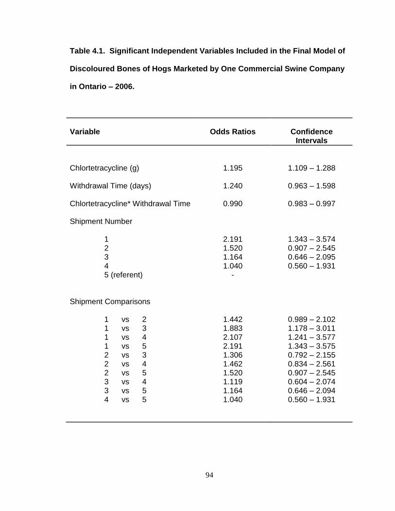
94
Table 4.1. Significant Independent Variables Included in the Final Model of
Discoloured Bones of Hogs Marketed by One Commercial Swine Company
in Ontario – 2006.
Variable
Odds Ratios
Confidence
Intervals
Chlortetracycline (g) Withdrawal Time (days) Chlortetracycline* Withdrawal Time Shipment Number 1 2 3 4 5 (referent) Shipment Comparisons 1 vs 2 1 vs 3 1 vs 4 1 vs 5 2 vs 3 2 vs 4 2 vs 5 3 vs 4 3 vs 5 4 vs 5
1.195
1.240
0.990
2.191 1.520 1.164 1.040
-
1.442 1.883 2.107 2.191 1.306 1.462 1.520 1.119 1.164 1.040
1.109 – 1.288
0.963 – 1.598
0.983 – 0.997
1.343 – 3.574 0.907 – 2.545 0.646 – 2.095 0.560 – 1.931
0.989 – 2.102 1.178 – 3.011 1.241 – 3.577 1.343 – 3.575 0.792 – 2.155 0.834 – 2.561 0.907 – 2.545 0.604 – 2.074 0.646 – 2.094 0.560 – 1.931

95
Figure 4.1. Proportion of Pig Carcasses with Discoloured Bones at
Slaughter Based on the Estimated Cumulative Chlortetracycline (CTC)
Consumption of Hogs Marketed by a Commercial Swine Company in
Ontario – 2006
n = Number of batches (a batch is the group of pigs that fills a barn at one time) a number of pigs declared as having discoloured bones b total number of pigs inspected for bone discolouration from that particular category of CTC consumption
0.1% 0.4%
2.4%
20.5%
2.1%
0%
5%
10%
15%
20%
25%
0-15g 15-30g 30-45g 45-66g Overall
Car
cass
es
wit
h D
isco
lou
red
Bo
nes
Chlortetracycline
n=5
(2a/2,530
b)
n=53
(96a/21,966
b)
n=82
(892a/37,576
b)
n=3
(282a/1,377
b)
n = 143
(1374a/64,232
b)

96
Figure 4.2. Proportion of Pig Carcasses with Discoloured Bones at
Slaughter, Based on Duration of Chlortetracycline (CTC) Treatment
Number of batches = 65 (a batch is the group of pigs that fills a barn at one time)
n = Number of batches (a batch is the group of pigs that fills a barn at one time) a number of pigs declared as having discoloured bones b total number of pigs inspected for bone discolouration from that particular category of CTC consumption duration
0.0%
1.0%
1.9%
2.8%
13.3%
0%
5%
10%
15%
3 4 5 6 7
Car
cass
es
wit
h D
isco
lou
red
Bo
nes
Treatment Duration (Weeks)
n = 3
(0a/1,301 b)
n = 21
(114a/11,354 b)
n = 23
(223a/11,759 b)
n = 5
(349a/1,972 b)
n = 13
(219a/7,922 b)
≥
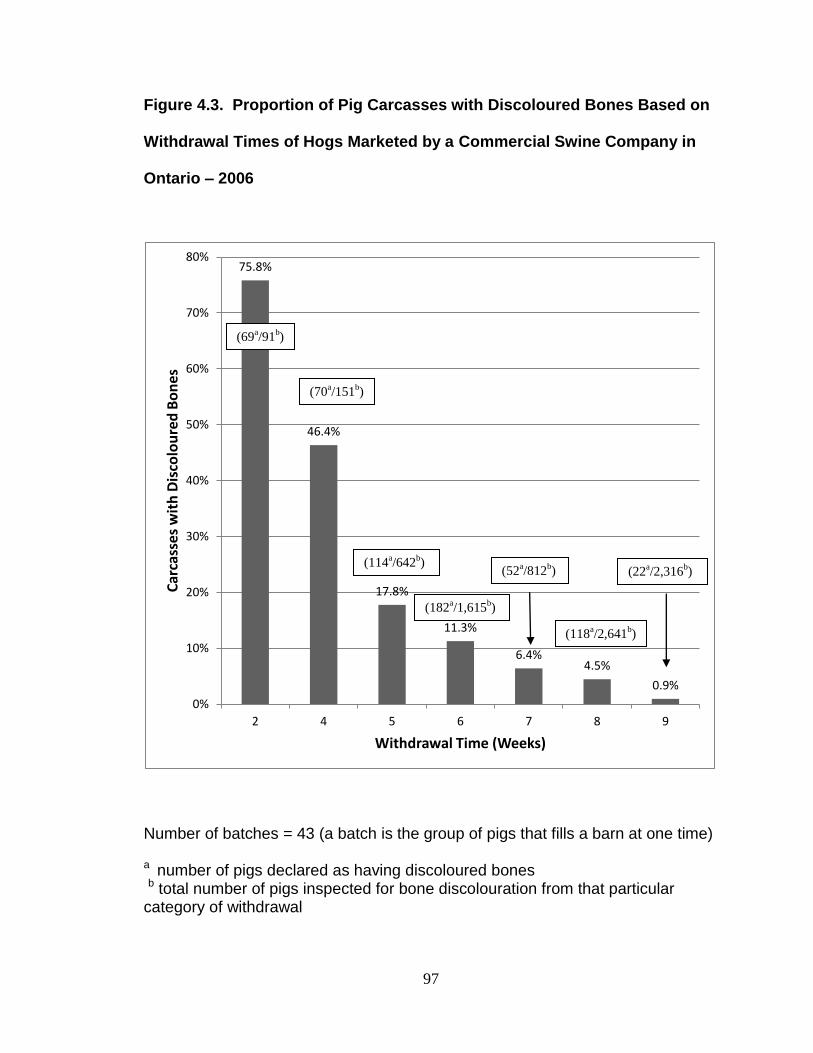
97
Figure 4.3. Proportion of Pig Carcasses with Discoloured Bones Based on
Withdrawal Times of Hogs Marketed by a Commercial Swine Company in
Ontario – 2006
Number of batches = 43 (a batch is the group of pigs that fills a barn at one time)
a number of pigs declared as having discoloured bones b total number of pigs inspected for bone discolouration from that particular category of withdrawal
75.8%
46.4%
17.8%
11.3%
6.4% 4.5%
0.9%
0%
10%
20%
30%
40%
50%
60%
70%
80%
2 4 5 6 7 8 9
Car
cass
es
wit
h D
isco
lou
red
Bo
nes
Withdrawal Time (Weeks)
(69a/91
b)
(70a/151
b)
(114a/642
b)
(182a/1,615
b)
(52a/812
b) (22
a/2,316
b)
(118a/2,641
b)

98
Figure 4.4. Proportion of Pigs Carcasses with Discoloured Bones at
Slaughter based on Shipment from the Grower-Finisher Barn
Number of batches = 43 (a batch refers to a group of pigs filling a barn) anumber of pigs declared as having discoloured bones b total number of pigs inspected for bone discolouration)
7.2%
5.3%
4.8%
1.0%
0%
1%
2%
3%
4%
5%
6%
7%
8%
1 2 3 4
Car
cass
es
wit
h D
isco
lou
red
Bo
nes
Shipment Number
(324a/4,511
b)
(60a/5,748
b)
(254a/5,299
b)
(320a/5,993
b)

99
Figure 4.5. Probability of Pig Carcasses Showing Bone Discolouration
According to Chlortetracycline (CTC) and Withdrawal Time Interaction*
*Data shown at the logit scale CTC = Chlortetracycline
-6
-4
-2
0
2
15 30 45 60 75 90
Logi
t (C
arca
sse
s w
ith
Dis
colo
ure
d B
on
es)
Withdrawal Time (days)
30g CTC 40g CTC 50g CTC 60g CTC

100
Chapter 5: General Discussion In Ontario, the discolouration of bones can be detected in hog carcasses
at the time of slaughter in a small number of pigs. Data collected during this
study, in 2006 and at various interval in the following 4 years, indicated that at
one large Ontario swine abattoir approximately 1 or 2 out of every 1,000 hogs
slaughtered are identified with bone discolouration. The discolouration appears to
be related to medication of pigs with tetracyclines, and this aspect was
investigated using feeding trials as well as examining the medication use of
farms that were identified as sources of the hogs identified at the abattoir in
2006.
Tetracyclines are widely used in pig production the world-over and have
been for over 60 years. Because modern pig production generally involves large
groups of animals under intensive confinement management, the use of
individual animal treatment is less commonly used as compared to mass
medication with tetracyclines in feed or water. However individual treatment
using an intramuscular injection of oxytetracycline might be used on occasion.
However labour requirements and the concerns with issues like broken needles
make this option less appealing than feed medication for many producers. The 3
molecular forms of tetracyclines used in pig production in Canada are
tetracycline, oxytetracycline and chlortetracycline. Pijpers and Verheijden (1992)
have reported that oxytetracycline is the most common form used in the Dutch
swine industry for medicating feed and that oxytetracycline is the molecule found
at the highest concentration in bones of pigs at slaughter. On the other hand

101
German researchers reported chlortetracycline as the most common form used
and the molecule most likely found in bone (Körner et al., 2001). In Canada
chlortetracycline has been reported to be more commonly used (Dunlop et al.,
1998) and was the tetracycline molecule identified at the highest concentration in
bones from cases of discoloured bones identified at slaughter that were analyzed
in the current study. In the current study, herds that were identified because they
had shipped a pig with discoloured bones used only the chlortetracycline form of
the antibiotic. The use of chlortetracycline in Canadian herds for feed medication
may reflect the reports that suggest the bioavailability of chlortetracycline is much
higher than oxytetracycline and tetracycline (del Castillo et al., 2000). In the
current study the rationale given by practitioners for using levels as high as 660
ppm in the feed for 3 or 6 weeks at the beginning of the grower-finisher phase
was to combat respiratory disease. Studies show that levels of over 500 ppm are
required to achieve serum and lung levels that will meet the MIC values for
common respiratory bacterial pathogens (del Castillo et al., 1998). Possibly the
preference for oxytetracycline in some studies may indicate that veterinarians are
using the antibiotic to manipulate gut flora or prevent clinical enteric diseases
such as porcine proliferative enteropathy caused by Lawsonia intracellularis and
not systemic or respiratory pathogens.
At the start of the current study involving investigating the tetracycline use
of pig farms that were identified as shipping pigs with discoloured bones, it was
thought that possibly the research would identify unusually high levels of drug
use or a disregard for withdrawal times. Previous work has identified that
102
tetracycline-related bone discolouration is influenced by the dosage and duration
of treatment, as well as the length of time from when medication stopped until the
animal is slaughtered. Guillot et al. (2010) have shown that the discolouration is
reversible if sufficient time is allowed to pass. Our study did reveal that the
likelihood of positive pigs (carcasses with discoloured bones) was reduced if
withdrawal time was 8 weeks or longer and if chlortetracycline was used for a
shorter duration (3 weeks versus 6 weeks) or at a lower dosage (330 ppm versus
660 ppm). The conclusion drawn from this study is that in order for bone
discolouration to be minimized, the use of chlortetracycline at therapeutic levels
should be restricted to the very early grower period.
The relationship between bone discolouration and tetracyclines was noted
as early as the 1950’s (Albert and Rees, 1956). Likewise, it was recognized that
tetracyclines form a tight bond with calcium ions and that during the process of
bone absorption and reformation, the calcium bound tetracycline was
reincorporated into bone, so that tetracycline residues persisted in bone long
after tetracyclines could no longer be detected in edible tissue (Körner et al.,
2001). In a controlled exposure trial in the current research, pigs were
administered tetracyclines via injection (oxytetracycline), and in water
(tetracycline) and in feed at various dosage levels and for different durations.
After lengthy withdrawal periods of more than 50 days, tetracyclines could be
detected in bone in all cases where pigs were exposed to the antibiotic
regardless of molecule used or method of administration. It is reported that the
calcium-tetracycline bond varies depending on the molecule with oxytetracycline

103
being the weakest bound and chlortetracycline the strongest and tetracycline
having an intermediate bond (Körner et al., 2001). The presence of residues of
tetracyclines in bone of animals at slaughter has been reported to be very
common, although the measurement of residues is difficult. The phenomenon of
tetracyclines in bone fluorescing under ultraviolet light has been used as a
convenient screening test for many years. Unfortunately tests to determine
accurate concentrations of tetracyclines present in bone are difficult to perform.
In the current study a high performance liquid chromatography test was
employed. The bones were ground and treated with acid to break the
tetracycline-calcium ion bonds. It is likely that only a portion of the bound
tetracyclines were released and the measurement may have been low,
particularly for chlortetracycline. One interesting finding, despite the limitations of
this analytic technique, is that bones with normal colour may contain high levels
of tetracyclines. The presence of residues of tetracyclines in bone of animals
passed by the meat inspection process for human consumption raises concerns
regarding food safety. Under Canadian law, bone is considered to be meat;
however the minimum allowable limits set for tetracyclines are based on analysis
of muscle, liver and kidney tissue. Bone is not included, possibly because of the
difficulty in measuring the levels or possibly because authorities don’t consider
bone to pose a risk. Jones et al. in a paper in the Canadian Veterinary Journal in
1977 raised the concern of oxytetracycline residues and food safety and their
conclusion was that bone residues need to be further investigated. German
researchers (Kühne et al., 2000) have demonstrated that microscopic particles of

104
bone are present in ground meat when mechanical deboning is used. Further
research (Kühne and Körner, 2001) has noted that gastric acidity can release
biologically active tetracyclines so that it is possible low levels of tetracyclines
present in ground meat when ingested may contribute to antimicrobial resistance
in gut flora. In addition when bone and meat meal derived from animals that were
medicated with tetracyclines, were fed to chickens, tetracycline residues were
found in the bones of the chickens. It was not within the scope of the current
study to investigate the aspect of tetracycline residues as a food safety concern
but clearly this is an area where further work should be performed.
There is a substantial economic cost of discoloured bones to the pig
industry if it results in the packer having to handle the processing of the
carcasses in a special manner such as removing the bones to make the cuts of
meat visually acceptable. Swine practitioners and producers need to be mindful
of the risk of bone discolouration when choosing medication regimens for the
grower-finisher period. In addition, there is the potential cost associated with
negative publicity regarding a food safety issue of antibiotic residue in pork, and
this could be of far greater concern. Further work is required to determine if
residues of tetracyclines in bone poses a health risk to consumers or to other
livestock via bone meal.

105
References
Albert, A., Rees, C.W. 1956. Avidity of the tetracyclines for the cations of metals. Nature 177: 433-434.
del Castillo, J. R. E., Elsener, J., Martineau, G. P. 1998. Pharmacokinetic modeling of in-feed tetracyclines in pigs using a meta-analytic compartmental approach. J Swine Health Prod 6:189-202.
del Castillo, J. R. E., Martineau, G. P., Hannon, M. R., Besner, J. G. 2000. Comparative pharmacokinetics and efficacy of aureomycin chlortetracycline granular premix and oxytetracycline premix against experimental pleuropneumonia in pigs. In: Proceedings of the 31st Annual Meeting of the American Association of Swine Practitioners. 31:89-94.
Dunlop, R. H., McEwen, S. A., Meek, A. H., Friendship, R. A., Clarke, R. C., Black, W. D. 1998. Antimicrobial drug use and related management practices among Ontario swine producers. Can Vet J 39:87-96.
Guillot, M., K. Alexander, C. Pomar, J. R. Del Castillo. 2011. In growing pigs, chlortetracycline induces a reversible green bone discoloration and a persistent increase of bone mineral density dependent of dosing regimen. Res Vet Sci 90(3):484-490.
Jones C.R.V., Usborne W.R., Tittiger F. 1977. Oxytetracycline residues in pigs fed experimentally. Can Vet J 18: 150-153.
Körner, U., Kühne, M., Wenzel, S. 2001. Tetracycline residues in meat and bone meals. Part 1: Methodology and examination of field samples. Food Addit Contam 18:293-302.
Kühne, M., Körner.U. 2001. Bound chlortetracycline residues in bones: release under acidic conditions. Food Chem 72(1):41-44.
Kühne, M., S. Wegmann, A. Kobe, R. Fries. 2000. Tetracycline residues in bones of slaughtered animals. Food Control 11(3):175-180.
Pijpers A., Verheijden F.H.M. 1992. Evaluation of antimicrobial treatment efficacy. Proceedings of the International Pig Veterinary Society Congress. 35-39.

106
Appendices

107
A.1.1. Letter from Animal Health Laboratory (AHL) - University of Guelph
reporting an outbreak of disease in Fall 2005.
Reproduced with permission of the Animal Health Laboratory (AHL) at the University of Guelph

108
Letter A.1.2. Meat Hygiene Directive - January 4, 2006: Disposition of
Carcasses with Bones Discoloured by Tetracyclines

109
Reproduced with permission of the Canadian Food Inspection Agency (CFIA)

110
Letter A.1.3. Meat Hygiene Directive - March 10, 2006: Disposition of
Carcasses with Bones Discoloured by Tetracyclines.
111
Reproduced with permission of the Canadian Food Inspection Agency (CFIA)