Embed Size (px)
Citation preview

Clinical Endocrinology (1999) 51, 205–215
205q 1999 Blackwell Science Ltd
Sleep deprivation effects on the activity of thehypothalamic–pituitary–adrenal and growth axes:potential clinical implications
Alexandros N. Vgontzas*, George Mastorakos†,Edward O. Bixler*, Anthony Kales*, Philip W. Gold‡and George P. Chrousos§*Sleep Research and Treatment Center, Department ofPsychiatry, Pennsylvania State University, Hershey,USA, †Endocrine Unit, Evgenidion Hospital, AthensUniversity, Athens, Greece, ‡ClinicalNeuroendocrinology Branch, National Institute of MentalHealth, Bethesda, USA and §DevelopmentalEndocrinology Branch, National Institute of Child Healthand Human Development, National Institutes of Health,Bethesda, USA
(Received 14 June 1998; returned for revision 18 December1998; finally revised 8 March 1999; accepted 9 March 1999)
Summary
OBJECTIVES Although several studies have shownthat sleep deprivation is associated with increasedslow wave sleep during the recovery night, the effectsof sleep deprivation on cortisol and growth hormone(GH) secretion the next day and recovery night havenot been assessed systematically. We hypothesizedthat increased slow wave sleep postsleep deprivationis associated with decreased cortisol levels and thatthe enhanced GH secretion is driven by the decreasedactivity of the HPA axis.DESIGN AND SUBJECTS After four consecutivenights in the Sleep Laboratory, 10 healthy youngmen were totally deprived of sleep during the fifthnight, and then allowed to sleep again on nightssix and seven. Twenty-four hour blood samplingwas performed serially every 30 minutes on thefourth day, immediately following the previous nightof sleep and on the sixth day, immediately after sleepdeprivation.MEASUREMENT Eight-hour sleep laboratory record-ing, including electroencephologram, electro-oculo-gram and electromyogram. Plasma cortisol and GHlevels using specific immunoassay techniques.
RESULTS Mean plasma and time-integrated (AUC)cortisol levels were lower during the postdeprivationnighttime period than on the fourth night ( P<0·05).Pulsatile analysis showed significant reduction ofboth the 24 h and daytime peak area ( P<0·05) andof the pulse amplitude ( P< 0·01), but not of the pulsefrequency. Also, the amount of time-integrated GHwas significantly higher for the first 4 h of the post-deprivation night compared to the predeprivationnight ( P< 0·05). Cross-correlation analyses betweenthe absolute values of the time-series of each hor-mone value and percentage of each sleep stage perhalf hour revealed that slow wave sleep was nega-tively correlated with cortisol and positively corre-lated with GH with slow wave sleep preceding thesecretion of these hormones. In contrast, indices ofsleep disturbance, i.e. wake and stage 1 sleep, werepositively correlated with cortisol and negativelycorrelated with GH.CONCLUSION We conclude that sleep deprivationresults in a significant reduction of cortisol secretionthe next day and this reduction appears to be, to alarge extent, driven by the increase of slow wave sleepduring the recovery night. We propose that reductionof CRH and cortisol secretion may be the mechanismthrough which sleep deprivation relieves depressiontemporarily. Furthermore, deep sleep has an inhibi-tory effect on the HPA axis while it enhances theactivity of the GH axis. In contrast, sleep disturbancehas a stimulatory effect on the HPA axis and a sup-pressive effect on the GH axis. These results areconsistent with the observed hypocortisolism in idio-pathic hypersomnia and HPA axis relative activationin chronic insomnia. Finally, our findings supportprevious hypotheses about the restitution andimmunoenhancement role of slow wave (deep) sleep.
It has been clearly demonstrated that sleep deprivation isassociated with increased slow wave sleep (SWS) and enhancedactivity in the delta band during the recovery night (Webb& Agnew, 1971; Borbelyet al., 1981). The effects of sleepdeprivation on cortisol secretion the next day and recoverynight have not been assessed systematically. Sleep and
Correspondence: Dr Alexandros N. Vgontzas, Sleep Research andTreatment Center, Department of Psychiatry Pennsylvania StateUniversity, College of Medicine, 500 University Drive, Hershey,PA 17033, USA. Fax:þ1 (717) 531 6491.
particularly SWS has an inhibiting effect on cortisol secretion(Weitzmanet al., 1983; Seifritzet al., 1995; Brandenbergeret al., 1996; Scheenet al., 1996; Bierwolfet al., 1997). Wehypothesized that increased slow wave sleep postsleep depri-vation is associated with decreased cortisol levels during thefirst recovery night compared to baseline.
Stress and the intracerebroventricular administration ofcorticotropin-releasing-hormone (CRH) in animals is asso-ciated with arousal, while glucocorticoid administration orhypercortisolism in humans are associated with sleep dis-turbance (Suttonet al., 1982; Chrousos & Gold, 1992;Chrousoset al., 1993). Furthermore, melancholic depressionin humans is associated with chronic hypersecretion of CRH,hypercortisolism, and sleep disturbance (Goldet al., 1988a, b).Pharmacological therapy of depression leads to normali-zation of CRH and cortisol secretion and correction of thesleep disturbance, while sleep deprivation temporarily relievesdepressive symptomatology (Goldet al., 1988a, b). We hypo-thesized that sleep deprivation is associated with decreasedactivity of the HPA axis the next day and night and that thiscould explain why sleep deprivation relieves depression.
Earlier studies have shown that cortisol secretion isnegatively correlated with GH secretion and that the HPAaxis has a suppressant effect on the activity of the GH axis(Ghizzoniet al., 1996). Several studies have shown that SWSsleep induced by sleep deprivation is associated with increasedamounts of GH secretion (Van Cauteret al., 1992; Scheenet al., 1996). We hypothesized that the enhanced GH secretionduring the recovery night is probably driven by the decreasedactivity of the HPA axis.
Methods
Subjects
Ten young healthy men 20–29 years of age (mean6 SE,23·36 0·8), body mass index 25·26 0·8, were recruited fromthe community and from the medical and technical staff andstudents of the Milton S. Hershey Medical Center. They werein good general health, had no sleep complaints or circadianabnormalities, were not taking any medications and werescreened in the Sleep Laboratory for sleep disordered breath-ing, nocturnal myoclonus or other primary sleep disorders.Also, a battery of clinical tests, including full blood count,urinalysis, thyroid indices and electro-cardiogram, werenegative for abnormal findings.
Protocol
Each subject participated in a sleep deprivation experiment thatlasted seven days. After four consecutive nights in the Sleep
Laboratory (1 adaptation night and 3 baseline nights), thesubjects were deprived of sleep during the entire fifth night,while they were allowed to sleep again on nights six andseven. The subjects stayed awake in the presence of nursingor technical staff and total wake time before the first recoverynight was 40 h. Twenty-four hour blood sampling was per-formed serially every 30 minutes on the fourth day (predepri-vation) and sixth day, the latter immediately following sleepdeprivation. An indwelling catheter was inserted in the ante-cubital vein about 30 minutes before the first blood sample.The catheter was kept patent with small amounts of heparin.During the sleep recording period, blood was collected outsidethe subjects’ room through a perforation in the wall, via extratubing, in order to decrease sleep disturbance from the bloodsampling technique. During the day, blood samples were takenin the Clinical Research Center of the University Hospital ofthe Milton S. Hershey Medical Center. The timing of thesubjects’ meals (lunch at 1200 h and dinner at 1800 h) as wellas the amount and composition of food intake were similaron the two days of blood sampling. The study was approvedby the Institutional Review Board and each subject signed awritten consent form.
Sleep recordings
Sleep laboratory recording was carried out in a sound-attenuated, light-and temperature-controlled room which hasa comfortable bedroom-like atmosphere. During this evalua-tion, each subject was monitored continuously for 8 hours(2200–0600 h). The sleep schedule in the sleep laboratorywas similar to the subjects’ normal sleep schedule. A standardsleep recording using the standardized 10–20 system, includingelectro-oculogram (EOG) recorded from the outer canthi ofboth eyes and referred to the same mastoid and to each other,was made (Rechtschaffen & Kales, 1968). The electromyogram(EMG) was recorded bipolarly from the submental muscle.The sleep recordings were amplified using standard clinicalpolygraphs (Grass Instrument Co., Model 78d & e). The sleeprecords were scored independently of any knowledge of theexperimental condition according to standardized criteria(Rechtschaffen & Kales, 1968).
Sleep parameters assessed from the sleep recordingsincluded sleep induction (sleep latency or SL); sleep main-tenance (wake time after sleep onset or WTASO); total waketime (TWT) which is the sum of sleep latency and WTASO;% ST which is total sleep time as percentage of time inbed; percentage of the various sleep stages (REM, 1, 2, 3 and 4combined [SWS]) which is calculated as the minutes ineach stage as the percentage of total sleep time; and REMlatency which is the interval from sleep onset to the firstREM period.
206 A. N. Vgontzas et al.
q 1999 Blackwell Science Ltd,Clinical Endocrinology, 51, 205–215

Hormone assays
Blood collected from the indwelling catheter was transferredto a heparinized tube and refrigerated until centrifugation(within 3 h). The supernatant was frozen at¹208 until assay forthe hormones. Cortisol and GH levels were measured byspecific immunoassay techniques as previously described(Chrousoset al., 1984; Magiakouet al., 1994). The low limitof detection was 17·66 nmol/l for cortisol and 0·2 mU/l for GH.The intra- and inter-assay coefficients were, respectively, 4·6%and 6% for cortisol and 3·3% and 5·1% for GH.
Pulse analysis
The plasma cortisol and GH concentrations obtained wereanalysed for the presence of pulses by the Detect programme(Oerter et al., 1986; Genazzani & Rodbard, 1991). Thedetection of pulses by Detect is based on the calculation of amathematical model of the predicted variance of the raw data.The most accurate way of calculating this model of varianceis based on the intra- and inter-assay coefficients of variationof the hormonal assay. This programme reveals significantpulses as follows: (1) analysis of first derivatives of data forthe detection of rapid events, and (2) fitting of linear seg-ments for the detection of slow events. Data from each subjectwere analysed using Detect, with a nominal value ofP¼ 0·01for positive peak detection. The programme calculates sepa-rately the one-tailed Student’st-value for the desiredP-value(in our caseP¼ 0·01) and number of degrees of freedom for1 derivative, 2, 3, or 4 derivatives in a row. Samples wereanalysed for 24-h mean serum hormone concentration, peakarea, peak amplitude, and number of peaks per unit of time.Also, the peak area was calculated separately for the daytimeperiod (0800–2200 h) and the nighttime period (2200–0600 h).
Time-integrated cortisol
The amount of time-integrated cortisol was calculated bythe trapezoid method (integrated AUC) for the 24-h period,the daytime period (0800–2200 h) and the nighttime period(2200–0600 h).
Cross-correlation analysis
To search for time-ordered relations between cortisol, GH andsleep stages, analysis of correlations between the absolutevalues of the time series of each hormone value and percentageof each sleep stage per 30 minutes time intervals of thecorresponding night was performed. These cross-correlationanalyses were computed between cortisol and GH valuesand percentage wakefulness, stage 1, and 2 sleep, REM sleep
and SWS in every 30 minutes interval at various time lagscovering the 8-h period of study. For example, if release of ahormone B is regulated by a sleep stage A (e.g. A is thereleasing sleep stage and B is the affected hormone), then onemight expect the concentration-time series of hormone B to lag(follow) in time, quantitatively, the percentage-time series ofsleep stage A. Cross-correlation was computed after lagging theconcentration time series of hormone B relative to thepercentage time series of sleep stage A. If we call rk
the coefficient of correlation between the time series of thehormone concentration and the percentage of sleep stage atlagtime k for one subject, then the mean rk of all subjects wasconsidered significant when it exceeded zero by more than2·00 SE (P<0·05 level of significance). The SE at each timepoint was calculated from the individual values of rks for the10 subjects at the lagtime k. All correlations were performedusing Statview software for the Macintosh computer (AbacusConcepts, Inc., Berkeley, CA).
Statistical analysis
Data were expressed as mean6 SE. Differences between pre-and post-sleep deprivation values were examined using pairedtwo-tailed Studentt-test.
Results
Sleep pre- and post-sleep deprivation
On postdeprivation (night 6) compared to predeprivation(nights 2–4), subjects demonstrated significantly shorter sleeplatencies and a higher percentage of slow wave sleep (Table 1).
Sleep deprivation, cortisol and GH 207
q 1999 Blackwell Science Ltd,Clinical Endocrinology, 51, 205–215
Table 1 Nighttime sleep pre- and post-sleep deprivation in normalmale volunteers
Nights 2–4 Night 6
SL 28·46 5·5 7·96 1·3**WTASO 43·36 10·3 37·26 12·5TWT 71·76 9·1 45·16 12·6% ST 85·16 1·9 90·66 2·6
% Stage 1 9·46 2·1 5·26 1·3%Stage 2 50·86 2·6 43·36 3·1% SWS 18·86 2·9 29·06 3·7*% REM 21·06 1·3 22·46 1·7
REM Latency 102·96 5·3 92·46 18·8# REM Periods 3·86 0·2 4·36 0·3REM Duration 23·76 1·7 23·36 2·3
Data represent mean6 SE. *P<0?05; ** P<0?01.

Also, total wake time and percentage of stage 1 sleep tendedto be lower postdeprivation (P¼ 0·1).
Twenty-four hour secretion of cortisol pre- andpost-sleep deprivation
Mean 24-h plasma cortisol levels were lower, but not signi-ficantly so, postdeprivation. During the postdeprivationnighttime period, mean plasma cortisol levels were signifi-cantly lower compared to the predeprivation night (4th night)(157·36 11·0 vs. 176·66 13·8 nmol/l, P< 0·05) (Table 2,Fig. 1). Pulsatile analysis showed significant reduction of the24 h peak area (7134·86 447·0 vs. 8323·96 491·1 nmol/l.minutes,P<0·05) and the pulse amplitude (1103·66 74·5vs.1534·06 99·3 nmol/l. minutes,P< 0·01), but not of the pulsefrequency (6·66 0·4 vs. 5·66 0·4, P>0·05). Finally, theamount of time-integrated cortisol, as calculated by the trape-zoid method (integrated AUC), was lower during the night-time postdeprivation than the nighttime predeprivation(71471·96 4745·5 vs. 80165·56 5989·8 nmol/l. minutes,P<0·05).
Twenty-four hour secretion of growth hormone pre- andpost-sleep deprivation
The growth hormone (GH) levels were increased during thenighttime postdeprivation period compared to the nighttimepredeprivation period (Table 3, Fig. 2). Specifically, meanplasma GH levels tended to be higher during the postdepriva-tion nighttime period than during the predeprivation nighttime(3·46 0·6 vs. 2·66 0·4 mU/l, P¼ 0·1). The difference was
stronger for the first 4-h of the nighttime period (5·46 1·2vs.3·86 0·8 mU/l,P¼ 0·07). Also, the amount of time-integratedGH, as calculated by the trapezoid method (integratedAUC), tended to be higher during the postdeprivation thanduring predeprivation nighttime (1676·46 276·2vs. 1331·66210·6 mU/l. minutes,P¼ 0·1). The difference was significantfor the first 4-h of the nighttime period (1285·86 294·8vs.886·26 163·6 mU/l. minutes,P<0·05). Pulsatile analysisshowed no significant differences between pre- and postdepri-vation with regard to 24-h peak area, pulse amplitude or pulsefrequency. The nighttime secretion of the hormone tendedto be higher during post- compared to pre-deprivation(50·06 9·2vs.36·66 6·6 mU/l,P<0·1).
Correlation analyses
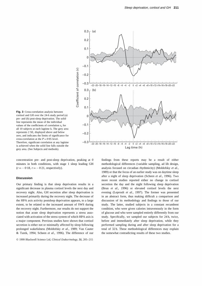
Cortisol and GH. A significant negative correlation wasobserved between cortisol and GH pre- and post-sleepdeprivation at lag times 0 (r ¼ ¹ 0·22) and 30 (r ¼ ¹ 0·25)minutes, respectively, with cortisol leading GH. Also, therewas a positive correlation between cortisol and GH pre- andpost-sleep deprivation at 6·5 (r ¼ 0·26) and 7·5 (r ¼ 0·25) hours,respectively, with GH leading cortisol (Fig. 3). Finally, therewas a positive correlation between cortisol and GH post-sleepdeprivation peaking at lag time 15 h, with cortisol leading GH(r ¼ 0·17).
Cortisol and sleep stages pre- and post-sleep deprivation.Nosignificant correlation was observed predeprivation betweenSWS and cortisol. However, a significant negative correlationwas observed postdeprivation between SWS and cortisol
208 A. N. Vgontzas et al.
q 1999 Blackwell Science Ltd,Clinical Endocrinology, 51, 205–215
Pre-deprivation Post-deprivation P-value
Mean values (nmol/l)24 h 231·86 13·8 212·46 8·3 ¼0·08Daytime (0800–2200 h) 240·06 19·3 226·26 13·8 nsNighttime (2200–0600 h) 176·66 13·8 157·36 11·0 <0·05
Time-integrated AUC (nmol/l. minutes)24 h 282334·06 18104·6 261933·96 9079·9 ¼0·1Daytime (0800–2200 h) 202165·76 16396.7 190462·06 11016·7 nsNighttime (2200–0600 h) 80165·56 5989·8 71471·96 4745·5 <0·05
Pulsatile analysis24 h peak AUC (nmol/l. minutes) 8323·96 491·1 7134·86 447·0 <0·05Daytime (0800–2200 h) 5244·96 355·9 4442·06 449·7 <0·05Nighttime (2200–0600 h) 1865·16 198·6 1713·36 173·8 nsaverage AUC/peak (nmol/l. minutes) 1534·06 99·3 1100·86 74·5 <0·01# peaks 5·66 0·4 6·66 0·4 ¼0·063
Data represent mean6 SE..
Table 2 Overall and pulsatile cortisol releasepre- and post-sleep deprivation

peaking at lag time 0 minutes, with SWS leading cortisol(r ¼¹ 0·46) (Fig. 4). Also, significant positive correlationswere observed postdeprivation between SWS and cortisol at lagtime 6·5 h with SWS leading cortisol (r ¼ 0·30) and at lag time3 h with cortisol leading SWS (r ¼ 0·16).
A positive correlation was observed between wake andcortisol pre- and post-sleep deprivation, peaking in bothconditions at lag time 7·5 h (r ¼ 0·26, r ¼ 0·19, respectively),with wake leading cortisol. Also, a negative correlation wasobserved between wake and cortisol pre- and post-sleep depri-vation, peaking in both conditions at lag time 1·5 h (r ¼ ¹ 0·25,r ¼ ¹ 0·23, respectively), with wake leading cortisol. In addi-tion, a positive correlation was observed between stage 1 sleepand cortisol pre- and post-sleep deprivation peaking at 1·5 h(r ¼ 0·19) and 2 h (r ¼ 0·12), respectively, with stage 1 sleepleading cortisol.
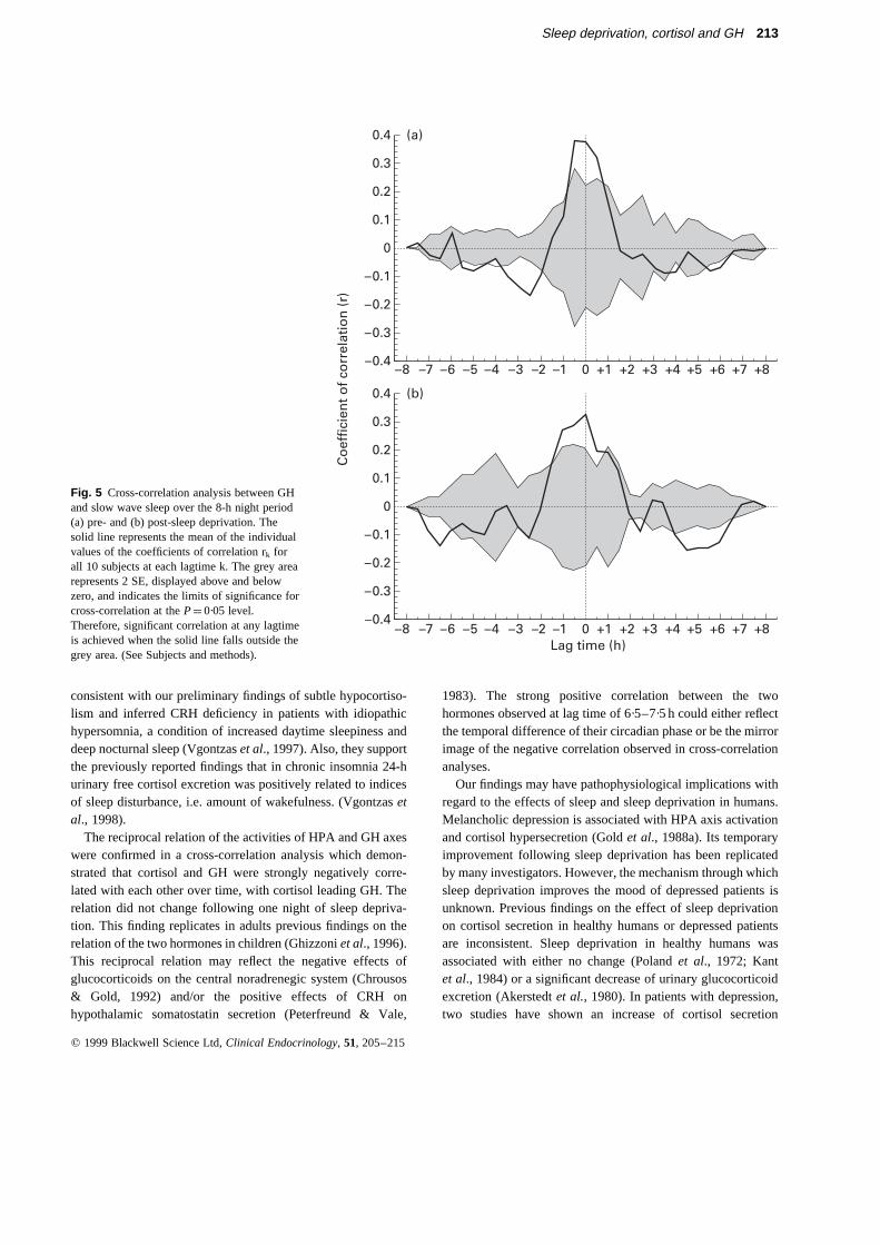
GH and sleep stages pre- and post-sleep deprivation.There wasa significant positive correlation between SWS and GH concen-tration pre- and post-sleep deprivation at lag times 30 (r ¼ 0·37)and 0 (r ¼ 0·32) minutes, respectively with SWS leading GH(Fig. 5). Also, there was a negative correlation between SWS andGH concentration pre- and post-sleep deprivation peaking at 2·5(r ¼ ¹ 0·10) hours and 6·5 (r ¼¹ 0·14) hours, respectively, withSWS leading GH. In addition, there was a negative correlationbetween SWS and GH concentration post-sleep deprivation,peaking at 4·5 h, with GH leading SWS (r ¼ ¹ 0·10).
There was a negative correlation between wake and GHpost-sleep deprivation, peaking at 0 minutes (r ¼ ¹ 0·20). Also,wake and GH concentration was positively correlated pre- andpost-sleep deprivation, peaking in both conditions at 2 h, withwake leading GH (r ¼ 0·28, r ¼ 0·23, respectively). Finally,there was a negative correlation between stage 1 sleep and GH
Sleep deprivation, cortisol and GH 209
q 1999 Blackwell Science Ltd,Clinical Endocrinology, 51, 205–215
Fig. 1 Diurnal variation (mean6 SE) in plasma cortisol concentrations in 10 healthy men pre- (X) and post- (W) sleep deprivation. Sampleswere taken every 30 minutes through an indwelling IV catheter. Inset, integrated nighttime AUC pre- (B) and post- (A) sleep deprivation.*P<0·05.

210 A. N. Vgontzas et al.
q 1999 Blackwell Science Ltd,Clinical Endocrinology, 51, 205–215
Pre-deprivation Post-deprivation P-value
Mean values (mU/l)24 h 1·46 0·2 1·86 0·4 nsDaytime (0800–2200 h) 0·86 0·2 1·06 0·6 nsNighttime (2200–0600 h) 2·66 0·4 3·46 0·6 ¼0·11st half of the night 3·86 0·8 5·46 1·2 ¼0·073
Time-integrated AUC (mU/l. minutes)24 h 2058·06 378·6 2636·06 594·6 nsDaytime (0800–2200 h) 656·66 224·6 883·86 495·8 nsNighttime (2200–0600 h) 1331·66 210·6 1676·46 276·2 ¼0·11st Half of the Night 886·26 163·6 1285·86 294·8 <0·05
Pulsatile analysis (mU/l. minutes)24 h peak AUC 56·66 12·4 77·06 18·6 nsDaytime (0800–2200 h) 29·06 16·0 69·46 49·8 nsNighttime (2200–0600 h) 36·66 6·6 50·06 9·2 ¼0·099Average AUC/peak 22·86 3·8 31·46 6·6 ns# Peaks 2·66 0·5 2·66 0·4 ns
Data represent mean6 SE.
Table 3 Overall and pulsatile GH release pre-and post-sleep deprivation
Fig. 2 Diurnal variation (mean6 SE) in plasma GH concentrations in 10 healthy men pre- (X) and post- (W) sleep deprivation. Samples weretaken every 30 minutes through an indwelling IV catheter. Inset, integrated AUC for the first half of the night pre- (B) and post- (A) sleepdeprivation. *P< 0·05.
concentration pre- and post-sleep deprivation, peaking at 0minutes in both conditions, with stage 1 sleep leading GH(r ¼¹ 0·18,r ¼ ¹ 0·21, respectively).
Discussion
Our primary finding is that sleep deprivation results in asignificant decrease in plasma cortisol levels the next day andrecovery night. Also, GH secretion after sleep deprivation isincreased primarily during the recovery night. The decrease ofthe HPA axis activity postsleep deprivation appears, to a largeextent, to be related to the increased amount of SWS duringthe recovery night. Furthermore, our results do not support thenotion that acute sleep deprivation represents a stress asso-ciated with activation of the stress system of which HPA axis isa major component. Previous studies have shown that cortisolsecretion is either not or minimally affected by sleep followingprolonged wakefulness (Moldofskyet al., 1989; Van Cauter& Turek, 1994; Scheenet al., 1996). The difference of our
findings from these reports may be a result of eithermethodological differences (variable sampling, ad lib design,analysis focused on circadian rhythmicity) (Moldofskyet al.,1989) or that the focus of an earlier study was on daytime sleepafter a night of sleep deprivation (Scheenet al., 1996). Twomore recent studies reported either no change in cortisolsecretion the day and the night following sleep deprivation(Brun et al., 1996) or elevated cortisol levels the nextevening (Leproultet al., 1997). The former was presentedin an abstract form, thus making difficult a comparison anddiscussion of its methodology and findings to those of ourstudy. The latter, studied subjects in a constant recumbentcondition, who were given calories intravenously in the formof glucose and who were sampled entirely differently from ourstudy. Specifically, we sampled our subjects for 24 h, twice,before and immediately after sleep deprivation, while theyperformed sampling during and after sleep deprivation for atotal of 32 h. These methodological differences may explainthe somewhat contradicting results of these two studies.
Sleep deprivation, cortisol and GH 211
q 1999 Blackwell Science Ltd,Clinical Endocrinology, 51, 205–215
(a)
(b)
Co
effi
cien
t o
f co
rrel
atio
n (
r)
0.3
0.2
0.1
0
–0.1
–0.2
–0.3
Lag time (h)–6 0–2–4–8–10–12–14–16–18–20–22 +2 +4 +6 +8 +10 +12+14+16+18 +20 +22
–6 0–2–4–8–10–12–14–16–18–20–22 +2 +4 +6 +8 +10 +12+14+16+18 +20 +22
0.3
0.2
0.1
0
–0.1
–0.2
–0.3
Fig. 3 Cross-correlation analysis betweencortisol and GH over the 24-h study period (a)pre- and (b) post-sleep deprivation. The solidline represents the mean of the individualvalues of the coefficients of correlation rk forall 10 subjects at each lagtime k. The grey arearepresents 2 SE, displayed above and belowzero, and indicates the limits of significance forcross-correlation at theP¼ 0·05 level.Therefore, significant correlation at any lagtimeis achieved when the solid line falls outside thegrey area. (See Subjects and methods).

Slow wave sleep, an index of sleep depth, was negativelycorrelated to cortisol levels over time, at lag time 0 h postsleepdeprivation, with SWS leading cortisol. In contrast, slow wavesleep was positively correlated to GH levels over time, at lagtime 30 and 0 minutes pre- and post-sleep deprivation,respectively, with SWS leading GH. These results indicatethat slow wave sleep has an inhibitory effect on cortisolsecretion, which is more pronounced in the first recovery nightfollowing sleep deprivation; in contrast, SWS enhances thesecretion of GH. It is possible that the enhanced GH secretionfollowing sleep deprivation is driven by the inhibition of theHPA axis activity induced by the increased amount of SWS,since HPA axis has been shown to regulate GH secretion undernormal conditions (Ghizzoniet al., 1996). The negative corre-lation between SWS and GH at lag time 2·5 h and 6·5 h pre- andpost-sleep deprivation, respectively, could be the mirror imageof the preceding positive correlation.
Stage 1 sleep, which is associated with sleep disturbance,
was positively correlated to cortisol levels over time, at a lagtime of 1·5–2·5 h, with stage 1 leading cortisol. The positivecorrelation between wake and cortisol at 7·5 h lag time couldreflect the fact that wake is high early at night, whereas cortisolplasma levels are high late at night. The strong negative corre-lation of wake and stage 1 with GH at 0 h lag time indicatesthat sleep disturbance is associated with inhibition of GH axisduring the nighttime. The positive correlation between wakeand GH at a lag time of 2 h reflects the lag between the wakebefore sleep onset and the occurrence of SWS. Indeed, SWSis positively correlated with GH at a lag time of only 30 minutes
Overall, our analysis indicates that deep sleep is associatedwith inhibition of the HPA axis, while it is associated withenhancement of the activity of the GH axis. The inhibitionof the HPA axis and the activation of the GH axis appear tocorrelate with the amount of SWS. In contrast, sleep distur-bance appears to be associated with activation of the HPA axisand suppression of the GH axis activity. These results are
212 A. N. Vgontzas et al.
q 1999 Blackwell Science Ltd,Clinical Endocrinology, 51, 205–215
(a)
(b)
Co
effi
cien
t o
f co
rrel
atio
n (
r)
Lag time (h)–8
0.4
0.3
0.2
0.1
0
–0.1
–0.2
–0.3
–0.4
0.4
0.3
0.2
0.1
0
–0.1
–0.2
–0.3
–0.4
–0.5–7 –6 –5 –4 –3 –2 –1 +10 +2 +3 +4 +5 +6 +7 +8
–8 –7 –6 –5 –4 –3 –2 –1 +10 +2 +3 +4 +5 +6 +7 +8
Fig. 4 Cross-correlation analysis betweencortisol and slow wave sleep over the 8-h nightperiod (a) pre- and (b) post-sleep deprivation.The solid line represents the mean of theindividual values of the coefficients ofcorrelation rk for all 10 subjects at each lagtimek. The grey area represents 2 SE, displayedabove and below zero, and indicates the limitsof significance for cross-correlation at theP¼ 0·05 level. Therefore, significantcorrelation at any lagtime is achieved when thesolid line falls outside the grey area. (SeeSubjects and methods).
consistent with our preliminary findings of subtle hypocortiso-lism and inferred CRH deficiency in patients with idiopathichypersomnia, a condition of increased daytime sleepiness anddeep nocturnal sleep (Vgontzaset al., 1997). Also, they supportthe previously reported findings that in chronic insomnia 24-hurinary free cortisol excretion was positively related to indicesof sleep disturbance, i.e. amount of wakefulness. (Vgontzasetal., 1998).
The reciprocal relation of the activities of HPA and GH axeswere confirmed in a cross-correlation analysis which demon-strated that cortisol and GH were strongly negatively corre-lated with each other over time, with cortisol leading GH. Therelation did not change following one night of sleep depriva-tion. This finding replicates in adults previous findings on therelation of the two hormones in children (Ghizzoniet al., 1996).This reciprocal relation may reflect the negative effects ofglucocorticoids on the central noradrenegic system (Chrousos& Gold, 1992) and/or the positive effects of CRH onhypothalamic somatostatin secretion (Peterfreund & Vale,
1983). The strong positive correlation between the twohormones observed at lag time of 6·5–7·5 h could either reflectthe temporal difference of their circadian phase or be the mirrorimage of the negative correlation observed in cross-correlationanalyses.
Our findings may have pathophysiological implications withregard to the effects of sleep and sleep deprivation in humans.Melancholic depression is associated with HPA axis activationand cortisol hypersecretion (Goldet al., 1988a). Its temporaryimprovement following sleep deprivation has been replicatedby many investigators. However, the mechanism through whichsleep deprivation improves the mood of depressed patients isunknown. Previous findings on the effect of sleep deprivationon cortisol secretion in healthy humans or depressed patientsare inconsistent. Sleep deprivation in healthy humans wasassociated with either no change (Polandet al., 1972; Kantet al., 1984) or a significant decrease of urinary glucocorticoidexcretion (Akerstedtet al., 1980). In patients with depression,two studies have shown an increase of cortisol secretion
Sleep deprivation, cortisol and GH 213
q 1999 Blackwell Science Ltd,Clinical Endocrinology, 51, 205–215
(a)
(b)
Co
effi
cien
t o
f co
rrel
atio
n (
r)
Lag time (h)–8
0.4
0.3
0.2
0.1
0
–0.1
–0.2
–0.3
–0.4
0.4
0.3
0.2
0.1
0
–0.1
–0.2
–0.3
–0.4–7 –6 –5 –4 –3 –2 –1 +10 +2 +3 +4 +5 +6 +7 +8
–8 –7 –6 –5 –4 –3 –2 –1 +10 +2 +3 +4 +5 +6 +7 +8
Fig. 5 Cross-correlation analysis between GHand slow wave sleep over the 8-h night period(a) pre- and (b) post-sleep deprivation. Thesolid line represents the mean of the individualvalues of the coefficients of correlation rk forall 10 subjects at each lagtime k. The grey arearepresents 2 SE, displayed above and belowzero, and indicates the limits of significance forcross-correlation at theP¼ 0·05 level.Therefore, significant correlation at any lagtimeis achieved when the solid line falls outside thegrey area. (See Subjects and methods).

during the night of sleep deprivation (Baumgartneret al., 1990;Bouhuyset al., 1990), while a third one showed no change ofplasma cortisol the morning after sleep deprivation (Ebertet al.,1994). However, these studies did not extend their measuresof cortisol the next day and night following sleep deprivation.Our findings that sleep deprivation leads to lower cortisollevels postdeprivation (primarily during the subsequent nightof sleep) suggest that lowering the level of HPA activity, whichis increased in depression, may be the mechanism throughwhich sleep deprivation improves the mood of depressedindividuals.
In addition, our findings shed new light on an old controversyin the sleep field which is whether sleep has a restitutive role(Adam & Oswald, 1983; Horne, 1983). Our findings tend tosupport that sleep, particularly SWS, by decreasing cortisol (acatabolic hormone) and increasing GH (an anabolic hormone)has a positive role on several systems and organs in terms ofthe daily ‘wear and tear.’ Previous findings have suggesteda possible link between SWS and the immune system (Kruegeret al., 1985; Moldofskyet al., 1986). Activation and sup-pression of the HPA axis, respectively, inhibits or enhancesthe immune-mediated inflammatory reaction (Chrousos, 1995).Our results suggest that increased amounts of SWS lead tolower levels of cortisol, which in turn may enhance immunefunction in humans.
Acknowledgements
We thank the nursing staff of the Clinical Research Centerat Hershey Medical Center for their assistance in completingthis study.
References
Adam, K. & Oswald, I. (1983) Protein synthesis, bodily renewal andsleep-wake cycle.Clinical Science, 65, 561–567.
Akerstedt, T., Palmblad, J. de la Torre, B., Marana, R. & Gillberg, M.(1980) Adrenocortical and gonadal steroids during sleep deprivation.Sleep, 3 (1), 23–30.
Baumgartner, A., Graf, K., Kurten, I., Meinhold, H. & Scholz, P. (1990)Neuroendocrinological investigations during sleep deprivation indepression I. Early morning levels of thyrotropin, TH, cortisol,prolactin, LH, FSH, estradiol, and testosterone.Biological Psychia-try, 28, 556–568.
Bierwolf, C., Struve, K., Marshall, L., Born, J. & Fehm, H. (1997) Slowwave sleep drives inhibition of pituitary-adrenal secretion in humans.Neuroendocrinology, 9, 479–484.
Borbely, A.A., Baumann, F., Brandeis, D., Strauch, I. & Lehmann, D.(1981) Sleep deprivation: effect on sleep stages and EEG powerdensity in man.Electroencephalograghy and Clinical Neurophysiol-ogy, 51, 483–493.
Bouhuys, A.L., Flentge, F. & Van den Hoofdakker, R.H. (1990)Effects of total sleep deprivation on urinary cortisol, self-ratedarousal, and mood in depressed patients.Psychiatry Research, 34,149–162.
Brandenberger, G., Buguet, A., Spiegel, K., Spiegel, K.,Stanghellini, A., Muanga, G., Bogui, P. & Dumas, M. (1996) Dis-ruption of endocrine rhythms in sleeping sickness with preservedrelationship between hormonal pulsatility and the REM-NREMsleep cycles.Journal of Biological Rhythms, 3, 258–267.
Brun, J., Khalfallah, Y., Chamba, G., Claustrat, B. & Sassolas, G.(1996) Effects of a 36H sleep deprivation (placebo versus modafinil)on rectal temperature, plasma levels of GH, cortisol, and melatoninin normal young men. 10th International Congress of Endocrinology,San Francisco, CA.
Chrousos, G.P., Schulte, H.M., Oldfield, E.H., Gold, P.W.,Cutler, G.B. Jr & Loriaux, D.L. (1984) The corticotropin-releasingfactor stimulation test: an aid in the evaluation of patients withCushing’s syndrome.New England Journal of Medicine, 310,622–626.
Chrousos, G.P. & Gold, P.W. (1992) The concept of stress and stresssystem disorders. Overview of physical and behavioral homeostasis.Journal of the American Medical Association, 267,1244–1252.
Chrousos, G.P., Kattah, J.C., Beck, R.W. & Cleary, P.A. (1993) & theOptic Neuritis Study Group. Side effects of glucocorticoid treatment.Journal of the American Medical Association, 269,2110–2112.
Chrousos, G.P. (1995) The hypothalamic-pituitary-adrenal axis andimmune-mediated inflammation.New England Journal of Medicine,332,1351–1362.
Ebert, D., Kaschka, W.P., Loew, T. & Beck, G. (1994) Cortisol andbeta-endorphin responses to sleep deprivation in major depression—the hyperarousal theories of sleep deprivation.Neuropsychobiology,29, 64–68.
Genazzani, A.D. & Rodbard, D. (1991) Use of receiver operatingcharacteristic curve to evaluate sensitivity, specificity, and accuracyof methods for detection of peaks in hormone time series.ActaEndocrinologica, 124,295–305.
Ghizzoni, L., Mastorakos, G., Vottero, A., Magiakou, M.A.,Chrousos, G.P. & Bernasconi, S. (1996) Spontaneous cortisol andgrowth hormone secretion interactions in patients with nonclassic21-hydroxylase deficiency (NCCAH) and control children.Journalof Clinical Endocrinology and Metabolism, 81, 482–487.
Gold, P.W., Goodwin, F.K. & Chrousos, G.P. (1988a) Clinical andbiochemical manifestations of depression: relationship to the neuro-biology of stress (part 1).New England Journal of Medicine, 319,348–353.
Gold, P.W., Goodwin, F.K. & Chrousos, G.P. (1988b) Clinical andbiochemical manifestations of depression: relationship to theneurobiology of stress (part 2).New England Journal of Medicine,319,413–420.
Horne, J. (1983) Human sleep and tissue restitution: some qualificationsand doubts.Clinical Science, 65, 569–579.
Kant, G.J., Genser, S.G., Thorne, D.R., Pfalser, J.L. & Mougey, E.H.(1984) Effects of 72 hour sleep deprivation on urinary cortisol andindices of metabolism.Sleep, 7 (2), 142–146.
Krueger, J., Walter, J. & Levin, C. (1985) Factor S. and relatedsomnogens: an immune theory for slow wave sleep. In:BrainMechanisms of Sleep(Eds. D.J. Mcginty, R. Drucker-Colin,A. Morrison, & P.L. Parmeggiani), pp. 253–269. Raven Press,New York.
Leproult, R., Copinschi, G., Buxton, O. & Van Cauter, E. (1997) Sleeploss results in an elevation of cortisol levels the next evening.Sleep,20, 865–870.
Magiakou, M.A., Mastorakos, G., Gomez, M.T., Rose, S.R. &Chrousos, G.P. (1994) Suppressed spontaneous and stimulatedgrowth hormone secretion in patients with Cushing’s disease
214 A. N. Vgontzas et al.
q 1999 Blackwell Science Ltd,Clinical Endocrinology, 51, 205–215
before and after surgical cure.Journal of Clinical Endocrinology andMetabolism, 78, 131–137.
Moldofsky, H., Lue, F.A., Eisen, J., Keystone, E. & Gorczynski, R.M.(1986) The relationship ofterleukin-1 and immune functions to sleepin humans.Psychosomatic Medicine, vol;48,xvol; 309–318.
Moldofsky, H., Lue, F.A., Davidson, J.R. & Gorczynski, R. (1989)Effects of sleep deprivation on human immune functions.FASEBJournal, 3, 1927–1977.
Oerter, K.E., Guardabasso, V. & Rodbard, D. (1986) Detection andcharacterization of peaks and estimation ofstantaneous secretory ratefor episodic pulsatile hormone secretion.Computers in BiomedicalResearch, 19, 170–175.
Peterfreund, R.A. & Vale, W.W. (1983) Ovine corticotropin-releasingfactor stimulates somatostatin secretion from cultured brain cells.Endocrinology, 112,1275–1278.
Poland, R.E., Rubin, R.T., Clark, B.R. & Gouin, P.R. (1972) Circadianpatterns of urine 17-OHC and VMA excretion during sleepdeprivation.Diseases of the Nervous System, 33, 456–458.
Rechtschaffen, A. & Kales, A. (Eds.) (1968)A manual of standardizedterminology, techniques, and scoring system for sleep stages ofhuman subjects.NIH publication 204, NIH, Bethesda.
Scheen, A.J., Byrne, M.M., Plat, L., Leproult, R. & Van Cauter. E.(1996) Relationships between sleep quality and glucose regulationin normal humans.American Journal of Physiology, 271, (Endo-crinology Metabolism., 34), E261–E270.
Seifritz, E., Hemmeter, U., Trachsel, L., Lauer, C.J., Hatzinger, M.,Emrich, H.M., Holsboer, F. & Holsboer-Trachsler, E. (1995) Effects
of flumazenil on recovery sleep and hormonal secretion after sleepdeprivation in male controls.Psychopharmacology, 120,449–456.
Sutton, R.E., Koob, G.F., LeMoal, M., Rivier, J. & Vale, W. (1982)Corticotropin-releasing factor produces behavioural activation inrats.Nature, 297,331–333.
Van Cauter, E., Kerkhofs, M., Caufriez, A., Van Onderbergen A.,Thorner, M.O. & Copinschi, G. (1992) A. quantitative estimationof growth hormone secretion in normal man: reproducibility andrelation to sleep and time of day.Journal of Clinical Endocrinologyand Metabolism, 74, 1441–1450.
Van Cauter, E. & Turek, F.W. (1994) Endocrine and other biologicalrhythms. In: Endocrinology, (Ed. L.J. Degroot), pp. 2487–2548.Saunders,Philadelphia.
Vgontzas, A.N., Papanicolaou, D.A., Bixler, E.O., Kales, A.,Vela-Bueno, A., Myers, D. & Chrousos, G.P. (1997)Evidence ofcorticotropin-releasing hormone (CRH) deficiency in patients withidiopathic hypersomnia: clinical and pathophysiological implica-tions. 79th Endocrine Society Meeting, Minneapolis, MN.
Vgontzas, A.N., Tsigos, C., Bixler, E.O., Stratakis, C.A., Zachman, K.,Kales, A., Vela-Bueno, A. & Chrousos, G.P. (1998) Chronicinsomnia and activity of the stress system: a preliminary study.Journal of Psychosomatic Research, 45, 21–31.
Webb, W.B. & Agnew, H.W. Jr (1971) Stage 4 sleep: influence oftime course variables.Science, 174,1354–1356.
Weitzman, E.D., Zimmerman, J.C., Czeisler, C.A. & Ronda, J. (1983)Cortisol secretion is inhibited during sleep in normal man.Journalof Clinical Endocrinology and Metabolism, 56, 352.
Sleep deprivation, cortisol and GH 215
q 1999 Blackwell Science Ltd,Clinical Endocrinology, 51, 205–215