Embed Size (px)
Citation preview
Slide 1 of 51
Circulatory system - YouTube
Copyright Pearson Prentice Hall
Slide 2 of 51
Copyright Pearson Prentice Hall
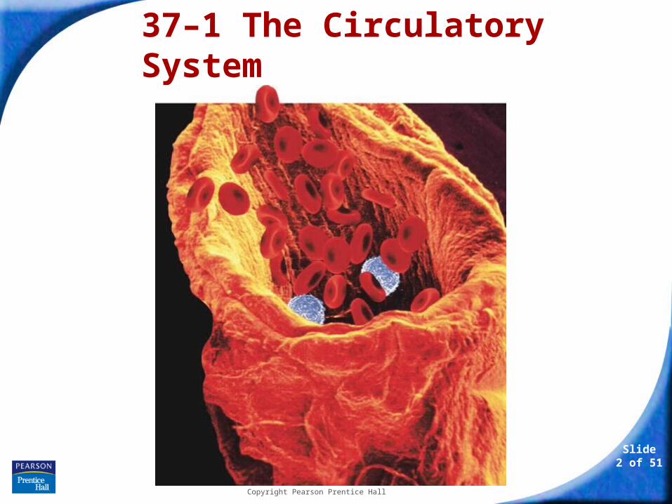
37–1 The Circulatory System
37–1 The Circulatory System
Slide 3 of 51
Copyright Pearson Prentice Hall
37–1 The Circulatory System
The circulatory system and respiratory system work together to supply cells with the nutrients and oxygen they need to stay alive.
37–1 The Circulatory System
Slide 4 of 51
Copyright Pearson Prentice Hall
Functions of the Circulatory System
Functions of the Circulatory System
Humans and other vertebrates have closed circulatory systems, meaning that the blood is contained within a system of vessels.

37–1 The Circulatory System
Slide 5 of 51
Copyright Pearson Prentice Hall
Functions of the Circulatory System
What are the structures of the circulatory system?
37–1 The Circulatory System
Slide 6 of 51
Copyright Pearson Prentice Hall
Functions of the Circulatory System
The human circulatory system consists of:
• the heart
• blood vessels
• blood
37–1 The Circulatory System
Slide 7 of 51
Copyright Pearson Prentice Hall
The Heart
The Heart
The heart is enclosed in a protective sac of tissue called the pericardium.
In the walls of the heart, two layers of epithelial and connective tissue form around a thick layer of muscle called the myocardium.
Contractions of the myocardium pump blood.
37–1 The Circulatory System
Slide 8 of 51
Copyright Pearson Prentice Hall
The Heart
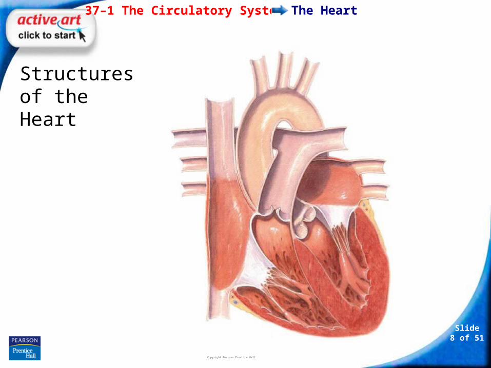
Structures of the Heart
37–1 The Circulatory System
Slide 9 of 51
Copyright Pearson Prentice Hall
The Heart
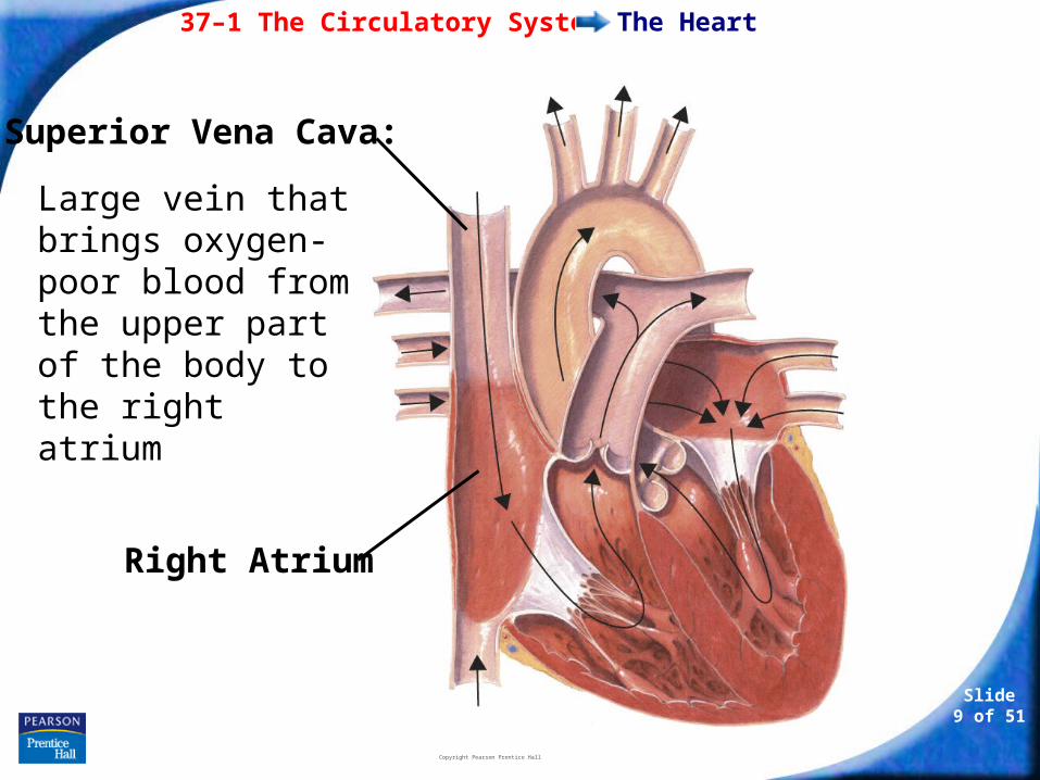
Large vein that brings oxygen-poor blood from the upper part of the body to the right atrium
Right Atrium
Superior Vena Cava:
37–1 The Circulatory System
Slide 10 of 51
Copyright Pearson Prentice Hall
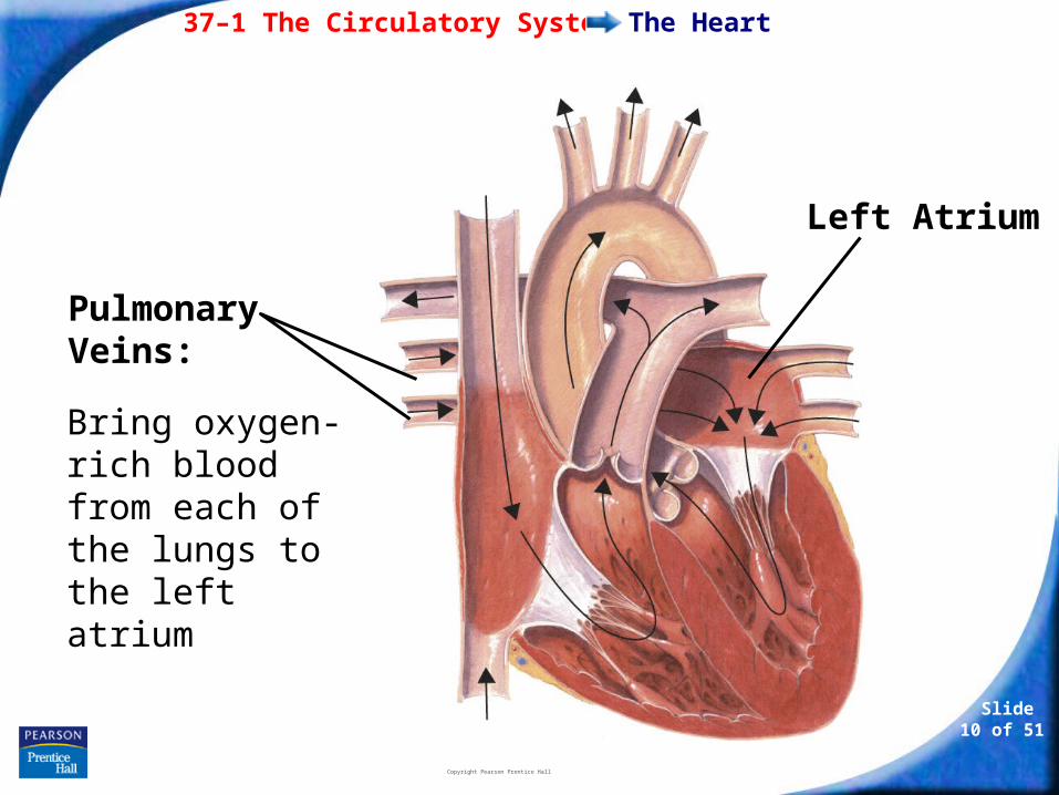
The Heart
Bring oxygen-rich blood from each of the lungs to the left atrium
Left Atrium
Pulmonary Veins:
37–1 The Circulatory System
Slide 11 of 51
Copyright Pearson Prentice Hall
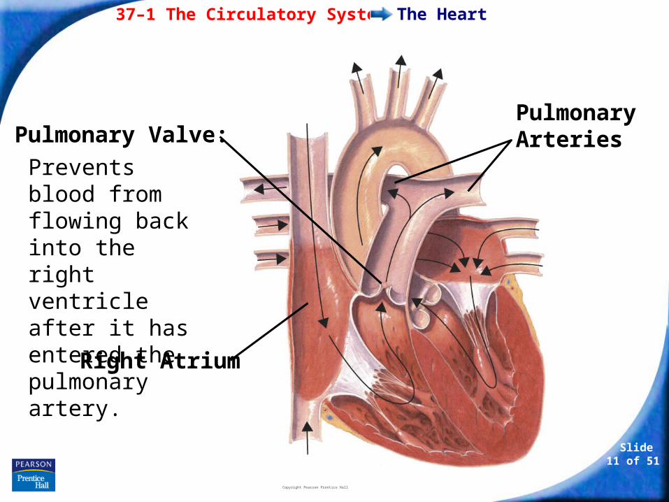
The Heart
Prevents blood from flowing back into the right ventricle after it has entered the pulmonary artery.
Right Atrium
Pulmonary ArteriesPulmonary Valve:
37–1 The Circulatory System
Slide 12 of 51
Copyright Pearson Prentice Hall
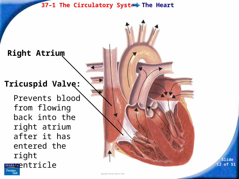
The Heart
Prevents blood from flowing back into the right atrium after it has entered the right ventricle
Right Atrium
Tricuspid Valve:
37–1 The Circulatory System
Slide 13 of 51
Copyright Pearson Prentice Hall
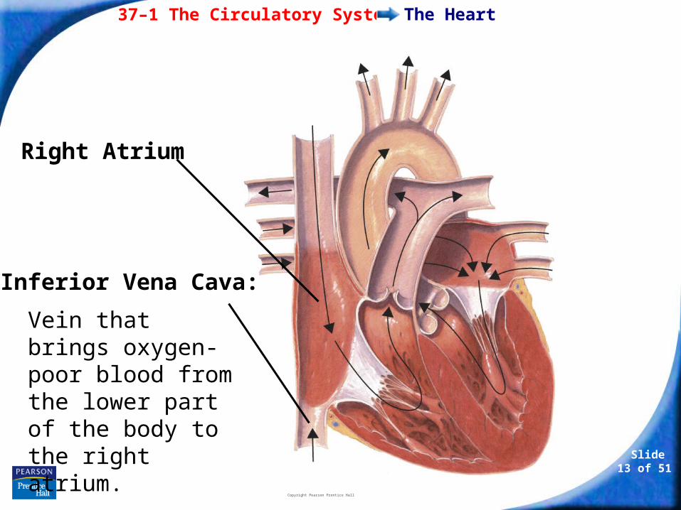
The Heart
Vein that brings oxygen-poor blood from the lower part of the body to the right atrium.
Right Atrium
Inferior Vena Cava:
37–1 The Circulatory System
Slide 14 of 51
Copyright Pearson Prentice Hall
The Heart
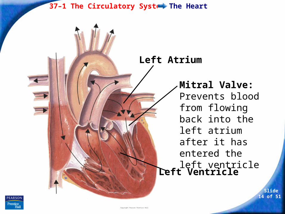
Mitral Valve: Prevents blood from flowing back into the left atrium after it has entered the left ventricle
Left Atrium
Left Ventricle
37–1 The Circulatory System
Slide 15 of 51
Copyright Pearson Prentice Hall
The Heart
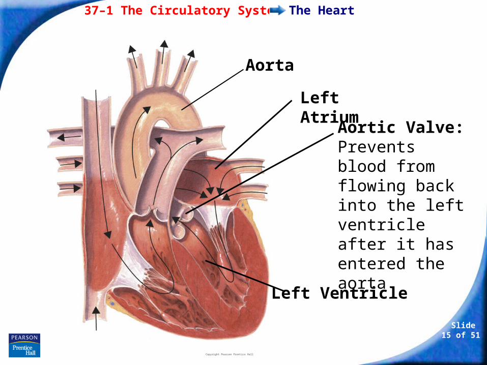
Aortic Valve: Prevents blood from flowing back into the left ventricle after it has entered the aorta
Left Atrium
Left Ventricle
Aorta
37–1 The Circulatory System
Slide 16 of 51
Copyright Pearson Prentice Hall
The Heart
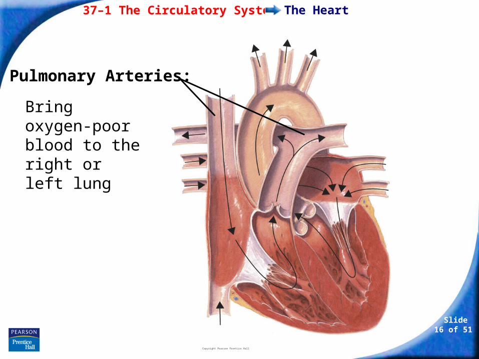
Bring oxygen-poor blood to the right or left lung
Pulmonary Arteries:
37–1 The Circulatory System
Slide 17 of 51
Copyright Pearson Prentice Hall
The Heart
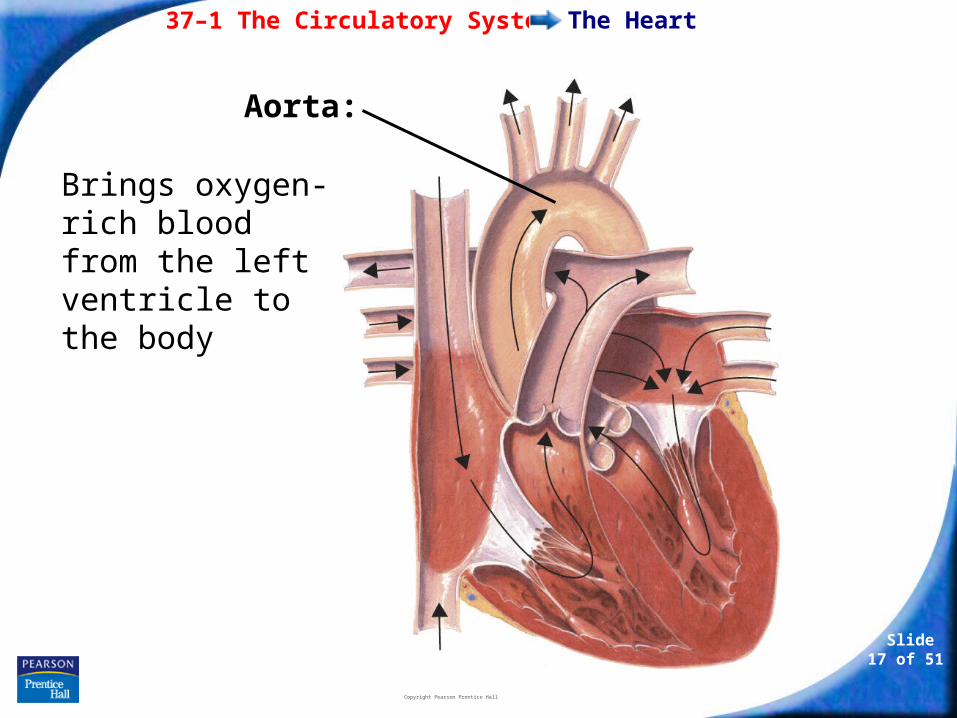
Brings oxygen-rich blood from the left ventricle to the body
Aorta:
37–1 The Circulatory System
Slide 18 of 51
Copyright Pearson Prentice Hall
The Heart
The septum divides the right side of the heart from the left.
It prevents the mixing of oxygen-poor and oxygen-rich blood.
37–1 The Circulatory System
Slide 19 of 51
Copyright Pearson Prentice Hall
The Heart
The heart has four chambers—two atria and two ventricles.
There are two chambers on each side of the septum.
The upper chamber, which receives the blood, is the atrium.
The lower chamber, which pumps blood out of the heart, is the ventricle.
37–1 The Circulatory System
Slide 20 of 51
Copyright Pearson Prentice Hall
The Heart
Circulation Through the Heart
Blood enters the heart through the right and left atria.
As the heart contracts, blood flows into the ventricles and then out from the ventricles to either the body or the lungs.
37–1 The Circulatory System
Slide 21 of 51
Copyright Pearson Prentice Hall
The Heart
There are flaps of connective tissue called valves between the atria and the ventricles.
When the ventricles contract, the valves close, which prevents blood from flowing back into the atria.
37–1 The Circulatory System
Slide 22 of 51
Copyright Pearson Prentice Hall
The Heart
At the exits from the right and left ventricles, valves prevent blood that flows out of the heart from flowing back in.
Blood leaves the left ventricle, and enters the aorta.
The aorta is one of the blood vessels that carry the blood through the body and back to the heart.
37–1 The Circulatory System
Slide 23 of 51
Copyright Pearson Prentice Hall
The Heart
Circulation Through the Body
The heart functions as two separate pumps.
37–1 The Circulatory System
Slide 24 of 51
Copyright Pearson Prentice Hall
The Heart
Pulmonary Circulation
One pathway circulates blood between the heart and the lungs.
This pathway is known as pulmonary circulation.
In the lungs, carbon dioxide leaves the blood and oxygen is absorbed. The oxygen-rich blood returns to the heart.
37–1 The Circulatory System
Slide 25 of 51
Copyright Pearson Prentice Hall
The Heart
Systemic Circulation
The second pathway circulates blood between the heart and the rest of the body.
This pathway is called systemic circulation.
After returning from the lungs, the oxygen-rich blood is pumped to the rest of the body.
37–1 The Circulatory System
Slide 26 of 51
Copyright Pearson Prentice Hall
The Heart
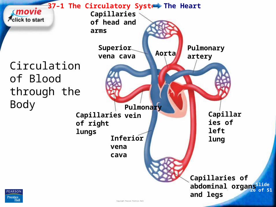
Circulation of Blood through the Body
Capillaries of head and arms
Superior vena cava Aorta
Pulmonary veinCapillaries of
right lungs
Inferior vena cava
Capillaries of abdominal organs and legs
Capillaries of left lung
Pulmonary artery
37–1 The Circulatory System
Slide 27 of 51
Copyright Pearson Prentice Hall
The Heart
Heartbeat
Each contraction begins in the sinoatrial (SA) node in the right atrium.
Because these cells start the wave of muscle contraction through the heart, they are called the pacemaker.
37–1 The Circulatory System
Slide 28 of 51
Copyright Pearson Prentice Hall
The Heart
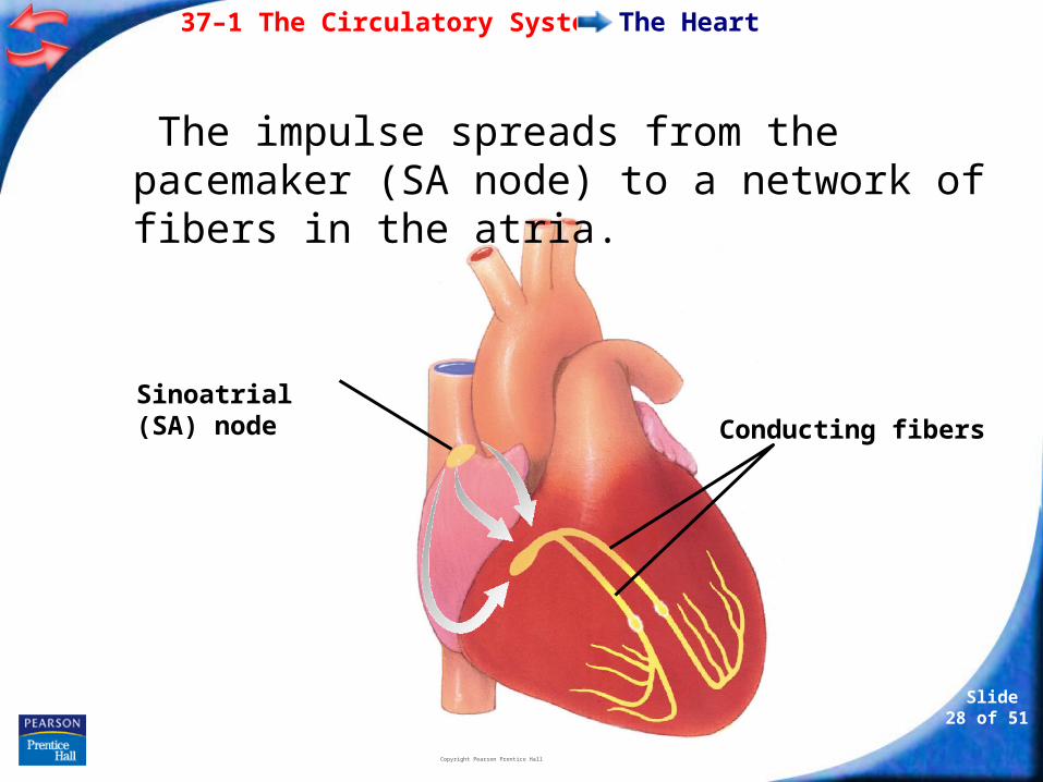
Sinoatrial (SA) node Conducting fibers
The impulse spreads from the pacemaker (SA node) to a network of fibers in the atria.
37–1 The Circulatory System
Slide 29 of 51
Copyright Pearson Prentice Hall
The Heart
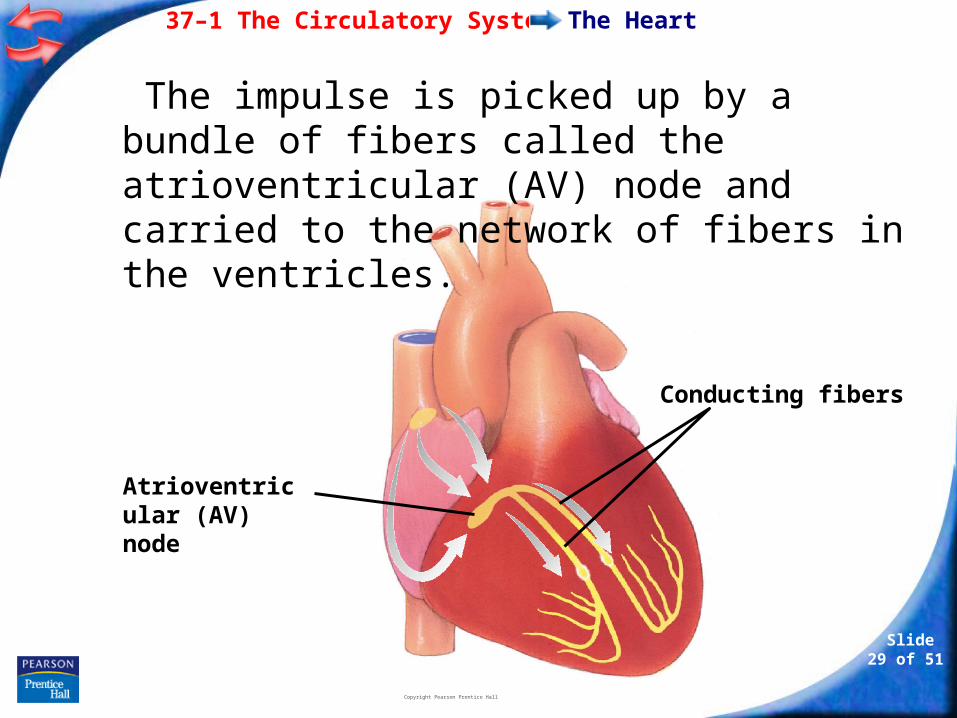
Conducting fibers
Atrioventricular (AV) node
The impulse is picked up by a bundle of fibers called the atrioventricular (AV) node and carried to the network of fibers in the ventricles.
37–1 The Circulatory System
Slide 30 of 51
Copyright Pearson Prentice Hall
The Heart
When the network in the atria contracts, blood in the atria flows into the ventricles.
When the ventricles contract, blood flows out of the heart.
37–1 The Circulatory System
Slide 31 of 51
Copyright Pearson Prentice Hall
Blood Vessels
Blood Vessels
What are the three types of blood vessels in the circulatory system?
37–1 The Circulatory System
Slide 32 of 51
Copyright Pearson Prentice Hall
Blood Vessels
As blood flows through the circulatory system, it moves through three types of blood vessels:
• arteries
• capillaries
• veins
37–1 The Circulatory System
Slide 33 of 51
Copyright Pearson Prentice Hall
Blood Vessels
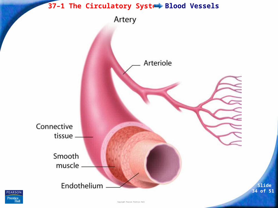
Arteries
Large vessels that carry blood from the heart to the tissues of the body are called arteries.
Except for the pulmonary arteries, all arteries carry oxygen-rich blood.
Arteries have thick walls.
They contain connective tissue, smooth muscle, and endothelium.
37–1 The Circulatory System
Slide 34 of 51
Copyright Pearson Prentice Hall
Blood Vessels
37–1 The Circulatory System
Slide 35 of 51
Copyright Pearson Prentice Hall
Blood Vessels
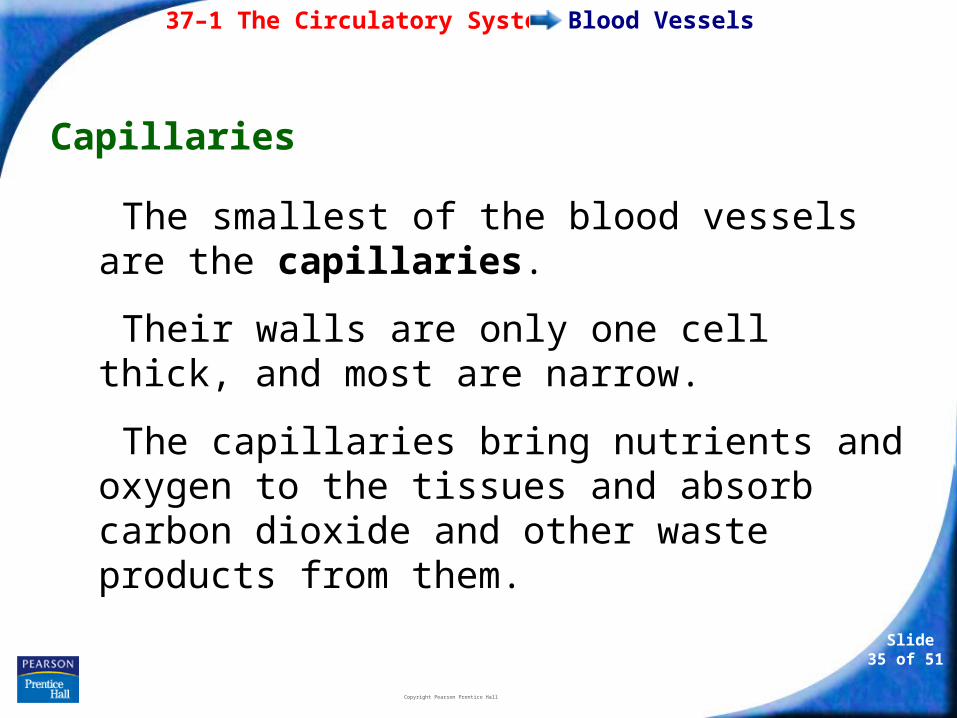
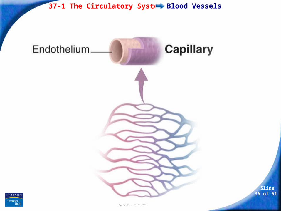
Capillaries
The smallest of the blood vessels are the capillaries.
Their walls are only one cell thick, and most are narrow.
The capillaries bring nutrients and oxygen to the tissues and absorb carbon dioxide and other waste products from them.
37–1 The Circulatory System
Slide 36 of 51
Copyright Pearson Prentice Hall
Blood Vessels
37–1 The Circulatory System
Slide 37 of 51
Copyright Pearson Prentice Hall
Blood Vessels
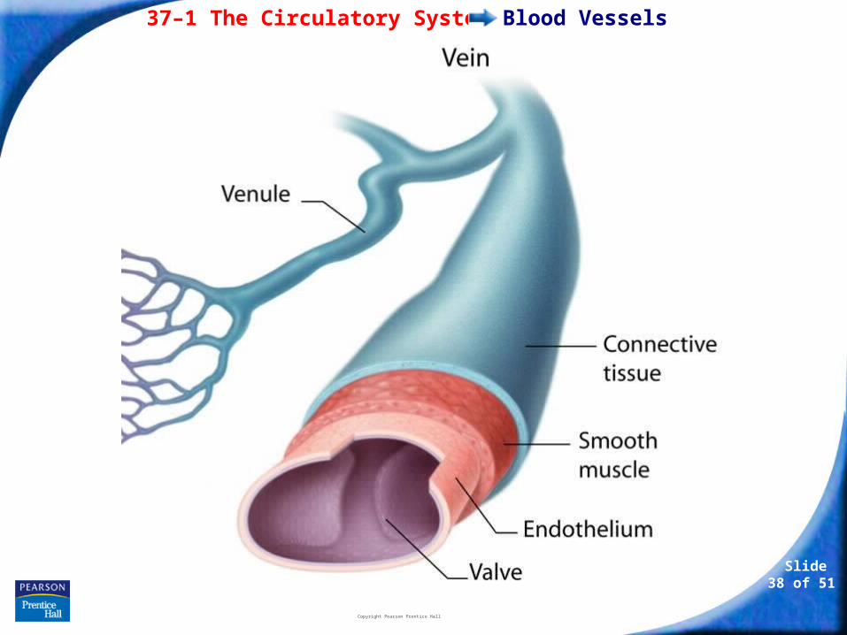
Veins
Blood vessels that carry blood back to the heart are veins.
Veins have thinner walls than arteries.
The walls of veins contain connective tissue and smooth muscle.
37–1 The Circulatory System
Slide 38 of 51
Copyright Pearson Prentice Hall
Blood Vessels
37–1 The Circulatory System
Slide 39 of 51
Copyright Pearson Prentice Hall
Blood Vessels
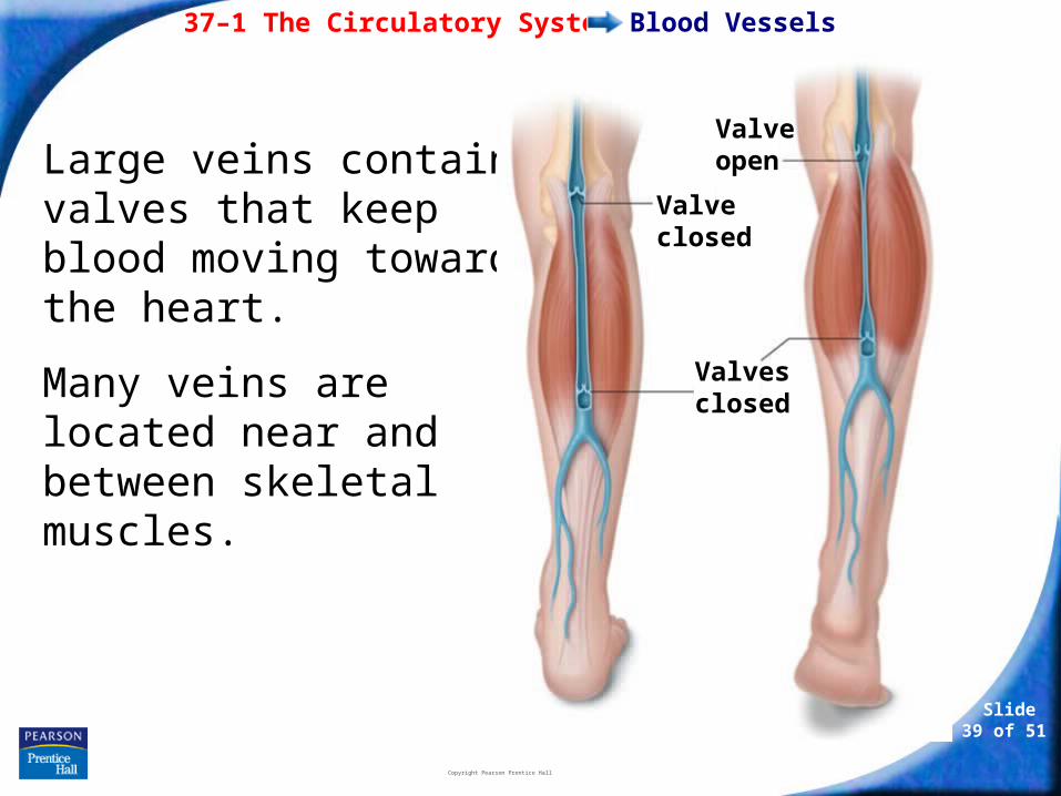
Large veins contain valves that keep blood moving toward the heart.
Many veins are located near and between skeletal muscles.
Valve open
Valve closed
Valves closed
37–1 The Circulatory System
Slide 40 of 51
Copyright Pearson Prentice Hall
Blood Pressure
Blood Pressure
When the heart contracts, it produces a wave of fluid pressure in the arteries.
The force of the blood on the arteries’ walls is blood pressure.
Blood pressure keeps blood flowing through the body.
37–1 The Circulatory System
Slide 41 of 51
Copyright Pearson Prentice Hall
Blood Pressure
Blood pressure is measured with a sphygmomanometer.
A typical blood pressure for a healthy person is 120/80.
37–1 The Circulatory System
Slide 42 of 51
Copyright Pearson Prentice Hall
Diseases of the Circulatory System
Diseases of the Circulatory System
Cardiovascular diseases are among the leading causes of death and disability in the U.S.
Atherosclerosis is a condition in which fatty deposits called plaque build up on the inner walls of the arteries.
High blood pressure is defined as a sustained elevated blood pressure of 140/90 or higher.
37–1 The Circulatory System
Slide 43 of 51
Copyright Pearson Prentice Hall
Diseases of the Circulatory System
Heart Attack and Stroke
If one of the coronary arteries becomes blocked, part of the heart muscle may begin to die from a lack of oxygen.
If enough heart muscle is damaged, a heart attack occurs.
37–1 The Circulatory System
Slide 44 of 51
Copyright Pearson Prentice Hall
Diseases of the Circulatory System
If a blood clot gets stuck in a blood vessel leading to the brain, a stroke occurs.
Brain cells die and brain function in that region may be lost.
37–1 The Circulatory System
Slide 45 of 51
Copyright Pearson Prentice Hall
Diseases of the Circulatory System
Circulatory System Health
Ways of avoiding cardiovascular disease include:
• getting regular exercise.
• eating a balanced diet.
• avoiding smoking.

- or -Continue to: Click to Launch:
Slide 46 of 51
Copyright Pearson Prentice Hall
37–1
Slide 47 of 51
Copyright Pearson Prentice Hall
37–1
The layer of muscle in the heart that pumps blood through the circulatory system is called the
a. myocardium.
b. atrium.
c. ventricle.
d. vena cava.
Slide 48 of 51
Copyright Pearson Prentice Hall
37–1
Oxygen-poor blood from the body enters the heart through the
a. left atrium.
b. left ventricle.
c. right atrium.
d. right ventricle.
Slide 49 of 51
Copyright Pearson Prentice Hall
37–1
Atherosclerosis is a condition in which
a. blood cells die from a lack of oxygen.
b. plaque builds up along the walls of the arteries.
c. blood pressure is too high.
d. the heart stops pumping blood.
Slide 50 of 51
Copyright Pearson Prentice Hall
37–1
The inner wall of all blood vessels is lined with
a. endothelium.
b. connective tissue.
c. smooth muscle.
d. myocardium.
Slide 51 of 51
Copyright Pearson Prentice Hall
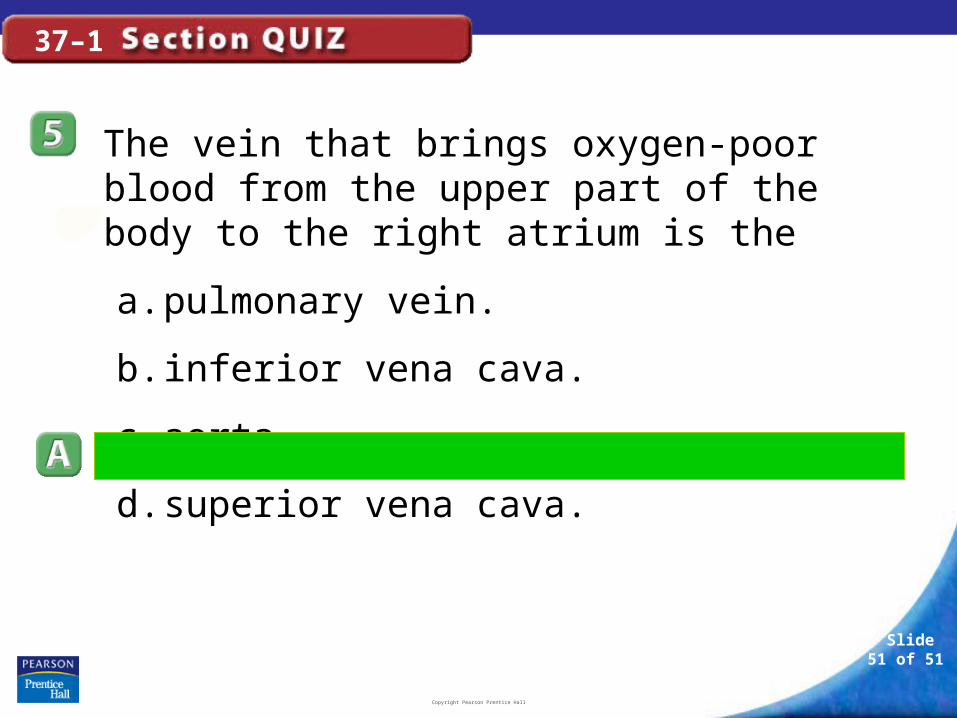
37–1
The vein that brings oxygen-poor blood from the upper part of the body to the right atrium is the
a. pulmonary vein.
b. inferior vena cava.
c. aorta.
d. superior vena cava.
END OF SECTION