Embed Size (px)
Citation preview


Slipped Capital
Femoral Epiphysis
Amy Leu, D.O.
UCSD Primary Care Sports Medicine Fellow
August 2009

General
Most common hip disorder in adolescents
Represents a combination of mechanical and constitutional factors involving the rapidly growing physis in the proximal femur
Involves the dislocation of the femoral head posteriorly and inferiorly relative to the femoral neck while remaining articulated with the acetabulum.
Accurate diagnosis and immediate treatment are paramount in avoiding significant morbidity associated with untreated cases, most importantly avascular necrosis of the hip.
Epidemiology
Most common group is growing adolescent males between 10-17 year of age (average age of 12)
Can occur in females, although much less prevalent (average age of 12)
Male:female 2-4:1
Affects the left hip more commonly than the right
Can become bilateral in 20-50% of cases; rarely simultaneous in presentation
Can occur during growth hormone therapy
Epidemiology
Endocrine abnormalities should certainly be considered when a child presents with bilateral SCFE.
Rarely before 11 in males and 9 in females, and when it does suggests other underlying processes such as hypothyroidism, delayed grown and bone age, panhypopituitaryism, gonadal conditions, and renal osteodystrophy

Pathophysiology
The proximal physis of the femur changes position from horizontal to oblique during preadolesence and adolesence.
This change redirects the stress on the plate from compression to shear forces.
This normal anatomic change along with rapid growth or weight gain can put excessive shearing forces across the plate resulting the the Salter-Harris type of fracture along the growth plate.
Pathophysiology
The femoral head then dislocates
posteriorly and inferiorly, while the
femoral neck and shaft extends and
externally rotates
The fracture is not usually associated
with acute trauma rather is a chronic
process associated with microfracture at
the physis.
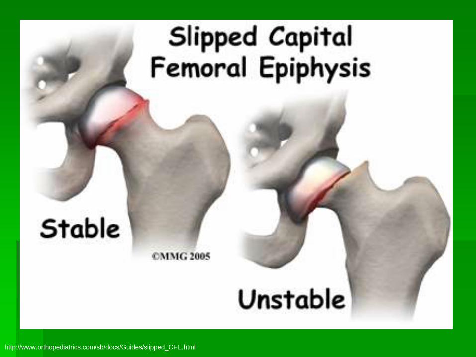
Classification
Stable (chronic) – allows for weight bearing, may have prolonged symptoms, and may have been minimal in injury
Unstable (acute) – acute physeal injury, too painful and unstable to allow weight-bearing. Risk for malunion and AVN
Acute on chronic - third category of patients who have had a stable slip for a variable period of time that is suddenly complicated by an acute physeal separation superimposed on a more chronic and more stable physeal deformation.
http://www.orthopediatrics.com/sb/docs/Guides/slipped_CFE.html
History
Knee pain is a common presenting symptom. Occurs as referred pain via the obturator nerve. Can lead to a delay in diagnosis if clinician fails to consider the hip as an etiology
Medial thigh pain
Hip or groin pain
Limp
Decreased ROM
Sxs < 3 weeks are considered acute
History
Symptoms are often vague, and pain
may not be present at all.
If there is any complaint, usually an
aching discomfort
Typically worse with physical activity.
Differential Diagnosis
Femoral head AVN
Femoral neck
fracture
Femoral neck stress
fracture
Femur injury
Groin injury
Osteitis Pubis
Knee injury
Chronic
developmental hip
dysplasia
Femoral hernia
Legg-Calve-Perthes
disease
Neoplastic processes
Septic joint
Synovitis
Physical Exam
A limp in gait can be present
Acutely, the hip can lie in extension, adduction, and external rotation. Any movement, active or passive, is usually painful.
In a chronic slip, little or no discomfort will occur during active or passive motion of the affected hip. Hip flexion is usually limited. At the end of hip flexion, the femur can drift into external rotation as the prominent anterior femoral neck abuts against the anterior acetabulum.
Diagnostic Imaging
Plain films: MUST have AP and lateral views
Widening and blurring of the proximal femoral physis is an early sign, even before the proximal femoral epiphysis begins its characteristic posterior tilting
Obvious discontinuity between the anterosuperior portion of the femoral neck and the anterolateral corner of the capital femoral epiphysis is commonly seen
Imaging: Plain Films
Degree of slippage:
Type I slippage is less than 33%
displacement.
Type II slippage is between 33% and 50%
displacement.
Type III slippage is greater than 50%
displacement.
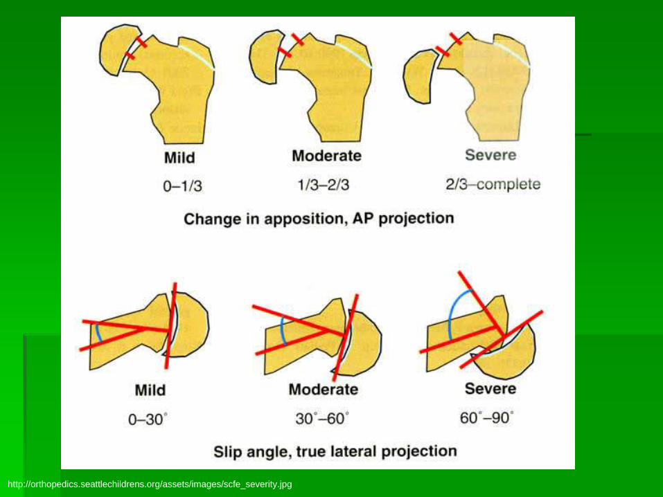
http://orthopedics.seattlechildrens.org/assets/images/scfe_severity.jpg

Imaging: Plain Films
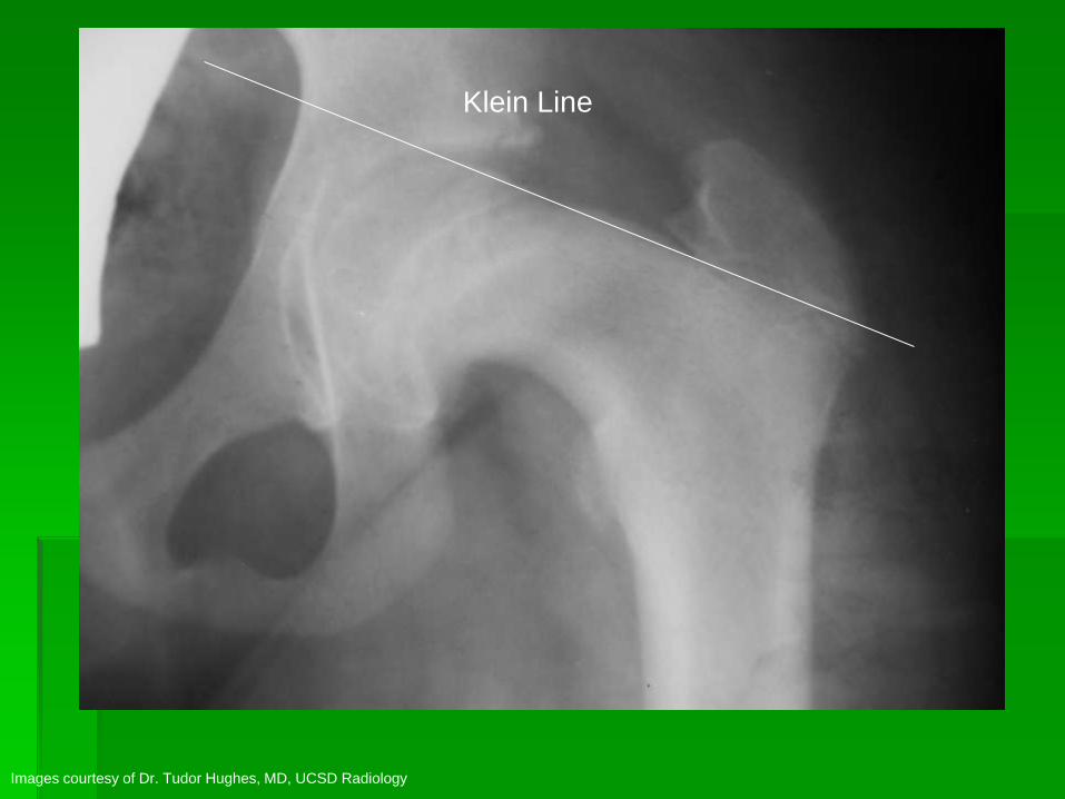
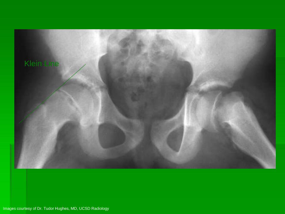
AP Films: A Klein line is a line drawn
along the superior border of the femoral
neck that would normally pass through a
portion of the femoral head. If not,
slipped capital femoral epiphysis is
diagnosed.
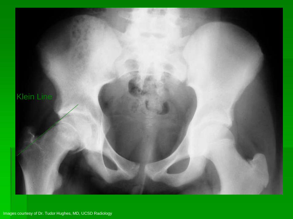
Klein Line
Images courtesy of Dr. Tudor Hughes, MD, UCSD Radiology
Klein Line
Images courtesy of Dr. Tudor Hughes, MD, UCSD Radiology
Klein Line
Images courtesy of Dr. Tudor Hughes, MD, UCSD Radiology

Imaging: Plain Films
Frog-leg views: A straight line through
the center of the femoral neck proximally
should be at the center of the epiphysis.
If not, and the line is anterior in the
epiphysis, it is likely an SCFE
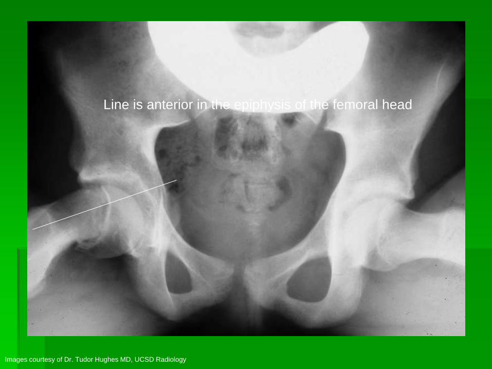
Line is anterior in the epiphysis of the femoral head
Images courtesy of Dr. Tudor Hughes MD, UCSD Radiology
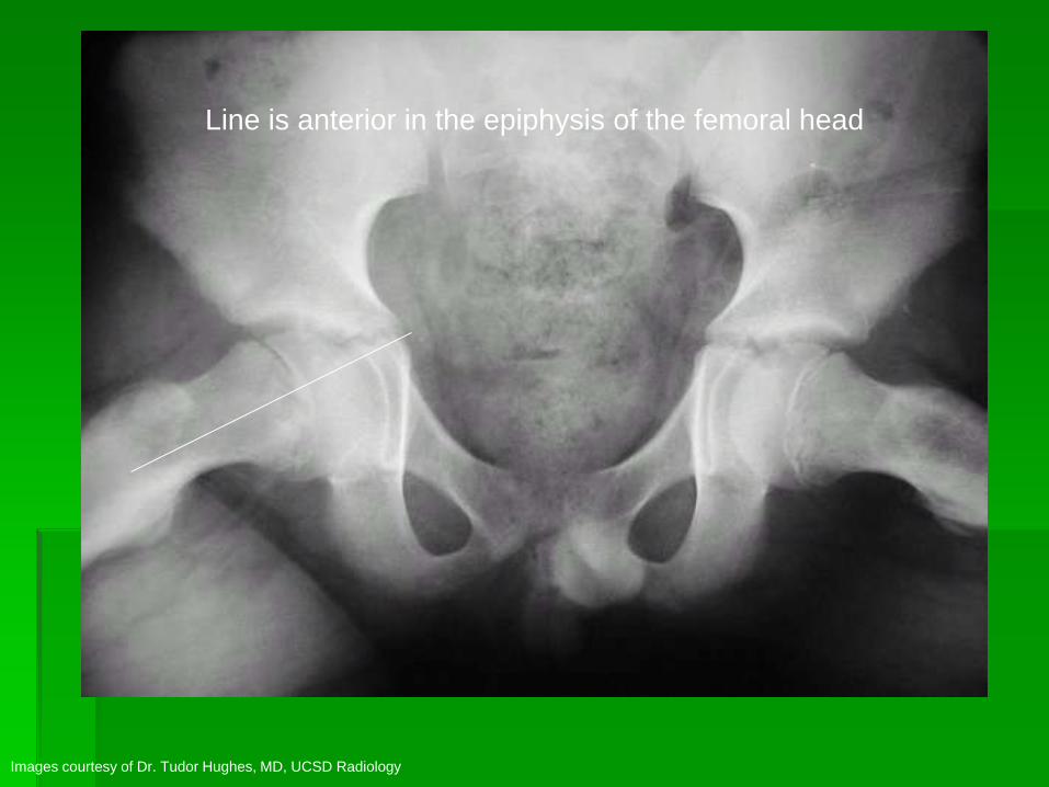
Images courtesy of Dr. Tudor Hughes, MD, UCSD Radiology
Line is anterior in the epiphysis of the femoral head
Advanced Imaging
Bone scans can show increased uptake
at the femoral neck
MRI can show epiphysis changes in the
early stage
Advanced imaging studies not routinely
used, however can aid in confirming the
diagnosis. Can also aid in the
measurement of the severity of the injury

Bone Scan
Images courtesy of Dr. Tudor Hughes MD, UCSD Radiology
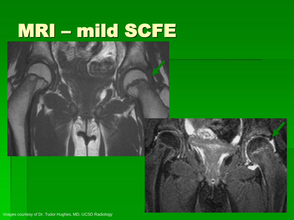
MRI – mild SCFE
Images courtesy of Dr. Tudor Hughes, MD, UCSD Radiology
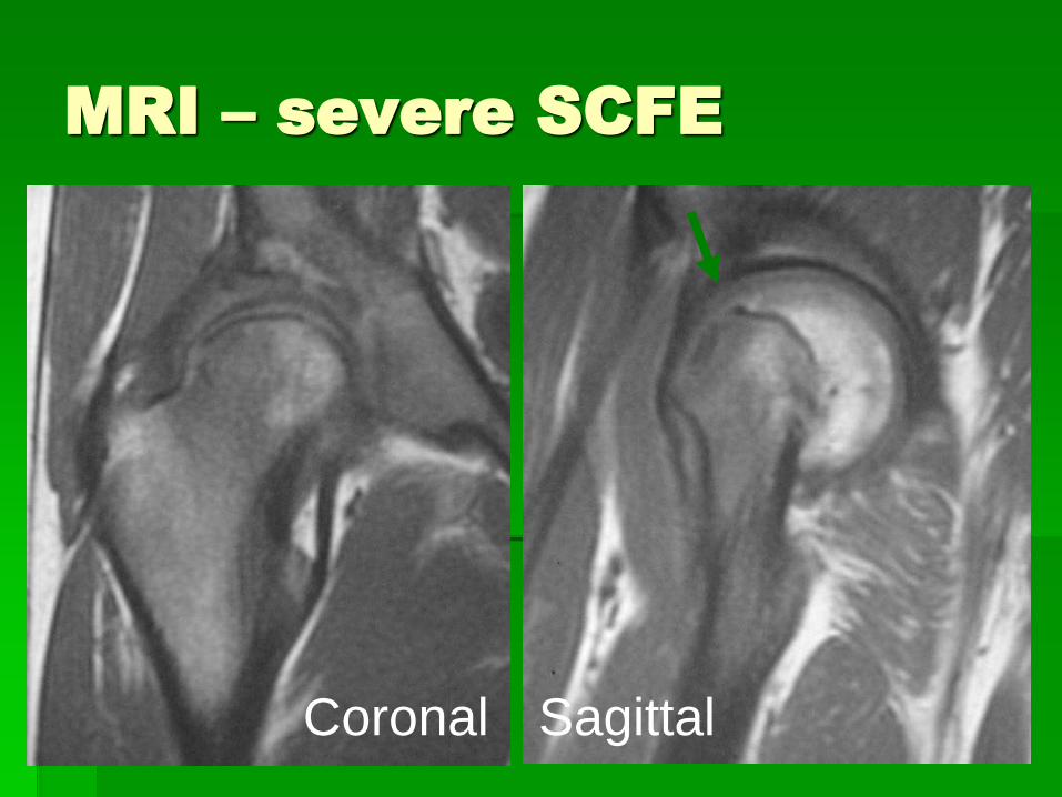
MRI – severe SCFE
Coronal Sagittal

MRI - moderate
Advanced Imaging
A report in the European Journal of Radiology suggests that pretreatment MRI in established cases of SCFE has a role with prognostic implications for the treatment approach and outcome of this condition. The investigators noted that synovitis, periphyseal edema, and joint effusion are regular features of SCFE; however, "the clinical history and findings are unreliable for the classification of SCFE," and "radiographs underestimate the severity of SCFE. MRI can potentially identify unstable, reducible slips. If the mode of surgical treatment depends on the particular nature of the SCFE then MRI contributes to surgical decision-making.“ [5]
Laboratory Evaluation
ROUTINE hormonal screening is NOT
indicated in children with SCFE
Workup can be initiated however in
cases if ATYPICAL presentations arise,
such as age < 10 or > 16, or presentation
with short stature (implication of
underlying congenital disease)

Treatment
VS.
Treatment
Casting has fallen out of favor due to high rate
of AVN and chondrolysis, as well as difficulty in
application and maintenance of casts
Classification:
Acute (< 3w) vs chronic (> 3w) vs acute on chronic
(> 3w but acute change)
Stable (wt bearing) vs unstable (non weight bearing)
Radiographic classification (Type I, II, III)
Usually immediate ORIF
Treatment
If the angle of the slip is >45 degrees can
consider a bone realignment procedure
to avoid significant risk to OA from
anterior impingement and dysfunction in
the form of limitation of flexion and
severe external rotation deformity
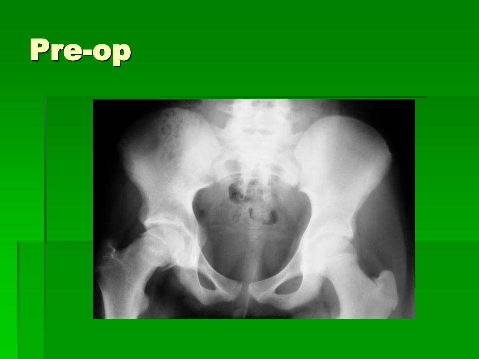
Pre-op
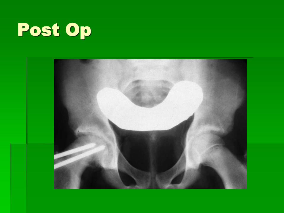
Post Op
Treatment
Prophylactic pinning of the contralateral asymptomatic hip is controversial.
May be considered in patients < 10 or with endocrinopathies that place them at higher risk for bilateral involvement
Also may be considered in patients / families that are unreliable, as close follow-up for monitoring the unaffected hip is of utmost importance
In Europe the majority of cases receive prophylactic fixation of the unaffected hip
Follow Up
Limited weight bearing 6-8 weeks
PT/rehab
Return to play when pain free and full
strength
Some say no return until physis has
closed
Complications
Can arise from disease process or treatment
Osteoarthritis from untreated deformity
AVN – up to 47% in unstable cases
?vascular compromise from initial injury vs. forceful reduction at time of surgery
Chondrolysis (treated or untreated)
Surgical complications (infection, loss of fixation, outgrowing screws)
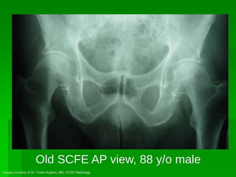
Old SCFE AP view, 88 y/o maleImages courtesy of Dr. Tudor Hughes, MD, UCSD Radiology
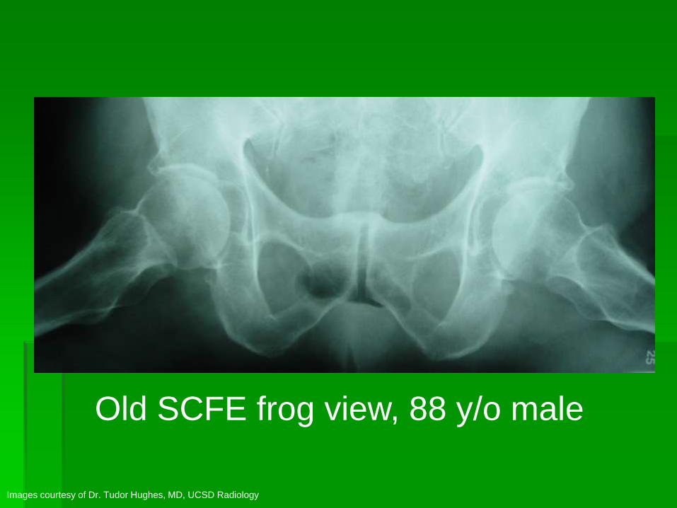
Old SCFE frog view, 88 y/o male
Images courtesy of Dr. Tudor Hughes, MD, UCSD Radiology
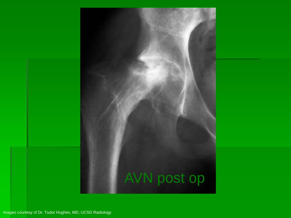
AVN post op
Images courtesy of Dr. Tudor Hughes, MD, UCSD Radiology
References
1. DeLee: DeLee and Drez's Orthopaedic Sports Medicine, 2nd ed, Saunders 2002
2. http://emedicine.medscape.com/article/91596-overview
3. http://www.wheelessonline.com/ortho/slipped_capital_femoral_epiphysis
4. Radiology of adolescent slipped capital femoral epiphysis: measurement of epiphyseal angles and diagnosis. Oper Orthop Traumatol. 2007 Oct;19(4):329-44.
5. Tins B, Cassar-Pullicino V, McCall I. The role of pre-treatment MRI in established cases of slipped capital femoral epiphysis. Eur J Radiol. 2009 Jun;70(3):570-8.