Embed Size (px)
Citation preview
398 J. BOUILLON et al.
116. – Leptomedusae. Campanulariidae. A to S: Orthopyxis. A to H: Orthopyxis crenata: A and B: different views of hydrothecae andpedicels, C: detail of hydrotheca, D: transverse section of a hydrotheca, E: gonotheca with male immature gonophore, F: gonotheca contain-ing male eumedusoid, G: gonothecae with immature female gonophore, H: spent female eumedusoid; I to S: Orthopyxis integra: I: generalaspect of a part of colony, J and K: different aspects of hydrothecae and pedicels, L: detail of hydrotheca, M: diagrams of hydrothecal rimand of basal chamber of a thickened hydrotheca, N: diagrams of hydrothecal rim and of basal chamber of unthickened hydrotheca, O: groovedwalled gonotheca, P: smooth walled gonotheca, Q: gonotheca with male gonophore, R: gonotheca with female gonophore, S: released eume-dusoid (A after Medel and Vervoort, 2000; B, E to H, K, Q and R after Hirohito, 1995; C and D after Millard, 1975; I, M to P, S after
Cornelius, 1995; J and L after Cornelius, 1982).
sm68s2Gfig3 14/10/04 15:38 Página 398
FAUNA OF THE MEDITERRANEAN HYDROZOA 399
117. – Leptomedusae. Campanulariidae. A: Pseudoclytia pentata: mature medusa. Limnomedusae. Armorhydridae. B and C: Armorhy-dra janowiczi: B: medusa; C: longitudinal histological section of a medusa. Microhydrulidae. D and E: Microhydrula pontica: D: histolog-ical section of a polyp; E: histological section of a frustule. F: Rhaptapagis cantacuzenei, histological section of a polyp (A after Kramp,1959a; B after Thiel, 1988; C after Lacassagne, 1968a; D to F after Bouillon and Deroux, 1967). cbl: cnidoblast; cnl: cnidocil; di: digestiveinclusion; ecd: ectoderm; end: endoderm; eng: endodermal gap; g: gonads; gc: gastric cavity; glc: glandular cell, ma: manubrium; meu:
microbasic eurytele; p: periderm; sc: subumbrellar cavity; seu: semiophoric eurytele; spt: septum; te: tentacle; v: velum.
sm68s2Gfig3 14/10/04 15:38 Página 399

400 J. BOUILLON et al.
FIG. 118. – Limnomedusae. Olindiidae. A to F: Craspedacusta sowerbii: A and B: two aspects of polyp colonies with a medusa bud; C: apolyp colony reducing itself in frustules and resting stages or cysts; D: fully-grown medusa; E: portion of umbrella showing the marginalcnidocyst ring and tentacular roots; F: portion of the velum with the centripetal tubes of the statocysts. G to I: Gonionemus vertens: G:hydranth; H and I: two hydranths developing a fustule. J: hydranth with a medusa bud (A and C after Damas, 1939; B, E and F after Russell,
1953, D after Tardent, 1978; G and I after Leloup, 1952; H and J after Werner, 1984). cy: cyst; fr: frustule; sta: statocyst, v: velum.
sm68s2Gfig3 14/10/04 15:38 Página 400
FAUNA OF THE MEDITERRANEAN HYDROZOA 401
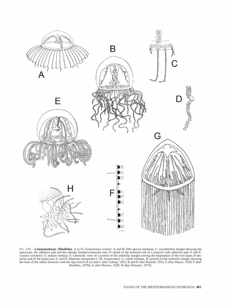
FIG. 119. – Limnomedusae. Olindiidae. A to D: Gonionemus vertens: A and B: fully-grown medusae, C: exumbrellar margin showing thestatocysts, the adhesive pad and the sharply bended tentacular end, D: detail of the terminal end of a tentacle with adhesive pad; E and F:Gossea corynetes: E: mature medusa, F: schematic view of a portion of the umbrella margin sowing the disposition of the two types of ten-tacles and of the statocysts; G and H: Maeotias marginata (= M. inexpectata): G: whole medusa, H: portion of the umbrella margin showingthe base of the oldest tentacles with the tips twitch of (A and C after Leloup, 1952; B and D after Russell, 1953; E after Mayer, 1910; F after
Bouillon, 1978a; G after Borcea, 1929, H after Denayer, 1973).
sm68s2Gfig3 14/10/04 15:38 Página 401
402 J. BOUILLON et al.
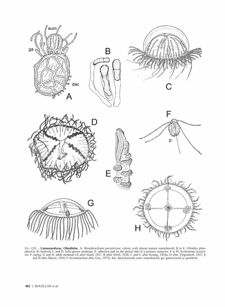
FIG. 120. – Limnomedusae. Olindiidae. A: Monobrachium parasiticum: colony with almost mature eumedusoid; B to E: Olindias phos-phorica: B: hydroid, C and D: fully-grown medusae; E: adhesive pad on the aboral side of a primary tentacles. F to H: Scolionema suvaen-sis: F: polyp; G and H: adult medusae (A after Hand, 1957, B after Weill, 1936; C and G after Kramp, 1959a; D after Trègouboff, 1957; E
and H after Mayer, 1910; F reconstruction after Goy, 1973). dac: dactylozooid; eum: eumedusoid; ga: gastrozooid; p: periderm.
sm68s2Gfig3 14/10/04 15:38 Página 402

FAUNA OF THE MEDITERRANEAN HYDROZOA 403
FIG. 121. – Limnomedusae. Olindiidae. A: Calpasoma dactyloptera: hydranths showing different stages of reproduction. Siphonophorae.Physaliidae. B to D: Physalia physalis: B: colony; C: cluster of persons from sexualy mature colony; D: small part of a gonodendron. Rhyso-physidae. E and F: Rhizophyla filiformis: E: colony; F: tentilla (A after Matthews, 1966; B and E after Pagès and Gili, 1992; C and D after
Hyman, 1940; F after Pugh, 1999).
sm68s2Gfig3 14/10/04 15:38 Página 403

404 J. BOUILLON et al.
FIG. 122. – Siphonophorae. Agalmatidae. A to I: Agalma: A to C: Agalma clausi: A: nectophore; B: bract; C: tentilla. D to F: Agalma ele-gans: D: detail of a lateral view of a nectophore; E: detail of an upper view of a nectophore; F: bracts. G to I: Agalma okeni: G and H: upperand lateral view of nectophore; I: bract (A to C after Bedot, 1888; D to E after Totton, 1965; G and I after Pugh, 1999; H after Gili, 1986).alr: apico-lateral ridge; aw: apical wings; ilr: infra-lateral ridge; lr: lateral ridge; n: nectophore; os: ostium; rc: radial canal; thb: thrust block;
vlr: vertical lateral ridge.
sm68s2Gfig3 14/10/04 15:38 Página 404
FAUNA OF THE MEDITERRANEAN HYDROZOA 405
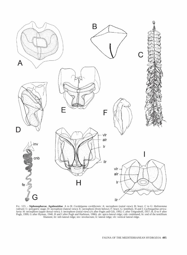
FIG. 123. – Siphonophorae. Agalmatidae. A to B: Cordalgama cordiformis: A: nectophore (ostial view); B: bract. C to G: Halistemmarubrum: C: polygstric stage; D: nectophore (lateral view); E: nectophore (from below); F: bract; G: tentillum. H and I: Lychnagalma utricu-laria: H: nectophore (upper dorsal view); I: nectophore (ostial view) (A after Pagès and Gili, 1992; C after Trègouboff, 1957; B, D to F afterPugh, 1999; G after Hyman, 1940, H and I after Pugh and Harbison, 1986). alr: apico-lateral ridge; cnb: cnidoband; fe: end of the tentillium
filament; ilr: infr-lateral ridge; inv: involucrum; lr: lateral ridge; vlr: vertical lateral ridge.
sm68s2Hfig4 14/10/04 15:39 Página 405
406 J. BOUILLON et al.
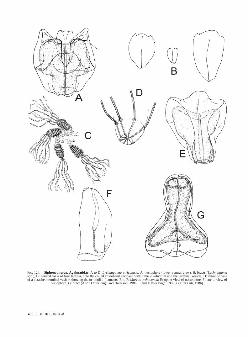
FIG. 124. – Siphonophorae. Agalmatidae. A to D: Lychnagalma utricularia. A: nectophore (lower ventral view), B: bracts (Lychnalgamaspp.), C: general view of four tentilla, note the coiled cnidoband enclosed within the involucrum and the terminal vesicle, D: detail of baseof a detached terminal vesicle showing the octoradial filaments. E to F: Marrus orthocanna: E: upper view of nectophore, F: lateral view of
nectophore, G: bract (A to D after Pugh and Harbison, 1986; E and F after Pugh, 1999; G after Gili, 1986).
sm68s2Hfig4 14/10/04 15:39 Página 406
FAUNA OF THE MEDITERRANEAN HYDROZOA 407
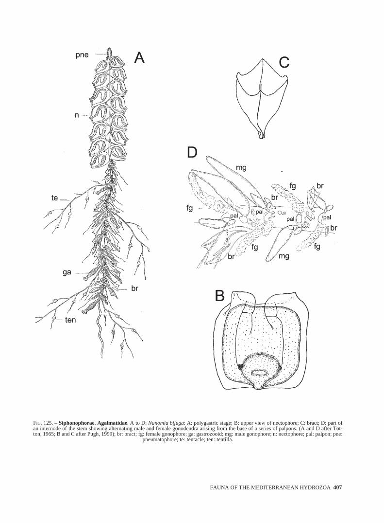
FIG. 125. – Siphonophorae. Agalmatidae. A to D: Nanomia bijuga: A: polygastric stage; B: upper view of nectophore; C: bract; D: part ofan internode of the stem showing alternating male and female gonodendra arising from the base of a series of palpons. (A and D after Tot-ton, 1965; B and C after Pugh, 1999); br: bract; fg: female gonophore; ga: gastrozooid; mg: male gonophore; n: nectophore; pal: palpon; pne:
pneumatophore; te: tentacle; ten: tentilla.
sm68s2Hfig4 14/10/04 15:39 Página 407

408 J. BOUILLON et al.
FIG. 126. – Siphonophorae. Apolemiidae. A to D: Apolemia uvaria: A: polygastric stage; B: nectophore (lateral view); C: nectophore (ostialview); D: bracts. Athorybiidae. E and F: Athorybia rosacea: E: polygastric stage (lateral view); F: dorsal view of a polygastric stage.Forskaliidae. G to K: Forskalia asymmetrica: G and H: inner and outer view of nectophores; I: inner and outer view of stem bratcs; J: innerview of bolster bract; K: inner view of knee-shaped bracts (A to D after Totton, 1965; E after Pugh, 1999; F after Trègouboff, 1957; G to K
after Pugh, 2003). lrc: lateral radial canal; os: ostium; ped: peduncle.
sm68s2Hfig4 14/10/04 15:39 Página 408
FAUNA OF THE MEDITERRANEAN HYDROZOA 409
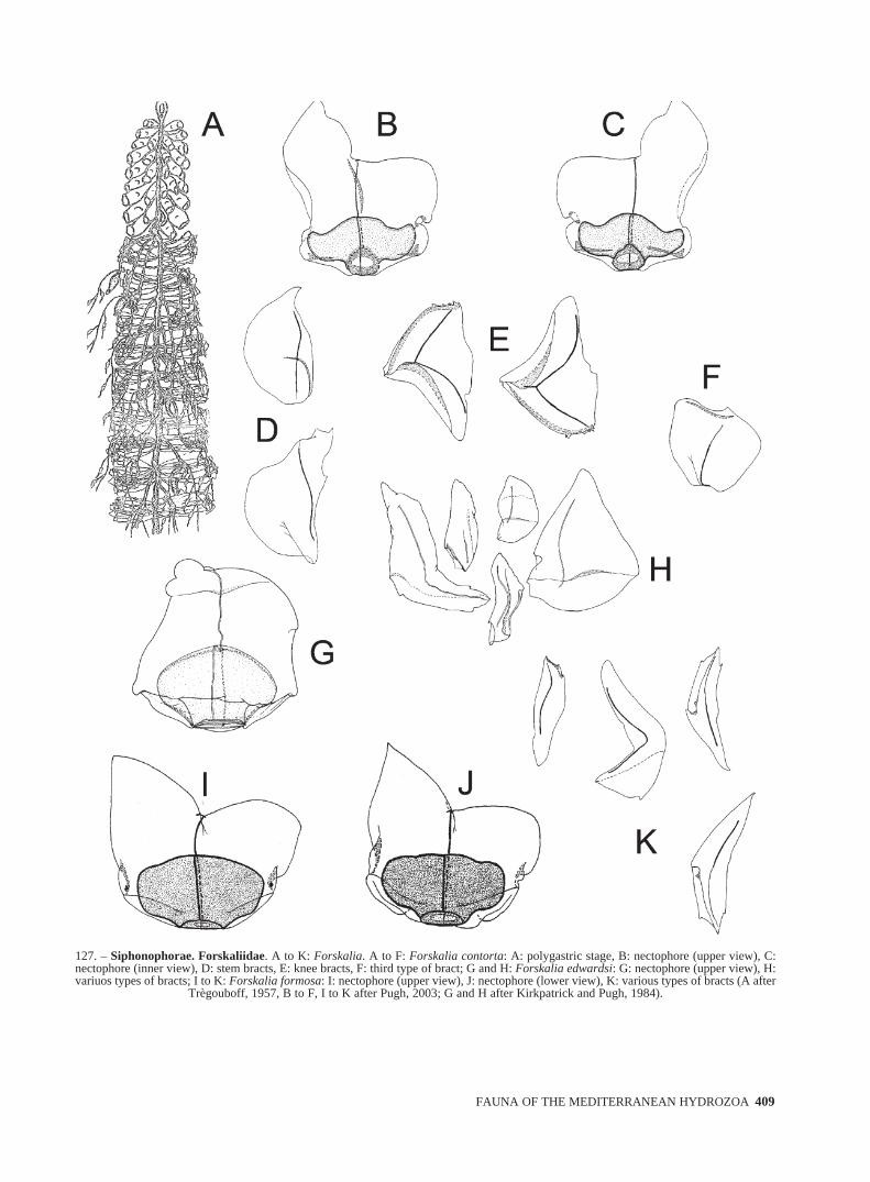
127. – Siphonophorae. Forskaliidae. A to K: Forskalia. A to F: Forskalia contorta: A: polygastric stage, B: nectophore (upper view), C:nectophore (inner view), D: stem bracts, E: knee bracts, F: third type of bract; G and H: Forskalia edwardsi: G: nectophore (upper view), H:variuos types of bracts; I to K: Forskalia formosa: I: nectophore (upper view), J: nectophore (lower view), K: various types of bracts (A after
Trègouboff, 1957, B to F, I to K after Pugh, 2003; G and H after Kirkpatrick and Pugh, 1984).
sm68s2Hfig4 14/10/04 15:39 Página 409

410 J. BOUILLON et al.
FIG. 128. – Siphonophorae. Physophoridae. A to C: Physophora hydrostatica: A: polygastric stage, B: nectophore (upper view), C: palponAbylidae. D to F: Abyla haeckeli: D: posterior nectophore, E: ventral and lateral views of anterior nectophore, F: eudoxid; G to K: Abylop-sis eschscholtzi: G: polygastric stage (lateral view), H: anterior nectophore, I: posterior nectophore, J: eudoxid, K: bract. (A to C after Kirkpatrick and Pugh, 1984; D and I after Gili, 1986; E, H and K after Pugh, 1999; F after Totton, 1965; G and J after Pagès and Gili, 1992).
sm68s2Hfig4 14/10/04 15:39 Página 410
FAUNA OF THE MEDITERRANEAN HYDROZOA 411
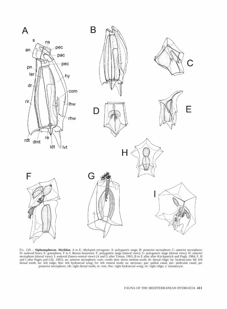
FIG. 129. – Siphonophorae. Abylidae. A to E: Abylopsis tetragona: A: polygastric stage; B: posterior nectophore; C: anterior nectophore;D: eudoxid bract; E: gonophore. F to I: Bassia bassensis: F: polygastric stage (lateral view); G: polygastric stage (dorsal view); H: anteriornectophore (dorsal view); I: eudoxid (latero-ventral view) (A and G after Totton, 1965; B to E after after Kirckpatrick and Pugh, 1984; F, Hand I after Pagès and Gili, 1992). an: anterior nectophore; com: comb; dmt: dorso median tooth; dr: dorsal ridge; hy: hydroecium; ldt: leftdorsal tooth; ler: left ridge; lhw: left hydroecial wing; lvt: left ventral tooth; ns: nectosac; pac: pallial canal; pec: pedicular canal; pn:
posterior nectophore; rdt: right dorsal tooth; re: rete; rhw: right hydroecial wing; rir: right ridge; s: somatocyst.
sm68s2Hfig4 14/10/04 15:39 Página 411
412 J. BOUILLON et al.
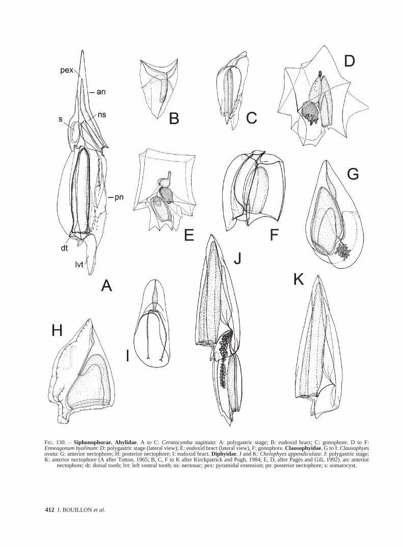
FIG. 130. – Siphonophorae. Abylidae. A to C: Ceratocymba sagittata: A: polygastric stage; B: eudoxid bract; C: gonophore. D to F:Enneagonum hyalinum: D: polygastric stage (lateral view); E: eudoxid bract (lateral view), F: gonophore. Clausophyidae. G to I: Clausophyesovata: G: anterior nectophore; H: posterior nectophore; I: eudoxid bract. Diphyidae. J and K: Chelophyes appendiculata: J: polygastric stage;K: anterior nectophore (A after Totton, 1965; B, C, F to K after Kirckpatrick and Pugh, 1984; E, D, after Pagès and Gili, 1992). an: anterior
nectophore; dt: dorsal tooth; lvt: left ventral tooth; ns: nectosac; pex: pyramidal extension; pn: posterior nectophore; s: somatocyst.
sm68s2Hfig4 14/10/04 15:39 Página 412

FAUNA OF THE MEDITERRANEAN HYDROZOA 413
FIG. 131. – Siphonophorae. Diphyidae. A to C: Chelophyes appendiculata: A: posterior nectophore, B: detail of mouth-plate of posteriornectophore (dorsal view), C: eudoxid stage (dorsal view); D to F: Chelophyes contorta: D: anterior nectophore (ventral view), E: anterior nec-tophore (lateral view), F: posterior nectophore; G to I: Dimophyes arctica: G: anterior nectophore, H: posterior nectophore, I eudoxid stage;J and K: Diphyes bojani: J: anterior nectophore (lateral view), K: posterior nectophore (lateral view). (A, B, C, G to I after Kirkpatrick and
Pugh, 1984; D to F, J and K after Pagès and Gili, 1992).
sm68s2Hfig4 14/10/04 15:39 Página 413
414 J. BOUILLON et al.
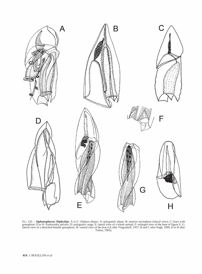
FIG. 132. – Siphonophorae. Diphyidae. A to C: Diphyes dispar: A: polygastric phase, B: anterior nectophore (lateral view), C: bract withgonophore; D to H: Eudoxoides spiralis: D: polygastric stage, E: lateral view of a whole animal, F: enlarged view of the base of figure E, G:lateral view of a detached femelle gonophore, H: ventral view of the bract (A after Trègouboff, 1957; B and C after Pugh, 1999; D to H after
Totton, 1965).
sm68s2Hfig4 14/10/04 15:39 Página 414

FAUNA OF THE MEDITERRANEAN HYDROZOA 415
FIG. 133. – Siphonophorae. Diphyidae. A to I: Lensia: A and B: Lensia campanella: A: anterior nectophore (lateral view); B: bract withgonophore. C to E: Lensia conoidea: C: anterior nectophore; D: posterior nectophore; E: eudoxid stage (lateral view). F to H: Lensia fowleri:F: anterior nectophore; G: posterior nectophore; H: eudoxid bract (lateral view). I: Lensia hotspur: anterior nectophore (lateral view) (A, B and
I after Pugh, 1999; C to H after Kirkpatrick and Pugh, 1984). br: bract; ga: gastrozooid; go: gonophore; nsh: neck-shield; phy: phyllocyst.
sm68s2Hfig4 14/10/04 15:39 Página 415
416 J. BOUILLON et al.
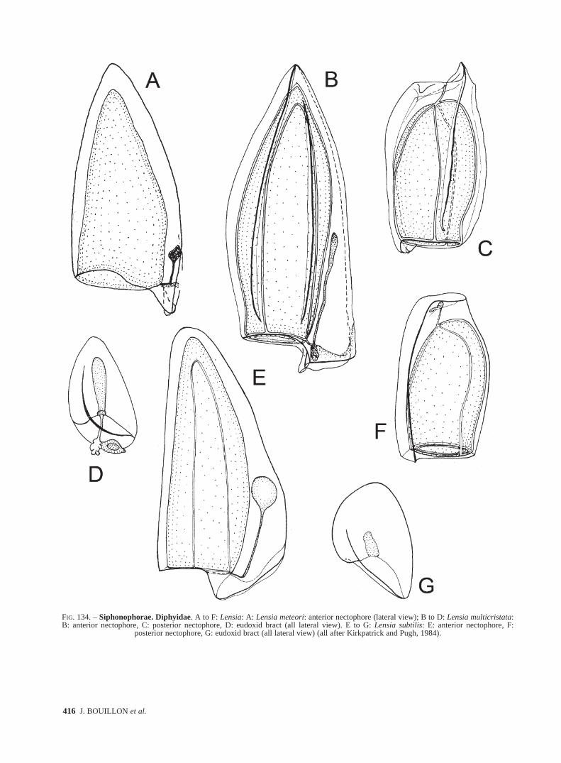
FIG. 134. – Siphonophorae. Diphyidae. A to F: Lensia: A: Lensia meteori: anterior nectophore (lateral view); B to D: Lensia multicristata:B: anterior nectophore, C: posterior nectophore, D: eudoxid bract (all lateral view). E to G: Lensia subtilis: E: anterior nectophore, F:
posterior nectophore, G: eudoxid bract (all lateral view) (all after Kirkpatrick and Pugh, 1984).
sm68s2Hfig4 14/10/04 15:39 Página 416
FAUNA OF THE MEDITERRANEAN HYDROZOA 417
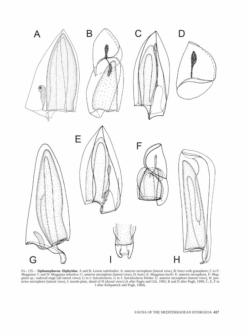
FIG. 135. – Siphonophorae. Diphyidae. A and B: Lensia subtiloides: A: anterior nectophore (lateral view), B: bract with gonophore; C to F:Muggiaea: C and D: Muggiaea atlantica: C: anterior nectophore (lateral view), D: bract; E: Muggiaea kochi: E: anterior nectophore, F: Mug-giaea sp.: eudoxid stage (all lateral view); G to I: Sulculeolaria. G to I: Sulculeolaria biloba: G: anterior nectophore (lateral view), H: pos-terior nectophore (lateral view), I: mouth-plate, detail of H (dorsal view) (A after Pagès and Gili, 1992; B and D after Pugh, 1999; C, E, F to
I after Kirkpatrick and Pugh, 1984).
sm68s2Hfig4 14/10/04 15:39 Página 417
418 J. BOUILLON et al.
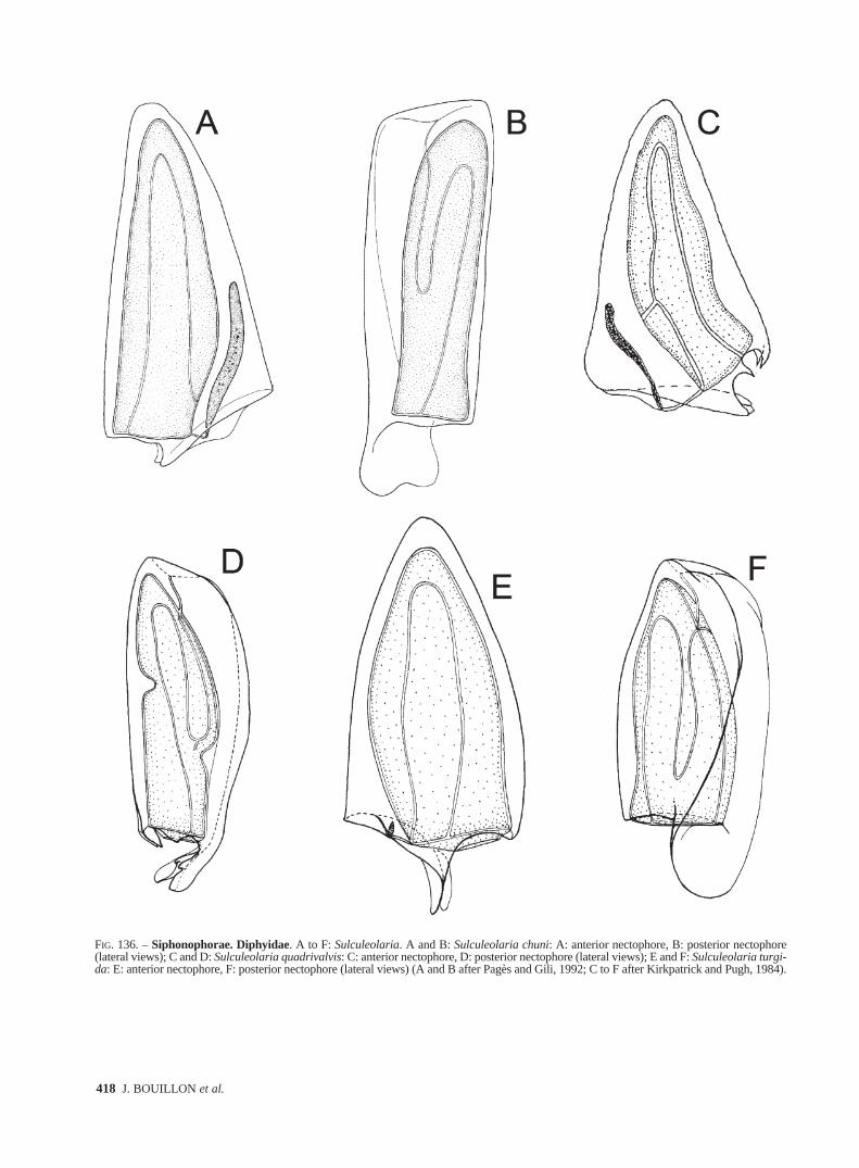
FIG. 136. – Siphonophorae. Diphyidae. A to F: Sulculeolaria. A and B: Sulculeolaria chuni: A: anterior nectophore, B: posterior nectophore(lateral views); C and D: Sulculeolaria quadrivalvis: C: anterior nectophore, D: posterior nectophore (lateral views); E and F: Sulculeolaria turgi-da: E: anterior nectophore, F: posterior nectophore (lateral views) (A and B after Pagès and Gili, 1992; C to F after Kirkpatrick and Pugh, 1984).
sm68s2Hfig4 14/10/04 15:39 Página 418

FAUNA OF THE MEDITERRANEAN HYDROZOA 419
FIG. 137. – Siphonophorae. Hippopodiidae. A to D: Hippopodius hippopus: A: polygastric stage; B: definitive nectophore; C: larval nec-tophore; D: schema of the disposition of the nectophores in a colony. E to H: Vogtia: E and F: Vogtia glabra: E: definitive nectophore (dorsalview); F: young stage; G: Vogtia pentacantha: definitive nectophore (dorsal view); H: Vogtia serrata: definitive nectophore (dorsal view) (A toC, E to H after Kirkpatrick and Pugh, 1984; D after Trègouboff, 1957). cm: cormidia; n1, n2, n3, n4, n5: nectophores; ol5: oleocyte of nectophore;
sto: stolon.
sm68s2Hfig4 14/10/04 15:39 Página 419
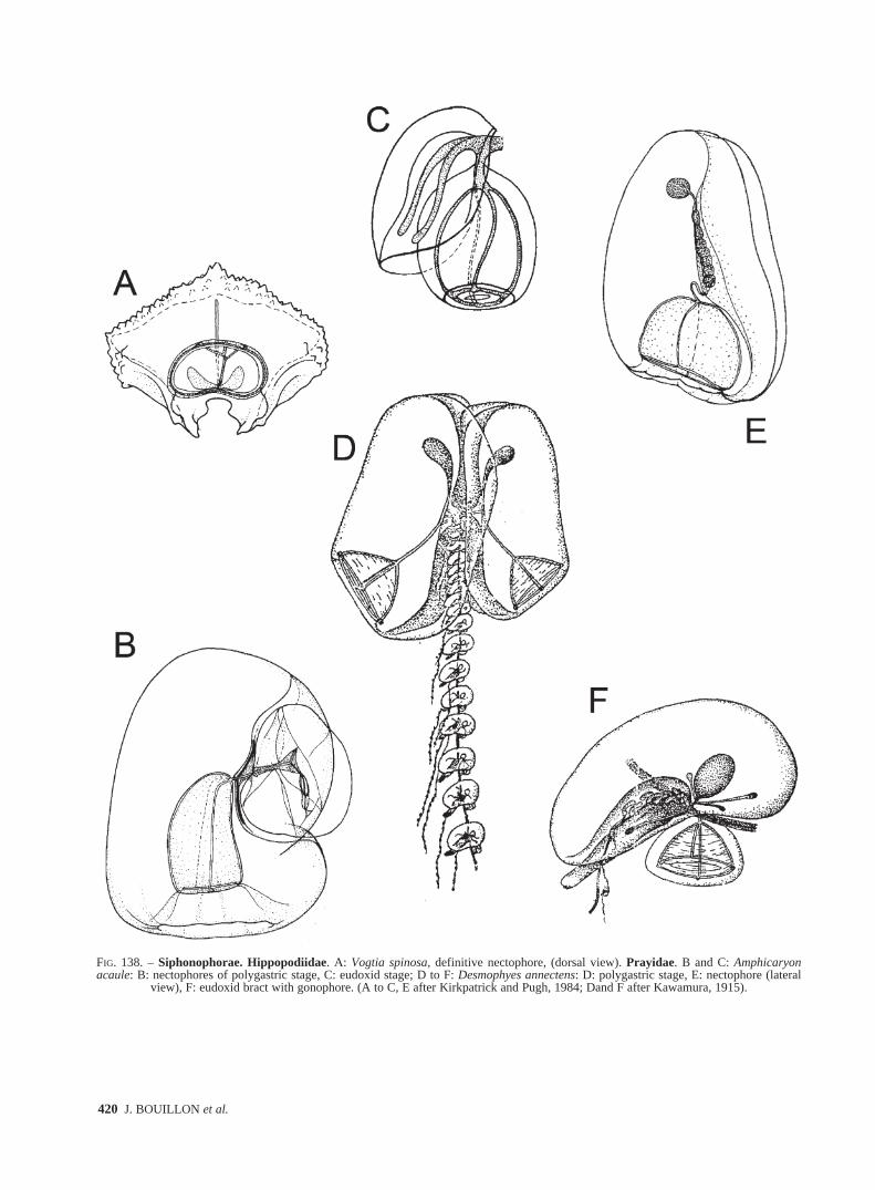
420 J. BOUILLON et al.
FIG. 138. – Siphonophorae. Hippopodiidae. A: Vogtia spinosa, definitive nectophore, (dorsal view). Prayidae. B and C: Amphicaryonacaule: B: nectophores of polygastric stage, C: eudoxid stage; D to F: Desmophyes annectens: D: polygastric stage, E: nectophore (lateral
view), F: eudoxid bract with gonophore. (A to C, E after Kirkpatrick and Pugh, 1984; Dand F after Kawamura, 1915).
sm68s2Hfig4 14/10/04 15:39 Página 420
![[CANCER RESEARCH 37, 2366-2372, July 1977] …cancerres.aacrjournals.org/content/canres/37/7_Part_2/2366.full.pdf · [CANCER RESEARCH 37, 2366-2372, July 1977] Summary Cancer involving](https://img.pdfslide.net/doc/110x75/5ad063c67f8b9a8b1e8dd9b8/cancer-research-37-2366-2372-july-1977-cancer-research-37-2366-2372-july.jpg)