Embed Size (px)
Citation preview
Physica 136B (1986) 252-255 North-Holland, Amsterdam
SMALL-ANGLE NEUTRON SCATTERING STUDIES FROM SOLUTIONS OF BOVINE NASAL CARTILAGE PROTEOGLYCAN
A. PATEL a, S.S. STIVALA a, S.P. DAMLE b, J.D. GREGORY b, G.J. BUNICK c and E.C. U B E R B A C H E R c aDepartment of Chemistry and Chemical Engineering, Stevens Institute of Technology, Hoboken, NJ 07030, USA bRockefeller University, New York, NY 10021, USA CUniversity of Tennessee - Oak Ridge Graduate School of Biomedical Sciences and Biology Division, Oak Ridge National Laboratory, Oak Ridge, TN37830, USA
Small-angle neutron scattering, SANS, of the proteoglycan subunit of bovine nasal cartilage in 0.15 N LiC1 at 25°C yielded the radius of gyration, Rg, radius of gyration of the cross-section, Rq, persistence length, a*, and the molecular weight, M. The following values were obtained: M = 3.9 × 10⁶, Rg = 745/k, Rq = 34.6 ,~ and a * = 35.2 A,. These values compare favorably with those that were obtained from small angle X-ray scattering, SAXS, of a similar extract. The scattering curve of the proteoglycan subunit in DzO showed a characteristic broad peak in the specified angular range similar to that observed from SAXS, thus confirming the polyelectrolyte nature of the proteoglycan.
1. Introduction
Proteoglycans are constituents of all types of connective tissues, such as blood vessels, skin, cartilage, and corneal stroma. In cartilage, a polypeptide of about 2000 amino acids forms the backbone of the proteoglycan molecule with many polysaccharide chains covalently attached as pendant groups [1]. These chains, comprising the glycosaminoglycans chondroitin sulfate and keratan sulfate, are linear sequences of alternat- ing carbohydrates bearing carboxyl groups and sulfate esters, and the hole proteoglycan is, there- fore, highly polyanionic.
Such molecules are essential components of connective tissues, but their structures and inter- actions are far from being understood completely. The structural patterns of proteoglycans have been shown to change in developing systems, for example, there are an increased ability to aggre- gate, increased length of keratan sulfate chains, decreased length of chondroitin sulfate chains, and changes in size of the proteoglycan, during the development of chick limb bud cell cultures [21.
Since physical methods such as SAXS and SANS can give information about macromolecular
structures in solution under mild conditions, they may eventually be valuable in establishing the significance of these properties to their changes in the organization and function of various tissues.
2. Experimental procedures
Bovine nasal cartilage was extracted with 4M guanidine hydrochloride in the presence of pro- tease inhibitors, and proteoglycan was isolated by sequential sedimentation equilibrium in CsC1 de- nsity gradients, first under associative and then under dissociative conditions, to give an A1D1 preparation as defined by Heinegard [3]. The material is referred to as proteoglycan subunit, PGS. PGS was further purified by passage through a column of sepharose CL-2B equilib- rated with 0.5 N lithium acetate, pH = 6.2 [3]. The PGS zone was divided into three parts in order to minimize the polydispersity of the sam- ple. It was dialyzed against water and lyophilized, and the third fraction of lowest molecular weight was used to prepare a solution of 11.925 mg/ml in 0.15N LiC1 in 99% D20. The solution was dialyzed against solvent several times and the dialyzate was used as buffer.
0378-4363/86/$03.50 © Elsevier Science Publishers B.V. (North-Holland Physics Publishing Division)
A, Patel et al. / Neutron scattering from proteoglycan 253
Neutron scattering measurements were per- formed on the 30-m small-angle neutron scatter- ing instrument at the National Center for Small- Angle Scattering Research, Oak Ridge National Laboratory. The instrument selects neutrons of A = 4.75 ~ with a AA/A of 6%. The detector to sample distance was 17.02 m which allowed meas- urement of a large radius of gyration using a suitable combination of slits. The short geometry had a sample to detector distance of 1.75 m and a 4 cm beam stop. This geometry permitted acquis- ition of high angle scattering data for the purpose of observing a characteristic peak previously seen in SAXS experiments [4]. The scattered neutrons are detected by a two-dimensional position sensi- tive 3He detector with 64 x 64cm 2 elements. Sample and buffer were run in a pair of matched quartz cells to avoid error due to cell thickness and transmission. The detailed procedure is as described in a previous paper [5].
the proteoglycan: f = 0.7295, T s = 0.857, T = 0.56, t = 0.2 cm, I(0) = 0.599, I i n c ( 0 ) = 0.1012, ~ b / N a = 6.58 X 10 -14 c m / d a l t o n , & = 6.36 x 10 m c m -2, V = 0.55 cm3/g.
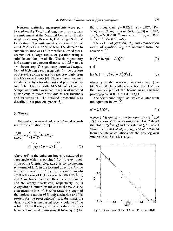
The radius of gyration, Rg, and cross-section radius of gyration, Rq, a re obtained from the equations [8]
In (I) = In I(0) - R~Q2/3 (2)
and
In (IQ) = In IQ(O) - R~Q2/2 , (3)
where I is the scattered intensity and Q = (47r/A) sin 0, the scattering vector. Fig. 1 shows the Guinier plot of the bovine nasal cartilage proteoglycan in 0.15 N LiC1-D20.
The persistence length, a*, was calculated from the equation below [8],
3. Theory
The molecular weight, M, was obtained accord- ing to the equation [6, 7]
L 1(__.__0)__ = f ( ~_S__~ )4 7r MNatc t .c(O)
x (Zb-psV) , (1)
where I(0) is the coherent intensity scattered at zero angle which is obtained from the extrapol- ation of the Guinier plot, line(0) is the incohenent scattering of H20 in the forward direction, f i s the correction factor for the anisotropy in the incoh- erent scattering of H20 for wavelength 4.75 A, T s and T are transmission coefficients of the sample and the empty quartz cell, respectively, N, is Avogadro's number, t is the cell thickness, c is the concentration in g/ml, b is the scattering length of the molecule (about 93% polysaccharide and 7% protein for the proteoglycan), Ps is the scattering density and V is the partial specific volume of the solute. The following parameter values were de- termined and used in assessing M from eq. (1) for
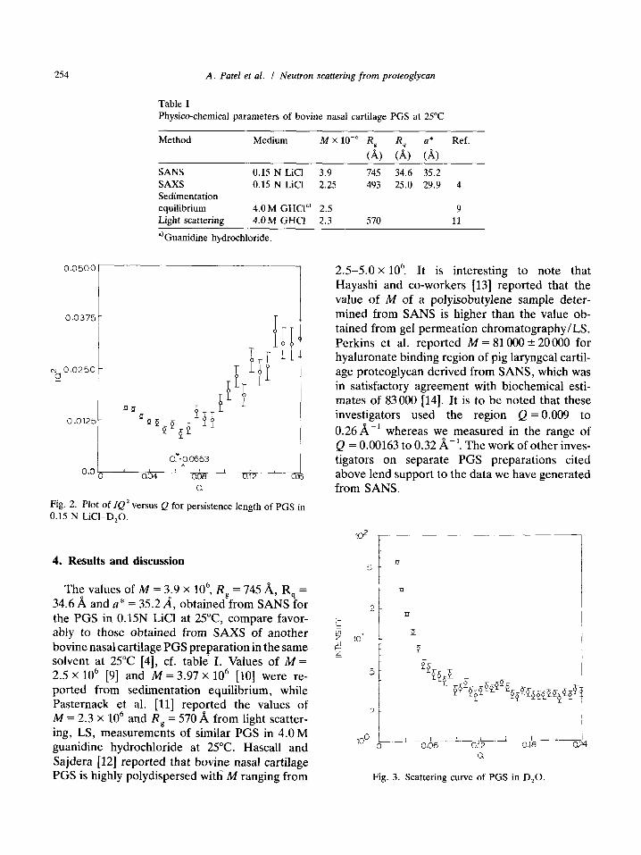
a* = 2 .3 /Q*, (4)
where Q* is the transition between the I /Q 2 and I /Q portions of the scattering curve. Fig. 2 shows the plot of lQ 2 vs. Q and the value of Q*. Table I shows the values of M, Rg, Rq, and a* obtained from the above equations for the proteoglycan subunit in 0.15 N LiCI-DzO.
29
0.%
- 1 . 0
!_
ii 0
- " O - - 3.2
Fig. 1. Guinier plot of the PGS in 0.15 N LiCI-D20.
254 A. Patel et al. / Neutron scattering f rom proteoglycan
Table I Physico-chemical parameters of bovine nasal cartilage PGS at 25°C
Method Medium M X 10 -6 Rg Rq a* Ref.
(A) (A) (A)
SANS 0.15 N LiCI 3.9 745 34.6 35.2 SAXS 0.15 N LiCI 2.25 493 25.0 29.9 Sedimentation equilibrium 4.0 M GHCP ~ 2.5 Light scattering 4.0 M GHCI 2.3 570
~)Guanidine hydrochloride.
4
9 11
0 .0500
0.037
t~ 0 .0250 0
0.0125
C:o.o653
o.o ' o .~ , I " o.&~ ' o.~2 ' o.1 Q,
Fig. 2. Plot of IQ 2 versus Q for persistence length of PGS in 0.15 N LiCI-D20.
2.5-5 .0x 106 It is interesting to note that Hayashi and co-workers [13] reported that the value of M of a polyisobutylene sample deter- mined from SANS is higher than the value ob- tained from gel permeation chromatography/LS. Perkins et al. reported M = 81 000 +-- 20 000 for hyaluronate binding region of pig laryngeal cartil- age proteoglycan derived from SANS, which was in satisfactory agreement with biochemical esti- mates of 83 000 [14]. It is to be noted that these investigators used the region Q=0.009 to 0.26,A, -1 whereas we measured in the range of Q = 0.00163 to 0.32/~-1. The work of other inves- tigators on separate PGS preparations cited above lend support to the data we have generated from SANS.
10 2
4. Results and discussion
The values of M = 3.9 x 10 6, Rg = 745/~, Rq =
34.6/~ and a* = 35.2A, obtained from SANS for the PGS in 0.15N LiCI at 25°C, compare favor- ably to those obtained from SAXS of another bovine nasal cartilage PGS preparation in the same solvent at 25°C [4], cf. table I. Values of M = 2.5 x 10 6 [9] and M = 3.97 x 10 6 [10] were re- ported from sedimentation equilibrium, while Pasternack et al. [11] reported the values of M = 2.3 x 10 6 a n d Rg = 570 ~ from light scatter- ing, LS, measurements of similar PGS in 4.0 M guanidine hydrochloride at 25°C. Hascall and Sajdera [12] reported that bovine nasal cartilage PGS is highly polydispersed witli M ranging from
101
5
5
100 ~____ 0
Zl
E
b-r
i ~ L i__ I ~--J 1 0,06 0.12 0.18 0.24
O,
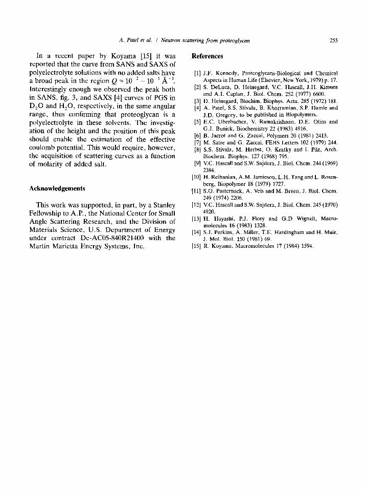
Fig. 3. Scattering curve of PGS in D20.
A. Patel et al. / Neutron scattering from proteoglycan 255
In a recent paper by K o y a m a [15] it was repor ted that the curve f rom S A N S and S A X S of polyelectrolyte solutions with no added salts have a b road peak in the region Q = 10 -2 - 10 -1 ~ -1 .
Interest ingly enough we observed the peak both in SANS, fig. 3, and S A X S [4] curves of PGS in D 2 0 and H 2 0 , respectively, in the same angular range, thus confirming that p ro teoglycan is a polyelectrolyte in these solvents. The investig- at ion of the height and the posi t ion of this peak should enable the es t imat ion of the effective cou lomb potential . This would require, however , the acquisition of scattering curves as a funct ion of molar i ty of added salt.
Acknowledgements
This work was suppor ted , in part , by a Stanley Fellowship to A.P. , the Nat ional Cente r for Small Angle Scattering Research , and the Division of Materials Science, U.S. D e p a r t m e n t of Ene rgy under contract De-AC05-840R21400 with the Mart in Marie t ta Ene rgy Systems, Inc.
References
[1] J.F. Kennedy, Proteoglycans-Biological and Chemical Aspects in Human Life (Elsevier, New York, 1979) p. 17.
[2] S. DeLuca, D. Heinegard, V.C. Hascall, J.H. Kimura and A.I. Caplan, J. Biol. Chem. 252 (1977) 6600.
[3] D. Heinegard, Biochim. Biophys. Acta. 285 (1972) 181. [4] A. Patel, S.S. Stivala, B. Khorramian, S.P. Damle and
J.D. Gregory, to be published in Biopolymers. [5] E.C. Uberbacher, V. Ramakrishnan, D.E. Olins and
G.J. Bunick, Biochemistry 22 (1983) 4916. [6] B. Jacrot and G. Zaccai, Polymers 20 (1981) 2413. [7] M. Satre and G. Zaccai, FEBS Letters 102 (1979) 244. [8] S.S. Stivala, M. Herbst, O. Kratky and I. Pilz, Arch.
Biocbem. Biophys. 127 (1968) 795. [9] V.C. Hascall and S.W. Sajdera, J. Biol. Chem. 244 (1969)
2384. [10] H. Reihanian, A.M. Jamieson, L.H. Tang and L. Rosen-
berg, Biopolymer 18 (1979) 1727. [11] S.G. Pasternack, A. Veis and M. Breen, J. Biol. Chem.
249 (1974) 2206. [12] 'v:C. Hascall and S.W. Sajdera, J. Biol. Chem. 245 (1970)
4920. [13] H. Hayashi, P.J. Flory and G.D Wignall, Macro-
molecules 16 (1983) 1328. [14] S.J. Perkins, A. Miller, T.E. Hardingham and H. Muir,
J. Mol. Biol. 150 (1981) 69. [15] R. Koyama, Macromolecules 17 (1984) 1594.