Embed Size (px)
Citation preview

Clays and Clay Minerals, Vol. 33, No. 3, 237-243, 1985.
SMALL-ANGLE X-RAY POWDER DIFFRACTION, MORPHOLOGY, AND STRUCTURE OF ALLOPHANE AND IMOGOLITE
S. J. VAN DER GAAST
Netherlands Institute for Sea Research, P.O. Box 59, Texel, The Netherlands
K. WADA, S.-I. WADA, AND Y. KAKUTO
Faculty of Agriculture, Kyushu University 46, Fukuoka 812, Japan
Abstract--Small-angle X-ray powder diffraction analyses and high-resolution electron microscopy of allophane samples (SiOJAlzO3 ratio, 1.12 to 1.68) showed that allophanes consist of nearly identical spherical particles with diameters of about 40 A and retain their characteristic "hollow" spherical mor- phology at different ambient moisture and even after dehydroxylation by heating at 500* to 6000C. Unheated allophane samples gave another X-ray powder diffraction band whose maximum position varied from 12.3 to 14.5 A depending on their SIO2/A1203 ratio. The appearance of this band may denote some long-range ordering in the structure of allophane. Unlike the spherical particles of allophane, the tube unit of imogolite collapsed on dehydroxylation. This observation suggests that imogolite and allo- phane are different in their framework structures and that a Si- or Si(A1)-tetrahedral sheet rather than an Al-octahedral sheet constitutes the framework structure ofallophane, irrespective of its SiO~/A12Oa ratio.
500 ~;WU 600~ ~ , ~ / I ~ OH / ~ G ~ ' ~ X * o , ~ � 9 " ~ " ~ r 1 6 2 ~ - ~ L ~ ? C o } J ~ , D C g ] o ~ o ? ~ Y ~ " Y - ~ - 2 ~ d ~ O ) ~ J < z f Y . ~ 12.3 ;b~ 1 4 . 5 h ~ L � 9
Key Words--Allophane, Dehydroxylation, Imogolite, Infrared spectroscopy, Morphology, Small angle scattering.
I N T R O D U C T I O N
Allophanes are present in many soils derived from volcanic ash and weathered pumices. High-resolution electron microscopy has shown that allophanes consist of "hollow" spherical particles or polyhedra with ex- ternal diameters of 55 A (Kitagawa, 1971) or 35 to 50
(Henmi and Wada, 1976). In the electron micro- scope, aUophane particles are exposed to high vacuum and electron bombardment; whether these conditions affect the morphology has not been confirmed. If al- lophane consists of nearly identical spherical particles as indicated by the electron microscopy, however, they should give a small-angle X-ray powder diffraction (XRD) band characteristic of such systems (Guinier, 1963). In the first part of the present paper, the small- angle X-ray powder diffraction (XRD) analyses of four allophane samples with different SIO2/A1203 molar ra- tios are reported. The analyses were carried out on unheated and heated samples at 200 ~ to 600~ and equilibrated at 0 and 100% relative humidi ty (r.h.) using the method developed by van der Gaast and Vaars (1981 ). The analyses provided information about the size and shape of allophane particles and their in- ternal structure.
Copyright �9 1985, The Clay Minerals Society
In the second part of the paper similar analyses are reported for a single imogolite sample. Imogolite is a unique paracrystalline clay mineral that consists of bundles of a long tube unit with an external diameter of about 20 /~ (Wada et al., 1970). It occurs with al- lophane in many soils derived from volcanic ash and weathered pumices. Cradwick et al. (1972) proposed a structure for the tube unit that consists of a single continuous gibbsite sheet with its inner-hydroxyl sur- face replaced by O3SiOH groups. This proposed struc- ture accounts for most features of the electron diffrac- tion patterns, the morphology, and the chemical composition of imogolite. There remain, however, problems regarding the interpretation of some of the XRD bands in relation to the size and arrangement of the tube at different ambient humidities and temper- atures.
Finally an argument exists about whether the wall of the allophane particles consists of a defect kaolin layer (Wada and Wada, 1977) or a defect imogolite layer (Parfitt et al., 1980; Parfitt and Henmi, 1980). The small-angle XRD analyses of heated allophane and imogolite samples brought fresh light to bear on this question.
237
238 van der Gaast, Wada, Wada, and Kakuto Clays and Clay Minerals
I i i I i i I i ~ I i i I i
Ki-P (20"C) /
(1% r.h.
% .
I i i I i i i , , i i i I I 15 12 9 6 3
I i i I i i I i i i i i i
VA(20*C) ~ '/
0% r .h .
15 12 9 6 3 ~ (CoKO~)
Figure 1. X-ray powder diffraction patterns of allophanes (Ki-P and VA) equilibrated at 0 and 100% relative humidity, respectively.
MATERIALS AND METHODS
Allophane samples were separated from three weath- ered pumices collected at Kitakami, Iwate (Ki-P); Ku- rayoshi, Tottori (Ku-P); and Choyo, Kumamoto (PA- P); and from one weathered volcanic ash collected at Choyo, Kumamoto (VA), Japan. One imogolite sample (Ki-G) was collected from gel films present in the Ki- takami pumice bed. All samples were treated with H202 to remove organic matter and dispersed at pH 4 for the Ki-P, Ku-P, and Ki-G samples, and at pH 10 for the PA-P and VA samples. Clay-size fractions (<0.2 izm) were collected by centrifugation, followed by floc- culation with NaC1, water washing, acetone washing, and air drying. Electron microscopy showed that the collected clay samples consisted of nearly pure allo- phane or imogolite. The SIO2/A1203 molar ratio of the samples Ki-P, Ku-P, PA-P, and VA allophane samples were 1.12, 1.38, 1.67, and 1.68, respectively.
For the XRD analyses, the samples suspended in water were suctioned through polished, porous ceramic plates to produce fiat aggregates and dried at room
temperature. The XRD analyses were carried out using a wide angle PW1050/25 goniometer (Philips) in com- bination with a broad focus Co tube (CoKa), a graphite monochromator, and a vacuum-hel ium device. The instrument used earlier by van der Gaast and Vaars (1981) was improved with a self-developed, computer- aided divergence slit system that enabled constant sam- ple-area i r r ad ia t ion i n d e p e n d e n t of the angle of incidence and accurate goniometer alignment. The go- niometer radius was increased from 173 m m to 214 ram. To avoid parasitic scattering at small angles, a 0.1-mm receiving slit and a l/:-antiscatter slit were used. The relative humidi ty in the sample chamber was kept constant at the desired values with a flow of moist or dry helium from a self-developed humidi ty generator. The humidi ty monitoring part of this device consisted ofa Humicap HMI 12 with a H MP 14 probe that controlled the humidity within the accuracy of 1%. The XRD patterns were recorded in stepscan mode with a counting time of 4 sec/0.02~ for CoKa and stored on floppy disc. The recorded XRD patterns were corrected with the Lorentz po la r i za t ion factor (MacEwan et al., 1961) and for the irradiated volume. A Ludox silica sample (grade, SM; particle size 70-80 /k) was used as reference material for the XRD anal- yses.
RESULTS A N D DISCUSSION
Allophane
As illustrated in Figure 1 for the Ki-P and VA sam- ples, the unheated allophane samples equilibrated at 0% r.h. gave a fairly well defined XRD band with max- imum at 2.95 to 3.10~ (CoKa) which was overlapped possibly by another broad band with max imum at 7.0 to 9.0~ (Table 1). At 100% r.h., a remarkable decrease in diffracted X-ray intensity occurred particularly for the first band showing a broadening toward the second band (Figure 1).
The relative enhancement of the first band at 0% r.h. suggests that it arose from the interference between the neighboring allophane particles. The latter interference increased with the removal of intervening water mol- ecules. The magnitude of the distance between the
Table 1. The positions of the first and second X-ray powder diffraction bands from allophanes equilibrated at 0 and 100% relative humidity.
Fi r s t b a n d ~ (*20; C o K a ) S e c o n d b a n d a (*20; C o K a )
R e l a t i v e h u m i d i t y (%) R e l a t i v e h u m i d i t y (%)
SampLe 2 SiO2/AI20~ ra t io 100 0 100 0
Ki-P 1.12 3.1 (33) 3.0 (34) 8.3 (12.4) 9.0 (11.4) Ku-P 1.38 3.1 (33) 2.95 (30) 8.0 (12.8) 8.5 (12.1) Pa-P 1.67 3.3 (31) 3.05 (33) 7.4 (13.9) 7.5 (13.7) VA 1.68 3.45 (30) 3.1 (33) 7.1 (14.4) 7.0 (14.7)
Figures in parentheses are d-spacings calculated using the Bragg formula (/~). 2 See text for locations.
Vol. 33, No. 3, 1985 Small-angle X-ray diffraction
! I ~ I I
I I I I I
6 3
~ (CoKr
Figure 2. X-ray powder diffraction patterns of Ludox silica at 0 and 100% relative humidity, respectively.
neighboring particles that corresponds to their d iam- eter d m w a s est imated using the theory of small-angle X-ray scattering for dense systems of identical spher- ical particles (Guinier, 1963). As for monatomic liq- uids, the following relation exists:
2 sin O/X = 1.1 to 1.2/dm, (1)
where 0 is the Bragg glancing angle and X the wavelength of the X-rays. The est imated d m value of 37.5 to 41 /~ is in agreement with the external d iameter of the al- lophane spherical particles, i.e., 35 to 50 A (Henmi and Wada, 1976). The 20, and hence the dm, values did not differ between the samples with different SIO2/ A1203 ratios (Table 1). This relationship was also ob- served by electron microscopy (Henmi and Wada, 1976). At 100% r.h. the first band max imum slightly shifted toward the high-angle side for the PA and VA samples with higher SiO2/AlzO3 ratios (Table 1). This shift probably resulted from the overlapping with the broad second band and does not indicate a variat ion in the size and morphology ofa l lophane particles with the ambient moisture coupled with a variat ion o f its chemical composi t ion. The decrease of diffracted X R D intensity suggests an intervening effect of water present between the allophane particles that caused irregular distances compared with the more regular packing of the particles at 0% r.h. A similar observat ion was made for a reference Ludox silica sample. I t gave a fairly well defined X R D band with max imum at 1.3~ (CoKa)
of allophane and imogolite 239
a b
K i - P K i - P
0 % r .h . 1 0 0 % r , h
6 0 W C
4 0 0 !1 3 0 0
_900"
15 12 9 6 3 15 12 9 6 3
~28 ( C o K e )
c
V A
0 % r .h .
5 0 0
400"
16 12 9 6 3
d
V A
1 0 0 % r . h .
500 '
4 0 0 ~
15 12 9 6 3
"20 ( C o K e )
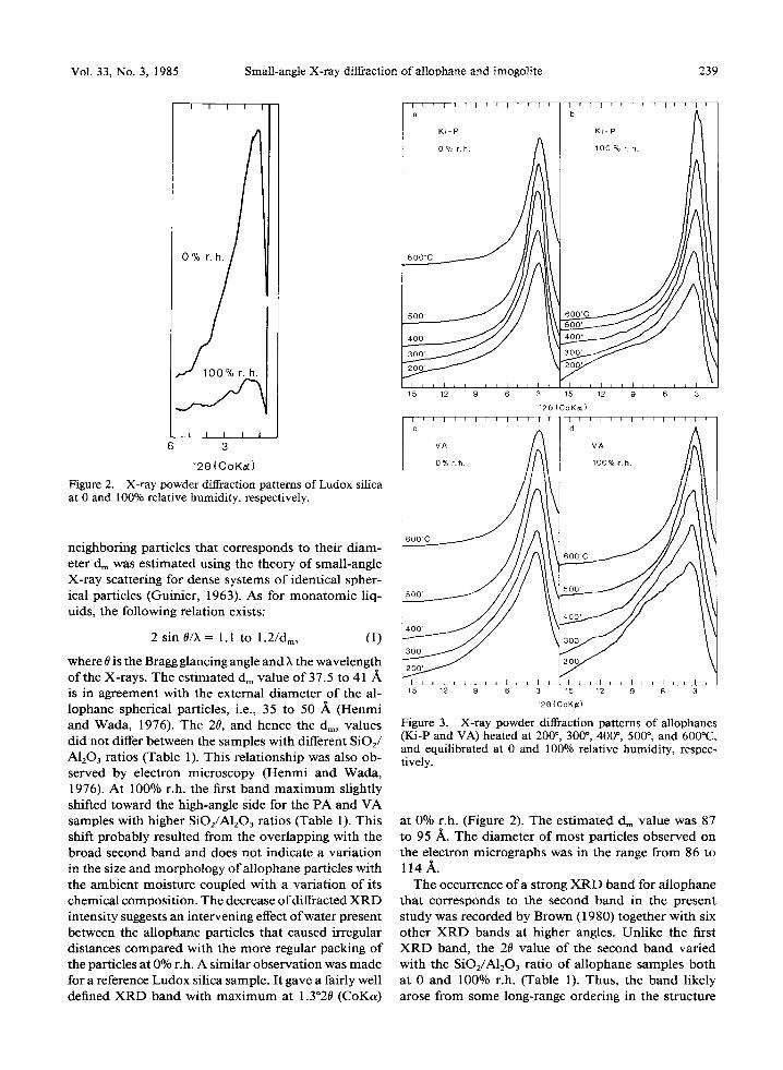
Figure 3. X-ray powder diffraction patterns of allophanes (Ki-P and VA) heated at 200 ~ 300 ~ 400 ~ 500 ~ and 600~ and equilibrated at 0 and 100% relative humidity, respec- tively.
at 0% r.h. (Figure 2). The est imated dm value was 87 to 95 Zk. The diameter of most particles observed on the electron micrographs was in the range from 86 to l 1 4 A .
The occurrence o f a strong X R D band for al lophane that corresponds to the second band in the present study was recorded by Brown (1980) together with six other X R D bands at higher angles. Unlike the first X R D band, the 20 value of the second band var ied with the SIO2/A1203 ratio of al lophane samples both at 0 and 100% r.h. (Table 1). Thus, the band likely arose from some long-range ordering in the structure
240 van der Gaast, Wada, Wada, and Kakuto Clays and Clay Minerals
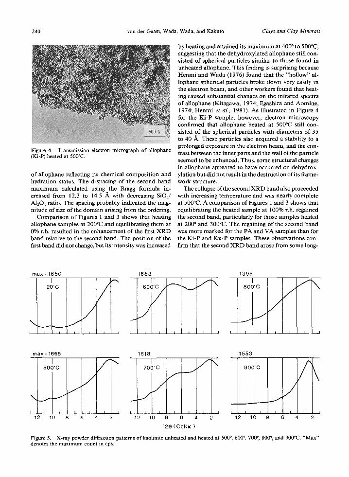
Figure 4. Transmission electron micrograph of allophane (Ki-P) heated at 500~
of allophane reflecting its chemical composition and hydration status. The d-spacing of the second band maximum calculated using the Bragg formula in- creased from 12.3 to 14.5 /~ with decreasing SiO2/ A1203 ratio. The spacing probably indicated the mag- nitude of size of the domain arising from the ordering.
Comparison of Figures 1 and 3 shows that heating allophane samples at 200~ and equilibrating them at 0% r.h. resulted in the enhancement of the first XRD band relative to the second band. The position of the first band did not change, but its intensity was increased
by heating and attained its max imum at 400 ~ to 500~ suggesting that the dehydroxylated allophane still con- sisted of spherical particles similar to those found in unheated allophane. This finding is surprising because Henmi and Wada (1976) found that the "hollow" al- lophane spherical particles broke down very easily in the electron beam, and other workers found that heat- ing caused substantial changes on the infrared spectra of allophane (Kitagawa, 1974; Egashira and Aomine, 1974; Henmi et al., 1981). As illustrated in Figure 4 for the Ki-P sample, however, electron microscopy confirmed that allophane heated at 500~ still con- sisted of the spherical particles with diameters of 35 to 40 A. These particles also acquired a stability to a prolonged exposure in the electron beam, and the con- trast between the inner parts and the wall of the particle seemed to be enhanced. Thus, some structural changes in allophane appeared to have occurred on dehydrox- ylation but did not result in the destruction of its frame- work structure.
The collapse of the second XRD band also proceeded with increasing temperature and was nearly complete at 500~ A comparison of Figures 1 and 3 shows that equilibrating the heated sample at 100% r.h. regained the second band, particularly for those samples heated at 200 ~ and 300~ The regaining of the second band was more marked for the PA and VA samples than for the Ki-P and Ku-P samples. These observations con- firm that the second XRD band arose from some long-
max = 1650 1683
I I I I I I I I l
1395
max = 1665 I
500~
I
2 10 8
1618
f \ I 700oC
. . . , . .
I I I I I i I I
6 4 2 12 10 8 6 2
f \ J
~
553
I 900~
12 C
0 8 6 4 I I
2
Figure 5. X-ray powder diffraction pattems of kaolinite unheated and heated at 500 ~ 600 ~ 700*, 800*, and 900~ "'Max'" denotes the maximum count in cps.

Vol. 33, No. 3, 1985 Small-angle X-ray diffraction of allophane and imogolite 241
:::, A
o
15 12 9 6 3
K i - G
100% r .h .
5o0 ~
15 12 9 6 3
h = 3
# # 3 0 0 ~ ~
~ooLqa>.---~
~20 (C O K N,.)
Figure 6. X-ray powder diffraction patterns ofimogolite (Ki- G) unheated and heated at 300", 400 ~ and 500~ and equil- ibrated at 0 and 100% relative humidity, respectively.
0 . 1 5
0 , 1 0 -
[dh ]-1
0 . 0 5 -
0 0 1 2 3
h
Figure 7. Plot of [dh]-1 VS. h for imogolite (Ki-G). See text for explanation.
Vainshtein (1966) showed that several (<7 to 10) cylindrical units aligned to form bundles of varying size gave "non-Bragg X R D maxima" and that their positions were governed solely by the separation be- tween the adjacent units, a. The following relationship exists:
range ordering in the structure of allophane that de- pended on the SiO2/A1203 ratio. Thus, water molecules must have had some role in developing the order as structural water.
Also, an XRD band similar to the second band of allophane was obtained for a kaolinite sample dehy- droxylated by heating. Figure 5 shows that the band with a maximum at 14.6 ~, (7.0~ CoKa) appeared on heating kaolinite at 600~ The appearance of this XRD band demonstrates the persistence of some long- range order in metakaolin as postulated by Brindley and Nakahira (1959), and its disappearance at higher temperatures possibly denotes the start of the trans- formation of metakaolin to A1-Si spinel.
Imogoli te
Figure 6 shows the XRD patterns of the unheated Ki-G sample at 0 and 100% r.h. Three nearly sym- metrical XRD bands with maxima at 2.9, 7.2, and 12.4~ (CoKa) are present; the band at 2.9~ (d = 30 /~) has not been reported earlier. The 7.2~ (d = 12.3 ~) and 12.4~ (d = 7.1 ~) bands were indexed as 020 and 030 reflections from the imogolite structure with a b-spacing of 23 ~k by Cradwick et al. (1972). The d-values of the three XRD bands, however, formed an irrational series. At 0% r.h., an additional, relatively sharp maximum developed at 5.5~ (d = 18.6 A), overlapping the second band. This feature was noted for natural (Wada and Yoshinaga, 1969) and synthetic (Farmer and Fraser, 1979) imogolite heated at 100~
[dh]-I = (h + O/a,
w h e r e d h is the d-spacing of the hth XRD ma x i mum and r is a small constant. A plot of [dh] ~ vS. h is shown in Figure 7. The predicted proportionality occurs for imogolite and indicates that a = 22 A , in agreement with the external diameter of the tube of about 20 observed in the electron microscope (Wada et al., 1970). The additional XRD maximum at 5.5~ (d = 18.6 A) at 0% r.h. can be explained by assuming that larger numbers of the tubes were arranged in a hexagonal close-packed arrangement, where the first X RD line with index 111 is predicted to appear at d = avrJ /2 = 19.1 ]k. A similar explanation for the additional band was given by Farmer and Fraser (1979).
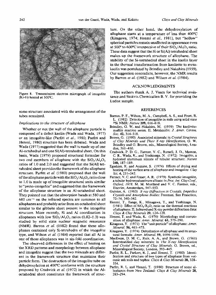
Figure 6 also shows the XRD patterns of the Ki-G samples heated at 200 ~ to 500~ Yoshinaga and Ao- mine (1962) showed by thermal analyses that the de- hydroxylation of imogolite starts at 400~ and ends at 500~ The XRD patterns shown in Figure 6 also in- dicate that the destruction of the tube started at 4000C and finished at 500~ The second and third XRD bands disappeared at 4000C, and the remaining addi- tional band with max imum at 18.6 A nearly disap- peared at 500~ Only the first band with max imum at about 40 ,~ remained at 500~ and showed a slight shift toward the low-angle side and a considerable in- crease in intensity. The electron micrograph of the Ki- G sample heated at 500~ (Figure 8) shows that most tubes broke down and changed to material showing a somewhat granular, poorly defined morphology, though
242 van der Gaast, Wada, Wada, and Kakuto Clays and Clay Minerals
ture. On the other hand, the dehydroxylation of allophane starts at a temperature of less than 400"C (Kitagawa, 1974; Henmi et al., 1981), but "hollow" spherical particles remain unaffected in appearance even at 500 ~ to 600~ irrespective of their SIO2/A1203 ratio. These data suggest that the Si or Si(A1) tetrahedral sheet makes up the framework structure of allophane. The stability of the Si-tetrahedral sheet in the kaolin layer in the thermal transformation from kaolinite to recta- kaolin was postulated by Brindley and Nakahira (1959). Our suggestion contradicts, however, the N M R results by Barron et al. (1982) and Wilson et al. (1984).
Figure 8. Transmission electron micrograph of imogolite (Ki-G) heated at 500"C.
ACKNOWLEDGMENTS
The authors thank A. J. Vaars for technical assis- tance and Stokvis Chemicalien B. V. for providing the Ludox sample.
some structure associated with the arrangement of the tubes remained.
Implications to the structure o f allophane
Whether or not the wall of the allophane particle is composed of a defect kaolin (Wada and Wada, 1977) or an imogolite-like (Parfitt et al., 1980; Parfitt and Henmi, 1980) structure has been debated. Wada and Wada (1977) suggested that the wall is made up of one Al-octahedral and one Si(A1)-tetrahedral sheet. On this basis, Wada (1979) proposed structural formulae for two end members of allophane with the SIO2/A1203 ratios of 1.0 and 2.0 and suggested that the Si(AI) tet- rahedral sheet provides the framework of the allophane structure. Parfitt et al. (1980) proposed that the wall of the allophane particle with the SiOJAI203 ratio close to 1.0 is made up of imogolite structural units similar to "proto-imogolite" and suggested that the framework of the allophane structure is an Al-octahedral sheet. They pointed out that the absorption bands at 580 and 680 cm -~ on the infrared spectra are common to all allophanes and probably arise from an octahedral sheet similar to the gibbsite sheet present in the imogolite structure. More recently, Si and AI coordination in allophanes with low SIO2/A1203 ratios (0.82-1.3) was studied by solid state nuclear magnetic resonance (NMR). Barton et al. (1982) found that these allo- phanes contained only Si-tetrahedra of the imogolite type, and Wilson et al. (1984) reported that all A1 in one of those allophanes was in six-fold coordination.
The observed differences in the effect of heating on the XRD patterns and morphology between allophane and imogolite suggest that the two minerals are differ- ent in the framework structure that maintains their particle form. The destruction of the imogolite tube on dehydroxylation at 400~ conforms with the structure proposed by Cradwick et aL (1972) in which the A1- octahedral sheet constitutes the framework of struc-
REFERENCES
Barron, P. F., Wilson, M. A., Campbell, A. S., and Frost, R. L. (1982) Detection ofimogolite in soils using solid state 29Si NMR: Nature 299, 616-618.
Brindley, G. W. and Nakahira, M. (1959) The kaolinite- mullite reaction series. II. Metakaolin: J. Amer. Ceram. Soc. 42, 314-318.
Brown, G. (1980) Associated minerals: in CrystalStructures of Clay Minerals and Their X-ray Identification, G. W. Brindley and G. Brown, eds., Mineralogical Society, Lon- don, 361-410.
Cradwick, P. D. G., Farmer, V. C., Russell, J. D., Masson, C. R., Wada, K., and Yoshinaga, N. (1972) Imogolite, a hydrated aluminum silicate of tubular structure: Nature 240, 187-189.
Egashira, K. and Aomine, S. (1974) Effects of drying and heating of the surface area ofallophane and imogolite: Clay Sci. 4, 231-242.
Farmer, V. C. and Fraser, A. R. (1979) Synthetic imogolite, a tubular hydroxyaluminum silicate: in Proc. Int. Clay Conf., Oxford, 1978, M. M. Mortland and V. C. Farmer, eds., Elsevier, Amsterdam, 547-553.
Guinier, A. (1963) X-ray Diffraction in Crystals, Imperfect Crystals and Amorphous Bodies: Freeman, San Francisco, 72-74, 340-342.
Henmi, T., Tange, K., Minagawa, T., and Yoshinaga, N. (1981) Effect of SiOz/R203 ratio on the thermal reactions ofallophane. II. Infrared and X-ray powder diffraction data: Clays & Clay Minerals 29, 124-128.
Henmi, T. and Wada, K. (1976) Morphology and compo- sition of allophane: Amer. Mineral. 61, 379-390.
Kitagawa, Y. (1971) The "unit particle" ofallophane: Amer. Mineral. $6, 465-475.
Kitagawa, Y. (1974) Dehydration ofallophane and its struc- tural formula: Amer. Mineral. 59, 1094-1098.
MacEwan, D. M. C., Ruiz, A. A., and Brown, G. (1961) Interstratified clay minerals: in The X-ray Identification and Crystal Structure of Clay Minerals, G. Brown, ed., Mineralogical Society, London, 393-445.
Parfitt, R. L., Furkert, R. J., and Henmi, T. (1980) Identi- fication and structure of two types of allophane from vol- canic ash soils and tephra: Clays & Clay Minerals 28, 328- 334.
Parfitt, R. L. and Henmi, T. (1980) Structure of some al- lophanes from New Zealand: Clays & Clay Minerals 28, 285-294.

Vol. 33, No. 3, 1985 Small-angle X-ray diffraction ofallophane and imogolite 243
Vainshtein, B. K. (1966) Diffraction of X-ray by Chain Mol- ecules: Elsevier, Amsterdam, 328-334.
van der Gaast, S. J. and Vaars, A. J. (1981) A method to eliminate the background in X-ray diffraction patterns of oriented clay mineral samples: Clay Miner. 16, 383-393.
Wada, K. (1979) Structural formulas ofallophanes: in Proc. Int. Clay Conf., Oxford, 1978, M. M. Mortland and V. C. Farmer, eds., Elsevier, Amsterdam, 537-545.
Wada, K. and Yoshinaga, N. (1969) The structure of"im- ogolite": Amer. Mineral 54, 50-71.
Wada, K., Yoshinaga, N., Yotsumoto, H., Ibe, K., and Aida, S. (1970) High resolution electron micrographs of imo- golite: Clay Miner. 8, 487-489.
Wada, S. and Wada, K. (1977) Density and structure of allophane: Clay Miner. 12, 289-298.
Wilson, M. A., Barron, P. F., and Campbell, A. S. (1984) Detection of aluminum coordination in soils and clay frac- tions using 27A1 magic angle spinning n.m.r.: J. Soil Sci. 35, 201-207.
Yoshinaga, N. and Aomine, S. (1962) Imogolite in some Ando soils: Soil ScL Plant Nutr. 8, 22-29.
(Received 4 August 1984; accepted 7 November 1984; Ms. 1389)