Embed Size (px)
Citation preview
RESEARCH Open Access
Small interfering RNA mediated Poly (ADP-ribose)Polymerase-1 inhibition upregulates the heatshock response in a murine fibroblast cell lineRajesh K Aneja1*, Hanna Sjodin2, Julia V Gefter3, Basilia Zingarelli4, Russell L Delude3
Abstract
Poly (ADP-ribose) polymerase-1 (PARP-1) is a highly conserved multifunctional enzyme, and its catalytic activity isstimulated by DNA breaks. The activation of PARP-1 and subsequent depletion of nicotinamide adeninedinucleotide (NAD+) and adenosine triphosphate (ATP) contributes to significant cytotoxicity in inflammation ofvarious etiologies. On the contrary, induction of heat shock response and production of heat shock protein 70(HSP-70) is a cytoprotective defense mechanism in inflammation. Recent data suggests that PARP-1 modulates theexpression of a number of cellular proteins at the transcriptional level. In this study, small interfering RNA (siRNA)mediated PARP-1 knockdown in murine wild-type fibroblasts augmented heat shock response as compared tountreated cells (as evaluated by quantitative analysis of HSP-70 mRNA and HSP-70 protein expression). Theseevents were associated with increased DNA binding of the heat shock factor-1 (HSF-1), the major transcriptionfactor of the heat shock response. Co-immunoprecipitation experiments in nuclear extracts of the wild type cellsdemonstrated that PARP-1directly interacted with HSF-1. These data demonstrate that, in wild type fibroblasts,PARP-1 plays a pivotal role in modulating the heat shock response both through direct interaction with HSF-1 andpoly (ADP-ribosylation).
IntroductionPoly (ADP-ribose) polymerase-1 (PARP-1) is a highlyconserved chromatin bound enzyme [1,2] and plays animportant role in DNA repair, gene transcription, cell-cycle progression, cell death, and maintenance of geno-mic integrity [3-5]. PARP-1 is activated by DNA breaksand cleaves nicotinamide adenine dinucleotide (NAD+)into nicotinamide resulting in ADP-ribose moieties;these moieties covalently attach to various acceptor pro-teins including PARP itself. The continued activation ofPARP leads to depletion of its substrate NAD+ withconsequent depletion of ATP, energy failure and celldeath [6].In addition to its influence on chromatin structure
and stability, recent studies indicate PARP-1 plays a rolein gene-specific transcription [7-9]. PARP-1 regulatestranscription by modifying chromatin-associated
proteins and acts as a cofactor for transcription factors,most notably NF-�B and AP-1 [10,11]. Genetic deletionof PARP-1 attenuates tissue injury after ischemia andreperfusion, streptozocin-induced diabetes, endotoxicand hemorrhagic shock, heat stroke and localized colo-nic inflammation [12-19]. The benefits conferred bypharmacological inhibitors of poly (ADP-ribosylation) indiverse experimental disease models further reiterate theimportance of PARP-1 as an important pharmacologicaltarget [20,21]Oxidative injury and ATP depletion also leads to
activation of heat shock factor (HSF)-1, a major tran-scription factor responsible for increased transcriptionof genes encoding heat shock proteins, particularlyheat shock protein-70 [22,23]. HSP-70 provides cyto-protection from a variety of inflammatory insults,including oxidative stress, viral infections and ische-mia-reperfusion injury [24,25]. Previously in an in vivomodel of myocardial ischemia/reperfusion injury, weshowed that cardioprotection conferred on PARP-1-/-
mice is associated with enhanced HSF-1 activity and
* Correspondence: [email protected] of Critical Care Medicine and Pediatrics, University ofPittsburgh School of Medicine and Children’s Hospital of Pittsburgh,Pittsburgh, PA 15213, USAFull list of author information is available at the end of the article
Aneja et al. Journal of Inflammation 2011, 8:3http://www.journal-inflammation.com/content/8/1/3
© 2011 Aneja et al; licensee BioMed Central Ltd. This is an Open Access article distributed under the terms of the Creative CommonsAttribution License (http://creativecommons.org/licenses/by/2.0), which permits unrestricted use, distribution, and reproduction inany medium, provided the original work is properly cited.
increased expression of HSP-70 as compared to wild-type mice [26].Similarly, Fossati et al. documented increased HSP-70
expression in murine PARP-1 deficient fibroblasts ascompared to wild type fibroblasts [27]. In gene knockoutcell lines, unexpected compensatory or redundantmechanisms develop in response to the missing geneand can confound experimental observations. To verifythat the upregulation of the heat shock response inPARP-1 deficient mice is not a compensatory responseto the missing PARP-1 gene, we employed post-transcriptional gene silencing technology by RNA inter-ference. Specifically, we utilized small interfering RNA(siRNA) to silence PARP-1 gene and hypothesized thatthe heat shock response is negatively modulated byPARP-1 activation in fibroblasts; therefore siRNAmediated PARP-1 inhibition would lead to augmentationof the heat shock response.
Material and methodsCell cultureMouse fibroblasts from wild-type mice were created byimmortalization by a standard 3T3 protocol [28]. Unlessnoted otherwise, all reagents were from Sigma-Aldrich(St. Louis MO). Cell monolayers were grown at 37°C in5% CO2 air in Dulbecco’s modified Eagle medium(DMEM) (Gibco Technologies, Grand Island, NY) con-taining 10% fetal bovine serum (FBS), penicillin (100 U/ml), and streptomycin (100 μg/ml). At 75-80% conflu-ence, fibroblasts were subjected to heat shock at 43°Cfor 45 min followed by recovery at 37°C up to 4 h. Ifneeded, cells were pretreated with PARP inhibitor 1, 5dihydroxyisoquinoline (DIQ, 100 μM; Sigma, St. Louis,MO) for 45 min in all experiments.
Nuclear protein extractionAll nuclear protein extraction procedures were per-formed on ice with ice-cold reagents. Cells were washedtwice with phosphate-buffered saline (PBS) and har-vested by scraping. Cells were pelleted in 1 ml of PBS at14,000 rpm for 1 min. The pellet was washed twice withPBS and resuspended in lysis buffer [10 mM Tris-HCl(pH 7.8), 10 mM KCl, 1 mM ethylene glycol tetra aceticacid (EGTA), 5 mM MgCl2, 1 mM dithiothreitol (DTT),and 0.5 mM phenylmethylsulfonyl fluoride (PMSF)].The suspension was incubated on ice for 15 min andNonidet P-40 was added followed by centrifugation at4°C at 2,000 rpm for 5 min. The supernatant was dis-carded and the cell pellet was dissolved in extractionbuffer (20 mM Tris-HCl, pH 7.8, 32 mM KCl, 0.2 mMEGTA, 5 mM MgCl2, 1 mM DTT, 0.5 mM PMSF and25% v/v glycerol) was added to the nuclear pellet andincubated on ice for 15 min. Nuclear proteins were iso-lated by centrifugation at 14,000 rpm at 4°C for 10 min.
Protein concentrations of the resultant supernatantswere determined using the Bradford assay. Nuclear pro-teins were stored at -70°C until used for electromobilitygel shift assays (EMSA).
EMSAEMSA were performed as previously described [29]. Anoligonucleotide probe corresponding to an HSF-1 con-sensus sequence (5’-GCC TCG ATT GTT CGC GAAGTT TCG-3’) was labeled with g-[32P] ATP using T4polynucleotide kinase (Promega) and purified in Bio-Spinchromatography columns (GE Healthcare, Buckingham-shire, UK). For each sample 4 μg of nuclear proteins wereincubated with Bandshift buffer (10 mM Tris, 40 mMKCl, 1 mM (ethylene diamine tetra acetic acid) EDTA,1 mM DTT, 50 ng/ml poly d(I-C), 10% glycerol) at roomtemperature with subsequent addition of the radiolabeledoligonucleotide probe for 30 min. Protein-nucleic acidcomplexes were resolved using a nondenaturing polya-crylamide gel consisting of 5% acrylamide (29:1 ratio ofacrylamide: bisacrylamide) and run in 0.25 X Tris/Borate/EDTA (TBE) (45 mM Tris, 45 mM boric acid,1 mM EDTA) for 1 h at 30 mA constant current. Gelswere transferred to Whatman 3 MM paper, dried undera vacuum at 80°C for 1 h, and used to expose to X-rayfilm at -70°C with an intensifying screen.
Real-time reverse transcriptase-PCR analysisFibroblasts were subjected to heat shock at 43°C for45 min followed by recovery at 37°C for 120 min. Cellswere harvested in 1 ml of TRI-Reagent as directed by themanufacturer (Molecular Research Center, Cincinnati,OH). Bromochloropropane was used for the extraction.The final RNA pellet was dissolved in nuclease - freewater and quantified using a GeneQuant Pro UV spectro-photometer (GE Healthcare). Extracted RNA (1 μg/reac-tion) was converted to single-stranded cDNA in a 20 μlreaction using the Reverse Transcriptase System Kit(Promega) as directed by the manufacturer. The mixturewas heated to 70°C for 10 min, maintained at 42°C for30 min, and then heated to 95°C for 5 min using a GeneAmp PCR System 9700 (Applied Biosystems, Foster City,CA). TaqMan Gene Expression Assays for HSP-70(GENBANK accession no. NM 010479), 18 S RNA(endogenous control) and real-time PCR reagents werepurchased from Applied Biosystems (Foster City, CA).Reaction mixtures for PCR were assembled as follows:10 μl TaqMan Universal PCR Master Mix, 1 μl of eachGene Expression Assay mix, 1 μl cDNA template and7 μl of water. PCR reactions were performed in anApplied Biosystems thermocycler 7300 Real Time PCRSystem by incubating at 50°C for 2 min, 95°C for 10 min,95°C for 15 s, and 60°C for 1 min; the two final condi-tions were repeated for 40 cycles. Each sample was
Aneja et al. Journal of Inflammation 2011, 8:3http://www.journal-inflammation.com/content/8/1/3
Page 2 of 9

assayed in duplicate and the values were averaged. A ΔΔCt relative quantification method was used to calculatemRNA levels for HSP-70 in the samples. Results werenormalized relative to 18 S rRNA expression.
SiRNA-mediated inhibition of PARP expressionStealth small interference RNA (siRNA) sequences forPARP (sequences) were designed using Invitrogen online software (Block-iT™RNAi Express) to target PARP-1 mRNA (accession number NM007415). Small interfer-ing RNA (siRNA)-mediated silencing of the PARP-1gene was performed using 21-bp siRNA duplexes pur-chased from Ambion (Austin, TX). The coding strandfor PARP-1 siRNA was 5’-AUG UCG GCA AAG UAGAUC CCU UUC C-3’. An unrelated siRNA sequence(catalog number 12935-113) was used as a control. Inthis experiment, cells were incubated for 6 h and trans-fected at approximately 40% confluency with 20nmsiRNA duplexes using Lipofectamine™2000 (Invitrogen,Carlsbad, CA) according to the manufacturer’s instruc-tions. All the experiments were performed 18 h aftertransfection. The efficiency and specificity of siRNAgene knockdown of PARP-1 was determined by realtime PCR for PARP-1 mRNA and Western blotting forPARP-1 expression.
Western blot analysisWestern blot analyses were performed as previouslydescribed [29]. Briefly, whole cell lysates containing30 μg of protein were boiled in equal volumes of loadingbuffer (125 mM Tris, pH 6.8, 4% sodium dodecyl sulfate(SDS), 20% glycerol, and 10% b-mercaptoethanol). Pro-teins were separated on 8-16% polyacrylamide gels andsubsequently transferred to polyvinylidene difluoride(PVDF) membranes (GE Healthcare, Buckinghamshire,UK). For immunoblotting, membranes were blockedwith 5% non-fat dried milk in PBS for 1 h. Primary anti-bodies against the inducible isoform of HSP-70 (Stress-gen, Victoria, BC, Canada) were applied at 1:2500dilution for 1 h. After washing twice with PBS contain-ing 0.5% Tween 20 (PBST), secondary antibody (horseradish peroxidase-conjugated goat anti- rabbit immuno-globulin G, Stressgen, Victoria, British Columbia) wasapplied at 1:4,000 dilution for 1 h. Blots were washed inPBST thrice for 10 min, incubated in Enhanced Chemi-luminescence Reagent (GE Healthcare), and used toexpose X-ray film (GE Healthcare).
ImmunoprecipitationNuclear extracts were incubated with normal mouse IgG-AC (20 μl Santa Cruz, sc-2343) and incubated for 30 minat 4°C. Anti-PARP antibody (10 μl Biomol, SA-250) orHSF-1 antibody (Stressgen, SPA-950) and non-specific
IgG was added to the supernatant for 1 h at 4°C. There-after, protein A/G PLUS-Agarose beads were added (20μl Santa Cruz Biotechnology, sc2003) and the sampleswere incubated overnight at 4°C. Beads were washedthree times in volume 1xPBS and resuspended in 2 XSDS- polyacrylamide gel electrophoresis (PAGE) samplebuffers and analyzed by 8% SDS-PAGE. The proteinswere then transferred onto PVDF membranes (GEHealthcare, Buckinghamshire, UK). The membraneswere blocked in 1X PBST containing 5% nonfat dry milkand incubated with an HSF-1 antibody (Stressgen, SPA-950) or Anti-PARP antibody (10 μl Biomol, SA-250). Themembranes were washed and incubated with a polyclonalrabbit anti -rat antibody conjugated to horseradish per-oxidase (Stressgen, SAB-200). Immunoreaction wasvisualized by chemiluminescence.
Data analysisAll values in the figures and text are expressed as mean± SEM. The results were examined by analysis ofvariance followed by the Bonferroni’s correction posthoc t test. A p-value less than 0.05 were consideredsignificant.
ResultsInhibition of PARP-1 expression by RNA interferenceaugments HSP-70 protein expressionTo investigate the biological consequences of PARP-1activation and its effect on the heat shock response, weemployed a siRNA based approach to selectively inhibitPARP-1 expression. As a first step, we treated fibroblastswith various siRNA concentrations (10 nm to 100 nm)and evaluated PARP-1 mRNA and protein expression 18h after transfection. The lowest concentration of PARP-1 siRNA resulting in efficient PARP-1 gene knockdown,as evidenced by a decrease in PARP-1 mRNA andprotein expression, was 20nm (Figure 1A and 1B).This concentration was employed in all subsequentexperiments.Cells were transfected with siRNA for 12 h, subjected
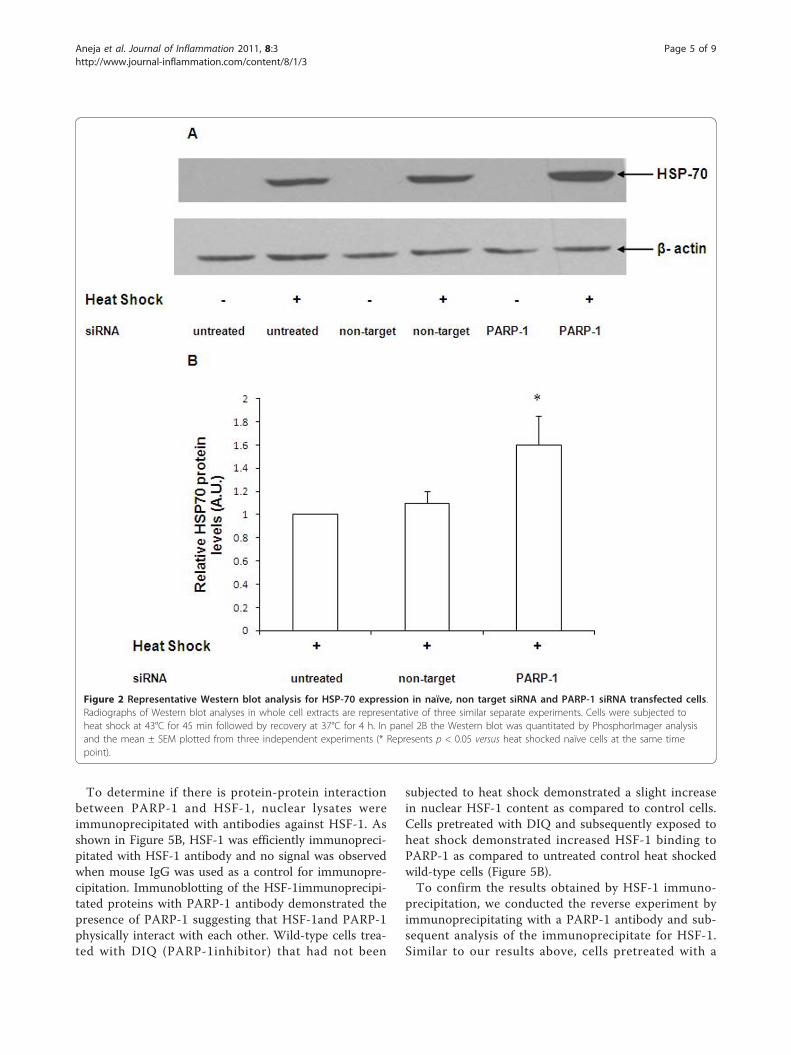
to heat shock for 45 min and allowed to recover for 4 h.The expression of HSP-70 was determined by immuno-blotting. After heat shock, naïve cells demonstrated asignificant increase in HSP-70 protein expression (Figure2A). HSP-70 protein expression in cells transfected withnon-target siRNA was comparable to naïve cells afterheat shock (Figure 2A and 2B). Using siRNA to silencePARP-1, we observed that HSP-70 protein expression inPARP-1 siRNA-transfected cells was markedly upregu-lated as compared to naïve or non-target siRNA trans-fected cells (Figure 2A and 2B). These data support theview that PARP-1 gene silencing leads to augmentationof HSP-70 protein expression after heat shock.
Aneja et al. Journal of Inflammation 2011, 8:3http://www.journal-inflammation.com/content/8/1/3
Page 3 of 9
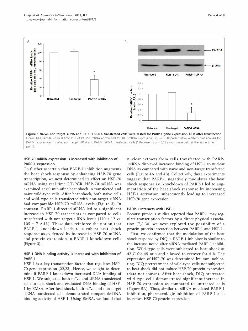
HSP-70 mRNA expression is increased with inhibition ofPARP-1 expressionTo further ascertain that PARP-1 inhibition augmentsthe heat shock response by enhancing HSP-70 genetranscription, we next determined its effect on HSP-70mRNA using real time RT-PCR. HSP-70 mRNA wasexamined at 60 min after heat shock in transfected andnaïve wild-type cells. After heat shock, both naïve cellsand wild-type cells transfected with non-target siRNAhad comparable HSP-70 mRNA levels (Figure 3). Incontrast, PARP-1 directed siRNA led to a significantincrease in HSP-70 transcripts as compared to cellstransfected with non-target siRNA levels (140 ± 12 vs.105 ± 7 A.U.). These data reinforce the notion thatPARP-1 knockdown leads to a robust heat shockresponse as evidenced by increase in HSP-70 mRNAand protein expression in PARP-1 knockdown cells(Figure 3).
HSF-1 DNA-binding activity is increased with inhibition ofPARP-1HSF-1 is a key transcription factor that regulates HSP-70 gene expression [22,23]. Hence, we sought to deter-mine if PARP-1 knockdown increased DNA binding ofHSF-1. We subjected both naïve and siRNA transfectedcells to heat shock and evaluated DNA binding of HSF-1 by EMSA. After heat shock, both naïve and non-targetsiRNA transfected cells demonstrated comparable DNAbinding activity of HSF-1. Using EMSA, we found that
nuclear extracts from cells transfected with PARP-1siRNA displayed increased binding of HSF-1 to nuclearDNA as compared with naïve and non-target transfectedcells (Figure 4A and 4B). Collectively, these experimentssuggest that PARP-1 negatively modulates the heatshock response i.e. knockdown of PARP-1 led to aug-mentation of the heat shock response by increasingHSF-1 activation, subsequently leading to increasedHSP-70 gene expression.
PARP-1 interacts with HSF-1Because previous studies reported that PARP-1 may reg-ulate transcription factors by a direct physical associa-tion [7,8,30] we next explored the possibility of aprotein-protein interaction between PARP-1 and HSF-1.First, we confirmed that the modulation of the heat
shock response by DIQ, a PARP-1 inhibitor is similar tothe increase noted after siRNA mediated PARP-1 inhibi-tion. Wild-type cells were subjected to heat shock at43°C for 45 min and allowed to recover for 4 h. Theexpression of HSP-70 was determined by immunoblot-ting. DIQ pretreatment of wild-type cells not subjectedto heat shock did not induce HSP-70 protein expression(data not shown). After heat shock, DIQ pretreatedwild-type cells demonstrated significant increase inHSP-70 expression as compared to untreated cells(Figure 5A). Thus, similar to siRNA mediated PARP-1inhibition, pharmacologic inhibition of PARP-1 alsoincreases HSP-70 protein expression.
Figure 1 Naïve, non target siRNA and PARP-1 siRNA transfected cells were tested for PARP-1 gene expression 18 h after transfection.Figure 1A-Quantitative Real time PCR of PARP-1 mRNA normalized for 18 S mRNA expression. Figure 1B-Representative Western blot analysis forPARP-1 expression in naïve, non target siRNA and PARP-1 siRNA transfected cells (* Represents p < 0.05 versus naïve cells at the same timepoint).
Aneja et al. Journal of Inflammation 2011, 8:3http://www.journal-inflammation.com/content/8/1/3
Page 4 of 9
To determine if there is protein-protein interactionbetween PARP-1 and HSF-1, nuclear lysates wereimmunoprecipitated with antibodies against HSF-1. Asshown in Figure 5B, HSF-1 was efficiently immunopreci-pitated with HSF-1 antibody and no signal was observedwhen mouse IgG was used as a control for immunopre-cipitation. Immunoblotting of the HSF-1immunoprecipi-tated proteins with PARP-1 antibody demonstrated thepresence of PARP-1 suggesting that HSF-1and PARP-1physically interact with each other. Wild-type cells trea-ted with DIQ (PARP-1inhibitor) that had not been
subjected to heat shock demonstrated a slight increasein nuclear HSF-1 content as compared to control cells.Cells pretreated with DIQ and subsequently exposed toheat shock demonstrated increased HSF-1 binding toPARP-1 as compared to untreated control heat shockedwild-type cells (Figure 5B).To confirm the results obtained by HSF-1 immuno-
precipitation, we conducted the reverse experiment byimmunoprecipitating with a PARP-1 antibody and sub-sequent analysis of the immunoprecipitate for HSF-1.Similar to our results above, cells pretreated with a
Figure 2 Representative Western blot analysis for HSP-70 expression in naïve, non target siRNA and PARP-1 siRNA transfected cells.Radiographs of Western blot analyses in whole cell extracts are representative of three similar separate experiments. Cells were subjected toheat shock at 43°C for 45 min followed by recovery at 37°C for 4 h. In panel 2B the Western blot was quantitated by PhosphorImager analysisand the mean ± SEM plotted from three independent experiments (* Represents p < 0.05 versus heat shocked naïve cells at the same timepoint).
Aneja et al. Journal of Inflammation 2011, 8:3http://www.journal-inflammation.com/content/8/1/3
Page 5 of 9
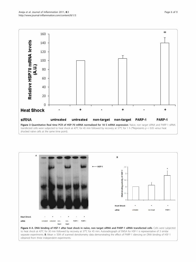
Figure 3 Quantitative Real time PCR of HSP-70 mRNA normalized for 18 S mRNA expression. Naïve, non target siRNA and PARP-1 siRNAtransfected cells were subjected to heat shock at 43°C for 45 min followed by recovery at 37°C for 1 h (*Represents p < 0.05 versus heatshocked naïve cells at the same time point).
Figure 4 A. DNA binding of HSF-1 after heat shock in naïve, non target siRNA and PARP-1 siRNA transfected cells. Cells were subjectedto heat shock at 43°C for 30 min followed by recovery at 37°C for 45 min. Autoradiograph of EMSA for HSF-1 is representative of 3 similarseparate experiments. B. Mean ± SEM of scanned densitometry data demonstrating the effect of PARP-1 silencing on DNA binding of HSF-1obtained from three independent experiments.
Aneja et al. Journal of Inflammation 2011, 8:3http://www.journal-inflammation.com/content/8/1/3
Page 6 of 9
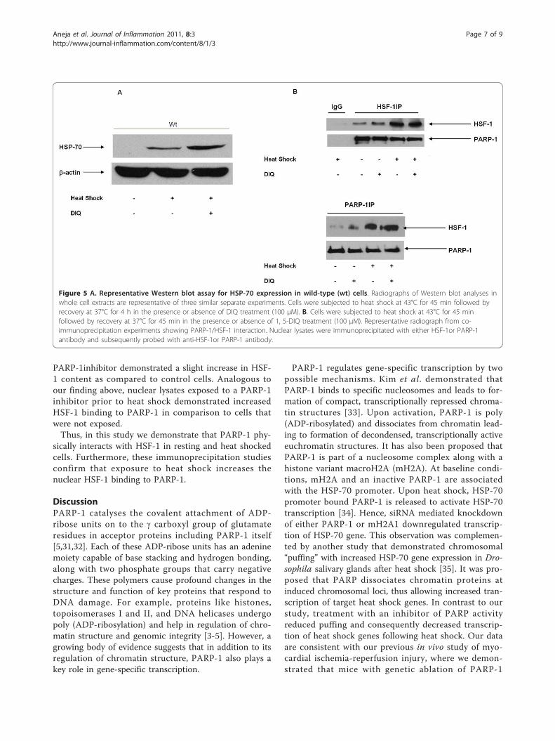
PARP-1inhibitor demonstrated a slight increase in HSF-1 content as compared to control cells. Analogous toour finding above, nuclear lysates exposed to a PARP-1inhibitor prior to heat shock demonstrated increasedHSF-1 binding to PARP-1 in comparison to cells thatwere not exposed.Thus, in this study we demonstrate that PARP-1 phy-
sically interacts with HSF-1 in resting and heat shockedcells. Furthermore, these immunoprecipitation studiesconfirm that exposure to heat shock increases thenuclear HSF-1 binding to PARP-1.
DiscussionPARP-1 catalyses the covalent attachment of ADP-ribose units on to the g carboxyl group of glutamateresidues in acceptor proteins including PARP-1 itself[5,31,32]. Each of these ADP-ribose units has an adeninemoiety capable of base stacking and hydrogen bonding,along with two phosphate groups that carry negativecharges. These polymers cause profound changes in thestructure and function of key proteins that respond toDNA damage. For example, proteins like histones,topoisomerases I and II, and DNA helicases undergopoly (ADP-ribosylation) and help in regulation of chro-matin structure and genomic integrity [3-5]. However, agrowing body of evidence suggests that in addition to itsregulation of chromatin structure, PARP-1 also plays akey role in gene-specific transcription.
PARP-1 regulates gene-specific transcription by twopossible mechanisms. Kim et al. demonstrated thatPARP-1 binds to specific nucleosomes and leads to for-mation of compact, transcriptionally repressed chroma-tin structures [33]. Upon activation, PARP-1 is poly(ADP-ribosylated) and dissociates from chromatin lead-ing to formation of decondensed, transcriptionally activeeuchromatin structures. It has also been proposed thatPARP-1 is part of a nucleosome complex along with ahistone variant macroH2A (mH2A). At baseline condi-tions, mH2A and an inactive PARP-1 are associatedwith the HSP-70 promoter. Upon heat shock, HSP-70promoter bound PARP-1 is released to activate HSP-70transcription [34]. Hence, siRNA mediated knockdownof either PARP-1 or mH2A1 downregulated transcrip-tion of HSP-70 gene. This observation was complemen-ted by another study that demonstrated chromosomal“puffing” with increased HSP-70 gene expression in Dro-sophila salivary glands after heat shock [35]. It was pro-posed that PARP dissociates chromatin proteins atinduced chromosomal loci, thus allowing increased tran-scription of target heat shock genes. In contrast to ourstudy, treatment with an inhibitor of PARP activityreduced puffing and consequently decreased transcrip-tion of heat shock genes following heat shock. Our dataare consistent with our previous in vivo study of myo-cardial ischemia-reperfusion injury, where we demon-strated that mice with genetic ablation of PARP-1
Figure 5 A. Representative Western blot assay for HSP-70 expression in wild-type (wt) cells. Radiographs of Western blot analyses inwhole cell extracts are representative of three similar separate experiments. Cells were subjected to heat shock at 43°C for 45 min followed byrecovery at 37°C for 4 h in the presence or absence of DIQ treatment (100 μM). B. Cells were subjected to heat shock at 43°C for 45 minfollowed by recovery at 37°C for 45 min in the presence or absence of 1, 5-DIQ treatment (100 μM). Representative radiograph from co-immunoprecipitation experiments showing PARP-1/HSF-1 interaction. Nuclear lysates were immunoprecipitated with either HSF-1or PARP-1antibody and subsequently probed with anti-HSF-1or PARP-1 antibody.
Aneja et al. Journal of Inflammation 2011, 8:3http://www.journal-inflammation.com/content/8/1/3
Page 7 of 9
exhibit significant cardioprotection, associated withenhanced upregulation of HSF-1 DNA binding in theheart [26]. The reasons for the different response of dip-terans and mammals to PARP-1 inhibition areunknown. It is plausible that factors including signalingmediators have evolved different effector proteins thatcan affect the heat shock response and its regulation byPARP-1.Another potential mechanism which may be more
relevant to this study entails the role of PARP as a genespecific transcription enhancer/promoter binding cofac-tor activity that can enhance or inhibit gene expression.PARP-1 has been shown to interact with the transcrip-tion factors NF-�B, HTLV Tax-1 and RAR and this wasassociated with increased expression from dependentpromoters [7-9]. Similarly, it has been suggested thatPARP-1 is a co-transcription factor for the mammalianachaete-scute homologue (MASH) gene. PARP-1 is pre-sent in an inactive state as part of a co-repressor com-plex. Upon activation, PARP-1 is required for thedismissal of the co-repressor complex, and a secondsubsequent event leads to activation of the targetMASH gene [36].Our experiments indicate that PARP-1 modulates the
heat shock response by functioning as a repressing factorof HSP-70 gene expression. We provide two lines of evi-dence in this regard. First, in this study we demonstratedthat knockdown of PARP-1 gene increased HSP-70 geneexpression as evidenced by increased DNA-binding activ-ity of HSF-1, HSP-70 mRNA and protein expression.Secondly, using co-immunoprecipitation we demon-strated that PARP-1 may also regulate HSF-1 activationthrough direct interaction with this transcription factor.Protein-protein interaction is recognized as a mechanismfor PARP-1 to function as a specific transcriptionalco-activator of NF-�B [37].Fossati et al. similarly documented increased HSP-70
expression in murine PARP-1 deficient fibroblasts ascompared to wild type fibroblasts [27]. In contrast toour findings, this study was unable to detect PARP-1and HSF-1 interaction by co-immunoprecipitation stu-dies. While the cell type utilized in the two studies wasremarkably similar, the duration of heat shock was dif-ferent. In our study, the cells were subjected to 45 minof heat shock in comparison to 30 min in the study byFossati et al. [27]. Other differences that could lead todifferent results may be the antibody type and protocoldesign for immunoprecipitation studies.Other studies have also proven that PARP-1 modulates
transcription by direct interaction with AP2 [38], Oct-1[39], YY-1 [40] and TEF-1 [41]. PARP has been alsoshown to alter RNA polymerase II dependent transcrip-tion [42] and to effectively prevent and reverse p53 bind-ing to the palindromic p53 consensus sequence [43].
Before HSF-1 is activated there are a series of processesthat involve phosphorylation, translocation from thecytosol to the nucleus, formation of a trimer, binding toheat shock elements (HSE), and initiating HSP-70 geneexpression [44-46]. It has been postulated that additionof long ADP-ribose tails to transcription factors can dis-able or dissociate the binding of transcription factors totheir DNA recognition sites, also in part by electrostaticrepulsion. This modification results in inhibition of tran-scription. Poly (ADP-ribosylation) of transcription factorsprevents their binding to DNA. On the contrary, inhibi-tion of PARP-1 enables the binding of the transcriptionfactors to their specific DNA sites [5]. Thus, it is possiblethat both physical interaction with PARP-1 and poly(ADP-ribosylation) of HSF-1 reduce the availability ofHSF-1 to initiate transcription. The increase in HSF-1content, albeit inactive with DIQ pretreatment furtherreinforces the notion that PARP-1 represses HSP-70gene transcription. Further studies need to be conductedto understand the precise mechanism as to how andwhere PARP-1 regulates HSF-1 activation.In conclusion, our results indicate that PARP-1 serves
as a repressing factor of the heat shock response by reg-ulating the expression of HSP-70. Both protein-proteininteraction and catalytic activity of the PARP proteinplay a key role in modulation of the heat shockresponse.
AcknowledgementsFunding for this study was provided by the National Institutes of Health toDr. Rajesh Aneja (grant K08GM076344), Dr. Basilia Zingarelli (grant R01 HL-60730), and Dr. Russell Delude (grant GM37631).
Author details1Departments of Critical Care Medicine and Pediatrics, University ofPittsburgh School of Medicine and Children’s Hospital of Pittsburgh,Pittsburgh, PA 15213, USA. 2Department of Critical Care Medicine, Universityof Pittsburgh School of Medicine Pittsburgh, PA 15213, USA. 3Departmentsof Critical Care Medicine and Pathology, University of Pittsburgh School ofMedicine, Pittsburgh, PA 15213, USA. 4Division of Critical Care Medicine,Cincinnati Children’s Hospital Medical Center and The University ofCincinnati College of Medicine, Cincinnati, Ohio 45229, USA.
Authors’ contributionsHS carried out the molecular studies. JG carried out the RT-PCR assays. BZ,RA conceived the study. RA, RLD participated in the design of the study. Allauthors read and approved the final manuscript.
Competing interestsThe authors declare that they have no competing interests.
Received: 20 January 2010 Accepted: 23 February 2011Published: 23 February 2011
References1. Ame JC, Spenlehauer C, de Murcia G: The PARP superfamily. Bioessays
2004, 26:882-93.2. Schreiber V, Dantzer F, Ame JC, de Murcia G: Poly(ADP-ribose): novel
functions for an old molecule. Nat Rev Mol Cell Biol 2006, 7:517-28.3. de Murcia G, Schreiber V, Molinete M, et al: Structure and function of poly
(ADP-ribose) polymerase. Mol Cell Biochem 1994, 138:15-24.
Aneja et al. Journal of Inflammation 2011, 8:3http://www.journal-inflammation.com/content/8/1/3
Page 8 of 9
4. de Murcia G, Menissier de Murcia J: Poly(ADP-ribose) polymerase: amolecular nick-sensor. Trends Biochem Sci 1994, 19:172-6.
5. D’Amours D, Desnoyers S, D’Silva I, Poirier GG: Poly(ADP-ribosyl)ationreactions in the regulation of nuclear functions. Biochem J 1999, 342(Pt2):249-68.
6. Chiarugi A: Poly(ADP-ribose) polymerase: killer or conspirator? The‘suicide hypothesis’ revisited. Trends Pharmacol Sci 2002, 23:122-9.
7. Cervellera MN, Sala A: Poly(ADP-ribose) Polymerase Is a B-MYBCoactivator. J Biol Chem 2000, 275:10692-6.
8. Anderson MG, Scoggin KE, Simbulan-Rosenthal CM, Steadman JA:Identification of poly(ADP-ribose) polymerase as a transcriptionalcoactivator of the human T-cell leukemia virus type 1 Tax protein. J Virol2000, 74:2169-77.
9. Pavri R, Lewis B, Kim TK, et al: PARP-1 determines specificity in a retinoidsignaling pathway via direct modulation of mediator. Mol Cell 2005,18:83-96.
10. Aguilar-Quesada R, Munoz-Gamez JA, Martin-Oliva D, et al: Modulation oftranscription by PARP-1: consequences in carcinogenesis andinflammation. Curr Med Chem 2007, 14:1179-87.
11. Hassa PO, Covic M, Hasan S, Imhof R, Hottiger MO: The enzymatic andDNA binding activity of PARP-1 are not required for NF-kappa Bcoactivator function. J Biol Chem 2001, 276:45588-97.
12. Zingarelli B, Szabo C, Salzman AL: Blockade of Poly(ADP-ribose)synthetase inhibits neutrophil recruitment, oxidant generation, andmucosal injury in murine colitis. Gastroenterology 1999, 116:335-45.
13. Zingarelli B, Salzman AL, Szabo C: Genetic disruption of poly (ADP-ribose)synthetase inhibits the expression of P-selectin and intercellularadhesion molecule-1 in myocardial ischemia/reperfusion injury. Circ Res1998, 83:85-94.
14. Zingarelli B, O’Connor M, Wong H, Salzman AL, Szabo C: Peroxynitrite-mediated DNA strand breakage activates poly-adenosine diphosphateribosyl synthetase and causes cellular energy depletion in macrophagesstimulated with bacterial lipopolysaccharide. J Immunol 1996, 156:350-8.
15. Eliasson MJ, Sampei K, Mandir AS, et al: Poly(ADP-ribose) polymerase genedisruption renders mice resistant to cerebral ischemia. Nat Med 1997,3:1089-95.
16. Liaudet L, Soriano FG, Szabo E, et al: Protection against hemorrhagicshock in mice genetically deficient in poly(ADP-ribose)polymerase. ProcNatl Acad Sci USA 2000, 97:10203-8.
17. Pieper AA, Brat DJ, Krug DK, et al: Poly(ADP-ribose) polymerase-deficientmice are protected from streptozotocin-induced diabetes. Proc Natl AcadSci USA 1999, 96:3059-64.
18. Virag L, Szabo C: The therapeutic potential of poly(ADP-ribose)polymerase inhibitors. Pharmacol Rev 2002, 54:375-429.
19. Mota RA, Hernandez-Espinosa D, Galbis-Martinez L, et al: Poly(ADP-ribose)polymerase-1 inhibition increases expression of heat shock proteins andattenuates heat stroke-induced liver injury. Critical care medicine 2008,36:526-34.
20. Veres B, Gallyas F Jr, Varbiro G, et al: Decrease of the inflammatoryresponse and induction of the Akt/protein kinase B pathway by poly-(ADP-ribose) polymerase 1 inhibitor in endotoxin-induced septic shock.Biochem Pharmacol 2003, 65:1373-82.
21. Jagtap P, Soriano FG, Virag L, et al: Novel phenanthridinone inhibitors ofpoly (adenosine 5’-diphosphate-ribose) synthetase: potentcytoprotective and antishock agents. Crit Care Med 2002, 30:1071-82.
22. Lindquist S, Craig EA: The heat-shock proteins. Annu Rev Genet 1988,22:631-77.
23. Goldenberg CJ, Luo Y, Fenna M, et al: Purified human factor activatesheat shock promoter in a HeLa cell-free transcription system. J Biol Chem1988, 263:19734-9.
24. Latchman DS: Heat shock proteins and cardiac protection. Cardiovasc Res2001, 51:637-46.
25. Moseley P: Stress proteins and the immune response.Immunopharmacology 2000, 48:299-302.
26. Zingarelli B, Hake PW, O’Connor M, et al: Differential regulation ofactivator protein-1 and heat shock factor-1 in myocardial ischemia andreperfusion injury: role of poly(ADP-ribose) polymerase-1. Am J PhysiolHeart Circ Physiol 2004, 286:H1408-15.
27. Fossati S, Formentini L, Wang ZQ, Moroni F, Chiarugi A: Poly(ADP-ribosyl)ation regulates heat shock factor-1 activity and the heat shock responsein murine fibroblasts. Biochem Cell Biol 2006, 84:703-12.
28. Wang ZQ, Auer B, Stingl L, et al: Mice lacking ADPRT and poly(ADP-ribosyl)ation develop normally but are susceptible to skin disease. GenesDev 1995, 9:509-20.
29. Chen PC, Wheeler DS, Malhotra V, et al: A green tea-derived polyphenol,epigallocatechin-3-gallate, inhibits IkappaB kinase activation and IL-8gene expression in respiratory epithelium. Inflammation 2002, 26:233-41.
30. Hassa PO, Hottiger MO: The functional role of poly(ADP-ribose)polymerase 1 as novel coactivator of NF-kappaB in inflammatorydisorders. Cell Mol Life Sci 2002, 59:1534-53.
31. Huletsky A, de Murcia G, Muller S, et al: The effect of poly(ADP-ribosyl)ation on native and H1-depleted chromatin. A role of poly(ADP-ribosyl)ation on core nucleosome structure. J Biol Chem 1989, 264:8878-86.
32. Ogata N, Ueda K, Kawaichi M, Hayaishi O: Poly(ADP-ribose) synthetase, amain acceptor of poly(ADP-ribose) in isolated nuclei. J Biol Chem 1981,256:4135-7.
33. Kim MY, Mauro S, Gevry N, Lis JT, Kraus WL: NAD+-dependent modulationof chromatin structure and transcription by nucleosome bindingproperties of PARP-1. Cell 2004, 119:803-14.
34. Ouararhni K, Hadj-Slimane R, Ait-Si-Ali S, et al: The histone variantmH2A1.1 interferes with transcription by down-regulating PARP-1enzymatic activity. Genes Dev 2006, 20:3324-36.
35. Tulin A, Spradling A: Chromatin loosening by poly(ADP)-ribosepolymerase (PARP) at Drosophila puff loci. Science 2003, 299:560-2.
36. Ju BG, Solum D, Song EJ, et al: Activating the PARP-1 sensor componentof the groucho/TLE1 corepressor complex mediates a CaMKinase IIdelta-dependent neurogenic gene activation pathway. Cell 2004, 119:815-29.
37. Hassa PO, Hottiger MO: A role of poly (ADP-ribose) polymerase in NF-kappaB transcriptional activation. Biol Chem 1999, 380:953-9.
38. Kannan P, Yu Y, Wankhade S, Tainsky MA: PolyADP-ribose polymerase is acoactivator for AP-2-mediated transcriptional activation. Nucleic Acids Res1999, 27:866-74.
39. Nie J, Sakamoto S, Song D, et al: Interaction of Oct-1 andautomodification domain of poly(ADP-ribose) synthetase. FEBS Lett 1998,424:27-32.
40. Oei SL, Griesenbeck J, Schweiger M, et al: Interaction of the transcriptionfactor YY1 with human poly(ADP-ribosyl) transferase. Biochem Biophys ResCommun 1997, 240:108-11.
41. Butler AJ, Ordahl CP: Poly(ADP-ribose) polymerase binds withtranscription enhancer factor 1 to MCAT1 elements to regulate muscle-specific transcription. Mol Cell Biol 1999, 19:296-306.
42. Oei SL, Griesenbeck J, Ziegler M, Schweiger M: A novel function of poly(ADP-ribosyl)ation: silencing of RNA polymerase II-dependenttranscription. Biochemistry 1998, 37:1465-9.
43. Valenzuela MT, Guerrero R, Nunez MI, et al: PARP-1 modifies theeffectiveness of p53-mediated DNA damage response. Oncogene 2002,21:1108-16.
44. Mosser DD, Kotzbauer PT, Sarge KD, Morimoto RI: In vitro activation ofheat shock transcription factor DNA-binding by calcium andbiochemical conditions that affect protein conformation. Proc Natl AcadSci USA 1990, 87:3748-52.
45. Sorger PK: Heat shock factor and the heat shock response. Cell 1991,65:363-6.
46. Van Why SK, Mann AS, Thulin G, et al: Activation of heat-shocktranscription factor by graded reductions in renal ATP, in vivo, in therat. J Clin Invest 1994, 94:1518-23.
doi:10.1186/1476-9255-8-3Cite this article as: Aneja et al.: Small interfering RNA mediated Poly(ADP-ribose) Polymerase-1 inhibition upregulates the heat shockresponse in a murine fibroblast cell line. Journal of Inflammation 20118:3.
Aneja et al. Journal of Inflammation 2011, 8:3http://www.journal-inflammation.com/content/8/1/3
Page 9 of 9
![Untersuchungen zum Wirkmechanismus von 6-Amino-11,12 ... · PARP Poly [ADP-ribose] polymerase PBGD Porphobilinogen deaminase PBS Phosphate buffered saline PCR Polymerase chain reaction](https://img.pdfslide.net/doc/110x75/5d5cbcc088c9939b368b7c27/untersuchungen-zum-wirkmechanismus-von-6-amino-1112-parp-poly-adp-ribose.jpg)