Embed Size (px)
Citation preview
University of Nebraska - LincolnDigitalCommons@University of Nebraska - LincolnFaculty Publications from the Department ofElectrical and Computer Engineering Electrical & Computer Engineering, Department of
2011
Small Molecule Inhibitors of Staphylococcus aureusRnpA Alter Cellular mRNA Turnover, ExhibitAntimicrobial Activity, and Attenuate PathogenesisPatrick D. OlsonUniversity of Nebraska Medical Center
Lisa J. KuechenmeisterUniversity of Nebraska Medical Center
Kelsi L. AndersonUniversity of Nebraska Medical Center
Sonja DailyUniversity of Arkansas for Medical Sciences
Karen E. BeenkenUniversity of Arkansas for Medical Sciences
See next page for additional authors
Follow this and additional works at: http://digitalcommons.unl.edu/electricalengineeringfacpub
Part of the Computer Engineering Commons, and the Electrical and Computer EngineeringCommons
This Article is brought to you for free and open access by the Electrical & Computer Engineering, Department of at DigitalCommons@University ofNebraska - Lincoln. It has been accepted for inclusion in Faculty Publications from the Department of Electrical and Computer Engineering by anauthorized administrator of DigitalCommons@University of Nebraska - Lincoln.
Olson, Patrick D.; Kuechenmeister, Lisa J.; Anderson, Kelsi L.; Daily, Sonja; Beenken, Karen E.; Roux, Christelle M.; Reniere,Michelle L.; Lewis, Tami L.; Weiss, William J,; Pulse, Mark; Nguyen, Phung; Simecka, Jerry W.; Morrison, John M.; Sayood, Khalid;Asojo, Oluwatoyin A.; Smeltzer, Mark S.; Skaar, Eric P.; and Dunman, Paul M., "Small Molecule Inhibitors of Staphylococcus aureusRnpA Alter Cellular mRNA Turnover, Exhibit Antimicrobial Activity, and Attenuate Pathogenesis" (2011). Faculty Publications fromthe Department of Electrical and Computer Engineering. 249.http://digitalcommons.unl.edu/electricalengineeringfacpub/249
AuthorsPatrick D. Olson; Lisa J. Kuechenmeister; Kelsi L. Anderson; Sonja Daily; Karen E. Beenken; Christelle M.Roux; Michelle L. Reniere; Tami L. Lewis; William J, Weiss; Mark Pulse; Phung Nguyen; Jerry W. Simecka;John M. Morrison; Khalid Sayood; Oluwatoyin A. Asojo; Mark S. Smeltzer; Eric P. Skaar; and Paul M.Dunman
This article is available at DigitalCommons@University of Nebraska - Lincoln: http://digitalcommons.unl.edu/electricalengineeringfacpub/249

Small Molecule Inhibitors of Staphylococcus aureusRnpA Alter Cellular mRNA Turnover, ExhibitAntimicrobial Activity, and Attenuate PathogenesisPatrick D. Olson1, Lisa J. Kuechenmeister1, Kelsi L. Anderson1, Sonja Daily2, Karen E. Beenken2,
Christelle M. Roux1, Michelle L. Reniere3, Tami L. Lewis1, William J. Weiss4, Mark Pulse4, Phung Nguyen4,
Jerry W. Simecka4, John M. Morrison1, Khalid Sayood5, Oluwatoyin A. Asojo1, Mark S. Smeltzer2, Eric P.
Skaar3, Paul M. Dunman1,6*
1 Department of Pathology and Microbiology, University of Nebraska Medical Center, Omaha, Nebraska, United States of America, 2 Department of Microbiology and
Immunology, University of Arkansas for Medical Sciences, Little Rock, Arkansas, United States of America, 3 Department of Microbiology and Immunology, Vanderbilt
University Medical Center, Nashville, Tennessee, United States of America, 4 Department of Molecular Biology and Immunology, University of North Texas Health Science
Center, Fort Worth, Texas, United States of America, 5 Department of Electrical Engineering, University of Nebraska, Lincoln, Nebraska, United States of America,
6 Department of Microbiology and Immunology, University of Rochester, Rochester, New York, United States of America
Abstract
Methicillin-resistant Staphylococcus aureus is estimated to cause more U.S. deaths annually than HIV/AIDS. The emergenceof hypervirulent and multidrug-resistant strains has further amplified public health concern and accentuated the need fornew classes of antibiotics. RNA degradation is a required cellular process that could be exploited for novel antimicrobialdrug development. However, such discovery efforts have been hindered because components of the Gram-positive RNAturnover machinery are incompletely defined. In the current study we found that the essential S. aureus protein, RnpA,catalyzes rRNA and mRNA digestion in vitro. Exploiting this activity, high through-put and secondary screening assaysidentified a small molecule inhibitor of RnpA-mediated in vitro RNA degradation. This agent was shown to limit cellularmRNA degradation and exhibited antimicrobial activity against predominant methicillin-resistant S. aureus (MRSA) lineagescirculating throughout the U.S., vancomycin intermediate susceptible S. aureus (VISA), vancomycin resistant S. aureus (VRSA)and other Gram-positive bacterial pathogens with high RnpA amino acid conservation. We also found that this RnpA-inhibitor ameliorates disease in a systemic mouse infection model and has antimicrobial activity against biofilm-associatedS. aureus. Taken together, these findings indicate that RnpA, either alone, as a component of the RNase P holoenzyme, and/or as a member of a more elaborate complex, may play a role in S. aureus RNA degradation and provide proof of principlefor RNA catabolism-based antimicrobial therapy.
Citation: Olson PD, Kuechenmeister LJ, Anderson KL, Daily S, Beenken KE, et al. (2011) Small Molecule Inhibitors of Staphylococcus aureus RnpA Alter CellularmRNA Turnover, Exhibit Antimicrobial Activity, and Attenuate Pathogenesis. PLoS Pathog 7(2): e1001287. doi:10.1371/journal.ppat.1001287
Editor: Ambrose Cheung, Dartmouth Medical School, United States of America
Received March 5, 2010; Accepted January 10, 2011; Published February 10, 2011
This is an open-access article distributed under the terms of the Creative Commons Public Domain declaration which stipulates that, once placed in the publicdomain, this work may be freely reproduced, distributed, transmitted, modified, built upon, or otherwise used by anyone for any lawful purpose.
Funding: S. aureus isolates NRS1, NRS3, VRS1 and VRS10 were obtained through the Network of Antimicrobial Resistance in Staphylococcus aureus (NARSA)program supported under NIAID/NIH Contract # HHSM272200700055C. This research was supported by NIH/NIAID award 1R01 AI073780-01, American HeartAssociation (AHA) Scientist Development Grant award 0535037N, and Nebraska Research Initiative award (P.M.D). K.L.A. was supported by AHA pre-doctoralfellowship award 0715547Z. Work in the EPS lab was funded by United States Public Health Service Grant AI69233 from the National Institute of Allergy andInfectious Diseases. M.L.R. was funded by NIH Training Grant in Mechanisms of Vascular Disease, 5 T32 HL07751. Work in the M.S.S. lab was funded by UnitedStates Public Health Service Grant AI43356 and AI074087 from the National Institute of Allergy and Infectious Disease. The funders had no role in study design,data collection and analysis, decision to publish, or preparation of the manuscript.
Competing Interests: The authors have declared that no competing interests exist.
* E-mail: [email protected]
Introduction
Staphylococcus aureus infections are often associated with high rates
of morbidity and mortality [1]. Indeed, reports estimate that in
2005 the organism caused more U.S. deaths than HIV/AIDS
[2,3]. The emergence of vancomycin-resistant and hypervirulent
strains has further accentuated the need for novel anti-staphylo-
coccal agents [4,5]. Bacterial RNA processing and degradation are
required cellular process that could be exploited for antibiotic drug
discovery.
Much of our understanding of bacterial RNA degradation
comes from studies of Escherichia coli where bulk mRNA decay is
thought to be catalyzed by a holoenzyme complex (RNA
degradosome), which consists of at least four subunits: RNase E
(rne), RNA helicase (rhlB), enolase (eno), and PNPase (pnpA) [6].
RNase E is an essential ribonuclease and a key component of the
degradosome complex; it serves as a scaffold for the assembly of
other members of the RNA degradosome and catalyzes the initial
endoribonucleolytic event during substrate degradation [7,8].
Based on its essentiality, RNase E could be considered an
appropriate target for antibiotic drug discovery. However, many
Gram-positive bacteria, including S. aureus, lack a RNase E
ortholog [9]. As a consequence, their degradation components and
mechanism(s) of mRNA decay are less understood.
Recent studies suggest that at least two ribonucleases, RNase J1
and RNase Y, contribute to bulk mRNA degradation within
PLoS Pathogens | www.plospathogens.org 1 February 2011 | Volume 7 | Issue 2 | e1001287
Bacillus subtilis, and presumably other Gram-positive bacteria. B.
subtilis ribonuclease J1 is a bifunctional ribonuclease, with 59
exonuclease and endonuclease activities, that mediates mRNA
degradation in vitro [10,11]. The enzyme has also been found to
interact with enolase (component of the E. coli RNA degradosome)
and RNase J1 depleted B. subtilis strains demonstrate a moderate
decrease in mRNA decay, suggesting that it may be the functional
equivalent to E. coli RNase E [10,12,13]. However, mRNA
turnover still occurs in RNase J1 diminished cells and RNA species
containing 59 strong-hairpin structures are not effectively degraded
by the enzyme, indicating that additional factors are likely to
contribute to B. subtilis cellular RNA degradation [14]. Ribonu-
clease Y is a recently identified endonuclease that can cleave
mRNA molecules containing high-order secondary structures,
globally affects cellular messenger RNA turnover and may
ostensibly work in concert with RNase J1 to mediate bulk RNA
decay [15]. Consistent with that possibility, recent two-hybridiza-
tion studies revealed that RNase J1 and RNase Y are likely to
interact with one another and with other proteins that are
presumably members of the B. subtilis degradosome, including 6-
phospho-fructokinase (Pfk), Enolase, PNPase, and the RNA
helicase CshA [12,16]. Both RNase J1 and RNase Y are essential
enzymes and, in that regard, could be considered targets for
antimicrobial drug discovery [17]. However, it remains to be seen
whether RNase J1, RNase Y, and/or previously uncharacterized
ribonucleases modulate mRNA decay within S. aureus.
In the current body of work we set out to empirically identify S.
aureus RNA degradation factors, with the expectation that they
would represent promising antimicrobial drug development
targets. To do so, we exploited the fact that S. aureus owes its
ability to cause infection, in part, to the temporal expression of an
expansive repertoire of virulence factors, many of which are
regulated in a cell density-dependent manner during laboratory
culture conditions [18]. This, combined with recent reports
indicating that bacterial pathogens, including S. aureus, govern
gene expression by modulating the mRNA turnover of target
transcripts [19,20,21] led to the prediction that growth phase
regulated changes in S. aureus virulence factor expression occur at
the level of mRNA degradation and that the proteins involved in
this process may include members of the organism’s RNA
degradation machinery. Accordingly, Affymetrix GeneChips were
used to compare the mRNA decay rates of well-characterized S.
aureus virulence factors during exponential- and stationary- phase
growth.
Results revealed that the mRNA turnover properties of many S.
aureus virulence factor transcripts differed between the two growth
phases. Furthermore, and of direct relevance to the current work,
the global mRNA decay properties of exponential and stationary
phase cells were found to be dramatically different; 884 S. aureus
mRNA species were stabilized during stationary phase growth.
Among the genes whose expression correlated with mRNA decay
was the protein component of ribonuclease P (RNase P), RnpA,
suggesting that it may play a role in bulk mRNA turnover.
Consistent with that possibility, we show that recombinant S. aureus
RnpA exhibits ribonuclease activity in vitro and RnpA depleted
cells exhibit reduced mRNA degradation, indicating that RnpA-
alone, RNase P, or RnpA in complex with other cellular factors
contributes to S. aureus mRNA degradation. Because RnpA is an
essential S. aureus enzyme with low amino acid conservation with
mammalian proteins, we hypothesized that it may be an
appropriate target for antimicrobial drug discovery. Accordingly,
high through-put and secondary screening assays were used to
identify small molecule inhibitors of RnpA-mediated in vitro RNA
degradation. One of these agents was shown to inhibit S. aureus
cellular mRNA turnover, exhibited antimicrobial activity against
MRSA, VISA and VRSA, as well as other Gram-positive
pathogens with high RnpA conservation, and limited pathogenesis
in a murine acute lethal model of infection. Collectively these
results suggest that RnpA alone, or as a member of a more
elaborate complex, may contribute to the mRNA degradation
properties of S. aureus and validate its utility as an antimicrobial
drug discovery target.
Results
Many S. aureus virulence factors are expressed in a cell density
dependent manner during growth in laboratory culture conditions.
In general, cell surface virulence determinants are predominantly
expressed during exponential phase growth, whereas secreted
virulence factors are primarily expressed during stationary phase
growth [18]. Recent studies indicate that regulated changes in S.
aureus mRNA turnover, in part, effect the expression of the
organism’s virulence determinants [22]. Accordingly, we predicted
that alterations between the mRNA turnover properties of
exponential and stationary phase cells may differ in a manner
that correlate with changes in virulence factor expression.
Growth-phase dependent alterations in S. aureus mRNAturnover
Affymetrix GeneChips were used to compare the mRNA
turnover properties of exponential- and stationary- phase S. aureus
cells. To do so, S. aureus strain UAMS-1 was cultured to either
mid-exponential or stationary phase growth. De novo transcript
synthesis was arrested by the addition of rifampin and aliquots
were removed at various post-transcriptional arrest time points.
The RNA from these samples was labeled, applied to Affymetrix
GeneChips, and the half-life of each mRNA species was
determined, as previously described [19,20]. As predicted, results
revealed that the mRNA turnover properties of many (41%)
virulence factor transcripts differed between the two growth
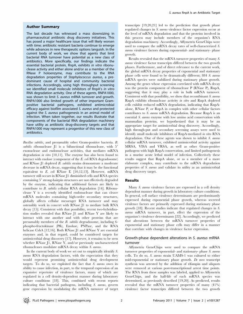
Author Summary
The last decade has witnessed a mass downsizing inpharmaceutical antibiotic drug discovery initiatives. Thishas posed a major healthcare issue that will likely worsenwith time; antibiotic resistant bacteria continue to emergewhile advances in new therapeutic options languish. In thecurrent body of work, we show that agents that limitbacterial RNA turnover have potential as a new class ofantibiotics. More specifically, our findings indicate theessential bacterial protein, RnpA, exhibits in vitro ribonu-clease activity and either alone and/or as a member of theRNase P holoenzyme, may contribute to the RNAdegradation properties of Staphylococcus aureus, a pre-dominant cause of hospital and community bacterialinfections. Accordingly, using high throughput screeningwe identified small molecule inhibitors of RnpA’s in vitroRNA degradation activity. One of these agents, RNPA1000,was shown to limit S. aureus mRNA turnover and growth.RNPA1000 also limited growth of other important Gram-positive bacterial pathogens, exhibited antimicrobialefficacy against biofilm associated S. aureus and protectedagainst the S. aureus pathogenesis in an animal model ofinfection. When taken together, our results illustrate thatcomponents of the bacterial RNA degradation machineryhave utility as antibiotic drug-discovery targets and thatRNPA1000 may represent a progenitor of this new class ofantibiotics.
S. aureus RnpA Is an Antibiotic Target
PLoS Pathogens | www.plospathogens.org 2 February 2011 | Volume 7 | Issue 2 | e1001287

phases, suggesting that regulated changes in mRNA turnover may
affect their expression (Supplementary Table S1.). Moreover, it
was observed that the organism produced at least five stationary
phase specific small stable RNAs (SSRs), a hypothesized class of
regulatory non-coding RNA molecules (Supplementary Table S2.;
[19,20]). Further, the global mRNA turnover properties of
exponential- and stationary-phase cells differed considerably.
Consistent with previous measurements, it was found that most
(90%) exponential phase transcripts are rapidly degraded (half life
of #5 min), 9% exhibit intermediate stability (half life of .5 min
but #30 min), and 1% are stable (half life of $30 min) [19,20].
However, during stationary phase growth, 76%, 21%, and 3% of
mRNA species exhibit short, intermediate, and stable half lives,
respectively (Figure 1). We anticipated that the observed stationary
phase-dependent stabilization of many mRNA species would
reflect diminished expression of a member(s) of the S. aureus RNA
decay machinery; neither RNase J1 or RNase Y were found to be
differentially expressed in a growth phase dependent manner.
Among the 367 genes repressed during stationary phase growth
was rnpA, which codes for the protein component of ribonuclease P
(RNase P; Supplementary Table S1).
S. aureus RnpA exhibits ribonuclease activityRNase P is an ubiquitous enzyme that catalyzes maturation of
the 59 end of precursor tRNAs [23,24,25]. The enzyme is unique
by virtue of the fact that it is a ribonucleoprotein complex, which
includes a single ribozyme RNA molecule and at least one protein
component. Within bacteria both the ribozyme (rnpB) and protein
(RnpA) components are required for cell viability; rnpB mediates
tRNA processing in vitro, whereas RnpA facilitates rnpB/substrate
binding at physiologically relevant magnesium concentrations
[26,27,28,29]. In addition to catalyzing tRNA maturation, E. coli
and B. subtilis RNase P have been found to digest certain double-
stranded RNA templates, such as guide-RNAs and 4.5s RNA [30].
Cleavage of those templates strictly requires RnpA [31,32], raising
the possibility that RNase P mediated RNA digestion may be
dependent on rnpB, RnpA, or both. Domain searches (http://
smart.embl.de/) [33,34] revealed that S. aureus RnpA residues 40–
111 best conform to a ribonuclease-like motif (data not shown) and
several RNA binding sites are embedded within this region [35].
Taking these observations into consideration, we predicted that
RnpA, either alone or, more likely, as a component of RNase P
holoenzyme or a more elaborate complex, affects S. aureus RNA
degradation.
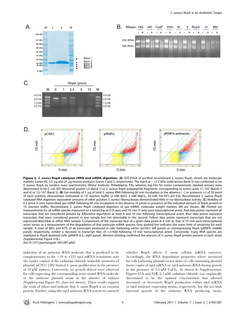
Consistent with those possibilities, recombinant S. aureus RnpA
was found to catalyze digestion of rRNA and staphylococcal
protein A (spa) mRNA (Figures 2B and 2C), as well as three other
mRNA species tested (data not shown). Other putative S. aureus
ribonucleases including RNase III, RNase HII, RNase HIII,
RNase Y, RNase J1, and BN did not exhibit equivalent RNA
degradation activity during these assay conditions (Figure 2B).
SDS-PAGE and matrix-assisted laser desorption/ionization
(MALDI) analysis confirmed that the observed ribonuclease
activity was associated with the presence of S. aureus RnpA
(Figure 2A). None the less, SDS-PAGE assessment of approxi-
mately 1000-fold excess (25 mg) of RnpA purification product used
in the aforementioned ribonuclease assays revealed trace amounts
of four additional polypeptides within the protein preparation,
raising the possibility that contaminating E. coli ribonucleases may
be present with our RnpA product. MALDI analysis revealed the
identity of these proteins to be E. coli ribosomal protein L3, and
three S. aureus RnpA fragments, presumably reflecting proteolytic
degradation of full length RnpA during protein preparation as
opposed to mature alternative translation products. Importantly,
no E. coli ribonucleases were detected, suggesting that the protein
preparation’s ribonucleolytic activity could be attributed to S.
aureus RnpA. Moreover, reverse transcriptase mediated PCR
revealed that E. coli rnpB was undetectable within our preparation,
establishing that RnpA ribonuclease activity was not due to the
formation of chimeric RNase P molecules consisting of S. aureus
RnpA and E. coli rnpB RNA. Indeed, in vitro synthesized E. coli rnpB
neither catalyzed S. aureus RNA degradation (alone) nor affected
the activity of RnpA-mediated RNA digestion during both
standard and elevated Mg+2 reaction conditions (data not shown).
S. aureus RnpA is an essential enzyme that affects cellularmRNA degradation
Small molecule inhibitors of essential bacterial RNA turnover
proteins are expected to interfere with bacterial growth and
represent a new class of antimicrobial agents. In that regard, S.
aureus RnpA is a reported essential enzyme [36,37] and thus could
be considered a target for chemotherapeutic development. Indeed,
Figure 1. S. aureus growth phase mRNA turnover measurements. Plotted are the percent of detectible mRNA species (Y-axis) with a half life of#2.5, 5, 15, 30, or .30 min during exponential- and/or stationary- phase growth (X-axis).doi:10.1371/journal.ppat.1001287.g001
S. aureus RnpA Is an Antibiotic Target
PLoS Pathogens | www.plospathogens.org 3 February 2011 | Volume 7 | Issue 2 | e1001287
induction of an antisense RNA molecule that is predicted to be
complementary to the 234 to +353 rnpA mRNA translation start
site (under control of the cadmium chloride inducible promoter of
plasmid, pCN51 [38]) limited S. aureus proliferation in the presence
of 10 mM inducer. Conversely, no growth defects were observed
for cells expressing the corresponding sense strand RNA molecule
or the antisense plasmid strain in the absence of inducer
(Supplemental Figure S1; data not shown). These results support
the work of others and indicate that S. aureus RnpA is an essential
protein. Further, using this rnpA antisense RNA system we assessed
whether RnpA affects S. aureus cellular mRNA turnover.
Accordingly, the RNA degradation properties where measured
for cells harboring plasmid vector alone or cells containing plasmid
borne copies of rnpA mRNA or rnpA antisense RNA during growth
in the presence of 2.5 mM CaCl2. As shown in Supplementary
Figures S1A and S1B, 2.5 mM cadmium chloride was empirically
determined to be the optimal concentration that allowed
increased- or decreased- RnpA production within rnpA mRNA
or rnpA antisense expressing strains, respectively, but did not limit
bacterial growth of the antisense RNA producing strain.
Figure 2. S. aureus RnpA catalyzes rRNA and mRNA digestion. (A) SDS-PAGE of purified recombinant S. aureus RnpA; shown are molecularmarkers (Lanes M), 2.5 mg and 25 mg elution products (Lanes 1 and 2, respectively). The band at ,17.2 kDa (solid arrow; Band 2) was confirmed to beS. aureus RnpA by tandem mass spectrometry (Wistar Institute; Philadelphia, PA), whereas top-hits for minor contaminants (dashed arrows) weredetermined to be E. coli 50S ribosomal protein L3 (Band 1) or S. aureus RnpA polypeptide fragments, corresponding to amino acids 11–107 (Bands 3and 4) or 12–107 (Band 5). (B) Gel-mobility of 1 mg of total S. aureus RNA following 60 min incubation in the absence (2) or presence (+) of 50 pmolof each putative ribonuclease (indicated) in 1X reaction buffer (2 mM NaCl, 2 mM MgCl2, 50 mM Tris-HCl, pH 6.0). Recombinant S. aureus RnpAcatalyzed RNA digestion; equivalent amounts of other putative S. aureus ribonucleases demonstrated little or no ribonuclease activity. (C) Mobility of0.5 pmol in vitro transcribed spa mRNA following 60 min incubation in the absence (0 pmol) or presence of the indicated amount of RnpA protein in1X reaction buffer. Recombinant S. aureus RnpA catalyzed digestion of spa mRNA; molecular weight markers (M) are shown. (D) Plotted aremeasurements for all mRNA species measured on a GeneChip at 0 (X-axis) and 10 min (Y-axis) post-transcriptional arrest. Red data points represent alltranscripts that are considered present by Affymetrix algorithms at both 0 and 10 min following transcriptional arrest. Blue data points representtranscripts that were considered present in one sample but not detectable in the second. Yellow data points represent transcripts that are notexpressed/detectible in either RNA sample. Comparisons of the transcript titer of a given data point at 0 min to that of 10 min post-transcriptionalarrest serves as a measurement of the degradation of that particular mRNA species. Grey dashed line indicates the lower limit of sensitivity for eachsample. A total of 88% and 87% of all transcripts produced in cells harboring vector (pCN51; left panel) or overexpressing RnpA (pRNPA; middlepanel), respectively, exhibit a decrease in transcript titer of $2-fold following 10 min transcriptional arrest. Conversely, many RNA species arestabilized in RnpA depleted cells (pRNPA-A.S.; right panel). Western blotting confirmed the amount of S. aureus RnpA protein present in each strain(Supplemental Figure S1B.).doi:10.1371/journal.ppat.1001287.g002
S. aureus RnpA Is an Antibiotic Target
PLoS Pathogens | www.plospathogens.org 4 February 2011 | Volume 7 | Issue 2 | e1001287

Accordingly, RNA turnover analyses revealed that diminished
RnpA levels correlated with the stabilization of many mRNA
species, suggesting that the enzyme either alone, as a member of
the RNase P holoenzyme, and/or as another RnpA-complex
contributes to bulk cellular RNA degradation (Figure 2D). More
specifically, it was found that 88% and 87% of all exponential
phase transcripts produced in RnpA overexpressing and vector
containing cells exhibited a half life of less than 10 min,
respectively. Conversely, 63% of transcripts produced in RnpA
depleted cells exhibited a half life of less than 10 min, suggesting
that the protein contributes to S. aureus mRNA turnover
(Figure 2D). The finding that RnpA overexpression did not
accelerate cellular RNA degradation suggests that either the
protein did not reach a concentration that effectively increases
RNA turnover or that the protein’s RNA degradation activity is
dependent on co-factors, which remain at wild type levels under
these experimental conditions.
Identification of small molecule inhibitors of RnpA-mediated RNA degradation
The above results indicate that S. aureus RnpA is an essential
enzyme that exhibits in vitro ribonuclease activity and either alone,
as a component of RNase P or in concert with other cellular
components participates in bulk RNA degradation. Moreover, the
protein is well conserved across Gram-positive bacteria but lacks
amino acid conservation with mammalian proteins, making it an
attractive target for novel antibiotic drug development. Accord-
ingly, we set out to exploit the protein’s in vitro ribonuclease activity
as a means to identify RnpA inhibitory agents; a fluorescence-
based high through-put assay was used to screen 29,066
commercial compounds (ActiProbe-25K and Natural product
libraries; Timtec; Newark, DE) for small molecule inhibitors of
RnpA-mediated in vitro RNA degradation (Figure 3A). In total,
fourteen molecules inhibited the enzyme’s RNA turnover activity
by $50%. A gel-based secondary assay confirmed that five of
these molecules were bona-fide inhibitors of RnpA-mediated RNA
degradation (Figure 3B). One of these compounds, RNPA1000
(Figure 3C; IC50 = 100–125 mM), did not affect the activity of the
commercially available E. coli RNase HI, RNase A, RNase I or in-
house purified S. aureus RNase J1 at any concentration tested (0–
750 mM), but did mildly inhibit E. coli RNase III activity (IC50 =
500–750 mM; data not shown). These and other data (see below)
suggest that RNPA1000 may have specificity for S. aureus RnpA,
yet as with any small molecule we cannot rule out the possibility
that the agent may also affect other S. aureus enzymes. To assess
whether RnpA-inhibitory agents exhibit potential as antimicrobi-
Figure 3. Identification of small molecule inhibitors of RnpA-mediated RNA degradation. (A) Representative screening effort results; darkblue arrow indicates substrate alone (negative control); grey arrow indicates enzyme (positive control); light-blue arrows indicate compounds thatinhibited RnpA activity by $50%. (B) An agarose gel-based assay was used to distinguish bona-fide RnpA inhibitors from primary screening artifacts.Shown is the gel mobility of molecular weight marker, spa mRNA in the absence (2) or presence (+) of 20 pmol RnpA and RnpA-mediated spa mRNAdegradation in the presence of increasing concentrations of RNPA1000, as described in Materials and Methods. (C) Structure of RnpA-inhibitorymolecule RNPA1000.doi:10.1371/journal.ppat.1001287.g003
S. aureus RnpA Is an Antibiotic Target
PLoS Pathogens | www.plospathogens.org 5 February 2011 | Volume 7 | Issue 2 | e1001287

als, a series of experiments were performed to evaluate whether
RNPA1000 inhibited S. aureus growth and could limit S. aureus
pathogenesis in a systemic model of infection.
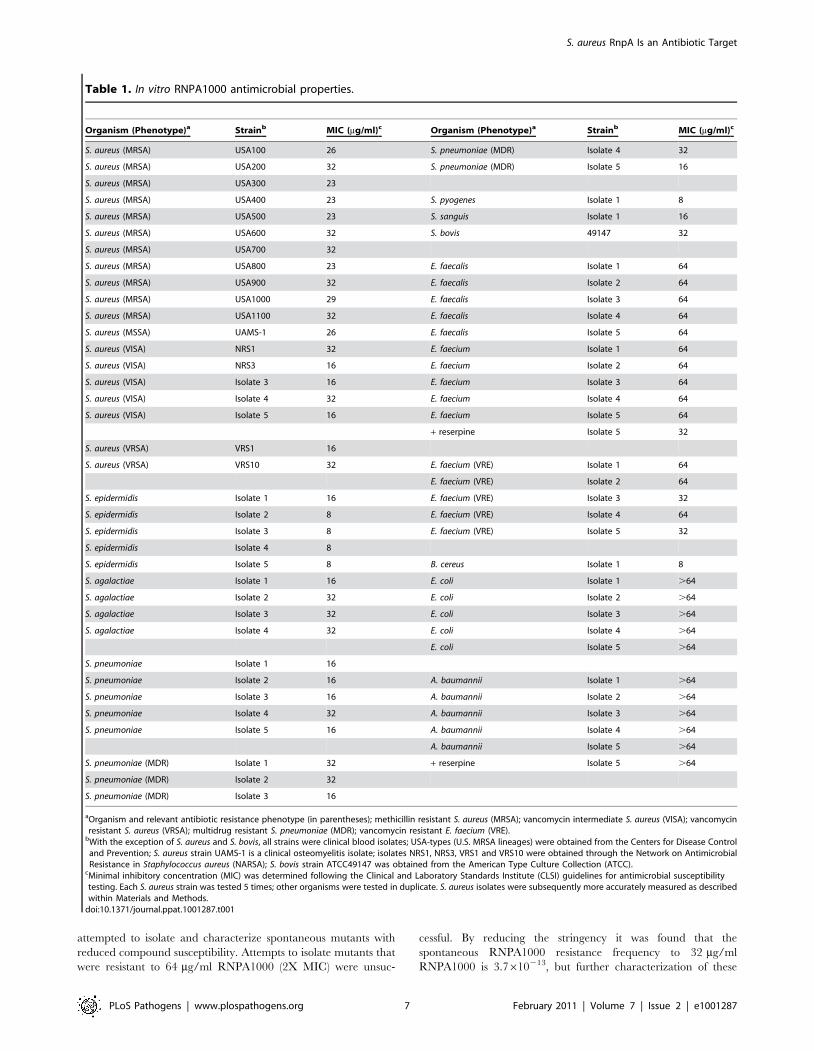
In vitro antimicrobial activity of RnpA-inhibitor RNPA1000As shown in Table 1, RNPA1000 demonstrated moderate
antimicrobial activity against two well-characterized genotypically
diverse S. aureus isolates, UAMS-1 (clinical osteomyelitis isolate;
MIC 26 mg/ml) and USA300–0114 [predominant cause of U.S.
community-associated methicillin resistant S. aureus infections
(MRSA); MIC 23 mg/ml], as well as representatives of other
major MRSA lineages circulating throughout the U.S. [39].
Likewise, RNPA1000 demonstrated antimicrobial activity against
vancomycin-intermediate susceptible S. aureus (VISA) and vanco-
mycin resistant S. aureus (VRSA). Time kill assays revealed that
RNPA1000 acts as a bacteriostatic agent (Supplemental Figure
S2A), and that it does not affect the antimicrobial activities of
other anti-staphylococcal agents, including vancomycin, dapto-
mycin, or rifampicin (data not shown), but does mildly increase the
potency of oxacillin (Supplemental Figure S2B and S2C). The
RnpA-inhibitor also exhibited antimicrobial activity against
Staphylococcus epidermidis, antibiotic susceptible and multi-drug
resistant Streptococcus pneumoniae, Streptococcus pyogenes, Streptococcus
agalactiae, and Bacillus cereus. RNPA1000 also showed mild activity
against Enterococcus faecalis, Enterococcus faecium and vancomycin
resistant E. faecium (VRE), but did not affect Escherichia coli or
Acinetobacter baumannii growth (Table 1). The latter was expected
because E. coli and A. baumannii RnpA share limited amino acid
identity (24% and 26%, respectively) with S. aureus RnpA
(Supplemental Figure S3). Moreover, purified A. baumannii RnpA
did not demonstrate ribonuclolytic activity in our in vitro assay
conditions (data not shown). Enterococci susceptibility to
RNPA1000 was increased from an MIC of 64 mg/ml to 32 mg/
ml in the presence of the efflux pump inhibitor reserpine,
suggesting that enterococci may be inherently susceptible to the
RnpA inhibitor. Conversely, the efflux inhibitor had no effect on
A. baumannii RNPA1000 susceptibility (Table 1). Taken together,
these results indicate that bacterial RNPA1000 susceptibility
correlates with amino acid similarity to S. aureus RnpA and the
enzyme’s in vitro RNA degradation activity.
In vivo antimicrobial efficacy of RnpA-inhibitor RNPA1000Next we assessed whether RnpA-inhibitory agent concentra-
tions corresponding to the effective bacterial MIC values (10–
50 mg/ml) elicited human cell cytotoxicity. MTT cell proliferation
assay measurements revealed that 24 hr RnpA-inhibitor exposure
did not cause human HepG2 cell toxicity at any concentration
tested (data not shown). However, extended RNP1000 exposure
(48 hr) elicited mild cytotoxicity at 25 mg/ml, which corresponds
to the minimum inhibitory concentration of most MRSA lineages
(Figure 4A), whereas higher concentrations exhibited increased
toxicity (data not shown).
Clearly, the observed toxicity associated with RNPA1000
precludes its direct utility as an antimicrobial agent. None the
less, we hypothesized that because RNPA1000 was not toxic
during short- and only mildly toxic during extended- HepG2
exposure, it could serve as an appropriate tool to assess whether
RnpA-inhibitory molecules are efficacious in a systemic mouse
infection model. As shown in Figure 4B, subcutaneous injection of
RNPA1000 limited the lethal effects of wild type S. aureus injected
(4.556105 cfu/animal) into the intraperitoneal cavity of CD-1
mice. Although this bacterial inoculum (equivalent to 10–100
LD50s) resulted in 100% death of non-treated control animals
within 24 hr, RNPA1000 provided protection in a dose-dependent
manner. Administration of the highest RnpA-inhibitor dose
(256 mg/kg) reproducibly resulted in 50% survival, whereas
128 mg/kg and 64 mg/kg resulted in 30% and 20% survival,
respectively, over the course of study (Figure 4B; Supplemental
Table S3). Notably, dosing regimens of compound (alone) did not
affect animal survival at any of the concentrations tested (32 mg/
kg, 64 mg/kg, 128 mg/kg, 256 mg/kg; Supplemental Table S3).
Taken together, these results suggest that RNPA1000 limits
bacterial pathogenicity within the acute lethal model of S. aureus
infection with a median effective dose (ED50) between 64–
256 mg/kg. While the combination of mild toxicity and high
effective dose would exclude the compound from consideration as
a therapeutic agent, RNPA1000 could be considered a platform
for medicinal chemistry-based generation of more potent deriva-
tives. More importantly, these results provide proof of concept that
RnpA inhibitory agents are efficacious in a systemic mouse
infection model and that RNPA1000 represents a tool to study the
contribution of RnpA to infection processes.
Antimicrobial efficacy of RnpA-inhibitor on biofilm-associated bacteria
The success of S. aureus as a bacterial pathogen can be
attributable, in part, to its ability to form biofilms on implanted
medical devices, which presumably provides a focus for bacterial
dissemination to secondary host sites. One of the complicating issues
in treating biofilm-associated infections is that biofilm-associated
bacteria are inherently recalcitrant to antibiotic treatment. For
instance, one recent in vitro study showed that despite using a strain
that was intrinsically susceptible to each antibiotic, 5X MIC of
daptomycin, linezolid, or vancomycin only reduced biofilm-
associated bacteria by ,2 logs following 24 hr treatment and none
of these antibiotics cleared biofilm-associated S. aureus even when
administered at 20X MIC over a course of 3 days [40].
Transcription profiling studies have revealed that despite being
physiologically unique, biofilm-associated S. aureus resemble plank-
tonic stationary phase cells [41]. Indeed, similar to stationary phase
bacteria, rnpA expression is diminished 4.3 and 6.2- fold in S. aureus
biofilm-associated and biofilm-detached bacteria, respectively, in
comparison to exponential phase cells (Dunman and Horswill,
unpublished). Because low levels of RnpA are likely to be present
within biofilm-associated bacteria, we hypothesized that fewer
RnpA-inhibitory molecules would be required to interfere with the
protein’s function and, consequently, antimicrobial activity. Thus,
biofilm-associated S. aureus may exhibit considerable susceptibility to
an RnpA-inhibitor, such as RNPA1000.
As shown in Figure 4C, treatment of biofilm-associated S. aureus
with 5X MIC RNPA1000 for 24 hr resulted in a 3-log decrease in
bacterial burden, suggesting that during short term exposure the
agent is equally, if not more potent, than daptomycin, vancomycin,
or linezolid. Further, while bacterial clearance was never achieved,
increasing the length of exposure or RNPA1000 concentration
enhanced antimicrobial activity. Maximal RNPA1000 antimicro-
bial potency (5-log reduction in biofilm-associated bacteria)
compared favorably with the activities of commercially available
antibiotics assessed in the same model and conditions (6-log
decrease daptomycin, 5-log decrease linezolid; 4-log decrease
vancomycin) [40]. Taken together, these results suggest that RnpA
plays an important biological role in S. aureus biofilm maintenance,
and that corresponding inhibitors may have expanded therapeutic
utility in treating biofilm-associated infections.
RNPA1000 affects S. aureus mRNA decayTo assess whether the susceptibility of S. aureus to RNPA1000
was attributable to the inhibition of cellular RnpA, we initially
S. aureus RnpA Is an Antibiotic Target
PLoS Pathogens | www.plospathogens.org 6 February 2011 | Volume 7 | Issue 2 | e1001287
attempted to isolate and characterize spontaneous mutants with
reduced compound susceptibility. Attempts to isolate mutants that
were resistant to 64 mg/ml RNPA1000 (2X MIC) were unsuc-
cessful. By reducing the stringency it was found that the
spontaneous RNPA1000 resistance frequency to 32 mg/ml
RNPA1000 is 3.7610213, but further characterization of these
Table 1. In vitro RNPA1000 antimicrobial properties.
Organism (Phenotype)a Strainb MIC (mg/ml)c Organism (Phenotype)a Strainb MIC (mg/ml)c
S. aureus (MRSA) USA100 26 S. pneumoniae (MDR) Isolate 4 32
S. aureus (MRSA) USA200 32 S. pneumoniae (MDR) Isolate 5 16
S. aureus (MRSA) USA300 23
S. aureus (MRSA) USA400 23 S. pyogenes Isolate 1 8
S. aureus (MRSA) USA500 23 S. sanguis Isolate 1 16
S. aureus (MRSA) USA600 32 S. bovis 49147 32
S. aureus (MRSA) USA700 32
S. aureus (MRSA) USA800 23 E. faecalis Isolate 1 64
S. aureus (MRSA) USA900 32 E. faecalis Isolate 2 64
S. aureus (MRSA) USA1000 29 E. faecalis Isolate 3 64
S. aureus (MRSA) USA1100 32 E. faecalis Isolate 4 64
S. aureus (MSSA) UAMS-1 26 E. faecalis Isolate 5 64
S. aureus (VISA) NRS1 32 E. faecium Isolate 1 64
S. aureus (VISA) NRS3 16 E. faecium Isolate 2 64
S. aureus (VISA) Isolate 3 16 E. faecium Isolate 3 64
S. aureus (VISA) Isolate 4 32 E. faecium Isolate 4 64
S. aureus (VISA) Isolate 5 16 E. faecium Isolate 5 64
+ reserpine Isolate 5 32
S. aureus (VRSA) VRS1 16
S. aureus (VRSA) VRS10 32 E. faecium (VRE) Isolate 1 64
E. faecium (VRE) Isolate 2 64
S. epidermidis Isolate 1 16 E. faecium (VRE) Isolate 3 32
S. epidermidis Isolate 2 8 E. faecium (VRE) Isolate 4 64
S. epidermidis Isolate 3 8 E. faecium (VRE) Isolate 5 32
S. epidermidis Isolate 4 8
S. epidermidis Isolate 5 8 B. cereus Isolate 1 8
S. agalactiae Isolate 1 16 E. coli Isolate 1 .64
S. agalactiae Isolate 2 32 E. coli Isolate 2 .64
S. agalactiae Isolate 3 32 E. coli Isolate 3 .64
S. agalactiae Isolate 4 32 E. coli Isolate 4 .64
E. coli Isolate 5 .64
S. pneumoniae Isolate 1 16
S. pneumoniae Isolate 2 16 A. baumannii Isolate 1 .64
S. pneumoniae Isolate 3 16 A. baumannii Isolate 2 .64
S. pneumoniae Isolate 4 32 A. baumannii Isolate 3 .64
S. pneumoniae Isolate 5 16 A. baumannii Isolate 4 .64
A. baumannii Isolate 5 .64
S. pneumoniae (MDR) Isolate 1 32 + reserpine Isolate 5 .64
S. pneumoniae (MDR) Isolate 2 32
S. pneumoniae (MDR) Isolate 3 16
aOrganism and relevant antibiotic resistance phenotype (in parentheses); methicillin resistant S. aureus (MRSA); vancomycin intermediate S. aureus (VISA); vancomycinresistant S. aureus (VRSA); multidrug resistant S. pneumoniae (MDR); vancomycin resistant E. faecium (VRE).
bWith the exception of S. aureus and S. bovis, all strains were clinical blood isolates; USA-types (U.S. MRSA lineages) were obtained from the Centers for Disease Controland Prevention; S. aureus strain UAMS-1 is a clinical osteomyelitis isolate; isolates NRS1, NRS3, VRS1 and VRS10 were obtained through the Network on AntimicrobialResistance in Staphylococcus aureus (NARSA); S. bovis strain ATCC49147 was obtained from the American Type Culture Collection (ATCC).
cMinimal inhibitory concentration (MIC) was determined following the Clinical and Laboratory Standards Institute (CLSI) guidelines for antimicrobial susceptibilitytesting. Each S. aureus strain was tested 5 times; other organisms were tested in duplicate. S. aureus isolates were subsequently more accurately measured as describedwithin Materials and Methods.
doi:10.1371/journal.ppat.1001287.t001
S. aureus RnpA Is an Antibiotic Target
PLoS Pathogens | www.plospathogens.org 7 February 2011 | Volume 7 | Issue 2 | e1001287

mutants revealed that the resistance phenotype was lost following
serial passage. As a second approach, we directly measured the
mRNA turnover properties of S. aureus that were challenged with a
sub-inhibitory concentration of RnpA-inhibitor (0.5X MIC).
Following 30 min treatment, RNPA1000 reduced the mRNA
degradation rate of S. aureus cells, in comparison to mock treated
cells (Figure 5A). Thus, RnpA-inhibitory compounds reduce
cellular mRNA degradation, presumably by limiting the enzyme’s
cellular function. While we cannot rule out the possibility that the
agent affects other enzymes or, in particular other S. aureus
ribonucleases, the mRNA turnover properties of RNPA1000
treated cells resembled that of RnpA depleted cells (Figure 2D),
suggesting that the agent may be affecting the enzyme. To more
directly determine whether RNPA1000’s antimicrobial effects are
mediated through cellular inhibition of RnpA we assessed the
RNPA1000 susceptibility of S. aureus RnpA over- and under-
producing cells. S. aureus harboring vector, or a plasmid copy of
wild type rnpA mRNA or rnpA antisense RNA under control of the
CdCl2 inducible promoter were grown in the presence of 2.5 mM
inducer and increasing concentrations of RNPA1000. As stated
above, this concentration of cadmium chloride induces mild
changes in RnpA protein expression (RnpA overproduction or
underproduction) but is modest enough that cellular growth is not
affected. As shown in Figure 5B, both vector containing- and
RnpA overproducing- cells exhibited an MIC of 32 mg ml21,
whereas the MIC of RnpA underproducing cells was 8 mg ml21.
The latter indicates that S. aureus’ RNPA1000 susceptibility
correlates to cellular RnpA levels and that the agent’s antimicro-
bial mode-of-action is, in part, RnpA dependent. It is currently
unclear why RnpA overexpression did not result in increased
RNPA1000 tolerance; presumably enzyme levels did not reach
concentrations needed to limit RNPA1000 effectiveness.
Discussion
The emergence of community-acquired methicillin-resistant S.
aureus (CA-MRSA) strains capable of causing serious infections in
otherwise healthy individuals make the organism a more formidable
pathogen now than perhaps any time since the beginning of the
antibiotic era. Indeed, it is estimated that in 2005 alone the number
of invasive MRSA infections in the U.S. reached approximately
94,360, with 18,650 resulting in fatal outcome [3]. Based on these
statistics, S. aureus has passed acquired-immunodeficiency syndrome
(AIDS) as a cause of death in the United States, further accentuating
the need for new anti-staphylococcal agents.
The success of S. aureus as a human pathogen can, in part, be
attributed to its ability to produce an expansive repertoire of
Figure 4. RnpA-inhibitor characterization. (A) MTT-cytotoxicity assay results of HepG2 cells exposed to compound solvent (DMSO; negativecontrol), Mitomycin C (positive control), and indicated amount of RNPA1000. (B) Shown are the average daily (X-axis) percent surviving animals (Y-axis) following no treatment (closed diamonds; negative control), vancomycin treatment (closed squares; 1 mg/kg; positive control), or RNPA1000treatment; 64 mg/kg (open circles); 128 mg/kg (open squares), 256 mg/kg (open triangles). Experiment was repeated twice; replicate measurementsare shown in Supplemental Table S3. (C) In vitro biofilm assay results; graphed are the number of catheter-associated S. aureus following 1, 2, or 3days of no antimicrobial treatment (untreated; UT) or exposure to 5, 10, or 20 times the MIC for RNPA1000. Boxes define the interval between the 25th
and 75th percentile. Bars extending upward indicate the boundary defined by the value 1.5X higher than the 75th percentile while those extendingdownward indicate the boundary defined by a value 1.5X lower than the 25th percentile. Filled circles indicate individual values outside these twoextremes.doi:10.1371/journal.ppat.1001287.g004
S. aureus RnpA Is an Antibiotic Target
PLoS Pathogens | www.plospathogens.org 8 February 2011 | Volume 7 | Issue 2 | e1001287

virulence factors, which collectively augment the organism’s ability
to colonize and invade host tissue, evade host immune responses,
and adapt to host-associated environmental challenges. In the
laboratory setting, many S. aureus cell surface virulence factors are
predominantly expressed during exponential phase growth,
whereas secreted virulence factors are predominantly expressed
as cultures transition to early stationary phase growth [18]. This
combined with the recent observation that the modulation of
mRNA turnover affects S. aureus virulence factor expression led to
the premise of the current study; the degradation properties of
virulence factors transcripts differ during exponential and
stationary phase growth [19,20,42]. Further, these changes in
mRNA turnover may, in part, account for growth-phase
dependent changes in virulence factor expression. A comparison
of the mRNA degradation properties of known and predicted
virulence factor transcripts indicates that this may be the case;
41% exhibited an alteration in mRNA turnover during exponen-
tial in comparison to stationary phase growth. Thus, growth
phase-dependent changes in transcript degradation are likely to
contribute to changes in the mRNA titers, and consequently
protein production, of these virulence determinants.
Our mRNA turnover measurements also revealed growth
phase-dependent alterations in mRNA turnover extend beyond
virulence factor mRNA species; 884 staphylococcal transcripts are
stabilized during stationary phase growth. This phenomenon
could be attributable to the stationary phase-dependent down-
regulation of components of the organism’s RNA degradation
machinery. An analysis of the transcript titers of known and
putative ribonucleases revealed that neither RNase J1 or RNase Y
were differentially expressed in a growth phase dependent manner,
Figure 5. RNPA1000 characterization. Plotted are all GeneChip detected transcript levels at 0 and 5 min post-transcriptional arrest. Red, blue,and yellow data points represent all GeneChip measurable transcripts that were considered present, present in one sample but absent in second, orabsent in both RNA samples, respectively. Grey dashed line indicates the lower limit of sensitivity for each sample. (A) A comparison of the mRNAlevels of DMSO treated cells; 90% of all transcripts decrease $2-fold at 5 min post-transcriptional arrest (Left Panel). A comparison of the mRNA levelsof cells challenged with sub-inhibitory concentration of RnpA-inhibitor; addition of the agent resulted in pronounced stabilization of S. aureustranscripts (Right Panel). (B) Microtiter plate assay illustrating the in vitro antimicrobial effects of indicated concentration of RNPA1000 (across top)against S. aureus RN4220 pRNPA-A.S. (RnpA depleted cells; top panel), RN4220 pRNPA (RnpA overexpressor cells; center panel) and RN4220 pCN51(vector; bottom panel) when grown in the presence of 2.5 mM CdCl2. All strains were assessed twice; arrows indicate MIC values.doi:10.1371/journal.ppat.1001287.g005
S. aureus RnpA Is an Antibiotic Target
PLoS Pathogens | www.plospathogens.org 9 February 2011 | Volume 7 | Issue 2 | e1001287
whereas RNase HIII and the protein component of RNase P,
RnpA, were down-regulated during stationary phase growth.
Bacterial RNase P holoenzymes are ribonucleoprotein com-
plexes consisting of a ribozyme (rnpB) and a protein component,
RnpA. Both rnpB and RnpA are required for bacterial survival,
thus an agent that inhibits either subunit’s activity could have
potential as an antimicrobial therapeutic agent [27,43]. Indeed,
the function of rnpB is well established; it catalyzes maturation of
precursor tRNA molecules, and progress has been made in
developing antimicrobial small molecule inhibitors of rnpB-
mediated tRNA processing [23,24,25]. RnpA augments cellular
rnpB-precursor tRNA binding [26], a phenotype that is difficult to
exploit for antimicrobial screening. In addition to mediating tRNA
processing, RNase P has also been shown to catalyze cleavage of
other double-stranded RNA substrates, such as guide RNAs, and
this activity requires RnpA [30,31,32].
Our data suggest that S. aureus RnpA exhibits pronounced in
vitro ribonuclease activity in comparison S. aureus RNase J1, RNase
Y and RNase HIII, in the conditions assessed here. We do not
mean to imply that the other putative ribonucleases assessed are
devoid of ribonucleolytic activity. Rather, their activity would
probably be better measured in differing buffering conditions or
with different RNA substrates. In fact, subsequent studies revealed
that S. aureus RNase J1 is a potent ribonuclease in differing
buffering conditions [10].
By exploiting the observed ribonuclease activity of RnpA, high
through-put screening and secondary assays identified small
molecule inhibitors of RnpA-mediated in vitro RNA degradation.
The inhibitory activity of one of these molecules, RNPA1000, was
found to have specificity for RnpA, as the agent did not interfere
with the activity of commercially available ribonucleases or
recombinant S. aureus RNase J1 (data not shown). RNPA1000
was shown to have increased antimicrobial activity against RnpA
depleted cells, indicating that the agent targets the enzyme in vivo.
None the less, we cannot exclude the possibility that RNPA1000
may also affect other essential S. aureus enzymes. While it remains
to be seen whether RnpA’s in vitro ribonuclease activity correlates
to what occurs within the cell, sub-inhibitory concentrations of
RNPA1000 and RnpA depleted cells were also found to limit S.
aureus RNA-degradation, suggesting that RnpA, either alone, as
RNase P cofactor, or as a component of a unrecognized complex,
may participate in bulk mRNA turnover, a function that may
contribute to enzyme’s role in maintaining cellular survival.
Because S. aureus RnpA is well conserved among many Gram-
positive bacteria, our findings may have expanded significance by
providing insight regarding the RNA degradation machinery of
additional bacterial species.
Our results also indicate that inhibitors of RnpA-mediated RNA
degradation may have promise as antimicrobial agents. Indeed,
RNPA1000 exhibited moderate antimicrobial activity against each
of the predominant S. aureus MRSA lineages circulating through-
out the U.S., VISA, VRSA, VRE, as well as bacterial pathogens
with high RnpA amino acid conservation. The agent also limited
the organism’s pathogenesis in a murine acute lethal model of
infection and limited biofilm-associated bacterial burden. While
the mode-of-action for RNPA1000 appears to, in part, effect S.
aureus RnpA mediated RNA processing we do not know if the
same is the case for other RNPA1000 susceptible pathogens.
Collectively, these data establish that RnpA represents an
attractive antimicrobial drug target and that RNA stabilizing
agents represent a new paradigm for treating bacterial pathogens
of immense health care concern. The RnpA-inhibitory molecule
identified in this study may represent a progenitor of this new class
of antimicrobials.
Methods
mRNA half-livesFor half life determinations, S. aureus strain UAMS-1, RN4220
(pCN51; plasmid containing CdCl2 inducible promoter), RN4220
(pRNPA; pCN51 capable of producing full length rnpA mRNA), or
RN4220 (pRNPA-A.S.; pCN51 capable of producing rnpA antisense
RNA) were grown to mid-exponential or stationary phase,
transcription was arrested by the addition of rifampin (200 mg/
ml), and aliquots were removed at 0-, 2.5-, 5-, 15- and 30- min post-
transcriptional arrest for strain UAMS-1. To conserve reagents,
aliquots were removed at 0 and 10 min post-transcriptional arrest
for RN4220 derivatives. Plating ensured cultures had not developed
rifampin resistance. Each strain and/or growth phase was assessed
twice, except for RN4220 pRNPA-A.S. cells which were assessed
four times. RNA was isolated from each aliquot, labeled, hybridized
to an S. aureus GeneChip (Affymetrix; Santa Clara, CA), duplicates
were averaged and the mRNA half-lives of all mRNA species were
determined, as previously described [19,20]. To measure the
mRNA turnover characteristics of RNPA1000 challenged cells,
exponential-phase S. aureus were treated with 0.5X MIC of the
RnpA inhibitor or equivalent volume compound solvent (DMSO)
for 30 min. Transcript synthesis was then arrested and the
transcript titers of mRNA species were measured at 0- and 5- min
post-transcriptional arrest [19,20].
Protein purificationEach putative S. aureus ribonuclease predicted open reading
frame was PCR amplified and inserted into the ligation-
independent cloning site of plasmid pET-30 Ek/LIC (Novagen;
Madison WI). Sequencing confirmed that this fused a hexahisti-
dine-tag to the N-terminus of each protein under the control of the
plasmid’s isopropyl b-D-1-thiogalactopyranoside (IPTG) inducible
promoter. Following transformation, each protein was purified
from E. coli BL21 (DE3) cells grown in the presence of IPTG (4 hr)
by Ni+2 affinity chromatography. More specifically, 10 g of cell
pellet was suspended in 50 ml of buffer A (300 mM NaCl, 50 mM
Na2HPO4, pH 7.4) containing a complete mini EDTA-free
protease inhibitor tablet (Roche; Branford, CT) and 20 mM
imidazole. Cells were ruptured by seven passes at 15,000 psi
through an Emulsifex-C3 microfluidizer (Avestin Inc.; Ottawa,
Canada). Cell debris was removed by centrifugation at 12,0006g
for 30 min and supernatants were loaded onto a 5 mL Ni-NTA
FF-crude affinity column (GE Healthcare Bio-Sciences; Piscat-
away, NJ) with an AKTA-FPLC high performance liquid
chromatography system (GE Healthcare Bio-Sciences; Pittsburgh,
PA). Proteins eluted in a single peak with a linear imidazole
gradient (80 mM to 500 mM) in buffer A. The presence of each
protein was assessed by Coomassie stained SDS-PAGE and
matrix-assisted laser desportion/ionization (MALDI) analysis
spectrometry (Wistar Institute; Philadelphia, PA).
PlasmidsPlasmids pRNPA-S and pRNPA-A.S. contain the putative rnpA
transcriptional unit including predicted Shine-Dalgarno sequence
in the sense and antisense orientation, respectively under control
of the CdCl2 inducible of the S. aureus shuttle-vector pCN51 [38].
Briefly, the rnpA open reading frame and 34 nt upstream sequence
was PCR amplified from S. aureus strain UAMS-1 using primers 59
GAATTCTCAAATAAAAACGATAAATAAGCGAGTGATG-
TTA (forward) and 59 GGTACCTTACTTAATCTTTTTAT-
TAAAAACTTTGGCAA (reverse) containing a 59 terminal EcoRI
and KpnI restriction enzyme site (underlined), respectively, or
primers in which the restriction enzyme sequence had been
S. aureus RnpA Is an Antibiotic Target
PLoS Pathogens | www.plospathogens.org 10 February 2011 | Volume 7 | Issue 2 | e1001287
reversed. Resulting PCR products were ligated into pCRII-
TOPO vector and transformed into E. coli INVaF’ cells for
propagation (Invitrogen, Carlsbad, CA). Plasmid DNA was
subsequently purified using QIAprep Spin Miniprep Kits
(Qiagen, Valencia, CA) then digested with EcoRI and KpnI to
liberate the plasmid inserts, which were gel purified using a
QIAquick Gel Extraction Kit (Qiagen) and ligated into EcoRI and
KpnI-digested pCN51. DNA sequencing confirmed the integrity of
plasmid pRNPA-S and pRNPA-A.S.
Western blottingAffinity purified PolyQuik rabbit S. aureus RnpA polyclonal
antibodies were generated by Invitrogen (Carlsbad, CA). Total
bacterial proteins were isolated from RN4220 cells containing
plasmid vector (pCN51), RnpA overexpressor plasmid (pRNPA-S)
or RnpA antisense RNA plasmid (pRNPA-A.S.) following 30 min
growth in TSB medium supplemented with 2.5 mM CdCl2 to induce
RNA expression and 10 mg/ml erythromycin for plasmid mainte-
nance. Resultant protein concentrations were determined by
conventional Bradford Assays and 2.0 mg of each protein sample
or purified S. aureus RnpA was electrophoresed in a 10% SDS
polyacrylamide gel and transferred to a polyvinylidene fluoride
membrane (Millipore, Billerica, MA). Membranes were blocked with
10% milk, washed, incubated with rabbit RnpA antibody (1:1000
dilution), washed, incubated with horseradish peroxidase-conjugated
anti-rabbit antibody (1:1000 dilution; GE Healthcare) and processed
using an Amersham ECL Western Blotting System, according to the
manufacturer’s recommendations (GE Healthcare).
RnpA inhibitorsMembers of the ActiProbe-25K and Natural Product libraries
(29,940 compounds total; TimTec Inc.; Newark, DE) were screened
for small molecule inhibitors of S. aureus RnpA mediated total
bacterial RNA degradation. All reactions (50 ml) were performed in
96-well format and contained 20 pmol RnpA, 200 ng S. aureus total
RNA, and ,5 mM of each compound in 1X reaction buffer (2 mM
NaCl, 2 mM MgCl2, 50 mM Tris-HCl, pH 6.0). Mixtures were
incubated at 37uC for 20 min at which time Quant-iT RiboGreen
(100 ml; Invitrogen) was added to quantify the amount of RNA
substrate remaining. Percent enzyme inhibition was calculated as
remaining substrate/starting substrate * 100. For inhibitory titration
assays, 1 pmol of spa mRNA was incubated with 20 pmol RnpA
alone (positive control) or in the presence of increasing amounts (0,
25, 50, 100, 125, 150, 200, 250, and 500 mM) RNP1000 for one
hour at 37uC. 20 ml of each reaction mixture were subjected to
electrophoresis in a 1.2% formaldehyde-containing agarose gel and
visualized by ethidium bromide staining.
Cytotoxicity assaysHepG2 human hepatocytes (105 cells) were seeded in individual
wells of a microtitre plate and incubated for 16 hr at 37uC with
5% carbon dioxide in Dulbecco’s Modified Eagle Media
supplemented with 10% fetal bovine serum. Cells were then
challenged with Mitomycin C (5 mg/ml; positive control) or 0, 25,
or 50 mg/ml RNPA1000 for either 24 or 48 hrs. Cell viability was
measured spectrophotometrically (570 nm) following the addition
and subsequent reduction of tetrazolium salt (MTT) within
metabolically active cells, as per the manufacturer’s recommen-
dations (American Type Culture Collection; Manassas, VA).
Antimicrobial susceptibility testingWith the exception of RN4220-derivatives, in vitro activities of
RNPA1000 against bacteria were determined by the broth
microdilution method according to the Clinical and Laboratory
Standards Institute (CLSI) guidelines using cation adjusted
Mueller-Hinton broth or MH broth supplemented with 5% lysed
horse blood (for testing Streptococcus spp.). Microtiter plates
containing serial dilutions of RNPA1000 (0, 4, 8, 16, 32, 64,
and 128 mg/ml) were inoculated with 105 colony forming units
(CFU)/ml and incubated for 18 hr at 37uC. The MIC for each
isolate was defined as the lowest concentration of RNPA1000 that
completely inhibited growth of the organism as detected by the
unaided eye. The MIC for each S. aureus strain was further refined
by repeat testing following the procedure described above, except
that microtiter wells contained 1 mg/ml incremental increases in
concentration of RNPA1000 spanning the lowest concentration
that initially did not completely inhibit growth (16 mg/ml) and the
concentration that completely inhibited growth (32 mg/ml). The
MIC value for each S. aureus strain was determined to be the
median score of replicate measurements (n = 5). Wells containing
concentrations of RNPA1000 $ MIC were plated for minimal
bactericidal measurement. Where possible, experiments with
VRSA strains were performed in a laminar flow hood to minimize
potential for equipment contamination. For RN4220 cells
containing plasmid vector (pCN51), RnpA overproducing plasmid
(pRNPA-S) or RnpA underproducing plasmid (pRNPA-A.S.) in
vitro antimicrobial activity of RNPA1000 was performed by the
microdilution method as described above, except that cells were
grown in Tryptic Soy Broth medium supplemented with 2.5 mM
CdCl2 and 0, 1, 2, 4, 8, 16, 32, 64, or 128 mg/ml RNPA1000.
Time-kill assays were also performed to monitor the antimicrobial
properties of RNPA1000 for S. aureus strain UAMS-1 in the
absence and presence of 0.25, 0.5, 2, and 4 times the strain’s MIC
for oxacillin (1 mg/ml), rifampicin (0.5 mg/ml), vancomycin (2 mg/
ml), or daptomycin (1 mg/ml). The indicated amount of
RNPA1000 and/or commercial antibiotic were added to mid-
exponential phase (26108 cfu/ml) S. aureus strain UAMS-1 cells
and incubated at 37uC. Aliquots were removed at 0, 2, 4, 8, and
24 hr post-antimicrobial challenge, serial diluted, and plated to
enumerate resulting cfu/ml. All time-kill assays were repeated at
least 3 times.
Biofilm assaysIn vitro biofilm assays were performed as previously described
[40]. Briefly, 1 cm segments of 14-gauge fluorinated ethylene
propylene Introcan Safety Catheters (B. Braun, Bethlehem, PA)
were coated with human plasma and placed in individual wells of a
12-well microtiter plate containing 2 ml biofilm medium and S.
aureus strain UAMS-1 at a final OD600 nm of 0.05. Following
overnight incubation at 37uC catheters were removed, rinsed in
phosphate buffered saline (PBS), and transferred to fresh biofilm
medium containing 0, 5, 10, or 20 times the S. aureus MIC for
RNPA1000. Catheters exposed to each dose (n = 3) were recovered
daily over a period of 3 days, with the medium being replaced
each day. After each recovery time point catheters were rinsed in
PBS and adherent bacteria were enumerated by sonication and
plating. Analysis of variance (ANOVA) of logarithmically-
transformed bacterial count data was used to evaluate the effect
of RNPA1000 exposure.
Acute lethal model of infectionFemale 5–6 week old CD-1 mice were challenged by
intraperitoneal injection (0.5 ml) of wild type S. aureus strain
Smith, resulting in a final inoculum of 4.556105 colony forming
units/animal; equivalent to 10–100 LD50s and resulted in death of
non-treated control animals (n = 5) within 24 hr post-inoculum.
RNPA1000 was solubilized in 1:1 mixture of DMSO and
S. aureus RnpA Is an Antibiotic Target
PLoS Pathogens | www.plospathogens.org 11 February 2011 | Volume 7 | Issue 2 | e1001287

PEG400; Vancomycin was prepared in water. Animals (5/dose
group) were administered 16, 64, and 256 mg/kg or 0.25, 1, 4,
and 16 mg/kg of RNPA1000 or Vancomycin, respectively, at
30 min post infection by subcutaneous injection (0.2 ml). The
percent surviving animals receiving no treatment, a single dose of
Vancomycin, or RnpA-inhibitor was recorded daily over the
course of the study (5 days).
Ethics statementAll animal studies were performed at the University of North
Texas Health Science Center (UNTHSC) at Fort Worth under the
principles and guidelines described in the Guide for the Care and
Use of Laboratory Animals. UNTHSC is an American Association
for Laboratory Animal Science (AALAS) and United States
Department of Agriculture (USDA) accredited facility using
Institutional Animal Care and Use Committee (IACUC) approved
protocol UNT 2006/07-09. The UNTHSC Animal Program is
registered with the Office of Laboratory Animal Welfare (OLAW
Animal Welfare Assurance No. A37711-01).
Supporting Information
Figure S1 RnpA expression. (A) Plotted are the growth
characteristics (optical density; Y-axis), for S. aureus strain
RN4220 containing vector (pCN51; dark blue diamonds), rnpA
sense RNA (pRNPA-S; dark blue triangles) and rnpA antisense
RNA (pRNPA-A.S.; red squares) when grown in the presence of
10 mM CdCl2. Plasmid capable of producing an RNA comple-
mentry to rnpA mRNA exhibited diminished growth for a period
of 4 hrs (X-axis) in the presence of inducer. This growth defect was
not observed when cells were grown in the absence of cadmium
chloride (not shown) or when grown in the presence of 2.5 mM
CdCl2 (hashed line and pink squares). (B) Western blotting results
for S. aureus strain RN4220 pCN51 (vector), RN4220 pRNPA
(overexpressor), and RN4220 pRNPA-A.S. (RnpA depleted) cells
grown in the presence of 2.5 mM CdCl2.
Found at: doi:10.1371/journal.ppat.1001287.s001 (0.29 MB TIF)
Figure S2 S. aureus time-kill assay results. (A) Mid-exponential
phase S. aureus strain UAMS-1 cells were treated with 0.25, 0.5, 1,
2, or 4 times the MIC for RNPA1000. Plotted are the average cfu/
ml at 0, 2, 4, 8, and 24 hr post-RNPA1000 addition for each drug
concentration tested (n = 3); standard deviation shown. (B) Plotted
are the average cfu/ml at 2, 4, 8, and 24 hr post-oxacillin
treatment (0.25, 0.5, 2, or 4 times the MIC; n = 3) of mid-
exponential phase cells. (C) Mid-exponential phase cells were
treated with 0.5 times the MIC for RNPA1000, oxacillin, or both
(RNPA1000 and oxacillin). Shown are the average cfu/ml of mid
exponential phase cells following 2, 4, 8, and 24 hr post treatment
(n = 3); standard deviation shown.
Found at: doi:10.1371/journal.ppat.1001287.s002 (0.30 MB TIF)
Figure S3 RnpA amino acid comparisons. Alignment of amino
acid sequences of RnpA using GramAlign (http://bioinfo.unl.
edu/gramalign.php) with default parameters. Conserved amino
acids are boxed.
Found at: doi:10.1371/journal.ppat.1001287.s003 (2.93 MB TIF)
Table S1 mRNA half-lives of S. aureus transcripts produceddur-
ing exponential or stationary phase growth. Shown are the
expression properties and RNA half-lives of all S. aureus
transcripts produced during exponential and/or stationary phase
growth.
Found at: doi:10.1371/journal.ppat.1001287.s004 (3.46 MB XLS)
Table S2 Characterization of stationary phase-specific small
stable RNAs. Characterization of Stationary Phase-Specific Small
Stable RNAs.
Found at: doi:10.1371/journal.ppat.1001287.s005 (0.03 MB XLS)
Table S3 In vivo efficiacy of RnpA-inhibitor. Acute-lethal
murine model of infection results of animals treated with the
RnpA-inhibitor RNPA1000, Vancomycin (positive control) and
mock treatment (negative control).
Found at: doi:10.1371/journal.ppat.1001287.s006 (0.02 MB XLS)
Acknowledgments
We thank members of the Dunman laboratory and Drs. Richard Novick,
and Paul Fey for critically evaluating this manuscript. We also would like to
thank Dr. Paul Fey for providing various clinical bacterial species.
Author Contributions
Conceived and designed the experiments: JWS MSS EPS PMD.
Performed the experiments: PDO LJK KLA SD KEB CMR MLR TLL
WJW MP PN JMM OAA. Analyzed the data: PDO LJK KLA KEB WJW
JMM KS MSS EPS PMD. Contributed reagents/materials/analysis tools:
OAA. Wrote the paper: PDO MSS EPS PMD.
References
1. Shorr AF, Tabak YP, Killian AD, Gupta V, Liu LZ, et al. (2006) Healthcare-
associated bloodstream infection: A distinct entity? Insights from a large U.S.
database. Crit Care Med 34: 2588–2595.
2. Bancroft EA (2007) Antimicrobial resistance: it’s not just for hospitals. Jama 298:
1803–1804.
3. Klevens RM, Morrison MA, Nadle J, Petit S, Gershman K, et al. (2007) Invasive
methicillin-resistant Staphylococcus aureus infections in the United States. Jama 298:
1763–1771.
4. Appelbaum PC (2007) Reduced glycopeptide susceptibility in methicillin-
resistant Staphylococcus aureus (MRSA). Int J Antimicrob Agents 30: 398–408.
5. Zetola N, Francis JS, Nuermberger EL, Bishai WR (2005) Community-acquired
meticillin-resistant Staphylococcus aureus: an emerging threat. Lancet Infect Dis 5:
275–286.
6. Carpousis AJ (2007) The RNA degradosome of Escherichia coli: an mRNA-
degrading machine assembled on RNase E. Annu Rev Microbiol 61: 71–87.
7. Mackie GA (1998) Ribonuclease E is a 59-end-dependent endonuclease. Nature
395: 720–723.
8. Vanzo NF, Li YS, Py B, Blum E, Higgins CF, et al. (1998) Ribonuclease E
organizes the protein interactions in the Escherichia coli RNA degradosome.
Genes Dev 12: 2770–2781.
9. Condon C (2003) RNA processing and degradation in Bacillus subtilis. Microbiol
Mol Biol Rev 67: 157–174, table of contents.
10. Even S, Pellegrini O, Zig L, Labas V, Vinh J, et al. (2005) Ribonucleases J1 and
J2: two novel endoribonucleases in B. subtilis with functional homology to E. coli
RNase E. Nucleic Acids Res 33: 2141–2152. Print 2005.
11. Mathy N, Benard L, Pellegrini O, Daou R, Wen T, et al. (2007) 59-to-39
exoribonuclease activity in bacteria: role of RNase J1 in rRNA maturation and
59 stability of mRNA. Cell 129: 681–692.
12. Commichau FM, Rothe FM, Herzberg C, Wagner E, Hellwig D, et al. (2009)
Novel activities of glycolytic enzymes in Bacillus subtilis: interactions with essential
proteins involved in mRNA processing. Mol Cell Proteomics 8: 1350–1360.
13. Mader U, Zig L, Kretschmer J, Homuth G, Putzer H (2008) mRNA processing
by RNases J1 and J2 affects Bacillus subtilis gene expression on a global scale. Mol
Microbiol 70: 183–196.
14. Yao S, Sharp JS, Bechhofer DH (2009) Bacillus subtilis RNase J1 endonuclease and 59
exonuclease activities in the turnover of DeltaermC mRNA. Rna 15: 2331–2339.
15. Shahbabian K, Jamalli A, Zig L, Putzer H (2009) RNase Y, a novel
endoribonuclease, initiates riboswitch turnover in Bacillus subtilis. Embo J 28:
3523–3533.
16. Lehnik-Habrink M, Pfortner H, Rempeters L, Pietack N, Herzberg C, et al.
(2010) The RNA degradosome in Bacillus subtilis: identification of CshA as the
major RNA helicase in the multiprotein complex. Mol Microbiol 21: 21.
17. Kobayashi K, Ehrlich SD, Albertini A, Amati G, Andersen KK, et al. (2003)
Essential Bacillus subtilis genes. Proc Natl Acad Sci U S A 100: 4678–4683.
18. Novick RP (2003) Autoinduction and signal transduction in the regulation of
staphylococcal virulence. Mol Microbiol 48: 1429–1449.
19. Anderson KL, Roberts C, Disz T, Vonstein V, Hwang K, et al. (2006)
Characterization of the Staphylococcus aureus heat-shock, cold-shock, stringent, and
SOS responses and their effects on log-phase mRNA turnover. J Bacteriol 188:
6739–6756.
S. aureus RnpA Is an Antibiotic Target
PLoS Pathogens | www.plospathogens.org 12 February 2011 | Volume 7 | Issue 2 | e1001287

20. Roberts C, Anderson KL, Murphy E, Projan SJ, Mounts W, et al. (2006)
Characterizing the Effect of the Staphylococcus aureus Virulence Factor Regulator,
SarA, on Log-Phase mRNA Half-Lives. J Bacteriol 188: 2593–2603.
21. Takayama K, Kjelleberg S (2000) The role of RNA stability during bacterial
stress responses and starvation. Environ Microbiol 2: 355–365.
22. Anderson KL, Dunman PM (2009) Messenger RNA Turnover Processes in
Escherichia coli, Bacillus subtilis, and Emerging Studies in Staphylococcus aureus.
Int J Microbiol 2009: 525491.
23. Frank DN, Pace NR (1998) Ribonuclease P: unity and diversity in a tRNA
processing ribozyme. Annu Rev Biochem 67: 153–180.
24. Kazantsev AV, Pace NR (2006) Bacterial RNase P: a new view of an ancient
enzyme. Nat Rev Microbiol 4: 729–740.
25. Walker SC, Engelke DR (2006) Ribonuclease P: the evolution of an ancient
RNA enzyme. Crit Rev Biochem Mol Biol 41: 77–102.
26. Hartmann RK, Gossringer M, Spath B, Fischer S, Marchfelder A (2009) The
making of tRNAs and more - RNase P and tRNase Z. Prog Mol Biol Transl Sci
85: 319–368.
27. Gossringer M, Kretschmer-Kazemi Far R, Hartmann RK (2006) Analysis of
RNase P protein (rnpA) expression in Bacillus subtilis utilizing strains with
suppressible rnpA expression. J Bacteriol 188: 6816–6823.
28. Schedl P, Primakoff P (1973) Mutants of Escherichia coli thermosensitive for the
synthesis of transfer RNA. Proc Natl Acad Sci U S A 70: 2091–2095.
29. Waugh DS, Pace NR (1990) Complementation of an RNase P RNA (rnpB) gene
deletion in Escherichia coli by homologous genes from distantly related eubacteria.
J Bacteriol 172: 6316–6322.
30. Lundblad EW, Xiao G, Ko JH, Altman S (2008) Rapid selection of accessible
and cleavable sites in RNA by Escherichia coli RNase P and random external
guide sequences. Proc Natl Acad Sci U S A 105: 2354–2357.
31. Liu F, Altman S (1994) Differential evolution of substrates for an RNA enzyme
in the presence and absence of its protein cofactor. Cell 77: 1093–1100.
32. Marvin MC, Engelke DR (2009) Broadening the mission of an RNA enzyme.
J Cell Biochem 108: 1244–1251.
33. Letunic I, Copley RR, Pils B, Pinkert S, Schultz J, et al. (2006) SMART 5:
domains in the context of genomes and networks. Nucleic Acids Res 34:D257–260.
34. Schultz J, Milpetz F, Bork P, Ponting CP (1998) SMART, a simple modular
architecture research tool: identification of signaling domains. Proc Natl AcadSci U S A 95: 5857–5864.
35. Spitzfaden C, Nicholson N, Jones JJ, Guth S, Lehr R, et al. (2000) The structureof ribonuclease P protein from Staphylococcus aureus reveals a unique binding site
for single-stranded RNA. J Mol Biol 295: 105–115.
36. Chaudhuri RR, Allen AG, Owen PJ, Shalom G, Stone K, et al. (2009)Comprehensive identification of essential Staphylococcus aureus genes using
Transposon-Mediated Differential Hybridisation (TMDH). BMC Genomics10: 291.
37. Ji Y, Zhang B, Van SF, Horn, Warren P, et al. (2001) Identification of criticalstaphylococcal genes using conditional phenotypes generated by antisense RNA.
Science 293: 2266–2269.
38. Charpentier E, Anton AI, Barry P, Alfonso B, Fang Y, et al. (2004) Novelcassette-based shuttle vector system for gram-positive bacteria. Appl Environ
Microbiol 70: 6076–6085.39. McDougal LK, Steward CD, Killgore GE, Chaitram JM, McAllister SK, et al.
(2003) Pulsed-field gel electrophoresis typing of oxacillin-resistant Staphylococcus
aureus isolates from the United States: establishing a national database. J ClinMicrobiol 41: 5113–5120.
40. Weiss EC, Spencer HJ, Daily SJ, Weiss BD, Smeltzer MS (2009) Impact of sarA
on antibiotic susceptibility of Staphylococcus aureus in a catheter-associated in vitro
model of biofilm formation. Antimicrob Agents Chemother 53: 2475–2482.41. Beenken KE, Dunman PM, McAleese F, Macapagal D, Murphy E, et al. (2004)
Global Gene Expression in Staphylococcus aureus Biofilms. J Bacteriol 186:
4665–4684.42. Huntzinger E, Boisset S, Saveanu C, Benito Y, Geissmann T, et al. (2005)
Staphylococcus aureus RNAIII and the endoribonuclease III coordinately regulatespa gene expression. EMBO J 24: 824–835. Epub 2005 Jan 2027.
43. Kole R, Baer MF, Stark BC, Altman S (1980) E. coli RNAase P has a required
RNA component. Cell 19: 881–887.
S. aureus RnpA Is an Antibiotic Target
PLoS Pathogens | www.plospathogens.org 13 February 2011 | Volume 7 | Issue 2 | e1001287