Embed Size (px)
Citation preview

SNC2 Biology Review

What is an Organism?
• An organism is a living thing. It is easy to recognise a living thing, but not so easy to define it. Animals and plants are organisms, obviously. Organisms are a biotic, or living, part of the environment. Rocks and sunshine are parts of the non-living environment abiotic.

Know the difference between• Quark: Simply energy. Can’t be seen under any microscope.
Atom: Made from quarks. Has protons, neutrons. These make molecules.
Molecule: These are made from Atoms. Examples of molecules are: Hydrogen, Oxygen.
Organelle: Made of molecules and are the small organs in each cell. Examples in animal cells are: Nucleus, Ribosome, Lysosome.
Cell: Made up of organelles. Cells are the building blocks of life.
Tissue: Made from similar cells. Each organ has specific types of cells that make that particular organ and its parts.
Organ: Similar types of tissue (example muscle tissue, heart tissue) makeup an organ.
Organ System: Organs that work together to help an organism execute a specific function (breathing, thinking, etc) are part of an organ system.
Organism: An organism is the person/thing.

Nucleus
• The nucleus is the large control centre of a cell, directing all of the cell's activities.
• Chromosomes that contain your DNA are stored in this organelle.
• The nucleus is separated from the rest of the cell by the nuclear membrane.

Ribosome
• Found on the endoplasmic reticulum or free in the cytoplasm.
• Proteins are made in this organelle.
Mitochondria
• The mitochondria is sometimes referred to as the powerhouse of the cell.
• Mitochondria are largely responsible for providing the cell with energy from glucose (sugar) through a process called cellular respiration.

Golgi Bodies
• Golgi bodies collect and process materials to be removed from the cell.
• They also make and secrete mucus.
• Cells that secrete a lot of mucus, such as cells lining the intestine, have many Golgi bodies.

Endoplasmic Reticulum
• The endoplasmic reticulum is a three-dimensional network of branching tubes and pockets.
• It extends throughout the cytoplasm from the nuclear membrane.
• These fluid-filled tubes transport materials, such as proteins, through the cell.

Chloroplast
• Looks like: green stacks of membranes that contain chlorophyll
• Job: perform photosynthesis (convert sunlight into energy)
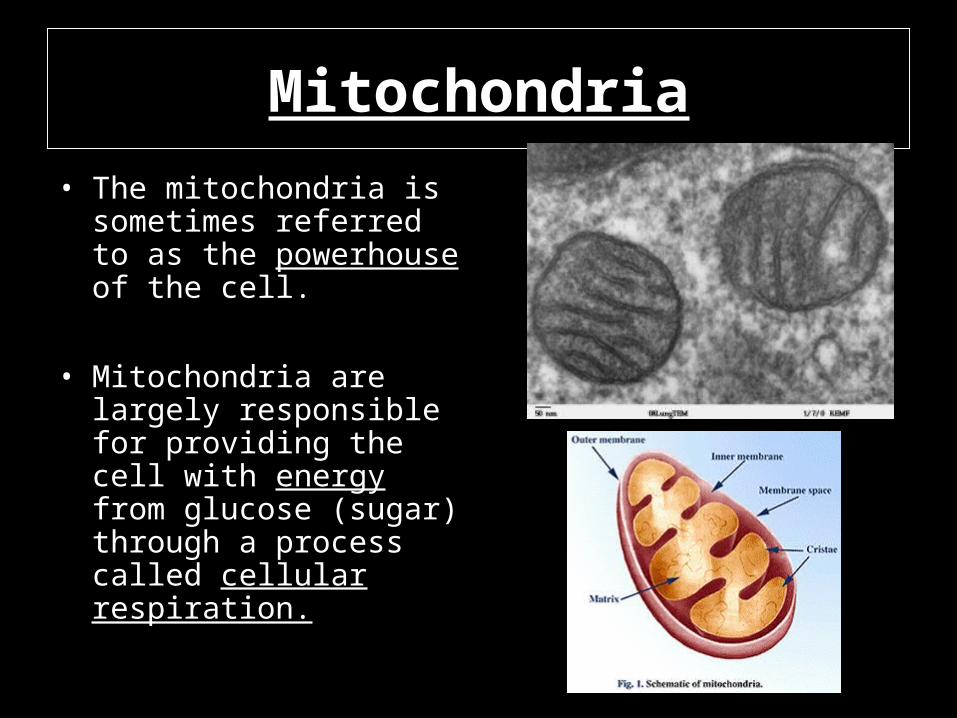
Vacuole• Looks like: sac-like
organ. HUGE in plant cells
• Job: stores water, food, and waste
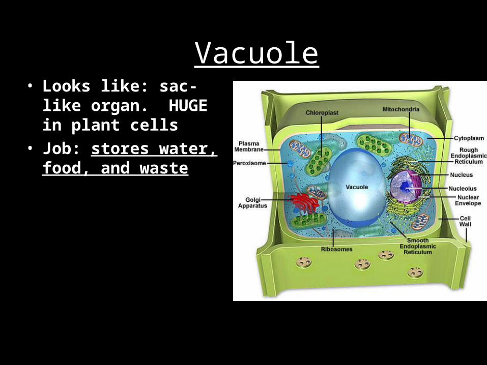
Vesicles
• A vesicle can be seen as a bubble of liquid within a cell.
• It is a small membrane-enclosed sack that can store or transport material.
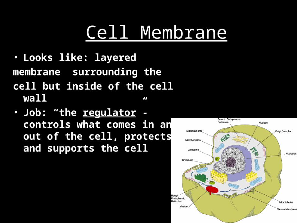
Cell Membrane• Looks like: layered
membrane surrounding the
cell but inside of the cell wall• Job: “the regulator”-controls what
comes in and out of the cell, protects, and supports the cell

Cell Wall
• Looks like: thick layer outside the plasma membrane• Job: structure and support

Nuclear Membrane

What is the Cell Cycle?
As eukaryotic cells grow and divide, they pass through a cell cycle that consists of 3 stages:• Interphase• Mitosis (cell division)• Cytokinesis (cell division)
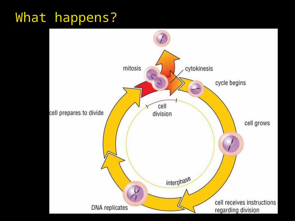
What happens?

STAGE of CELL CYCLE

Stage 1: Interphase
• Interphase is the longest stage of the cell cycle
• The cell performs its normal functions and, in preparation for cell division, duplicates its genetic material (DNA)
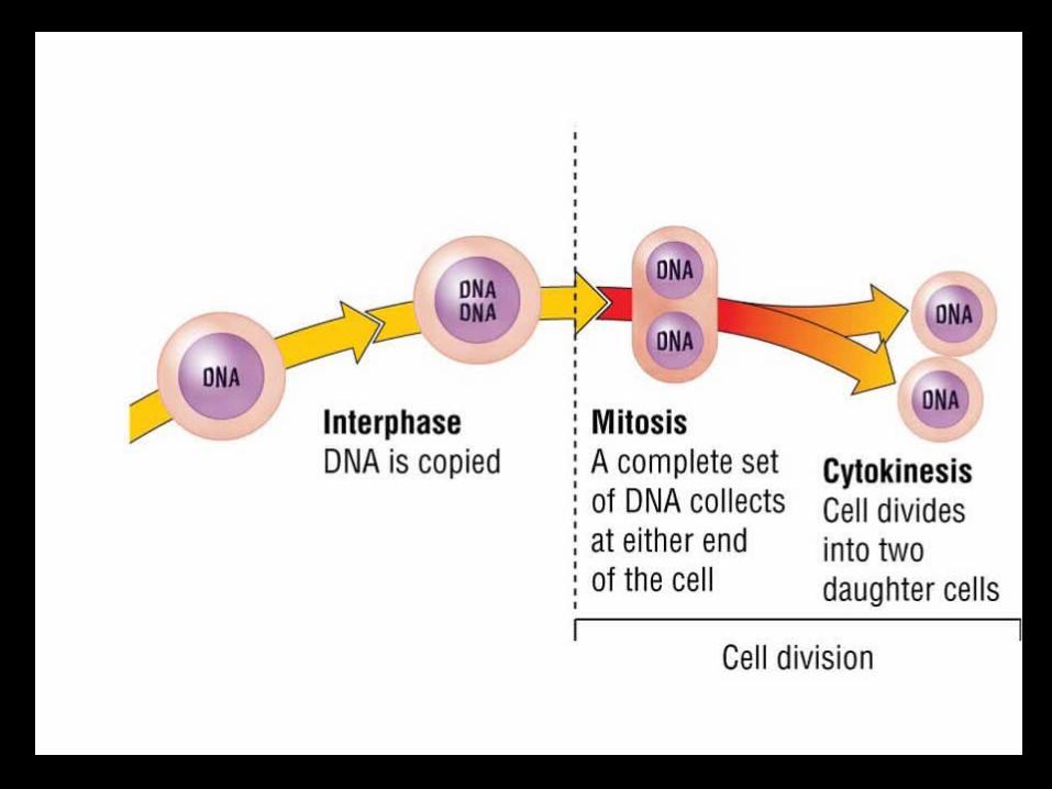

Stage 2: Cell Division - Mitosis
Consists of 4 phases:Prophase
MetaphaseAnaphaseTelophase

Mitosis Phase 1:Prophase
• DNA compacts into visible form as chromosomes
• Each chromosome contains two strands called sister chromatids.
• Sister chromatids are held together by a centromere
• The nuclear membrane breaks down

Mitosis Phase 2: Metaphase
• Chromosomes line up in the middle of the cell.

Mitosis Phase 3: Anaphase
• The centromere splits and sister chromatids separate.
• They are now called daughter chromosomes.
• They move to opposite ends of the cell, pulled by spindle fibres

Mitosis Phase 4: Telophase
• Final phase of mitosis.
• Chromosomes stretch out and are no longer visible.
• A new nuclear membrane forms around each group of daughter chromosomes.

Stage 3: Cell Division - Cytokinesis
• Final stage of cell division and cell cycle
• The cytoplasm divides and two genetically identical cells are formed. In a plant cell, a plate develops into a new cell wall
• In an animal cell, the cell membrane is pinched off in the centre

Plant vs Animal (Cytokinesis)
• In a plant cell, a plate develops into a new cell wall
• In an animal cell, the cell membrane is pinched off in the centre forming two membranes,
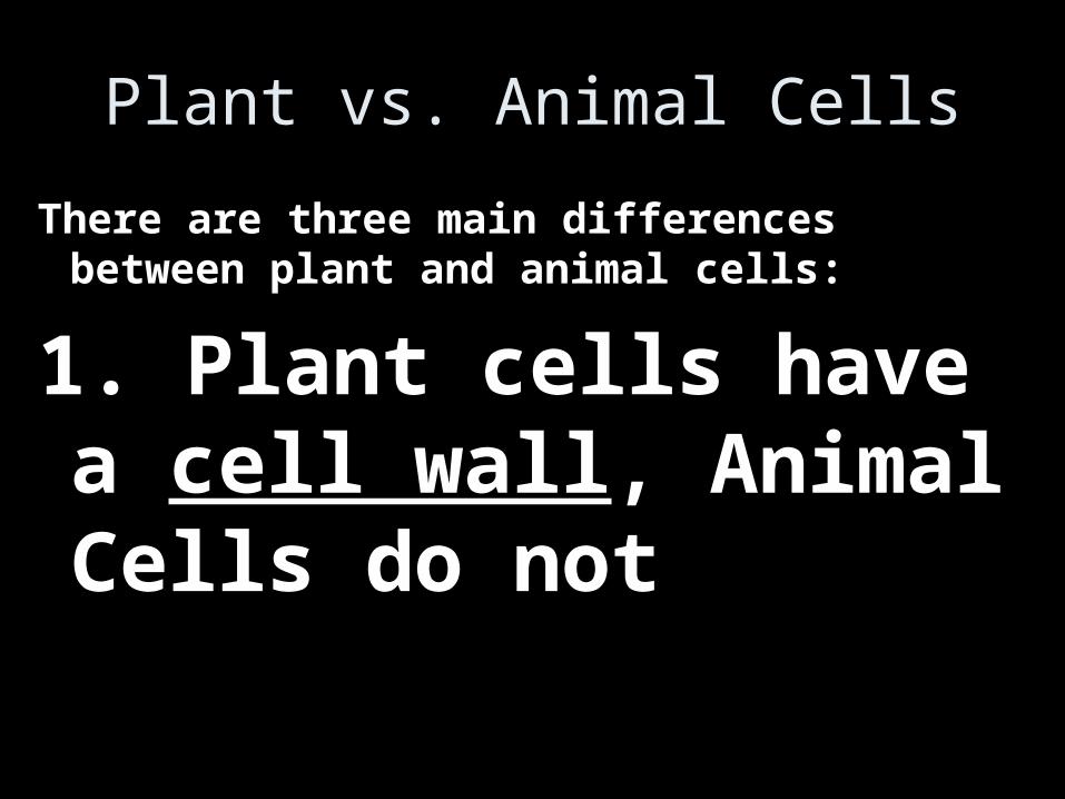
Plant vs. Animal Cells
There are three main differences between plant and animal cells:
1. Plant cells have a cell wall, Animal Cells do not

Plant vs. Animal Cells
2. Plant cells have chloroplasts and Animal Cells do not
Plant vs. Animal Cells
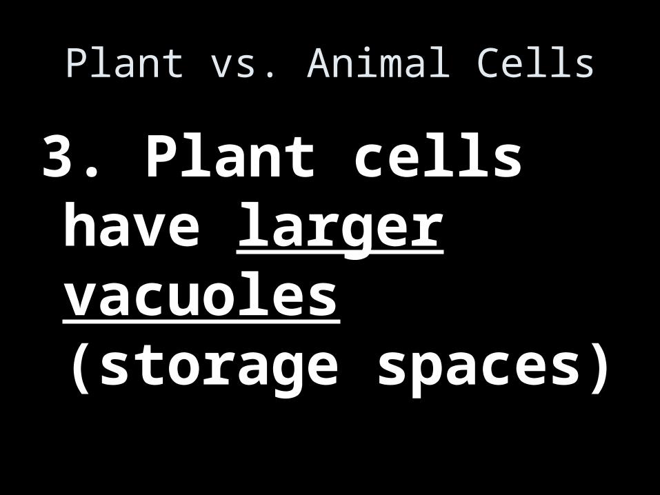
3. Plant cells have larger vacuoles (storage spaces)

Plant vs. Animal Cells
Plant Cells
1.Chloroplasts
2.Cell wall
3.Large vacuole
Animal Cells
1.No Chloroplasts
2.No Cell wall
3.Small Vacuole

Plant vs. Animal Cells
Different Shapes (Plants are rectangles, and Animals are round)

Stages of Pregnancy and Stages of Pregnancy and DevelopmentDevelopment
Fertilization
Embryonic development - Cleavage
Fetal - Differentiation
Growth – Development
Childbirth

FertilizationFertilization
The egg is viable for 12 to 24 hours after ovulation
Sperm are viable for 12 to 48 hours after ejaculation
Sperm cells must make their way to the uterine tube for fertilization to be possible

ZYGOTE
1. The sperm and egg join to form a zygote: the first cell of a new individual.
2. Zygote results of the fusion of DNA from sperm and egg
2. Fertilization occurs in the Fallopian Tubes3. The zygote begins rapid mitotic cell division4. Beginning of human development

EMBRYO0.5 cms20 days after fertilization
•Embryo begins to form organs during the third week.•Cannot tell if it is human or other vertebrate. Tall visible.
DifferentiationDifferentiation

EMBRYO1 months 0.6 cms
• Ears, nose and eyes not visible• Small arm and leg buds, backbone seen• Heart beats.
DifferentiationDifferentiation

FETUS2 months 3 cms
• During the second month most of the major organ systems form, limb buds develop.
• Limbs distinct with fingers and toes bones begin to form, eyes far apart.
• The embryo becomes a fetus by the seventh week.
DifferentiationDifferentiation

DifferentiationDifferentiation All organ systems are
formed by the end of the eighth week
Activities of the fetus are growth and organ specialization
A stage of tremendous growth and change in appearance
Fetus at nine weeks3 cm

FETUS2 months 3 cms
DifferentiationDifferentiation
•Beginning the eighth week, the sexually neutral fetus activates gene pathways for sex determination, forming testes in XY fetuses and ovaries in XX fetuses. •External genitalia develop.

Developmental Aspects of the Developmental Aspects of the Reproductive SystemReproductive System
Slide 16.64a
Copyright © 2003 Pearson Education, Inc. publishing as Benjamin Cummings
Gender is determined at fertilization
Males have XY sex chromosomes
Females have XX sex chromosomes
Gonads do not begin to form until the eighth week

Four Basic Kinds of Tissues
• Epithelial Tissue
• Connective Tissue
• Muscle Tissue
• Nervous Tissue

Epithelial Tissue• Epithelial Tissue Locations:
– Covers the body– Lines the cavities, tubes, ducts and blood
vessels inside the body– Covers the organs inside body cavities
• Epithelial Tissue Functions:– Protection from physical & chemical injury,– Protection against microbial invasion,• Examples: Skin, small intestine

Connective Tissue
• Connective Tissue:– Most abundant & widely distributed tissue
• Connective Tissue Functions:– Connects, binds and supports structures,
• Tendons, ligaments, etc.– Protects & cushions organs and tissues,– Insulates (fat) and– Transports substances (blood).
Example: Bone and Blood.

Muscle Tissue
• Muscle Tissue:– Associated with the bones of the skeleton, the heart and in the
walls of the hollow organs of the body.• Muscle Tissue Functions:
– Movement– Locomotion– Maintains posture– Produces heat– Facial expressions– Pumps blood– PeristalsisExample: arm, stomach, heart

Nervous Tissue• Nervous Tissue:
– Main component of the nervous system, ie., brain, spinal cord & nerves.
• Nervous Tissue Functions:– Regulates & controls body functions– Generates & transmits nerve impulses – Supports, insulates and protects impulse generating neurons.
There are three types of neurons, sensory neuronsmotor neurons.
connector neurons

Cell Specialization
• All cells carry the same DNA information but they do not perform the same functions and may not look the same.
• All cells start their lives as identical cells called stem cells.
• The process of a cell becoming a particular type of cell is called cell differentiation.

• Groups of cells that function together and are specialized for common tasks are called tissues.
• In simple terms, they are groups of cells that work together to do the same job

Human Organ Systems

Levels of Organization
Remember, the human body is organized in several levels, from the simplest to the most complex. . .
Cells – the basic unit of life
Tissues – clusters of cells performing a similar function
Organs – made of tissues that perform one specific function
Organ Systems – groups of organs that perform a specific purpose in the human body
***The purpose of the 11 organ systems is for the human body to maintain homeostasis.

Homeostasis
• Homeostasis is: The ability or tendency of an organism or cell to maintain internal equilibrium by adjusting its physiological processes.

The 11 Human Body Systems
The 11 human body systems are as follows:
-- nervous system -- integumentary system
-- respiratory system -- digestive system
-- excretory system -- skeletal system
-- muscular system -- circulatory system
-- endocrine system -- reproductive system
-- lymphatic (immune) system

The Digestive SystemPurpose: to convert food particles into simpler
micromolecules that can be absorbed into the bloodstream and used by the body
Major Organs and their Functions:
Mouth – to chew and grind up food
-- saliva also begins the chemical breakdown
Esophagus – pipe connecting mouth to stomach
Stomach – secretes an extraordinarily strong acid (pH = 2) that leads to breakdown of food
-- once the food is broken down in the stomach and mixed with digestive juices, it is called chyme

Pancreas – produces the hormone insulin that regulates blood sugar levels
-- also help neutralize stomach acid
Liver – produces bile, which breaks down fats in foods
Gallbladder – pouch-like organ that stores bile for future use
Small Intestine – after digestion is complete, the chyme enters the small intestine where it is absorbed into the bloodstream
-- the chyme is propelled along by folded surfaces called villi, on the intestine
Large Intestine – removes water from the chyme and gets the waste ready for excretion

The Digestive System

The Respiratory SystemPurpose: to provide the body with a fresh supply of oxygen for cellular respiration and remove the waste product carbon dioxide
Major Organs and Their Functions
Nose – internal entry and exit point for air
Pharynx – serves as a passage way for both air and food at the back of the throat
Larynx – your “voicebox”, as air passes over your vocal chords, you speak
Trachea – the “windpipe”, or what connects your pharynx to your lungs
-- a piece of skin, called the epiglottis, covers the trachea when you swallow, preventing food from entering

Bronchi – the two large passageways that lead from the trachea to your lungs (one for each lung)
-- the bronchi are further subdivided into bronchioles
-- eventually, the further subdivisions lead to tiny air sacs called alveoli
-- alveoli are in clusters, like grapes
-- capillaries surrounding each alveolus is where the exchange of gases with the blood occurs
The diaphragm is the muscle that causes you to breath
-- hiccups are involuntary contractions of the diaphragm

Image of the Respiratory System

The Circulatory SystemPurpose: to deliver oxygenated blood to the various cells and organ systems in your body so they can undergo cellular respiration
Major Organs and Their Functions
Heart – the major muscle of the circulatory system
-- pumps blood through its four chambers (two ventricles and two atria)
-- pumps deoxygenated blood into the lungs, where it gets oxygenated, returned to the heart, and then pumped out through the aorta to the rest of the body
-- valve regulate the flow of blood between the chambers

Arteries – carry blood away from the heart and to the major organs of the body
Veins – carry blood back to the heart away from the major organs of the body
Capillaries – small blood vessels where gas exchange occurs
Blood – the cells that flow through the circulatory system
-- red blood cells contain hemoglobin, an iron-rich protein that carries oxygen
-- white blood cells function in the immune system
-- platelets help in blood clotting
Spleen – helps to filter out toxins in the blood

Image of the Circulatory System

THE HUMAN HEART
The human heart consists of 4 chambers
there are two types of chambers: the atrium and the ventricle
there are two of each type in the heart
the heart weighs a mere 300 grams

HEART VALVES
The human heart contains valves which prevent blood from flowing back into the heart chambers after it has contracted
atrioventricular valves are found between the atrium and ventricle of each side of the heart
semilunar valves are found in the arteries leaving the heart to prevent blood from flowing back into the ventricles

HEART AT WORK
Each minute of the day the heart pumps 5 litres of blood
each time the heart beats it sends deoxygenated blood to the lungs and oxygenated blood to the body
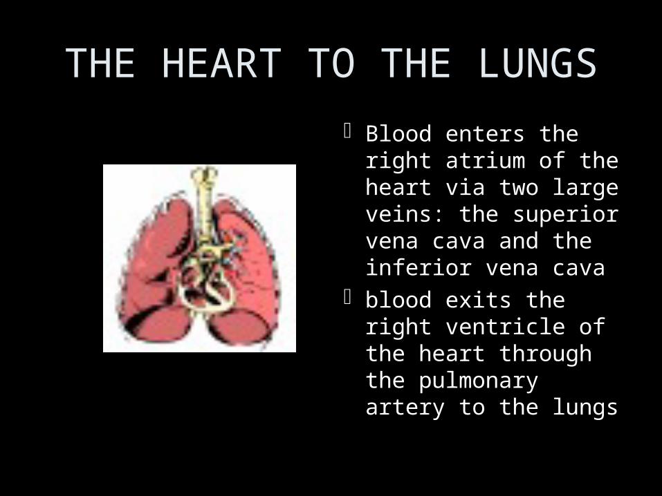
THE HEART TO THE LUNGS
Blood enters the right atrium of the heart via two large veins: the superior vena cava and the inferior vena cava
blood exits the right ventricle of the heart through the pulmonary artery to the lungs

Direction of Blood

THE HEART TO THE BODY (Circulatory and Respiratory)
Blood reenters the left artrium of the heart via the pulmonary vein
blood on the left side of the heart is oxygenated and is pumped into the body by the left ventricle through the aorta

What is cancer?
• Caner is defined as the continuous uncontrolled growth of cells.
• A tumor is a any abnormal proliferation of cells.• Benign tumors stays confined to its original
location• Malignant tumors are capable of invading
surrounding tissue or invading the entire body• Tumors are classified as to their cell type• Tumors can arise from any cell type in the body

What is Melanoma?What is Melanoma?1.1. A type of skin cancerA type of skin cancer2.2. Some risk factors Some risk factors
include:include:1.1. Sun exposure -Sun exposure -
depleting ozone layer depleting ozone layer 2.2. Presence of many or Presence of many or
unusual molesunusual moles3.3. Skin typesSkin types4.4. Genetics Genetics
predispositionpredisposition

benignmalignant
skin


Diagnosis and Diagnosis and Medical Imaging Medical Imaging
TechnologyTechnologySNC2DSNC2D
DiagnosisDiagnosis
• The interdependence of our organ systems can The interdependence of our organ systems can sometimes make it difficult to pinpoint the source sometimes make it difficult to pinpoint the source of a medical problem.of a medical problem.
• Doctors are trained to look for symptoms that Doctors are trained to look for symptoms that are characteristic of specific problems.are characteristic of specific problems.

DiagnosisDiagnosis
• The interdependence of our organ systems can The interdependence of our organ systems can sometimes make it difficult to pinpoint the source sometimes make it difficult to pinpoint the source of a medical problem.of a medical problem.
• Doctors are trained to look for symptoms that Doctors are trained to look for symptoms that are characteristic of specific problems (e.g. are characteristic of specific problems (e.g. swollen lymph nodes are a symptom of swollen lymph nodes are a symptom of infection).infection).

Tools of the TradeTools of the Trade
To collect information about what’s going on To collect information about what’s going on inside the body, doctors may use devices inside the body, doctors may use devices like the stethoscope (to listen to the heart like the stethoscope (to listen to the heart and lungs)and lungs)

Tools of the TradeTools of the Trade
To collect information about what’s going on To collect information about what’s going on inside the body, doctors may use devices inside the body, doctors may use devices like the stethoscope (to listen to the heart like the stethoscope (to listen to the heart and lungs) and a sphygmomanometer (to and lungs) and a sphygmomanometer (to measure blood pressure).measure blood pressure).

Tests of the TradeTests of the Trade
Doctors may also order tests of material Doctors may also order tests of material collected from the body (such as blood collected from the body (such as blood and urine) which may be analyzed by a and urine) which may be analyzed by a separate lab.separate lab.
E.g. Blood may be tested to determine the levels E.g. Blood may be tested to determine the levels of red blood cells, white blood cells, sugar, and of red blood cells, white blood cells, sugar, and hormones – the chemicals that carry messages hormones – the chemicals that carry messages through the body to regulate the functioning of through the body to regulate the functioning of organs.organs.

Medical Imaging TechnologiesMedical Imaging Technologies
Diagnostic imaging tests can provide Diagnostic imaging tests can provide doctors with even more information: an doctors with even more information: an actual visual picture of the structure and actual visual picture of the structure and functioning of organs.functioning of organs.
However, these technologies are often However, these technologies are often expensive, and the effectiveness of each expensive, and the effectiveness of each technology is limited by its properties.technology is limited by its properties.

EndoscopyEndoscopy
An endoscope is a thin, flexible tube that An endoscope is a thin, flexible tube that has a bright light and a video camera that has a bright light and a video camera that can be used to image the digestive tract, can be used to image the digestive tract, as in a colonoscopy.as in a colonoscopy.

ThermographyThermography
In thermograms, infrared light cameras are In thermograms, infrared light cameras are used to diagnose problems with used to diagnose problems with circulation.circulation.
NormalNormal Raynaud’s syndromeRaynaud’s syndrome
X-RaysX-Rays
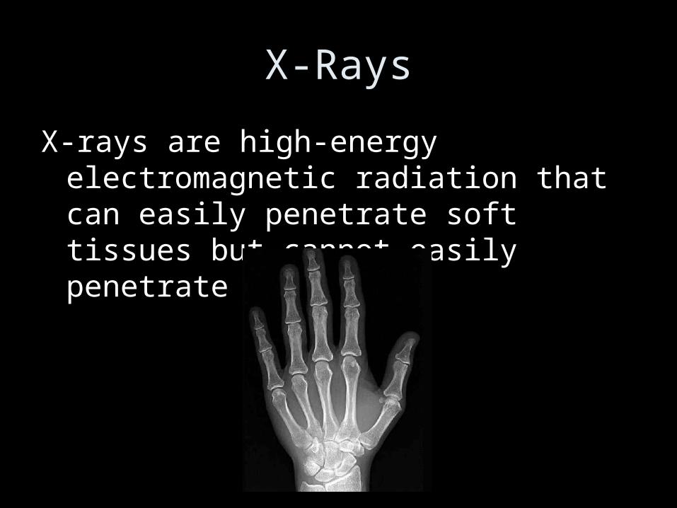
X-rays are high-energy electromagnetic X-rays are high-energy electromagnetic radiation that can easily penetrate soft radiation that can easily penetrate soft tissues but cannot easily penetrate bone.tissues but cannot easily penetrate bone.

X-RaysX-Rays
X-rays are used to check for cancers (e.g. X-rays are used to check for cancers (e.g. mammograms), to diagnose problems in mammograms), to diagnose problems in the circulatory and respiratory systems, the circulatory and respiratory systems, and to check for broken bones.and to check for broken bones.
They are quick, painless, and non-invasive They are quick, painless, and non-invasive but exposure to x-rays can damage cells but exposure to x-rays can damage cells and increases cancer risk.and increases cancer risk.

Body ScannersBody Scanners
Note that this is also true for the body Note that this is also true for the body scanners that are used at airports, scanners that are used at airports, especially the new “naked body scanners.”especially the new “naked body scanners.”

Body ScannersBody Scanners
Since the radiation emitted by these body Since the radiation emitted by these body scanners is absorbed at the level of the scanners is absorbed at the level of the skin, it is your skin cancer risk that is most skin, it is your skin cancer risk that is most increased – and the scans cannot find increased – and the scans cannot find anything concealed in any body cavity.anything concealed in any body cavity.
Also, like most x-rays, they do not show Also, like most x-rays, they do not show contrast that may be used to identify soft contrast that may be used to identify soft materials (like plastics and chemical materials (like plastics and chemical explosives).explosives).
In other words, they’re completely ineffective.In other words, they’re completely ineffective.

Computed TomographyComputed Tomography
Computed tomography Computed tomography (CT) scans, also called (CT) scans, also called computer-assisted computer-assisted tomography (CAT) tomography (CAT) scans, use x-rays to scans, use x-rays to produce images at produce images at different angles through different angles through the body so that a 3D the body so that a 3D image can be image can be constructed.constructed.

Computed TomographyComputed Tomography
CT scans may be used to diagnose cancers, CT scans may be used to diagnose cancers, skeletal abnormalities and vascular skeletal abnormalities and vascular diseases (affecting blood vessels).diseases (affecting blood vessels).
But since CT scans use x-rays, they also But since CT scans use x-rays, they also increase your cancer risk.increase your cancer risk.

FluoroscopyFluoroscopy
Fluoroscopy is a technique in which a Fluoroscopy is a technique in which a continuous beam of x-rays is used to continuous beam of x-rays is used to produce moving images. produce moving images.
It is used to show movement in the digestive It is used to show movement in the digestive system (which may require ingestion of a system (which may require ingestion of a high-contrast liquid such as barium) and high-contrast liquid such as barium) and the circulatory system (angiograms).the circulatory system (angiograms).

UltrasoundUltrasound
Ultrasound is high-frequency sound waves Ultrasound is high-frequency sound waves produced by a device called a transducer produced by a device called a transducer that are reflected back to the transducer that are reflected back to the transducer by internal body structures. by internal body structures.

UltrasoundUltrasound
Ultrasound is used to study soft tissues and Ultrasound is used to study soft tissues and organs, especially the heart organs, especially the heart (echocardiograms) and especially during (echocardiograms) and especially during pregnancy.pregnancy.
Because the presence of gas can distort Because the presence of gas can distort images, ultrasound is not often used for images, ultrasound is not often used for imaging the respiratory or digestive imaging the respiratory or digestive systems.systems.

Magnetic Resonance Imaging Magnetic Resonance Imaging (MRI)(MRI)
Magnetic Resonance Imaging (MRI) is a Magnetic Resonance Imaging (MRI) is a technique that uses strong magnets and technique that uses strong magnets and radio waves that interact with the radio waves that interact with the hydrogen atoms in your body (esp. in hydrogen atoms in your body (esp. in water). A computer is used to construct water). A computer is used to construct an image from the signal received from the an image from the signal received from the atoms.atoms.

Magnetic Resonance Imaging Magnetic Resonance Imaging (MRI)(MRI)
MRI is used to image the structure and MRI is used to image the structure and function of the brain, heart, soft tissue, and function of the brain, heart, soft tissue, and the inside of bones; to diagnose cancers, the inside of bones; to diagnose cancers, brain diseases, and problems with the brain diseases, and problems with the circulatory system.circulatory system.
But it is also extremely expensive and the But it is also extremely expensive and the availability of machines/technicians is availability of machines/technicians is limited.limited.

Positron Emission Tomography Positron Emission Tomography (PET)(PET)
PET scans are a type of nuclear medicine is PET scans are a type of nuclear medicine is which a patient is given a radioisotope that which a patient is given a radioisotope that emits positron radiation; the radioisotope emits positron radiation; the radioisotope is attached to a chemical absorbed by is attached to a chemical absorbed by certain tissues or organs.certain tissues or organs.
It is used to detect cancers, It is used to detect cancers,
heart disease, heart disease,
and some brain disorders and some brain disorders
(such as Alzheimer’s).(such as Alzheimer’s).

• End!!!!


![[PPT]mstrainorbiology.files.wordpress.com · Web viewAn Organism is a Living Thing But What Makes Something ALIVE??? Basic unit of “organization” is the cell Contains DNA, the](https://img.pdfslide.net/doc/110x75/5ae8636e7f8b9ae157906811/ppt-viewan-organism-is-a-living-thing-but-what-makes-something-alive-basic.jpg)