Embed Size (px)
Citation preview
ORIGINAL RESEARCH
Sodium glucose transporter-2 inhibition has norenoprotective effects on non-diabetic chronic kidneydiseaseQiuyue Ma*, Stefanie Steiger* & Hans-Joachim Anders
Medizinische Klinik und Poliklinik IV, Klinikum der Universit€at M€unchen, Munich, Germany
Keywords
Crystal, diabetic nephropathy,
glomerulosclerosis, progression.
Correspondence
Hans-Joachim Anders, Medizinische Klinik
und Poliklinik IV, Klinikum der Universit€at
M€unchen-LMU, Ziemssenstr. 1, 80336
Munich, Germany.
Tel: +49-89-440053583
Fax: +49-89-440053379
E-mail: [email protected]
Funding Information
This work was supported by grants from the
Deutsche Forschungsgemeinschaft to HJA
(AN372/16-1 and AN372/20-1) and to SS
(STE2437/2-1).
Received: 14 December 2016; Revised: 16
February 2017; Accepted: 2 March 2017
doi: 10.14814/phy2.13228
Physiol Rep 5 (7), 2017, e13228,
doi: 10.14814/phy2.13228
*Contributed equally to this work.
Abstract
Sodium glucose transporter (SGLT)-2 inhibition has renoprotective effects in
diabetic kidney disease. Whether similar effects can be achieved also in non-
diabetic kidney disease is speculative. Chronic kidney disease was induced in
C57BL/6N mice by feeding an oxalate-rich diet for 14 days, known to induce
nephrocalcinosis-related tubular atrophy and interstitial fibrosis without
directly affecting the glomerular compartment. Empagliflozin treatment started
from day 0 of oxalate feeding had no effect on the decline of glomerular filtra-
tion rate, crystal deposition, blood urea nitrogen or serum creatinine levels on
day 7 and 14. Tissue morphometry of tubular injury and kidney mRNA levels
of kidney injury molecule-1 or tissue inhibitor of metalloproteinase-2 were
comparable between empagliflozin- and vehicle-treated mice with oxalate
nephropathy on day 7 and 14. Similarly, empagliflozin did not affect markers
of interstitial fibrosis, including silver, alpha smooth muscle actin (aSMA) and
collagen 1 staining, and mRNA levels of fibronectin-1, collagen 1a1, fibrob-last-specific protein-1, and transforming growth factor (TGF)-b2 on day 7
and 14. Thus, the specific renoprotective mechanisms-of-action of SGLT2
inhibition in diabetic kidney disease do not apply to chronic oxalosis, a non-
diabetic form of chronic kidney disease.
Introduction
Chronic kidney disease (CKD) is a global health care
problem of increasing dimension (Eckardt et al. 2013).
CKD progression is the result of progressive loss of
nephrons being replaced by fibrous extracellular matrix
components, that is interstitial fibrosis, which stabilizes
the remaining nephrons. Therapeutic interventions aiming
to retard or stop the progressive loss of nephrons and the
associated decline of renal excretory function can be
divided into two categories: Firstly, abolishing the specific
cause of disease, an approach that is feasible in renal
infections or toxic kidney injury, which is still impossible
in genetic kidney disease or aging nephropathy, and
which is notoriously difficult in complex disease states,
such as metabolic syndrome. Secondly, targeting unspeci-
fic mechanisms of CKD progression, for example reduc-
ing glomerular hyperfiltration and hypertension with
inhibitors of the renin-angiotensin system (RAS)
(Remuzzi et al. 2006).
Recently, inhibitors of the sodium-glucose transporter
(SGLT)-2 were found to elicit renoprotective effects in
kidney disease in diabetic mice and rats (Gembardt et al.
2014; Ojima et al. 2015 Gallo et al. 2016;). Indeed, a large
ª 2017 The Authors. Physiological Reports published by Wiley Periodicals, Inc. on behalf of
The Physiological Society and the American Physiological Society.
This is an open access article under the terms of the Creative Commons Attribution License,
which permits use, distribution and reproduction in any medium, provided the original work is properly cited.
2017 | Vol. 5 | Iss. 7 | e13228Page 1
Physiological Reports ISSN 2051-817X

randomized controlled trial demonstrated a profound
renoprotective effect of the SGLT2 inhibitor empagliflozin
in addition to RAS blockade in patients with type 2 dia-
betes (Wanner et al. 2016). Empagliflozin somewhat
improved glycemic control and hypertension, but these
effects alone could not explain the immediate drop in the
glomerular filtration rate (GFR) without any further
decline during the following 3 years (Wanner et al. 2016).
While the underlying mechanisms of renoprotection
remain debated these findings raise the question whether
SGLT2 inhibition can elicit renoprotective effects also in
non-diabetic kidney disease. To address this question, we
tested empagliflozin versus vehicle treatment in a murine
model of progressive non-diabetic CKD based on oxalate-
induced nephropathy mimicking primary hyperoxaluria-
related nephrocalcinosis.
Materials and Methods
Animals and experimental design
Eight week-old male C57BL/6N mice (Charles River Lab-
oratories, Sulzfeld, Germany) were maintained under
standard housing conditions with 10 mice per group, and
free access to food and water. High-oxalate diet was pre-
pared by adding sodium oxalate (50 lmoles/g) to a cal-
cium-free diet or calcium-free diet without sodium
oxalate (control, both from Ssniff, Soest, Germany), as
previously described (Knauf et al. 2013). Removal of cal-
cium from the diet increases the amount of soluble oxa-
late available for absorption as previously shown (Knauf
et al. 2013). Empagliflozin was dissolved in hydroxy ethyl
cellulose (HEC) and administered by oral gavage (10 mg/
kg, 200 lL) to the oxalate-treated mice once daily for 7
or 14 days, whereas the vehicle group was given the same
volume of HEC alone. All animal experiments were per-
formed in accordance with the European protection laws
of animal welfare, and with approval by the local govern-
ment authorities Regierung von Oberbayern (reference
number: 55.2-1-54-2532-189-2015).
Measurement of plasma BUN, creatinine,and glucose
Mouse blood was collected from the tail vein of mice on
the day of sacrifice. Blood samples were centrifuged at
8000 rpm for 8 min and plasma transferred to 1.5 mL
plastic Eppendorf tubes, stored at �20°C until analysis.
Plasma creatinine and blood urea nitrogen (BUN) were
measured using the Creatinine FS kit and Urea FS kit
from DiaSys Diagnostic Systems, Germany. Plasma glu-
cose was measured using Glucose GOD FS kit (DiaSys)
according to the protocol provided by the manufacturer.
Measurement of urine oxalate, calcium, andglucose
Mouse urine samples were collected at different time
points and stored at �20°C until analysis. Urine oxalate
concentration was assessed with a colorimetric, enzymatic
assay (Oxalate assay kit, Libios) in 96-well plate according
to the manufacturer’s instructions. Urine calcium concen-
tration was assessed using calcium colorimetric assay kit
(Sigma-Aldrich), and urinary glucose levels were deter-
mined using Glucose GOD FS kit (DiaSys).
Assessment of renal histology
Kidneys were fixed in 4% formaldehyde solution for his-
tology. 2 lm thick sections were stained with periodic
acid-Schiff (PAS) reagent and for the kidney injury mar-
ker-1 (KIM-1). The renal tubular injury was evaluated by
assessing the percentage of necrotic tubules, dilation
tubules and presence of tubular casts (59 and 209 mag-
nification). Fibrotic areas were identified by silver, aSMA
and collagen 1 stain, and counted in 15 high power fields
(hpf) per section using Image J software. Calcium oxalate
(CaOx) crystals were visualized by Pizzolato staining and
crystal deposit areas evaluated using Image J. All assess-
ments were performed by an observer blinded to the
experimental conditions.
mRNA isolation and real-time quantitative–PCR
Total RNA was isolated and purified from murine kidneys
with a Qiagen RNA extraction kit (Qiagen, Germany)
according to the manufacturer’s instructions. mRNA was
reverse transcribed to cDNA and carried out by reverse
transcriptase (Superscript II, Invitrogen, USA). Real time
RT-PCR was performed using SYBR Green PCR master
mix and analyzed with a Light Cycler 480 (Roche, Ger-
many). The relative expression of target genes was normal-
ized to 18s RNA control. The gene-specific primer
sequences were: KIM-1 Forward, TCAGCTCGGGAATG-
CACAA; KIM-1 Reverse, TGGTTGCCTTCCGTGTCTCT;
TIMP-2 Forward, CAGACGTAGTGATCAGAGCCAAA;
TIMP-2 Reverse, ACTCGATGTCTTTGTCAGGTCC;
collagen 1a1 Forward, ACATGTTCAGCTTTGTGGACC;
collagen 1a1 Reverse, TAGGCCATTGTGTATGCAGC;
fibronectin-1 Forward, GGAGTGGCACTGTCAACCTC;
fibronectin-1 Reverse, ACTGGATGGGGTGGGAAT; TNFaForward, CCACCACGCTCTTCTGTCTAC; TNFa Reverse,
AGGGTCTGGGCCATAGAACT; NLRP3 Forward, CCA-
CAGTGTAACTTGCAGAAGC; NLRP3 Reverse, GGTGT
GTGAAGTTCTGGTTGG; FSP-1 Forward, CAG CAC TTC
CTC TCT CTT GG; FSP-1 Reverse, TTT GTG GAA GGT
2017 | Vol. 5 | Iss. 7 | e13228Page 2
ª 2017 The Authors. Physiological Reports published by Wiley Periodicals, Inc. on behalf of
The Physiological Society and the American Physiological Society.
SGLT2 Inhibition in Non-Diabetic CKD Q. Ma et al.

GGA CAC AA; TGFb2 Forward, GAT AAT TGC TGC CTT
CGC CC; TGFb2 Reverse, GGC TGA GGA CTT TGG TGT
GT; 18S RNA Forward, GCAATTATTCCCCATGAACG;
18S Reverse, AGGGCCTCACTAAACCATCC; IGFBP7 For-
ward, AAGAGGCGGAAGGGTAAAGC; IGFBP7 Reverse,
TGGGGTAGGTGATGCCGTT. The data were analyzed
using the 2�DDCT method.
Transcutaneous measurement of glomerularfiltration rate (GFR) in conscious mice
For GFR measurement, mice where anesthetized with
isoflurane and a miniaturized imager device built from
two light-emitting diodes, a photodiode and a battery
(Medibeacon, Mannheim, Germany) was mounted via a
double-sided adhesive tape onto the shaved animal’s
neck (Schreiber et al. 2012). The fluorescent dye FITC-
Sinistrin (150 mg/kg, Mannheim Pharma & Diagnostics
GmbH, Germany) was injected intravenously, and each
mouse was conscious and kept in a single cage through-
out 1.5 h. After 1.5 h, the imager device was removed
and the data analyzed using MPD Lab software (Man-
nheim Pharma & Diagnostics GmbH, Germany). The
GFR (lL/min) was calculated from the decrease of fluo-
rescence intensity over time (i.e., plasma half-life of
FITC-Sinistrin) using a two-compartment model, body
weight of mouse, and an empirical conversion factor
(Schreiber et al. 2012).
Statistical data analysis
Data are shown as means � SEM between-group differ-
ences were analyzed by two-tailed Student t-test or one-
way ANOVA, with Tukey’s post hoc test, in which
P < 0.05 was considered statistically significant.
Results
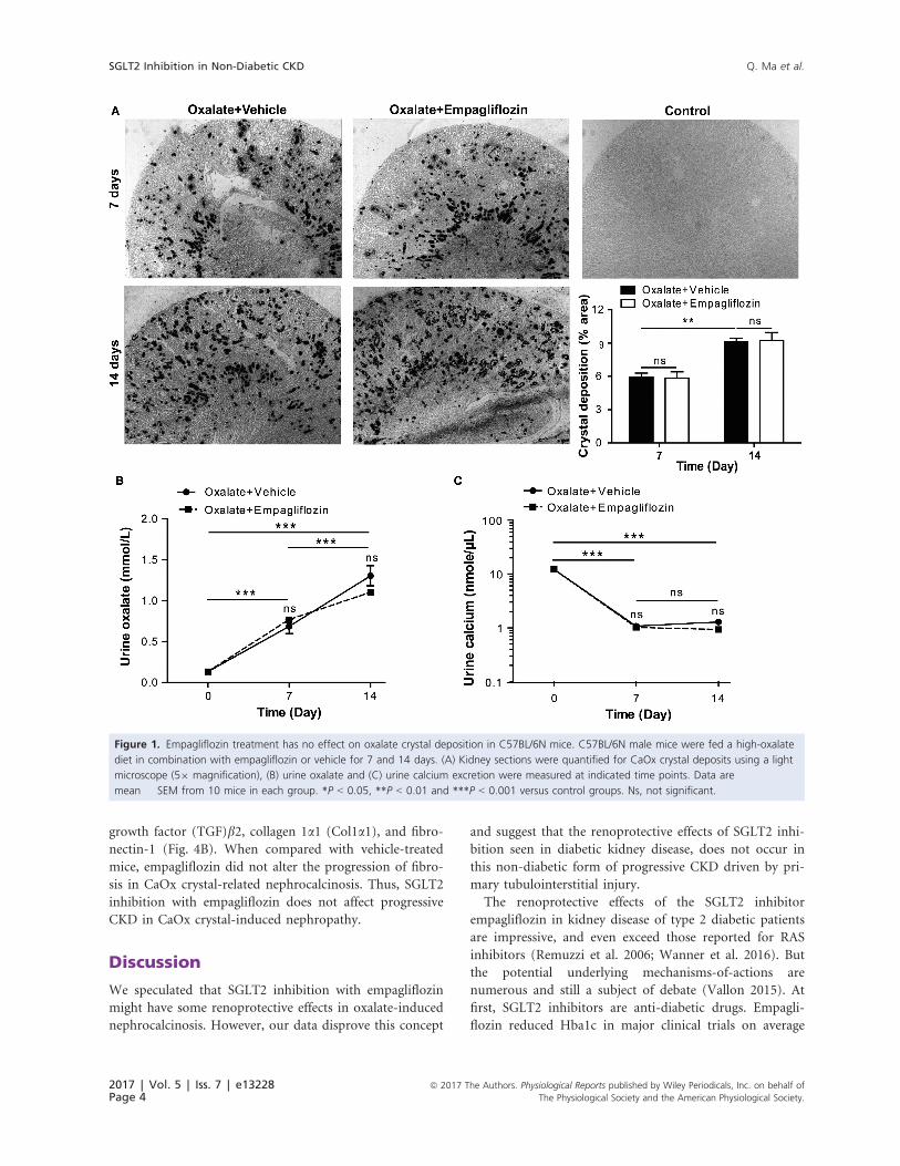
Empagliflozin has no effect on renal oxalatecrystal deposition
Feeding a diet rich in oxalate but depleted of calcium to
C57BL/6N mice represents a reliable model of non-dia-
betic nephrocalcinosis and progressive CKD (Knauf et al.
2013; Mulay 2016a). Indeed, oxalate feeding resulted in
the progressive deposition of calcium oxalate (CaOx)
crystals in the renal medulla and cortex after 7 and
14 days (Fig. 1A). Treatment with empagliflozin had no
effect on CaOx crystal deposition compared to vehicle, as
indicated by Pizzolato staining and the % area of crystal
deposition (Fig. 1A). Urinary oxalate levels significantly
increased over time following oxalate feeding (Fig. 1B).
Urinary calcium levels rather declined upon feeding a
calcium-depleted diet (Fig. 1C), but empagliflozin treat-
ment did neither affect oxaluria nor calciuria (Fig. 1B
and 1C). This enabled us to assess the impact of empagli-
flozin treatment in comparison to untreated control mice
with identical oxalate-induced kidney injury.
Empagliflozin does not attenuate theprogressive loss of renal function
Hyperoxaluria-induced nephrocalcinosis resulted in a pro-
gressive decline in the GFR (Fig. 2A), and in a significant
increase in plasma BUN and creatinine levels (Fig. 2C)
after 7 and 14 days. Treatment with the SGLT-2 inhibitor
empagliflozin did not attenuate the progressive loss of
renal function, as indicated by GFR, plasma BUN, and
creatinine compared to vehicle treatment (Fig. 2A and
C). No difference in the kidney/body weight ratio
between empagliflozin and vehicle treatment was observed
in oxalate-treated mice (Fig. 2B). However, empagliflozin
had no significant effect on serum glucose levels
(Fig. 2D), despite inducing significant glucosuria com-
pared to vehicle-treated control mice (Fig. 2E). Together,
these data imply that inhibiting SGLT-2 with empagliflo-
zin does not affect the progressive decline of renal func-
tion in CaOx crystal-induced nephropathy.
Empagliflozin and renal pathology in CaOxcrystal-induced CKD
Tubular atrophy and interstitial fibrosis are hallmark fea-
tures of progressive CKD regardless of the underlying dis-
ease trigger. We next examined the effect of empagliflozin
on renal injury in CaOx crystal-induced CKD at various
time points. Renal CaOx crystal deposition was associated
with progressive tubular atrophy in vehicle- and empagli-
flozin-treated mice after 7 and 14 days compared to con-
trol diet, as indicated by PAS staining and tubular injury
score (Fig. 3A). Intrarenal mRNA expression of the kid-
ney injury marker KIM-1, metallopeptidase inhibitor-2
(TIMP-2), and insulin like growth factor binding protein
7 (IGFBP7) (Fig. 3B), as well as that of the pro-inflam-
matory mediators tumor necrosis factor (TNF)a and the
inflammasome protein NLRP3 (Fig. 3C) was increased in
oxalate-fed mice compared to control diet. This was also
the case for the protein expression of KIM-1 in hyperox-
aluric mice compared to control diet (Fig. 3D). However,
empagliflozin had no renoprotective effect regarding
tubular injury and inflammation in CaOx crystal-induced
nephropathy.
Finally, progressive interstitial fibrosis was assessed by
aSMA and collagen 1 staining (Fig. 4A), silver staining
(Fig. S1), and mRNA expression profiling for the fibrosis
marker fibroblast-specific protein (FSP)-1, transforming
ª 2017 The Authors. Physiological Reports published by Wiley Periodicals, Inc. on behalf ofThe Physiological Society and the American Physiological Society.
2017 | Vol. 5 | Iss. 7 | e13228Page 3
Q. Ma et al. SGLT2 Inhibition in Non-Diabetic CKD
growth factor (TGF)b2, collagen 1a1 (Col1a1), and fibro-
nectin-1 (Fig. 4B). When compared with vehicle-treated
mice, empagliflozin did not alter the progression of fibro-
sis in CaOx crystal-related nephrocalcinosis. Thus, SGLT2
inhibition with empagliflozin does not affect progressive
CKD in CaOx crystal-induced nephropathy.
Discussion
We speculated that SGLT2 inhibition with empagliflozin
might have some renoprotective effects in oxalate-induced
nephrocalcinosis. However, our data disprove this concept
and suggest that the renoprotective effects of SGLT2 inhi-
bition seen in diabetic kidney disease, does not occur in
this non-diabetic form of progressive CKD driven by pri-
mary tubulointerstitial injury.
The renoprotective effects of the SGLT2 inhibitor
empagliflozin in kidney disease of type 2 diabetic patients
are impressive, and even exceed those reported for RAS
inhibitors (Remuzzi et al. 2006; Wanner et al. 2016). But
the potential underlying mechanisms-of-actions are
numerous and still a subject of debate (Vallon 2015). At
first, SGLT2 inhibitors are anti-diabetic drugs. Empagli-
flozin reduced Hba1c in major clinical trials on average
Figure 1. Empagliflozin treatment has no effect on oxalate crystal deposition in C57BL/6N mice. C57BL/6N male mice were fed a high-oxalate
diet in combination with empagliflozin or vehicle for 7 and 14 days. (A) Kidney sections were quantified for CaOx crystal deposits using a light
microscope (59 magnification), (B) urine oxalate and (C) urine calcium excretion were measured at indicated time points. Data are
mean � SEM from 10 mice in each group. *P < 0.05, **P < 0.01 and ***P < 0.001 versus control groups. Ns, not significant.
2017 | Vol. 5 | Iss. 7 | e13228Page 4
ª 2017 The Authors. Physiological Reports published by Wiley Periodicals, Inc. on behalf of
The Physiological Society and the American Physiological Society.
SGLT2 Inhibition in Non-Diabetic CKD Q. Ma et al.

by 0.62% (Wanner et al. 2016). Glycemic control with
other drugs for up to 3, 7 years in the Action to Control
Cardiovascular Risk in Diabetes (ACCORD) trial resulted
in decreased albuminuria, but not in progressive CKD
after 5 years (Ismail-Beigi et al. 2010). Controlling glyce-
mic patients had no effect on the pre-specified renal out-
comes in the UK Prospective Diabetes Study (UKPDS)
trial (Bilous 2008). These data imply that empagliflozin‘s
anti-diabetic effect alone does not account for its pro-
found renoprotective effects. Similarly, empagliflozin
reduced systolic and diastolic blood in major clinical trials
on average by 4.19 and 1.88 mmHg, respectively (Liakos
et al. 2014). However, an even more rigorous blood
pressure control in patients with diabetes did not reveal
renoprotective effects comparable to those seen in the
EMPA-REG trial (UK Prospective Diabetes Study Group,
1998; de Galan et al. 2009; Group, A.S., et al. 2010;
Ismail-Beigi et al. 2012). Reactivation of the tubu-
loglomerular feedback (TGF) is another possible mecha-
nism-of-action, because glomerular hyperfiltration and
hypertension in diabetes are rather a direct consequence
of SGLT2-mediated increased sodium reabsorption in the
proximal tubule, which reduces sodium delivery in the
macula densa, a process that deactivates the TGF and
causes vasodilation of the afferent glomerular arteriole
(Carlstrom et al. 2015; Skrtic and Cherney 2015; Vallon
2015). This mechanism seems specific for hyperglycemic
states, because only increased glucose filtration would
serve as a trigger and driver of decreased sodium delivery
to the macula densa (Anders et al. 2016). In contrast, in
the absence of hyperglycemia SGLT2 is not induced and
contributes only minimally to proximal sodium
Figure 2. Empagliflozin does not prevent from oxalate-induced renal dysfunction. C57BL/6N male mice received high-oxalate diet with
empagliflozin or vehicle daily for 7 and 14 days. (A) Glomerular filtration rate (GFR), (B) kidney/body weight ratio, (C) plasma BUN and plasma
creatinine levels, and (D) plasma and (E) urine glucose levels were measured at indicated time points. Data are mean � SEM from 10 mice in
each group. *P < 0.05, **P < 0.01 and ***P < 0.001 versus control groups. Ns, not significant.
ª 2017 The Authors. Physiological Reports published by Wiley Periodicals, Inc. on behalf ofThe Physiological Society and the American Physiological Society.
2017 | Vol. 5 | Iss. 7 | e13228Page 5
Q. Ma et al. SGLT2 Inhibition in Non-Diabetic CKD
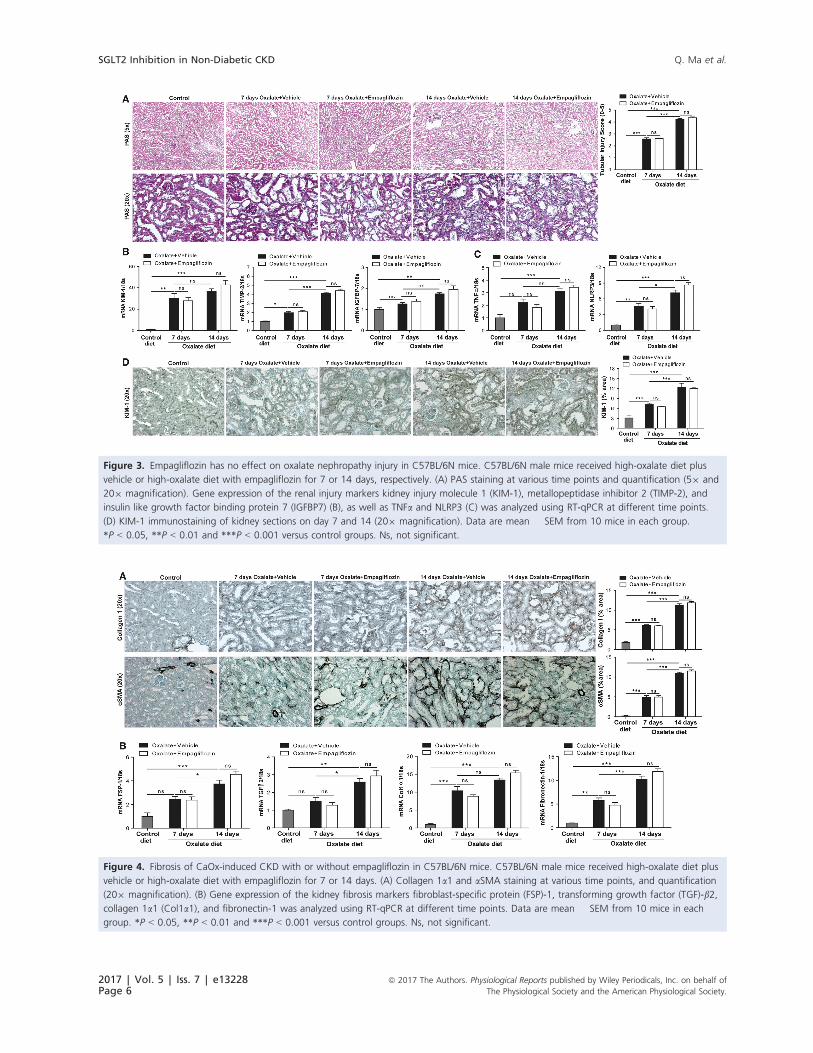
Figure 3. Empagliflozin has no effect on oxalate nephropathy injury in C57BL/6N mice. C57BL/6N male mice received high-oxalate diet plus
vehicle or high-oxalate diet with empagliflozin for 7 or 14 days, respectively. (A) PAS staining at various time points and quantification (59 and
209 magnification). Gene expression of the renal injury markers kidney injury molecule 1 (KIM-1), metallopeptidase inhibitor 2 (TIMP-2), and
insulin like growth factor binding protein 7 (IGFBP7) (B), as well as TNFa and NLRP3 (C) was analyzed using RT-qPCR at different time points.
(D) KIM-1 immunostaining of kidney sections on day 7 and 14 (209 magnification). Data are mean � SEM from 10 mice in each group.
*P < 0.05, **P < 0.01 and ***P < 0.001 versus control groups. Ns, not significant.
Figure 4. Fibrosis of CaOx-induced CKD with or without empagliflozin in C57BL/6N mice. C57BL/6N male mice received high-oxalate diet plus
vehicle or high-oxalate diet with empagliflozin for 7 or 14 days. (A) Collagen 1a1 and aSMA staining at various time points, and quantification
(209 magnification). (B) Gene expression of the kidney fibrosis markers fibroblast-specific protein (FSP)-1, transforming growth factor (TGF)-b2,
collagen 1a1 (Col1a1), and fibronectin-1 was analyzed using RT-qPCR at different time points. Data are mean � SEM from 10 mice in each
group. *P < 0.05, **P < 0.01 and ***P < 0.001 versus control groups. Ns, not significant.
2017 | Vol. 5 | Iss. 7 | e13228Page 6
ª 2017 The Authors. Physiological Reports published by Wiley Periodicals, Inc. on behalf of
The Physiological Society and the American Physiological Society.
SGLT2 Inhibition in Non-Diabetic CKD Q. Ma et al.
reabsorption, hence, SGLT2 inhibition may not signifi-
cantly increase distal sodium delivery and elicit hemody-
namic effects on glomerular filtration.
In contrast to the critical role of SGLT2 in the pathogene-
sis of progressive diabetic kidney disease, our data imply that
the progression of oxalate nephropathy does not involve
SGLT2. Chronic oxalosis is driven by intrarenal supersatura-
tion of oxalate and calcium ions that promote crystal forma-
tion inside the tubular lumen, especially in segments of
calcium secretion and urine concentration (Cochat and
Rumsby 2013). Calcium oxalate crystal adhesion depends
on the luminal expression of several crystal adhesion mole-
cules, such as CD44, annexin II, and osteopontin (Asselman
et al. 2003) which are induced by TNF receptor signaling in
tubular epithelial cells (Mulay 2016b). Crystal adhesion pro-
motes further crystal growth to crystal plugs that obstruct
the tubular lumen leading to intrarenal nephron obstruction
(Mulay 2016b). Tubule obstruction is then followed by
tubular atrophy and replacement with fibrous tissue. Hence,
progressive nephrocalcinosis is associated with a progressive
decline in renal function, as illustrated by a decline in GFR
(Mulay 2016a). SGLT2 does not seem to interfere with any
of these known pathomechanisms, probably mainly because
of the TGF deactivation, and the consecutive glomerular
hypertension and hyperfiltration that are not features of this
form of progressive CKD. However, glomerular hyperten-
sion and hyperfiltration may not respond to SGLT2 inhibi-
tion in the absence of diabetes. For example, Zhang et al.
(2016) evaluated the effects of the SGLT2 inhibitor dapagli-
flozin in a rat model of renal mass ablation by 5/6 nephrec-
tomy, which induces progressive glomerulosclerosis. In the
absence of hyperglycemia dapagliflozin treatment did not
show any renoprotective effect (Zhang et al. 2016).
In summary, SGLT2 inhibition with empagliflozin does
not affect CKD progression in oxalate-related nephrocal-
cinosis. These data do not support SGLT2 as a therapeu-
tic target in non-diabetic forms of CKD, especially when
driven by tubulointerstitial injury.
Acknowledgments
We thank Jana Mandelbaum and Dan Draganovic for
their expert technical assistance.
Conflict of Interest
The authors have nothing to disclose.
References
Anders, H. J., J. M. Davis, and K. Thurau. 2016. Nephron
protection in diabetic kidney disease. N. Engl. J. Med.
24:2096–2098.
Asselman, M., A. Verhulst, M. E. De Broe, and C. F.
Verkoelen. 2003. Calcium oxalate crystal adherence to
hyaluronan-, osteopontin-, and CD44-expressing injured/
regenerating tubular epithelial cells in rat kidneys. J. Am.
Soc. Nephrol. 14:3155–3166.Bilous, R. 2008. Microvascular disease: what does the UKPDS
tell us about diabetic nephropathy? Diabetes Med. 25(Suppl
2):25–29.Carlstrom, M., C. S. Wilcox, and W. J. Arendshorst. 2015.
Renal autoregulation in health and disease. Physiol. Rev.
95:405–511.
Cochat, P., and G. Rumsby. 2013. Primary hyperoxaluria. N.
Engl. J. Med. 369:649–658.
Eckardt, K. U., J. Coresh, O. Devuyst, R. J. Johnson, A.
Kottgen, A. S. Levey, et al. 2013. Evolving importance of
kidney disease: from subspecialty to global health burden.
Lancet 382:158–169.
de Galan, B. E., V. Perkovic, T. Ninomiya, A. Pillai, A.
Patel, A. Cass, et al. 2009. Lowering blood pressure
reduces renal events in type 2 diabetes. J. Am. Soc.
Nephrol. 20:883–892.
Gallo, L. A., M. S. Ward, A. K. Fotheringham, A. Zhuang,
D. J. Borg, N. B. Flemming, et al. 2016. Once daily
administration of the SGLT2 inhibitor, empagliflozin,
attenuates markers of renal fibrosis without improving
albuminuria in diabetic db/db mice. Sci. Rep. 6:26428.
Gembardt, F., C. Bartaun, N. Jarzebska, E. Mayoux, V. T.
Todorov, B. Hohenstein, et al. 2014. The SGLT2 inhibitor
empagliflozin ameliorates early features of diabetic
nephropathy in BTBR ob/ob type 2 diabetic mice with and
without hypertension. Am. J. Physiol. Renal Physiol. 307:
F317–F325.Group, A. S., W. C. Cushman, G. W. Evans, R. P. Byington,
D. C. Goff Jr , R. H. Grimm Jr., et al. 2010. Effects of
intensive blood-pressure control in type 2 diabetes mellitus.
N. Engl. J. Med. 362:1575–1585.Ismail-Beigi, F., T. Craven, M. A. Banerji, J. Basile, J. Calles, R.
M. Cohen, et al. 2010. Effect of intensive treatment of
hyperglycaemia on microvascular outcomes in type 2
diabetes: an analysis of the ACCORD randomised trial.
Lancet 376:419–430.Ismail-Beigi, F., T. E. Craven, P. J. O’Connor, D. Karl, J.
Calles-Escandon,I. Hramiak, et al. 2012. Combined intensive
blood pressure and glycemic control does not produce an
additive benefit on microvascular outcomes in type 2
diabetic patients. Kidney Int. 81:586–594.
Knauf, F., J. R. Asplin, I. Granja, I. M. Schmidt, G. W.
Moeckel, R. J. David, et al. 2013. NALP3-mediated
inflammation is a principal cause of progressive renal failure
in oxalate nephropathy. Kidney Int. 84:895–901.
Liakos, A., T. Karagiannis, E. Athanasiadou, M. Sarigianni, M.
Mainou, K. Papatheodorou, et al. 2014. Efficacy and safety
of empagliflozin for type 2 diabetes: a systematic review and
meta-analysis. Diabetes Obes. Metab. 16:984–993.
ª 2017 The Authors. Physiological Reports published by Wiley Periodicals, Inc. on behalf ofThe Physiological Society and the American Physiological Society.
2017 | Vol. 5 | Iss. 7 | e13228Page 7
Q. Ma et al. SGLT2 Inhibition in Non-Diabetic CKD
Mulay, S. R., J. N. Eberhard, J. Desai, J. A. Marschner, S. V.
Kumar, M. Weidenbusch, et al. 2016. Oxalate-induced
chronic kidney disease with its uremic and cardiovascular
complications in C57BL/6 mice. Am. J. Physiol. Renal
Physiol. 310:F785–F795.Mulay, S. R., J. N. Eberhard, V. Pfann, J. A. Marschner, M. N.
Darisipudi, C. Daniel, et al. 2016. Hyperoxaluria requires
TNF receptors to initiate crystal adhesion and kidney stone
disease. J. Am. Soc. Nephrol. 28:761–768
Ojima, A., T. Matsui, Y. Nishino, N. Nakamura, and S.
Yamagishi. 2015. Empagliflozin, an inhibitor of sodium-
glucose cotransporter 2 exerts anti-inflammatory and
antifibrotic effects on experimental diabetic nephropathy
partly by suppressing AGEs-receptor axis. Horm. Metab.
Res. 47:686–692.
Remuzzi, G., A. Benigni, and A. Remuzzi. 2006.
Mechanisms of progression and regression of renal
lesions of chronic nephropathies and diabetes. J. Clin.
Invest. 116:288–296.
Schreiber, A., Y. Shulhevich, S. Geraci, J. Hesser, D.
Stsepankou, S. Neudecker, et al. 2012. Transcutaneous
measurement of renal function in conscious mice. Am. J.
Physiol. Renal Physiol. 303:F783–F788.
Skrtic, M., and D. Z. Cherney. 2015. Sodium-glucose
cotransporter-2 inhibition and the potential for renal
protection in diabetic nephropathy. Curr. Opin. Nephrol.
Hypertens. 24:96–103.
UK Prospective Diabetes Study Group. 1998. Tight blood
pressure control and risk of macrovascular and
microvascular complications in type 2 diabetes: UKPDS 38.
BMJ 317:703–713.Vallon, V. 2015. The mechanisms and therapeutic potential of
SGLT2 inhibitors in diabetes mellitus. Annu. Rev. Med.
66:255–270.
Wanner, C., S. E. Inzucchi, J. M. Lachin, D. Fitchett, von
Eynatten M., M. Mattheus, et al. 2016. Empagliflozin and
progression of kidney disease in type 2 diabetes. N. Engl. J.
Med. 375:1801–1802
Zhang, Y., K. Thai, D. M. Kepecs, and R. E. Gilbert. 2016.
Sodium-glucose linked cotransporter-2 inhibition does not
attenuate disease progression in the rat remnant kidney
model of chronic kidney disease. PLoS ONE 11:e0144640.
Supporting Information
Additional Supporting Information may be found online
in the supporting information tab for this article:
Fig S1. Fibrosis in oxalate nephropathy with or without
empagliflozin.
2017 | Vol. 5 | Iss. 7 | e13228Page 8
ª 2017 The Authors. Physiological Reports published by Wiley Periodicals, Inc. on behalf of
The Physiological Society and the American Physiological Society.
SGLT2 Inhibition in Non-Diabetic CKD Q. Ma et al.


![Research Article Influence of a Diester …downloads.hindawi.com/journals/jvm/2014/492735.pdfichi Sankyo, Tokyo) ug/kg was injected intravenously [ ]. e serum was collected before](https://img.pdfslide.net/doc/110x75/5f7ba9d22a14f7750765cf5a/research-article-influence-of-a-diester-ichi-sankyo-tokyo-ugkg-was-injected-intravenously.jpg)


![Therapeutic Interventions for Obsessive-Compulsive Disorder...transmission scan for attenuation correction using a 68Ge/ Ga source, α-[11C]MTrp was injected intravenously over 2min](https://img.pdfslide.net/doc/110x75/6001d0d34ca7d7703956d1d9/therapeutic-interventions-for-obsessive-compulsive-disorder-transmission-scan.jpg)