Embed Size (px)
Citation preview

HAL Id: hal-01404054https://hal.archives-ouvertes.fr/hal-01404054
Submitted on 28 Nov 2016
HAL is a multi-disciplinary open accessarchive for the deposit and dissemination of sci-entific research documents, whether they are pub-lished or not. The documents may come fromteaching and research institutions in France orabroad, or from public or private research centers.
L’archive ouverte pluridisciplinaire HAL, estdestinée au dépôt et à la diffusion de documentsscientifiques de niveau recherche, publiés ou non,émanant des établissements d’enseignement et derecherche français ou étrangers, des laboratoirespublics ou privés.
Soft Tissue Finite Element Modeling and Calibration ofthe Material Properties in the Context ofComputer-Assisted Medical Interventions
Yohan Payan
To cite this version:Yohan Payan. Soft Tissue Finite Element Modeling and Calibration of the Material Properties inthe Context of Computer-Assisted Medical Interventions. S. Avril and S. Evans. Material ParameterIdentification and Inverse Problems in Soft Tissue Biomechanics., 573, Springer, pp.133-144, 2016,CISM International Centre for Mechanical Sciences, �10.1007/978-3-319-45071-1_6�. �hal-01404054�

SOFT TISSUES FINITE ELEMENT MODELING AND CALIBRATION OF THE
MATERIAL PROPERTIES IN THE CONTEXT OF COMPUTER ASSISTED MEDICAL
INTERVENTIONS
Yohan Payan, TIMC-IMAG Laboratory, Univ. Grenoble Alpes & CNRS, F-38000 Grenoble, France. Phone : +33 4 56 52 00 01, Fax : +33 4 56 52 00 55, Email : [email protected]
Key words: Finite Element modeling; Computer assisted surgical planning; constitutive law.
ABSTRACT
This chapter aims at illustrating how patient-specific models of human organs / soft tissues
can be implemented into Finite Element (FE) packages. First is addressed the question of the
generation of patient-specific FE models compatible with the clinical constraints. Then is discussed
the calibration of the material properties, with choices that should be done between calibrations
based on ex vivo or in vivo tissues loadings. The example of computer assisted maxillofacial surgery
is addressed and results based on patients’ data are provided.
1. INTRODUCTION
Computer-Assisted Medical Interventions (CAMI) is now a mature domain, with pioneering works
dating from the 80’s. Researchers, clinicians and industrial partners have developed CAMI
applications by building links with classical domains such as Computer Science, Robotics, Image
Processing and Mathematics. The connection to Biomechanics is more recent and began with the
study of bony structures. This was motivated by the fact that the pioneering CAMI devices were
dedicated to orthopedics, where bones are the main anatomical structures of interest and were
assumed to be non-deformable with a 3D shape easily reconstructible from Computed Tomography
(CT) imaging. The main idea for such biomechanical modeling was to define a theoretical and
numerical framework that provided information about the mechanics of the bones after a clinical
treatment or a surgical intervention. For example, a patient-specific Finite Element (FE) model of the
femur could be designed to estimate the internal stresses generated by a hip prosthesis and therefore
to help limit fracture risks. In these continuous biomechanical models, bones were usually
considered as linear elastic material that underwent small deformations, which permitted easy
calculation of numerical solutions.

More recently, CAMI has addressed a larger spectrum of clinical domains such as cardiology,
neurosurgery, maxillofacial surgery, urology or abdominal surgery. For these applications,
biomechanics faces a new challenge since the involved tissues are required to move and be deformed
by stresses generated by clinical actions. Moreover, soft tissues are difficult to model accurately
since they typically exhibit complex, patient-specific, time dependent, non-linear, inhomogeneous
and anisotropic behaviors. Most of the corresponding biomechanical models need to include large
deformation effects and visco-hyperelastic constitutive laws. Such models are very computationally
demanding which explain why most of them are limited to pre-operative use, since the simulations
often require many minutes or hours to compute.
This chapter aims at illustrating soft tissues modeling in the context of maxillofacial surgery where
Finite Element models of the facial tissues have to be developed to predict the impact on facial
aesthetics depending on the bone repositioning maxillofacial surgery plan. The following questions
will be addressed (1) “How to generate a patient-specific Finite Element model in a framework
compatible with the clinical constraints?” and (2) “How to calibrate the material properties for the
modeled soft tissues?”
2. COMPUTER ASSISTED ORTHOGNATHIC SURGERY
2.1 Clinical context
Orthognathic surgery (the “surgery to create straight jaws”, see Richter et al. (1998)) involves a
wide variety of surgical procedures performed to reposition maxilla, mandible and the dento-alveolar
segments to achieve facial and occlusal balance. This may be necessary due to congenital
abnormalities, growth disturbances or trauma. Such corrections are largely achieved by osteotomies,
surgical techniques by which parts of the jaw(s) are cut to create separate fragments, which can then
be moved to new positions while preserving their blood supply (figure 1a). Correction of these
abnormalities generally normalizes patients’ dental occlusion and temporo-mandibular joint
function, and results in improvement in functions such as chewing, speaking and breathing, while
often enhancing facial aesthetics.
A model of the patient face used to simulate the morphological modifications following bone
repositioning could greatly improve the planning of the intervention, for both the surgeon and the
patient.

2.2 Patient-specific biomechanical model of the human face
Various models of the face soft tissue were proposed in the literature. Whereas the first works were
based on discrete models such as mass-spring models (Delingette et al., 1994 ; Waters, 1996 ; Keeve
et al., 1996 ; Teschner et al., 1999 ; Barré et al., 2000), many continuous Finite Element models
were then proposed in the literature (Koch et al., 1996 ; Keeve et al., 1998 ; Koch et al., 1999 ;
Zachow et al., 2000 ; Gladilin et al., 2001 ; Teschner, 2001 ; Chabanas et al., 2003 ; Mollemans et
al., 2007).
Building a Finite Element model of an organ requires an accurate observation of the anatomy of that
organ with a description of the various structures that constitute the organ. In particular, a focus
should be given to the mechanical differences that can be observed in terms of constitutive
behaviors, from one sub-structure to another one. Facial tissues are composed of a complex
interweaving of muscular fibers, fat, glands and mucosa (Rouviere and Delmas, 1997). The facial
skin structure is basically made of three layers: the epidermis, the dermis and the hypo-dermis. The
epidermis is a superficial 0.1 mm thick layer, mainly composed of dead cells. The underlying dermis
layer, which is much thicker (0.5–4 mm), is composed of elastin fibers mainly responsible for the
global elastic behaviour of the skin. Finally, the hypodermis layer, mainly fat tissues and mucosa,
can slightly slide over the bones of the skull and jaw. The muscular structure (mostly flat muscles
contained in the hypodermis layer) that connects these skin layers to the skull is extremely complex,
with insertion points, orientations and interweaving fibers allowing great facial dexterity (mimics,
expressions, lip gestures for speech production).
Our group has developed a “generic” 3D Finite Element face model implemented in the ANSYS
software (Chabanas 2002 ; Chabanas et al., 2003 ; Nazari et al., 2010). The volume defined by the
facial tissues located between the skull and the external skin surface of the face was manually
meshed, as regularly as possible, with an 8 nodes hexahedra-dominant mesh representing the three
layers of dermis and sub-dermis tissues with muscles courses identified inside the corresponding
elements (figure 1b). The mesh is symmetrical with respect to the mid-sagittal plane. Contacts are
added to formulate inter-lip and lip/teeth collisions.
In the framework of computer-assisted maxillofacial surgery, a model of each patient is required. A
3D mesh adapted to each patient’s morphology must therefore be defined. Most of the existing Finite
Element face models are built from patient CT scans using automatic meshing methods. However,
these methods usually lack robustness because of the complex geometry and topology of the face
soft tissues, and the time needed to generate such meshes is generally several days, which is not
compatible with clinical use. In addition, these models are limited in terms of biomechanical
modeling. Indeed, the unstructured organization of the elements does not allow one anatomical

structure to be distinguished from another within the mesh. The face soft tissues are thus modeled as
a single entity, without distinctions between dermis layers, fat, muscles and mucosa. To overcome
these limitations, our group has proposed a methodology called the Mesh-Match-and-Repair
algorithm (Couteau et al., 2000 ; Bucki et al., 2010) consisting first, in manually defining a
structured 3D mesh, in order to build one “generic” model of the face. Emphasis is given to the
design of the generic mesh, so that the elements inside the mesh can be associated to anatomical
entities (dermis layers, fat, muscles, mucosa…). Specific mechanical properties can therefore be
explicitly given to such substructures. The next step of our methodology consists of matching the
generic model to each patient’s morphology, using an automatic elastic registration method: a
structured Finite Element model of each patient is then automatically generated (figure 1c).
Figure 1 illustrates an example of the predictions provided by a Finite Element model adapted to the
geometry of a patient. The bone repositioning planned by the surgeon (figure 1a) is put as an input to
the model by imposing the corresponding displacements to the nodes that are supposed to be
attached to the mandible, chin and maxillary segments (figure 1d). The results provided by the
simulation can therefore be compared with the actual outcome of the surgery (figure 1e).
Figure 1a : A patient with a mandibular dysmorphosis (left) and the planned bone cuttings (right)
Figure 1b: Generic Finite Element model: dermis and hypodermis fixed to the skull
(center), course (left) et action (right) of the muscular fibers (Chabanas et al., 2003).

Figure 1c: Registration generic model � morphology
of the patient.
Left: generic model (blue) and 3D reconstruction of
the patient’s morphology (yellow).
Right: Result of the elastic registration.
Figure 1d: Predictions of the skin deformations due
to the planned bone repositioning.
Figure 1e: Post-operative data (left) and comparison with the predictions provided by the model (right)
3. CALIBRATION OF THE MATERIAL PROPERTIES
The example provided in figure 1 needs the facial soft tissues material properties to be calibrated to
run the simulations. This can be done assuming a mean generic values for such parameters (e.g.
through ex vivo testing) or trying to fit the actual behavior of patient’s soft tissues (in vivo
measurements).
3.1 Generic ex vivo calibration of the material properties
Some years ago, our group has proposed to characterize the mechanical behavior of the human
cheek. For this, an indentation experiment was provided, by measuring the mechanical response of
cheek tissues removed from the fresh cadaver of a 74 year old woman (Gerard et al., 2005). The
experiment was run less than 24 h after death, in order to limit tissue deterioration. The method

consists in exerting a calibrated pressure onto the material and simultaneously recording the
corresponding deformations (figure 2).
The indentation experiment itself does not provide the constitutive law of the material. Indeed, the
measurements only give the non-linear relationship between the local force applied to the external
surface of the body and the resulting displacement. To get the constitutive law from this indentation
experiment, i.e. the global relationship that can be assumed between strain and stress inside the body,
an optimization algorithm based on an “analysis by synthesis” strategy was elaborated. It consisted
in (1) assuming a given constitutive law, (2) building a finite element analysis (FEA) of the
indentation experiment, (3) comparing the simulations provided by this FEA with the indentation
measurements, (4) using this comparison to propose a change of the constitutive law that should
make the FEA simulations and the measurements closer, and (5) starting again with (2) up to the
point where the comparison carried out in (3) gives satisfactory results.
According to Fung (1993), a hyperelastic material seems to be well adapted for human soft tissues.
A material is said to be hyperelastic if it is possible to find a function W, called the “strain-energy
function”, the derivative of which with respect to the strain tensor equals the stress tensor. Among
the various strain-energy functions which can describe the non-linear mechanical response measured
during our indentation experiment, we have proposed to use the incompressible two parameter Yeoh
strain-energy function (Gerard et al., 2005). Such a constitutive law can therefore be used as an input
to our ANSYS simulations to provided results such as those illustrated on figure 1d.
Figure 2: Indentation on a cheek specimen and the corresponding axisymmetric FE analysis

3.2 In vivo calibration of the material properties
Regarding the question of mechanical properties, it is now undoubtedly established that the
constitutive behaviors of human soft tissues are patient-specific and cannot be obtained by a generic
value deduced from any standard ex vivo measurements. Indeed, it has clearly been shown (Kerdok
et al., 2006; Ottensmeyer, 2002; Gefen and Margulies, 2004) that the mechanical behavior of soft
tissue can differ significantly between in vivo and ex vivo conditions, for a number of reasons,
including the vascularization of the tissue.
The various excitation methods range from suction to ultrasound with a variety of devices in each
category. Aspiration is probably the most widely used technique. Starting from the pioneering work
by Grahame and Holt (1969), several authors proposed suction cups differing mostly in the way the
aspirated height is measured (optically Kauer et al., 2002, using ultrasound (Diridollou et al., 2000))
or by their ability to accurately measure the dynamic response. Other excitation methods include
indentation (Ottensmeyer, 2001; Carter et al., 2001; Samur et al., 2007) using a handheld or robotic
indenter, and torsion (Agache et al., 1980) or ballistometer (Jemec et al., 2001) which consists of
striking the tissue with a known mass and a known force. Ultrasound measurement (Chen et al.,
1996; Gennisson et al., 2004) is another method linked with the emerging field of elastography.
Aspiration is probably the most widespread method because it is non-invasive and proved to be quite
reliable. However, most of the papers rarely address the important issue of sterilization to any great
depth of detail whereas such sterilization can be very important in case of the aspiration of human
living tissues. In such a perspective, our group has developed a sterilizable device (figure 3), called
Light Aspiration device for in vivo Soft TIssue Characterization (for LASTIC), based on the
aspiration technique and originaly introduced by Schiavone et al. (2009) while quantifying the
human brain behavior. LASTIC is a 33x34 mm2 metal cylinder composed of two compartments. The
lower one is an airtight chamber, open at the bottom by a 10 mm diameter circular aperture and
closed at the top by a glass window. The upper compartment holds the electronic part consisting of a
miniature 2 megapixel digital camera and a LED used as a light source. The aspiration chamber is
connected to a programmable syringe pump that can generate a negative pressure (measured by a
manometer) which deforms the tissues on which LASTIC is laid on. This deformation is imaged by
the camera via a 45° inclined mirror which provides a view of the tissue from the side. The height of
the tissue deformation is segmented on the recorded image. Measuring the deformation height
corresponding to several steps of increasing negative pressures can give an estimation of the
behavior of the tissues. Like for the indentation experiment (figure 2), the deformation/pressure
measurements are then processed through an inverse Finite Element analysis to estimate the tissues
constitutive laws.

Figure 3a : Principle of the in vivo aspiration method
Figure 3b: LASTIC’s two compartments. The lower part is the aspiration chamber with the mirror and the
upper part contains the camera that images the deformation (Schiavone et al., 2010)
To be able to improve the planning of the outcome of an aesthetic or maxillo-facial surgery, it is
necessary to estimate the stiffness of the facial soft tissues in several places. Four locations with
presumably different tissue thicknesses and different amounts of fat and muscle were consequently
chosen: the cheek, the cheekbone, the forehead, and the lower lip. To study the possible variations
between subjects, the stiffness estimation was performed on a group of 16 healthy subjects, eight
males and eight females, of different ages and body mass indexes. LASTIC was used to estimate the
stiffness of the soft tissues at these four locations for these 16 subjects (figure 4).

Figure 4: Facial tissues equivalent Young moduli (kPa) measured at four locations (Luboz et al., 2014)
Significant intra-subject differences in tissue stiffness are highlighted by these estimations. They also
show important inter-subject variability for some locations even when mean stiffness values show no
statistical difference. This study stresses the importance of using a measurement device capable of
evaluating the patient specific tissue stiffness during an intervention.
The corresponding constitutive parameters can therefore be entered into our ANSYS environment to
simulate face deformations due to bone repositioning. With a dedicated Usermat functionality of
ANSYS, a transversely isotropic material was implemented to model muscle contraction (Nazari et
al., 2011a). The activation is accounted for by adding an active stress to the Cauchy stress in the
direction of fibers. An additional isochoric term is included in the strain energy function, to account
for lateral normal stiffness. Figure 5 illustrates such muscle action with the deformations induced by
the activation of the orbicularis oris peripheral muscle which is a sphincter muscle running around
the lips and responsible for lip protrusion (Nazari et al., 2011b ; Stavness et al., 2013)).
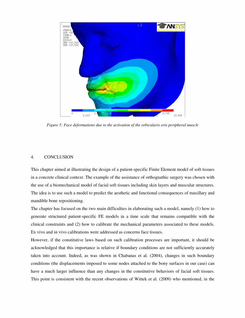
Figure 5: Face deformations due to the activation of the orbicularis oris peripheral muscle
4. CONCLUSION
This chapter aimed at illustrating the design of a patient-specific Finite Element model of soft tissues
in a concrete clinical context. The example of the assistance of orthognathic surgery was chosen with
the use of a biomechanical model of facial soft tissues including skin layers and muscular structures.
The idea is to use such a model to predict the aesthetic and functional consequences of maxillary and
mandible bone repositioning.
The chapter has focused on the two main difficulties in elaborating such a model, namely (1) how to
generate structured patient-specific FE models in a time scale that remains compatible with the
clinical constraints and (2) how to calibrate the mechanical parameters associated to these models.
Ex vivo and in vivo calibrations were addressed as concerns face tissues.
However, if the constitutive laws based on such calibration processes are important, it should be
acknowledged that this importance is relative if boundary conditions are not sufficiently accurately
taken into account. Indeed, as was shown in Chabanas et al. (2004), changes in such boundary
conditions (the displacements imposed to some nodes attached to the bony surfaces in our case) can
have a much larger influence than any changes in the constitutive behaviors of facial soft tissues.
This point is consistent with the recent observations of Wittek et al. (2009) who mentioned, in the

case of a brain FE model deformed by nodes’ imposed displacements, the “unimportance of
constitutive models in computing brain deformation for image-guided surgery”.
ACKNOWLEDGMENTS
Many scientific colleagues and clinicians have participated to the works listed in this chapter. I
would like to thank them for their important inputs. Hoping that I won’t forget anyone, I’d like to list
Georges Bettega, Franck Boutault, Marek Bucki, Matthieu Chabanas, Jean-Michel Gerard, Vincent
Luboz, Christophe Marécaux, Mohammad Nazari, Jacques Ohayon, Pascal Perrier, Emmanuel
Promayon and Patrick Schiavone.
REFERENCES
Agache, P.G., Monneur, C., Leveque, J.L., Rigal, J.De., 1980. Mechanical properties and Young’s
modulus of human skin in vivo. Arch. Dermatol. Res. 269 (3), 221–232.
Barré, S., Fernandez, C., Paume, P., Subrenat, G., 2000. Simulating facial surgery. Proc. IS&T /
SPIE Electron. Imaging 3960, 334–34.
Bucki M., Lobos C. & Payan Y. (2010). A Fast and Robust Patient Specific Finite Element Mesh
Registration Technique: Application to 60 Clinical Cases. Medical Image Analysis, Vol. 14, pp.
303–317.
Carter, F.J., Frank, T.G., Davies, P.J., McLean, D., Cuschieri, A., 2001. Measurements and
modelling of the compliance of human and porcine organs. Med. Image Anal. 5 (4), 231–236.
Chabanas M. (2002). Modélisation des tissus mous de la face pour la chirurgie orthognatique
assistée par ordinateur, PhD de l’Université Joseph Fourier, Grenoble 1.
Chabanas M., Luboz V. & Payan Y. (2003). Patient specific Finite Element model of the face soft
tissue for computer-assisted maxillofacial surgery, Medical Image Analysis, Vol. 7, Issue 2, pp. 131-
151.
Chabanas M., Payan Y., Marécaux C., Swider P. & Boutault F. (2004). Comparison of linear and
non-linear soft tissue models with post-operative CT scan in maxillofacial surgery. Lecture Notes in
Computer Science, vol. 3078, pp. 19-27.

Chen, E.J., Novakofski, J., Jenkins, W.K., O’Brien Jr., W.D., 1996. Young’s modulus measurements
of soft tissues with application to elasticity imaging. IEEE Trans. Ultrason. Ferroelectr. Freq.
Control 43 (1), 191–194.
Couteau B., Payan Y. & Lavallée S. (2000). The Mesh-Matching algorithm: an automatic 3D mesh
generator for finite element structures, Journal of Biomechanics, vol. 33/8, 1005-1009.
Delingette, H., Subsol, G., Cotin, S., Pignon, J., 1994. A Craniofacial Surgery Simulation
Testbed. INRIA, Technical Report 2199.
Diridollou, S., Patat, F., Gens, F., Vaillant, L., Black, D., Lagarde, J.M., Gall, Y., Berson, M., 2000.
In vivo model of the mechanical properties of the human skin under suction. Skin Res. Technol. 6
(4), 214–221.
Fung, Y.C., 1993. Biomechanics: Mechanical Properties of Living Tissues. Springer, New
York.
Gefen A., Margulies S., 2004. Are in vivo and in situ brain tissues mechanically similar? J. Biomech.
37 (9), 1339–1352.
Gennisson, Jean-Luc, Baldeweck, Thérèse, Tanter, Mickaël, Catheline, Stefan, Fink, Mathias,
Sandrin, Laurent, Cornillon, Céline, Querleux, Bernard, 2004. Assessment of elastic parameters
of human skin using dynamic elastography. IEEE Trans. Ultrason. Ferroelectr. Freq. Control 51 (8),
980–989.
Gerard J.M., Ohayon J., Luboz V., Perrier P. & Payan Y. (2005). Non linear elastic properties of the
lingual and facial tissues assessed by indentation technique. Application to the biomechanics of
speech production, Medical Engineering & Physics, Vol. 27 (10), pp. 884-892.
Gladilin, E., Zachow, S., Hege, H.C., Deuflard, P., 2001. FE-based heuristic approach for the
estimation of person-specific facial mimics. In: Middleton, I., Jones, M.L., Shrive, N.G. (Eds.), 5th
International Symposium on Computer Methods in Biomechanics and Biomedical Engineering,
Rome, Italy, November 2001.
Grahame, R., Holt, P.J., 1969. The influence of ageing on the in vivo elasticity of human skin.
Gerontologia 15 (2), 121–139.
Jemec, G.B., Selvaag, E., Agren, M., Wulf, H.C., 2001. Measurement of the mechanical properties
of skin with ballistometer and suction cup. Skin Res. Technol. 7 (2), 122–126.
Kauer, M., Vuskovic, V., Dual, J., Szekely, G., Bajka, M., 2002. Inverse finite element
characterization of soft tissues. Med. Image Anal. 6 (3), 275–287.

Keeve, E., Girod, S., Girod, B., 1996. Computer-aided craniofacial surgery. In: Lemke,
H.U. et al. (Ed.), Computer Assisted Radiology, Elsevier, pp. 757–763.
Keeve, E., Girod, S., Kikinis, R., Girod, B., 1998. Deformable modelling of facial tissue for
craniofacial surgery simulation. Computed Aided Surgery, 3 (5), 228–238.
Kerdok, Amy E., Ottensmeyer, Mark P., Howe, Robert D., 2006. Effects of perfusion on the
viscoelastic characteristics of liver. J. Biomech. 39 (12), 2221–2231.
Koch, R.M., Gross, M.H., Carls, F.R., von Büren, D.F., Frankhauser, G., Parish, Y.I.H., 1996.
Simulating facial surgery using finite element models. Proceedings of SIGGRAPH. ACM Press, pp.
421–428.
Koch, R.M., Roth, S.H.M., Gross, M.H., Zimmermann, A.P., Sailer, H.F., 1999. A framework for
facial surgery simulation. CS Technical Report #326, 18 June 1999. ETH Zurich, Switzerland.
Luboz V., Promayon E. & Payan Y. (2014). Linear Elastic Properties of the Facial Soft Tissues
Using an Aspiration Device: Towards Patient Specific Characterization. Annals of Biomedical
Engineering, Vol. 42 (1), pp. 2369-2378. DOI: 10.1007/s10439-014-1098-1.
Mollemans, W, Schutyser, F, Nadjmi ,N, Maes, F., & Suetens, P. (2007). Predicting soft tissue
deformations for a maxillofacial surgery planning system: From computational strategies to a
complete clinical validation, Medical Images Analysis, 11, 282–301.
Nazari M., Perrier P., Chabanas M. & Payan Y. (2010). Simulation of dynamic orofacial movements
using a constitutive law varying with muscle activation. Computer Methods in Biomechanics &
Biomedical Engineering, Vol. 13(4), pp. 469–482.
Nazari M.A., Perrier P., Chabanas M. & Payan Y. (2011a). A 3D Finite Element Muscle Model and
its Application in Driving Speech Articulators. Proceedings of the 23rd Congress of the
International Society of Biomechanics, ISB'2011, Brussels, Belgium.
Nazari M., Perrier P., Chabanas M. & Payan Y. (2011b). Shaping by stiffening: a modeling study for
lips. Motor Control, Vol. 15(1), pp. 141-168.
Ottensmeyer, Mark Peter., 2001. Minimally Invasive Instrument for in vivo Measurement of Solid
Organ Mechanical Impedance. Ph.D. Thesis, Massachusetts Institute of Technology, Dept. of
Mechanical Engineering.
Ottensmeyer M., 2002. In vivo measurement of solid organ visco-elastic properties. Stud. Health
Technol. Inform. 85, 328–333.

Richter M., Mossaz C., Laurent F., Goudot P. Chirurgie correctrice des malformations
maxillomandibulaires : approche diagnostique et plan de traitement. Encycl. Méd. Chir. (Elsevier,
Paris), Stomatologie, 22-066-D-10, 1998, 16p. 1998.
Rouviere, H., Delmas, A. (1997). In: Tete et Cou, 14th Edition. Anatomie Humaine Descriptive,
Topographique et Fonctionnelle, Vol. 1. Masson, Paris.
Samur, E., Sedef, M., Basdogan, C., Avtan, L., Duzgun, O., 2007. A robotic indenter for minimally
invasive measurement and characterization of soft tissue response. Med. Image Anal. 11 (4), 361–
373.
Schiavone P., Chassat F., Boudou T., Promayon E., Valdivia F. & Payan Y. (2009). In vivo
measurement of human brain elasticity using a light aspiration device. Medical Image Analysis, Vol.
13, pp. 673–678.
Schiavone P., Promayon E. & Payan Y. (2010). LASTIC: a Light Aspiration device for in vivo Soft
TIssue Characterization. Lecture Notes in Computer Science, Vol. 5958, pp. 1-10.
Stavness I., Nazari M., Perrier P., Demolin D. & Payan Y. (2013). A Biomechanical Modeling Study
of the Effects of the Orbicularis Oris Muscle and Jaw Posture on Lip Shape. Journal of Speech,
Language, and Hearing Research, Vol. 56, pp. 878-890.
Teschner, M., Girod, S., Girod, B., 1999. Optimization approaches for soft-tissue prediction in
craniofacial surgery simulation. In: 2nd International Conference on Medical Image Computing and
Computer Assisted Intervention, MICCAI’99. LNCS, Vol. 1679. Springer, pp. 1183–1190.
Teschner, M., 2001. Direct Computation of Soft-tissue Deformation in Craniofacial Surgery
Simulation. Ph.D. Thesis, Shaker Verlag, Aachen, Germany (January 2001).
Waters, K., 1996. Synthetic muscular contraction on facial tissue derived from computer
tomography data. In: Taylor, R., Lavallee, S., Burdea, G., Mosges, R. (Eds.), Computer
Integrated Surgery. MIT Press, Cambridge, MA, pp. 191–200.
Wittek A., Hawkins T. & Miller K. (2009). On the unimportance of constitutive models in
computing brain deformation for image-guided surgery. Biomech Model Mechanobiol., Vol 8(1), pp.
77-84.
Zachow, S., Gladiline, E., Hege, H.-C., Deuflhard, P., 2000. Finite element simulation of soft tissue
deformation. In: Proceedings of CARS, pp. 23–28.