Embed Size (px)
Citation preview
British Journal ofOphthalmology, 1983, 67, 826-829
Solitary choroidal mass as the presenting sign insystemic sarcoidosisR. JOSEPH OLK,' MATTHEW J. LIPMANN,2 H. CLAY CUNDIFF,3AND JOHN DANIELS'
From the 'Washington University School of Medicine, St Louis, Missouri; 2University of Cincinnati School ofMedicine, Cincinnati, Ohio; and 3St Francis Medical Center, Cape Girardeau, Missouri, USA
SUMMARY A solitary choroidal mass with an overlying neurosensory retinal detachment was seenin an otherwise healthy 25-year-old Caucasian female. Ocular and general physical examinations,serum chemistry, and pathological examination of a lymph node biopsy confirmed sarcoidosis asthe cause for the choroidal mass. Treatment with systemic steroids resulted in resolution of thelesion and return of normal visual acuity.
Case report
A 25-year-old Caucasian female was seen by her localophthalmologist and referred for retinal examinationbecause of central blurring in her left eye for theprevious 3 days. The patient noticed no redness, pain,or photophobia. Ocular history revealed one sisterwho was noted to have a unilateral eye defect whichby history was suggestive of a coloboma. The sisterwas not examined. Past medical history was negativeexcept for an upper respiratory infection one monthprior to ocular examination. Four months previouslythe patient had given birth to a full-term, healthychild, delivered by caesarean section because ofprolonged labour. After delivery the patient had araised white blood count, the cause of which wasnever determined. There was no history of fever,weight loss, or anorexia. The patient abused neitherdrugs nor alcohol.
Ocular examination revealed best corrected visualacuity of 20/20 OD, 20/100 OS. Applanation intra-ocular tensions were 12/13. External examination wasnormal. The pupils were 4 mm, equal, round, andreacted to light directly and consensually without anafferent defect. A muscle balance test showedorthophoria. Motility tests revealed full ductions andversions. A photo stress test showed a delay on theleft greater than one minute compared with the right.American Optical colour plates showed 12 of 15correct OD, 0 of 15 OS. An Amsler grid test showed acentral scotoma OS. Slit-lamp examination of theanterior segment was unremarkable in both eyes.Correspondence to Dr R. J. Olk, 4949 Barnes Hospital Plaza, StLouis, Missouri 63141, USA.
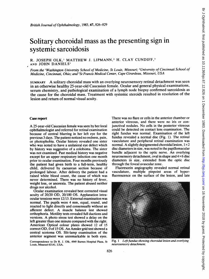
There was no flare or cells in the anterior chamber oranterior vitreous, and there were no iris or con-junctival nodules. No cells in the posterior vitreouscould be detected on contact lens examination. Theright fundus was normal. Examination of the leftfundus revealed a normal disc (Fig. 1). The retinalvasculature and peripheral retinal examination wasnormal. A slightly depigmented choroidal lesion, 1 x 2disc diameters in size, was noted in the papillomacularbundle adjacent to the optic nerve. An overlyingneurosensory detachment, oval in shape and 4 x 6 discdiameters in size, extended from the optic discthrough the foveal avascular zone.
Fluorescein angiography revealed normal retinalvasculature, multiple pinpoint areas of hyper-fluorescence on the surface of the lesion, and late
Fig. 1 LeftJunaus snowing cnoroiaal lesion ana overlyingneurosensory detachment.
826
on 16 Novem
ber 2018 by guest. Protected by copyright.
http://bjo.bmj.com
/B
r J Ophthalm
ol: first published as 10.1136/bjo.67.12.826 on 1 Decem
ber 1983. Dow
nloaded from

Solitary choroidal mass as the presenting sign in systemic sarcoidosis
Fig. 2 Red-free photograph, OS: late phase, showingdiffuse staining over lesion and collection ofdye underneurosensory detachment.
diffuse staining of the entire lesion with collection ofdye under the neurosensory detachment (Fig. 2).Goldmann visual fields showed a caecocentral defectin the left eye (Fig. 3). Orbital high-resolution CTscan showed a mild thickening of the soft tissues in theposterior aspect of the left globe which was enhancedby infusion (Fig. 4). Ocular ultrasound by quantitativeA-scan and B-scan confirmed elevation of the neuro-sensory retina with fluid in the extrascleral spaceadjacent to the temporal aspect of the optic nerve, apattern thought to be consistent with localised in-flammatory disease.The patient had a complete general physical exam-
ination. Chest x-ray showed right hilar adenopathyand an enlarged azygous node with scattered granulo-mata in both lung fields. A complete blood count andurine analysis were normal. The sedimentation ratewas 63 mg/h (Westergren). Tests of serum chemistryshowed the following abnormal values: alkaline
Fig. 3 Goldmann visualfields OS, showing caecocentraldefect.
Fig. 4 High resolution orbital CT scan, post-infusion.
phosphatase (339 IU/l), 5' nucleotidase (32 IU/l), andSGOT (38 IU/l). Angiotensin I converting enzyme(ACE) was 73 nmol min-' ml-' (normal 20-68), andserum lysozyme was 15 ng/ml (normal 2 8-8). (SIconversion: nmol/ml=,umol/l; ng/ml=,ug/l.) Serumprotein electrophoresis, antinuclear antibodies, andrheumatoid factor tests were normal. Serological testsfor fungi and syphilis were negative. IntermediatePPD was nonreactive. A liver-spleen scan showed anormal liver, an enlarged spleen, and increasedactivity in the spleen in comparison with the liver.
.*.Fig. 5 Mediastinal lymph node biopsy showingnoncaseating granulomatous tubercle surrounded bylymphocytes and giant cells. (Haematoxylin and eosin,x 58).
827
on 16 Novem
ber 2018 by guest. Protected by copyright.
http://bjo.bmj.com
/B
r J Ophthalm
ol: first published as 10.1136/bjo.67.12.826 on 1 Decem
ber 1983. Dow
nloaded from

R. Joseph Olk, Matthew J. Lipmann, H. Clay Cundiff, and John Daniels
The patient had mediastinoscopy and a lymph nodebiopsy. The operative report stated that the visualisedmediastinal nodes were enlarged and succulent,homogeneous, and grey in colour, characteristic ofsarcoid. A histopathological examination revealedmultiple noncaseating granulomata with early fibrosisand surrounding lymphocytes (Fig. 5). The biopsywas believed to be consistent with a diagnosis ofsarcoidosis, and the patient was begun on a taperingdose of systemic steroids over a 6-week period.Examination after one month of therapy revealed a
visual acuity of 20/30 OS and marked reduction of thechoroidal lesion, with complete resolution of theoverlying sensory retinal detachment. Retinal striaeinto the foveal avascular zone were still evident. Fivemonths after initial presentation the visual acuity was20/20 OS, and examination ofthe fundus showed onlypigmentary changes at the level of the retinal pigmentepithelium in the area of the previous lesion. Visualacuity one year after therapy remains 20/20 OU.
Discussion
Sarcoidosis is a chronic noncaseating granulomatousdisorder of unknown aetiology affecting variousorgan systems, with protean symptoms. In the last 2decades it has replaced syphilis as the 'great imitator.'Reports vary, but the disease appears to be unusuallycommon in blacks andwomen and usually presents inthe third and fourth decades. 1-4The known clinical ocular manifestations of
sarcoidosis include nut-shaped nodules in the peri-orbital skin, pseudochalazia, enlargement of thelacrimal glands, keratitis sicca, nodular infiltrates ofthe palpebral conjunctiva, nummular and band-shaped calcification of the cornea, episcleral nodules,iridocyclitis, iris nodules, nodular vitreous opacities,periphlebitis, chorioretinitis 'en taches de bougie'(candle wax spots), optic atrophy, optic nerve andorbital tumours, papilloedema, macular oedema,extraocular muscle paralysis, and visual fielddefects.3' -10 Although there are many case reports ofocular and orbital sarcoidosis, it is extremely rare tohave isolated involvement of the posterior segmentof the eye.
Ocular involvement in systemic sarcoidosis has anaverage estimated incidence of 25-40% 11-3 but hasbeen reported to be as high as 63%.'1 The mostcommon ocular presentation involves the uveal tractmore often than the sclera, cornea, or conjunctiva.Uveal involvement characteristically is much morefrequent anteriorly in the iris rather than in the ciliarybody or choroid. Posterior uveitis in sarcoid is rareand is usually overshadowed by concomitant anterioruveitis.'5 Gould and Kaufman stated that only one-third of all reported cases of posterior segment
sarcoidosis had no apparent anterior uveitis.'6 Theleast common sites of sarcoid infiltration are said tobe the choroid and retina," and King emphasisedthat sarcoid must be an extremely rare cause oflocalised chorioretinitis. 18
Chotioretinitis was first reported in associationwith systemic sarcoidosis by Hudelo and Rabut,'9 butthis patient had accompanying iridocyclitis andbilateral choroidal infiltrates. Others13 15 20-23 havereported cases having retinal and choroidal in-volvement with sarcoidosis, but in all cases therewere other associated ocular findings. Goldberg andNewell reported 2 cases with single lesions of thefundus but primarily involving retinal infiltration withaccompanying retinal vascular changes. 15 Theyspeculated that perhaps in those cases of retinalinfiltration with sarcoid the retinal vascular changesmight precede the appearance of the visible fundusmass, as was directly observed in one of their cases.Chumbley and Kearns reported 4 cases in which
retinal findings were the exclusive ocular manifesta-tion of sarcoidosis.23 They emphasised that inposterior segment sarcoidosis retinal involvement wasmuch more frequent than choroiditis, choroidal oroptic nerve granulomas, or lesions of the vitreous.Many cases of optic disc infiltration and optic disc
oedema secondary to ocular sarcoid as well as sar-coid tumours of the optic nerve have beenreported.' 22 24-29 In addition the presence of achorioretinal mass projecting into the vitreous hasbeen reported, 13-15 30 but in all these cases there havebeen associated changes in the overlying retinalvasculature or other associated findings consistentwith ocular sarcoidosis.A recent report3' cites 2 cases of sarcoid granuloma
of the choroid. However, one case occurred in apatient with previously diagnosed systemic sar-coidosis. The authors are not aware of any otherpreviously published case reports of a solitarychoroidal granuloma as the sole presenting sign insystemic sarcoidosis. The differential diagnosis mustinclude primary and metastatic disease, includingHodgkin's lymphoma, inflammatory conditionsassociated with collagen-vascular and rheumato-logical disorders, Eales's disease, Behcet's disease, aswell as infectious diseases such as syphilis, tuberculo-sis, cytomegalic inclusion retinitis, herpes retinitis,brucellosis, fungal disease, and toxoplasmosis.A complete medical evaluation including appro-
priate laboratory and serological tests as well as atissue biopsy was crucial to the diagnosis in this caseand to the initiation of prompt therapy, whichresulted in complete resolution of the lesion andreturn to normal visual acuity. Knowledge thatsystemic sarcoidosis can present as a solitary choroidalmass may be of importance to ophthalmologists.
828
on 16 Novem
ber 2018 by guest. Protected by copyright.
http://bjo.bmj.com
/B
r J Ophthalm
ol: first published as 10.1136/bjo.67.12.826 on 1 Decem
ber 1983. Dow
nloaded from
Solitary choroidal mass as the presenting sign in systemic sarcoidosis
The authors acknowledge the assistance of Morton Smith, MD, whoreviewed the histopathology from the lymph node biopsy, andWilliam A. Hart, MD, who performed the quantitative A- andB-scan ultrasonography.
References1 Nielsen RH. Ocular sarcoidosis. Arch Ophthalmol 1959; 61:657-63.
2 Obenauf CD, Shaw HE, Sydnor CF, Klintworth GK. Sarcoidosisand its ophthalmic manifestations. Am J Ophthalmol 1978; 86:648-55.
3 Crick RP, Hoyle C, Smellie H. The eyes in sarcoidosis. Br JOphthalmol 1961; 45:461-81.
4 Iwata K, Nanba K, Sobue K, Abe H, et al. Ocular sarcoidosis:evaluation of intraocular findings. Ann NY Acad Sci 1976; 278:445-54.
5 Zimmerman LE, Maumenee AE. Ocular aspects of sarcoidosis.Am Rev Resp Dis 1961; 84: 38-44.
6 Wilmer WH. Tubercle-like nodules of episclera and eyelids,bilateral. Am J Ophthalmol 1934; 17: 99-106.
7 James DG. Ocular sarcoidosis. Am J Med 1959; 26: 331-9.8 Levitt JM. Boeck's sarcoid with ocular localization. ArchOphthalmol 1941; 26: 358-88.
9 Franceschetti A, Babel J. La chorioretinite en 'taches de bougie'manifestation de la maladie de Besnier-Boeck. Ophthalmologica1949; 118:701-10.
10 James DG, Zatouroff MA, Trowell J, Rose FC. Papilloedema insarcoidosis. Br J Ophthalmol 1967; 51: 526-9.
11 Klein M, Calvert RJ, Joseph WE, Smith E. Rarities in ocularsarcoidosis. Br J Ophthalmol 1955; 39: 416-21.
12 Letocha CE, Shields JA, Goldberg RE. Retinal changes insarcoidosis. Can J Ophthalmol 1975; 10: 184-92.
13 Walsh FB. Ocular importance of sarcoid. Its relation touveoparotid fever. Arch Ophthalmol 1939; 21: 421-38.
14 Ozazewski JC, Bennett V. Ocular sarcoidosis. Report of threecases. Am J Ophthalmol 1952; 35: 547-50.
15 Goldberg MS, Newell FW. Sarcoidosis with retinal involvement.Arch Ophthalmol 1944; 32: 93-6.
16 Gould H, Kaufman HE. Sarcoid of the fundus. Arch Ophthalmol1961; 65:453-6.
17 Laval J. Ocular sarcoidosis. Am J Ophthalmol 1952; 35: 551-4.18 King MJ. Ocular lesions of Boeck's sarcoid. Trans Am Acad
Ophthalmol Soc 1939; 37: 422-58.19 Hudelo L, Rabut R. Lupoides disseminees de Boeck. Bull Soc Fr
Derm Syph 1925; 32: 108-9.20 Horton R, Lincoln NS, Pinner M. Noncaseating tuberculosis.Am Rev Respir Dis 1939; 39: 186-203.
21 Meyer FW. Augentuberkulose und lymphogranulomatosisbenigna. Klin Monatsbl Augenhedkd 1939; 102: 76-88.
22 ReisW, Rothfeld J. Tuberkulide des Schnerven als Komplikationvon Hautsarkoiden vom Typus Darier-Roussy. Arch Ophthalmol1931; 126: 357-66.
23 Chumbley LC, Kearns TP. Retinopathy of sarcoidosis. Am JOphthalmol 1972; 73: 123-31.
24 Statton R, Blodi FC, Hanigan J. Sarcoidosis of the optic nerve.Arch Ophthalmol 1964; 71: 834-6.
25 Morax PV. Les localisations neuro-oculaires de la reticulo-endotheliose de Besnier-Boeck-Schaumann. Ann Oculist (Puris)1956; 189:73-91.
26 Kojima K. Fundus involvement in sarcoidosis. J Clin Ophthalmol1969; 23: 790-3.
27 von Mackensen G. Veranderungen am Augenintergrund beiBesnier-Boeck-Schaumannscher Erkemkung. Klin MonatsblAugenheilkd 1958; 121: 51-63.
28 Brunste E. Ocular sarcoidosis. Dan Med Bull 1958; 5: 217-27.29 Laties AM, Scheie HG. Sarcoid granuloma of the optic disk:
evolution of multiple small tumors. Trans Am Ophthalmol Soc1970:68:219-33.
30 Woods AC, Guyton JS. Role of sarcoidosis and of brucellosis inuveitis. Arch Ophthalmol 1944; 31: 469-80.
31 Marcus DF, Bovino JA, Burton TC. Sarcoid granuloma of thechoroid. Ophthalmology 1982; 89: 1326-30.
829
on 16 Novem
ber 2018 by guest. Protected by copyright.
http://bjo.bmj.com
/B
r J Ophthalm
ol: first published as 10.1136/bjo.67.12.826 on 1 Decem
ber 1983. Dow
nloaded from


![Comparison of Retinal and Choroidal Involvement in ...BIB_A1EC8B55A4F4.P001/REF.pdf · Workshop on Ocular Sarcoidosis (IWOS). [1] Such criteria may have a higher sensitivity in the](https://img.pdfslide.net/doc/110x75/60094a82fb923b4ee61df82c/comparison-of-retinal-and-choroidal-involvement-in-biba1ec8b55a4f4p001refpdf.jpg)