Embed Size (px)
Citation preview
Somatostatin and dopamine receptors
as molecular targets for the medical
treatment of Cushing’s disease
Christiaan de Bruin
Somatostatin and dopamine receptors as molecular targets for the medical treatment of Cushing’s disease.© 2009 C. de Bruin
ISBN: 978-90-8559-545-8
Cover: U.S. postal stamp of Harvey CushingCover design: OGC
No part of this thesis may be reproduced, stored in a retrieval system or transmitted in any other form or by any means, without permission from the author or, when appropri-ate, from the publishers of the publications.
Printed by Optima Grafische Communicatie, Rotterdam, the Netherlands
Somatostatin and dopamine receptors as molecular targets for the medical
treatment of Cushing’s disease
Somatostatine en dopamine receptoren als moleculaire doelwitten voor de medische behandeling van de ziekte
van Cushing
Proefschrift
ter verkrijging van de graad van doctor aan de Erasmus Universiteit Rotterdam
op gezag van de rector magnificus
Professor dr. S.W.J. Lamberts en volgens besluit van het College voor Promoties
De openbare verdediging zal plaatsvinden op woensdag 1 juli 2009 om 15.45 uur door
Christiaan de Bruin
geboren te Zevenaar
PromotieCommissie:
Promotor: Prof.dr. S.W.J. Lamberts
Overige leden: Prof.dr. A.J. van der Lely Prof.dr.ir. T.J. Visser Prof.dr. J.A. Romijn
Co-promotor: Dr. L.J. Hofland
The studies described in this thesis were performed at the Department of Internal Medi-cine, Division of Endocrinology, Erasmus MC, Rotterdam, the Netherlands.
Printing of this thesis was financially supported by Novartis Oncology, IPSEN Pharmaceu-tica, Pfizer, Novo Nordisk, Goodlife Healthcare, Boehringer Ingelheim and the Erasmus University Rotterdam.
Contents
Chapter 1 Introduction and aims of thesis 7
Chapter 2 Expression and functional analysis of dopamine receptor subtype 2 and somatostatin receptor subtypes in canine Cushing’s disease
35
Chapter 3 Differential regulation of human dopamine D2 and somatostatin receptor subtype expression by glucocorticoids in vitro
59
Chapter 4 Co-expression of dopamine and somatostatin receptor subtypes in corticotroph adenomas
79
Chapter 5 Early clinical experiences with combined somatostatin analogue, dopamine agonist and/or low-dose ketoconazole therapy in human Cushing’s disease
97
Chapter 6 Somatostatin receptor expression in a patient with Cushing’s syndrome due to ectopic adrenocorticotropin secretion after successful mifepristone therapy
107
Chapter 7 Functional characterization of the dopamine somatostatin chimeric molecule BIM-23A760
121
Chapter 8 General discussion 133
Chapter 9 Summary / Samenvatting 147
Appendix 157
Dankwoord 167
Curriculum Vitae 171


Chapter 1
introduction and aims of thesis
Based on: C. de Bruin, R. A. Feelders, S. W. J. Lamberts and L. J. Hofland
Rev Endocr Metab Disord. 2008 Jul 19 [Epub ahead of print]

Introduction and aims of thesis 9
Chap
ter 1
A. HArvey W. CusHing
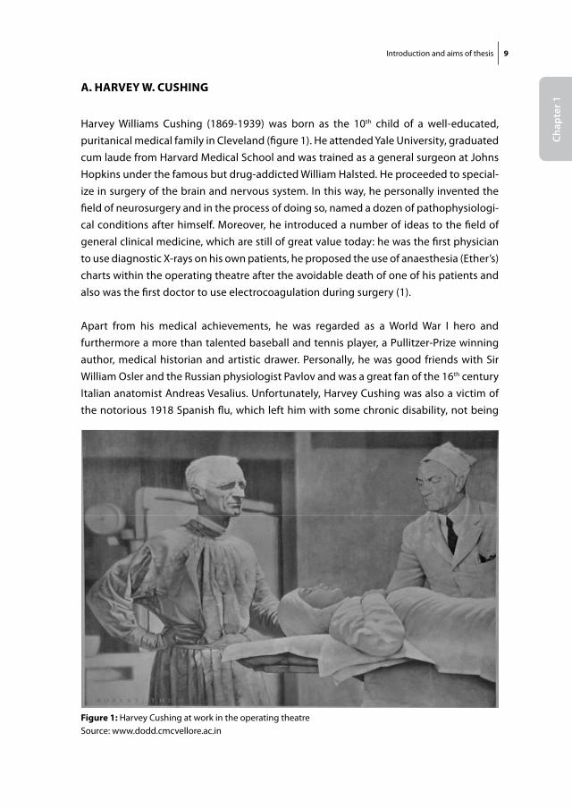
Harvey Williams Cushing (1869-1939) was born as the 10th child of a well-educated, puritanical medical family in Cleveland (figure 1). He attended Yale University, graduated cum laude from Harvard Medical School and was trained as a general surgeon at Johns Hopkins under the famous but drug-addicted William Halsted. He proceeded to special-ize in surgery of the brain and nervous system. In this way, he personally invented the field of neurosurgery and in the process of doing so, named a dozen of pathophysiologi-cal conditions after himself. Moreover, he introduced a number of ideas to the field of general clinical medicine, which are still of great value today: he was the first physician to use diagnostic X-rays on his own patients, he proposed the use of anaesthesia (Ether’s) charts within the operating theatre after the avoidable death of one of his patients and also was the first doctor to use electrocoagulation during surgery (1).
Apart from his medical achievements, he was regarded as a World War I hero and furthermore a more than talented baseball and tennis player, a Pullitzer-Prize winning author, medical historian and artistic drawer. Personally, he was good friends with Sir William Osler and the Russian physiologist Pavlov and was a great fan of the 16th century Italian anatomist Andreas Vesalius. Unfortunately, Harvey Cushing was also a victim of the notorious 1918 Spanish flu, which left him with some chronic disability, not being
Fig 1
Figure 1: Harvey Cushing at work in the operating theatre Source: www.dodd.cmcvellore.ac.in
10 Chapter 1
able to walk more than a few steps at a time. Despite this disability, he proceeded to perform more than 2000 brain surgeries and wrote over 330 books and articles.
He was a hard-working, stern man who was both respected and feared by his residents and nursing staff for his sarcastic remarks and stormy outbursts. Not much of a family man to his wife and 5 children, he consistently placed his medical work ahead of anything else in life and could mourn for days after the death of a patient, blaming only himself. At the age of 70 he died in a way, which is both ironic as well as symbolic for the life he has lived. When working on a manuscript about Vesalius, he died of a myocardial infarction, presumably induced by the lifting of one of the heavy folio volumes of Vesalius’ work. At autopsy, he had a posterior coronary artery occlusion, as well as a 1 cm colloid cyst of the third ventricle, in line with the common belief that doctors tend to develop the disease in which they have specialized.
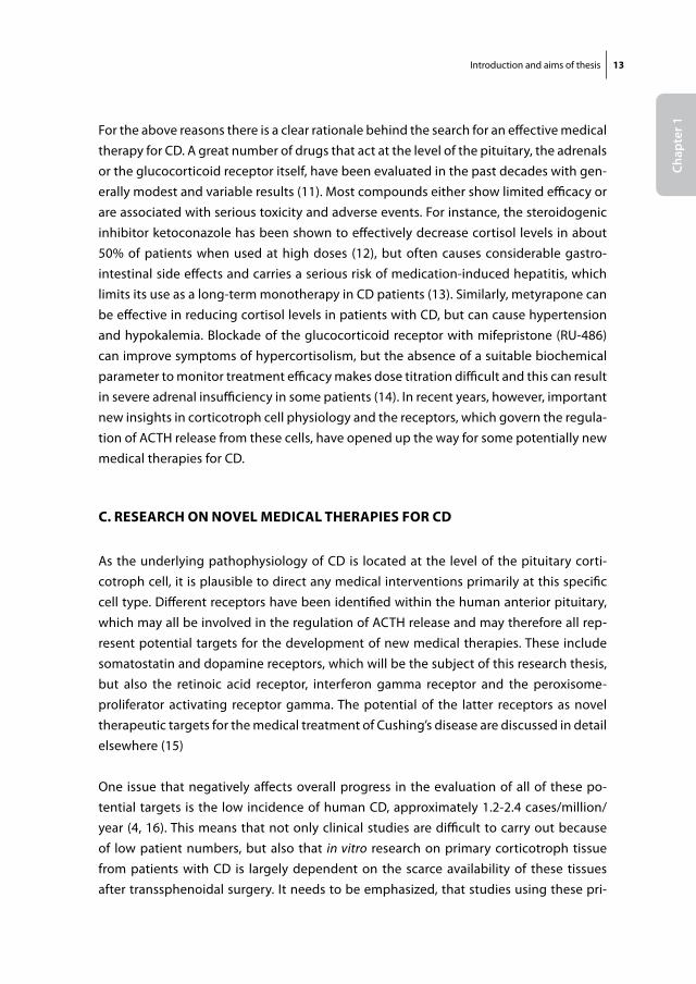
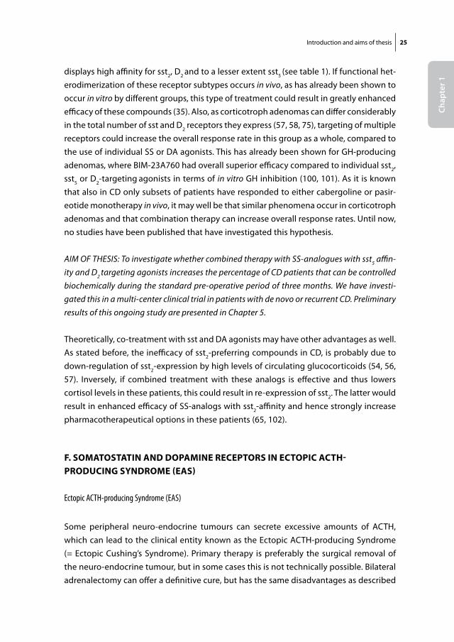
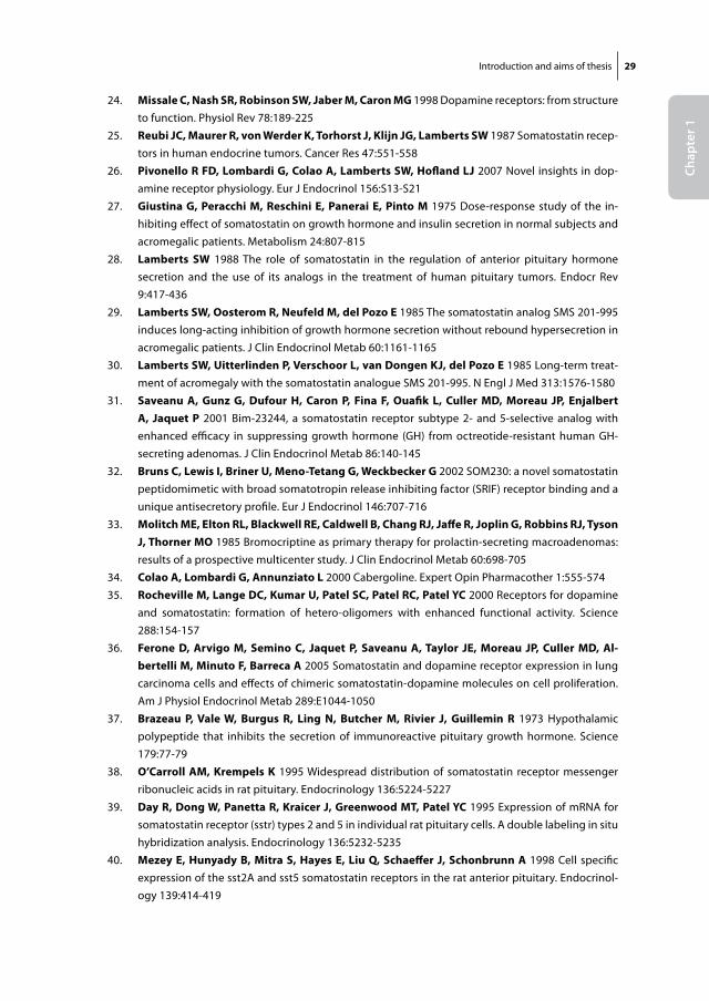
B. CusHing’s DiseAse: PAtHoPHysiology AnD Current treAtment
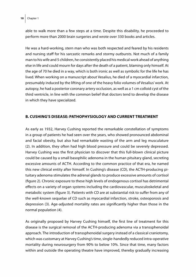
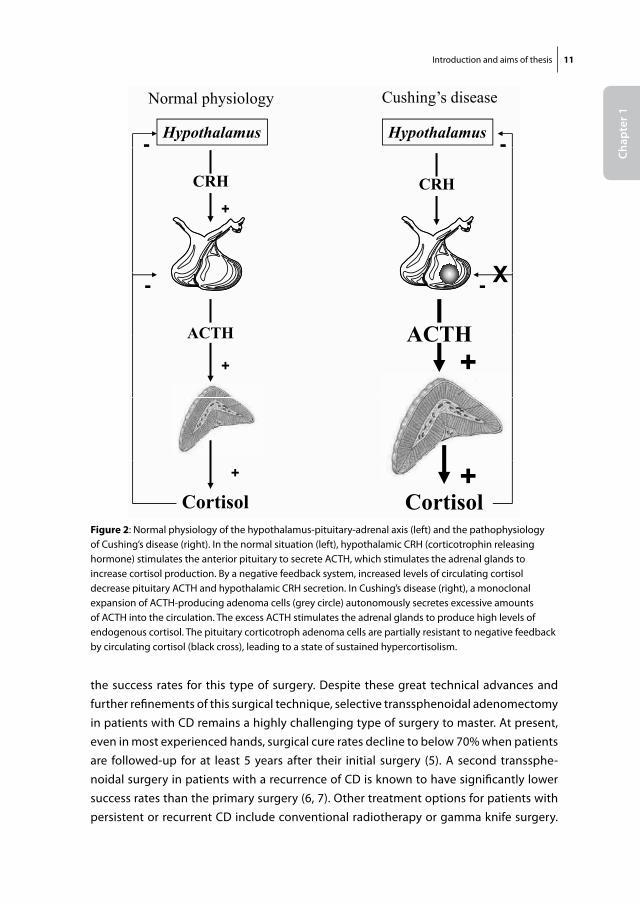
As early as 1932, Harvey Cushing reported the remarkable constellation of symptoms in a group of patients he had seen over the years, who showed pronounced abdominal and facial obesity, but also had remarkable wasting of the arm and leg musculature (2). In addition, they often had high blood pressure and could be severely depressed. Harvey Cushing was the first physician to discover that this full-blown clinical picture could be caused by a small basophilic adenoma in the human pituitary gland, secreting excessive amounts of ACTH. According to the common practice of that era, he named this new clinical entity after himself. In Cushing’s disease (CD), the ACTH-producing pi-tuitary adenoma stimulates the adrenal glands to produce excessive amounts of cortisol (figure 2). Chronic exposure to these high levels of endogenous cortisol has detrimental effects on a variety of organ systems including the cardiovascular, musculoskeletal and metabolic system (figure 3). Patients with CD are at substantial risk to suffer from any of the well-known sequelae of CD such as myocardial infarction, stroke, osteoporosis and depression (3). Age-adjusted mortality rates are significantly higher than those in the normal population (4). As originally proposed by Harvey Cushing himself, the first line of treatment for this disease is the surgical removal of the ACTH-producing adenoma via a transsphenoidal approach. The introduction of transsphenoidal surgery instead of a classical craniotomy, which was customary at Harvey Cushing’s time, single-handedly reduced intra-operative mortality during neurosurgery from 90% to below 10%. Since that time, many factors within and outside the operating theatre have improved, thereby gradually increasing
Introduction and aims of thesis 11
Chap
ter 1
the success rates for this type of surgery. Despite these great technical advances and further refinements of this surgical technique, selective transsphenoidal adenomectomy in patients with CD remains a highly challenging type of surgery to master. At present, even in most experienced hands, surgical cure rates decline to below 70% when patients are followed-up for at least 5 years after their initial surgery (5). A second transsphe-noidal surgery in patients with a recurrence of CD is known to have significantly lower success rates than the primary surgery (6, 7). Other treatment options for patients with persistent or recurrent CD include conventional radiotherapy or gamma knife surgery.
Fig 2
Normal physiology Cushing’s disease
Hypothalamus Hypothalamus- -
CRH CRH++
X
ACTH ACTH
- X-
ACTH ACTH+ +
Cortisol Cortisol++
Figure 2: Normal physiology of the hypothalamus-pituitary-adrenal axis (left) and the pathophysiology of Cushing’s disease (right). In the normal situation (left), hypothalamic CRH (corticotrophin releasing hormone) stimulates the anterior pituitary to secrete ACTH, which stimulates the adrenal glands to increase cortisol production. By a negative feedback system, increased levels of circulating cortisol decrease pituitary ACTH and hypothalamic CRH secretion. In Cushing’s disease (right), a monoclonal expansion of ACTH-producing adenoma cells (grey circle) autonomously secretes excessive amounts of ACTH into the circulation. The excess ACTH stimulates the adrenal glands to produce high levels of endogenous cortisol. The pituitary corticotroph adenoma cells are partially resistant to negative feedback by circulating cortisol (black cross), leading to a state of sustained hypercortisolism.
12 Chapter 1
Both are effective at reducing ACTH hypersecretion in the majority of patients, but have a slow onset of action, with an average time until remission of 9-24 months (8). In addi-tion, radiotherapy is accompanied by a significant risk of inducing secondary pituitary dysfunction, cranial nerve damage or secondary brain tumours (8-10). As a definitive cure, patients with persistent CD can undergo bilateral adrenalectomy, but this does have important implications in terms of lifelong dependence on hormone replacement therapy and the risk of future Addisonian crises, as well as Nelson’s syndrome.
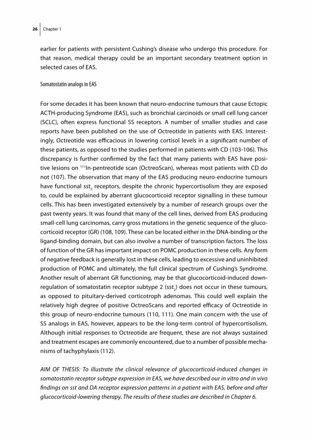
Fig 3
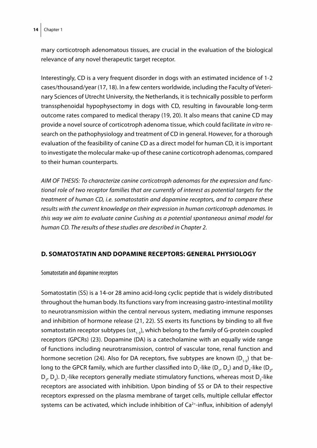
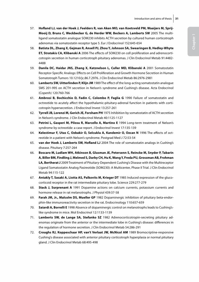
Figure 3: Clinical characteristics of patients with Cushing’s disease. Source: Emanuel Rubin, john L. Farber, Pathology, 3rd edition, Lippincott-Raven Publishers (1999)
Introduction and aims of thesis 13
Chap
ter 1For the above reasons there is a clear rationale behind the search for an effective medical
therapy for CD. A great number of drugs that act at the level of the pituitary, the adrenals or the glucocorticoid receptor itself, have been evaluated in the past decades with gen-erally modest and variable results (11). Most compounds either show limited efficacy or are associated with serious toxicity and adverse events. For instance, the steroidogenic inhibitor ketoconazole has been shown to effectively decrease cortisol levels in about 50% of patients when used at high doses (12), but often causes considerable gastro-intestinal side effects and carries a serious risk of medication-induced hepatitis, which limits its use as a long-term monotherapy in CD patients (13). Similarly, metyrapone can be effective in reducing cortisol levels in patients with CD, but can cause hypertension and hypokalemia. Blockade of the glucocorticoid receptor with mifepristone (RU-486) can improve symptoms of hypercortisolism, but the absence of a suitable biochemical parameter to monitor treatment efficacy makes dose titration difficult and this can result in severe adrenal insufficiency in some patients (14). In recent years, however, important new insights in corticotroph cell physiology and the receptors, which govern the regula-tion of ACTH release from these cells, have opened up the way for some potentially new medical therapies for CD.
C. reseArCH on novel meDiCAl tHerAPies For CD
As the underlying pathophysiology of CD is located at the level of the pituitary corti-cotroph cell, it is plausible to direct any medical interventions primarily at this specific cell type. Different receptors have been identified within the human anterior pituitary, which may all be involved in the regulation of ACTH release and may therefore all rep-resent potential targets for the development of new medical therapies. These include somatostatin and dopamine receptors, which will be the subject of this research thesis, but also the retinoic acid receptor, interferon gamma receptor and the peroxisome-proliferator activating receptor gamma. The potential of the latter receptors as novel therapeutic targets for the medical treatment of Cushing’s disease are discussed in detail elsewhere (15)
One issue that negatively affects overall progress in the evaluation of all of these po-tential targets is the low incidence of human CD, approximately 1.2-2.4 cases/million/year (4, 16). This means that not only clinical studies are difficult to carry out because of low patient numbers, but also that in vitro research on primary corticotroph tissue from patients with CD is largely dependent on the scarce availability of these tissues after transsphenoidal surgery. It needs to be emphasized, that studies using these pri-
14 Chapter 1
mary corticotroph adenomatous tissues, are crucial in the evaluation of the biological relevance of any novel therapeutic target receptor.
Interestingly, CD is a very frequent disorder in dogs with an estimated incidence of 1-2 cases/thousand/year (17, 18). In a few centers worldwide, including the Faculty of Veteri-nary Sciences of Utrecht University, the Netherlands, it is technically possible to perform transsphenoidal hypophysectomy in dogs with CD, resulting in favourable long-term outcome rates compared to medical therapy (19, 20). It also means that canine CD may provide a novel source of corticotroph adenoma tissue, which could facilitate in vitro re-search on the pathophysiology and treatment of CD in general. However, for a thorough evaluation of the feasibility of canine CD as a direct model for human CD, it is important to investigate the molecular make-up of these canine corticotroph adenomas, compared to their human counterparts.
AIM OF THESIS: To characterize canine corticotroph adenomas for the expression and func-tional role of two receptor families that are currently of interest as potential targets for the treatment of human CD, i.e. somatostatin and dopamine receptors, and to compare these results with the current knowledge on their expression in human corticotroph adenomas. In this way we aim to evaluate canine Cushing as a potential spontaneous animal model for human CD. The results of these studies are described in Chapter 2.
D. somAtostAtin AnD DoPAmine reCePtors: generAl PHysiology
Somatostatin and dopamine receptors
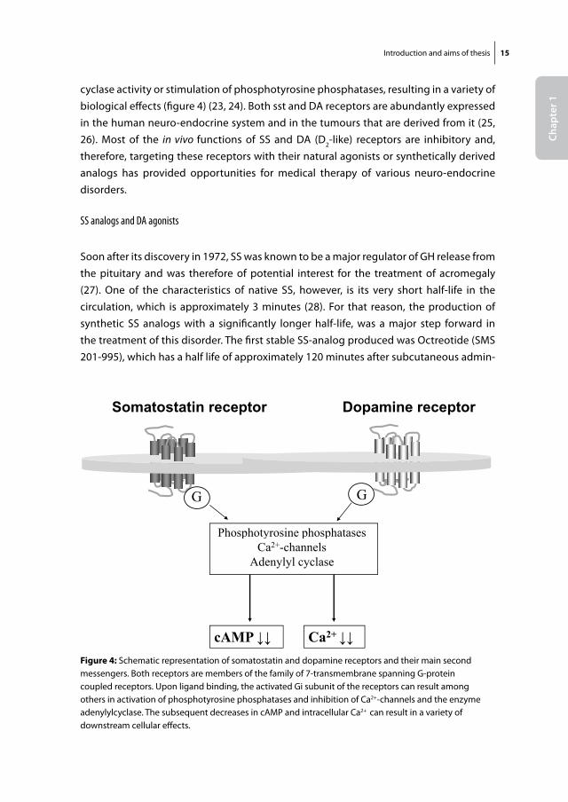
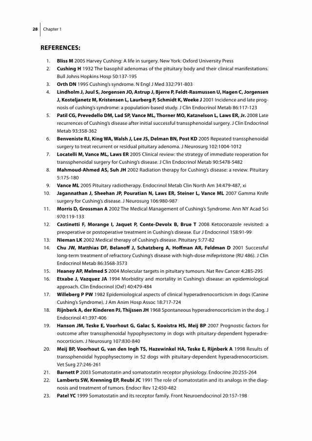
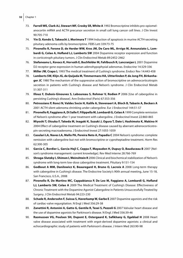
Somatostatin (SS) is a 14-or 28 amino acid-long cyclic peptide that is widely distributed throughout the human body. Its functions vary from increasing gastro-intestinal motility to neurotransmission within the central nervous system, mediating immune responses and inhibition of hormone release (21, 22). SS exerts its functions by binding to all five somatostatin receptor subtypes (sst1-5), which belong to the family of G-protein coupled receptors (GPCRs) (23). Dopamine (DA) is a catecholamine with an equally wide range of functions including neurotransmission, control of vascular tone, renal function and hormone secretion (24). Also for DA receptors, five subtypes are known (D1-5) that be-long to the GPCR family, which are further classified into D1-like (D1, D5) and D2-like (D2, D3, D4). D1-like receptors generally mediate stimulatory functions, whereas most D2-like receptors are associated with inhibition. Upon binding of SS or DA to their respective receptors expressed on the plasma membrane of target cells, multiple cellular effector systems can be activated, which include inhibition of Ca2+-influx, inhibition of adenylyl
Introduction and aims of thesis 15
Chap
ter 1
cyclase activity or stimulation of phosphotyrosine phosphatases, resulting in a variety of biological effects (figure 4) (23, 24). Both sst and DA receptors are abundantly expressed in the human neuro-endocrine system and in the tumours that are derived from it (25, 26). Most of the in vivo functions of SS and DA (D2-like) receptors are inhibitory and, therefore, targeting these receptors with their natural agonists or synthetically derived analogs has provided opportunities for medical therapy of various neuro-endocrine disorders.
SS analogs and DA agonists
Soon after its discovery in 1972, SS was known to be a major regulator of GH release from the pituitary and was therefore of potential interest for the treatment of acromegaly (27). One of the characteristics of native SS, however, is its very short half-life in the circulation, which is approximately 3 minutes (28). For that reason, the production of synthetic SS analogs with a significantly longer half-life, was a major step forward in the treatment of this disorder. The first stable SS-analog produced was Octreotide (SMS 201-995), which has a half life of approximately 120 minutes after subcutaneous admin-
Somatostatin receptorFig 4
Dopamine receptorp p p
G G
Phosphotyrosine phosphatases
G G
p y p pCa2+-channels
Adenylyl cyclase
cAMP Ca2+
Figure 4: Schematic representation of somatostatin and dopamine receptors and their main second messengers. Both receptors are members of the family of 7-transmembrane spanning G-protein coupled receptors. Upon ligand binding, the activated Gi subunit of the receptors can result among others in activation of phosphotyrosine phosphatases and inhibition of Ca2+-channels and the enzyme adenylylcyclase. The subsequent decreases in cAMP and intracellular Ca2+ can result in a variety of downstream cellular effects.
16 Chapter 1
istration and was shown to reduce GH and IGF-1 levels in approximately two thirds of acromegalic patients (29, 30).
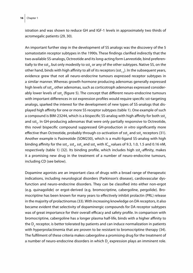
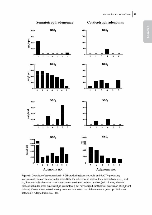
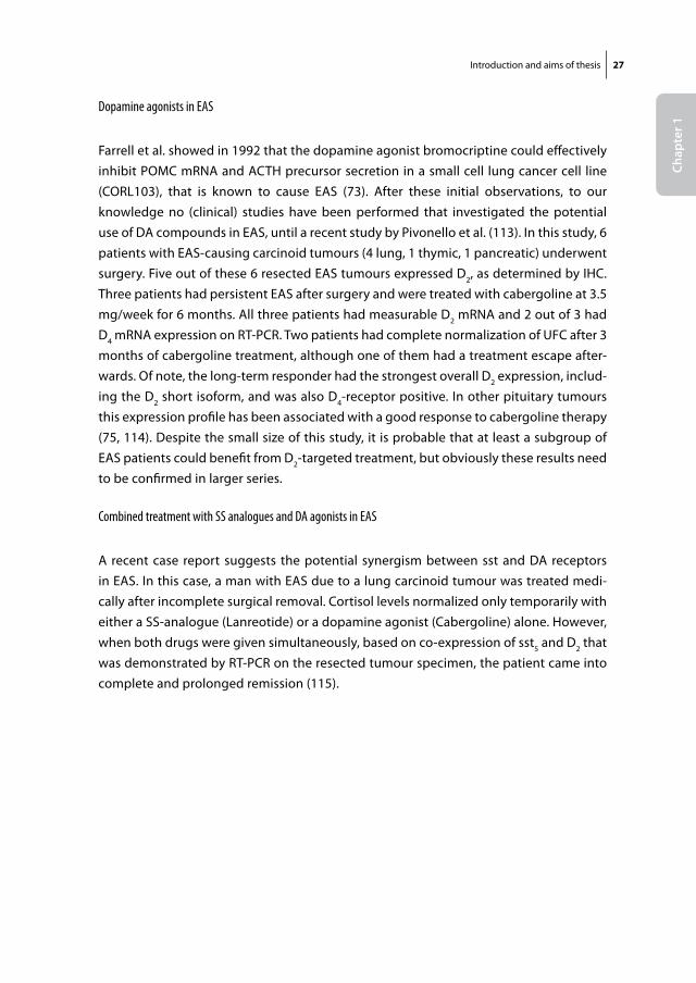
An important further step in the development of SS analogs was the discovery of the 5 somatostatin receptor subtypes in the 1990s. These findings clarified indirectly that the two available SS-analogs, Octreotide and its long-acting form Lanreotide, bind preferen-tially to the sst2, but only modestly to sst5 or any of the other subtypes. Native SS, on the other hand, binds with high affinity to all of its receptors (sst1-5). In the subsequent years, evidence grew that not all neuro-endocrine tumours expressed receptor subtypes in a similar manner. Whereas growth-hormone producing adenomas generally expressed high levels of sst2, other adenomas, such as corticotroph adenomas expressed consider-ably lower levels of sst2 (figure 5). The concept that different neuro-endocrine tumours with important differences in sst expression profiles would require specific sst-targeting analogs, sparked the interest for the development of new types of SS-analogs that dis-played high affinity for one or more SS receptor subtypes (table 1). One example of such a compound is BIM-23244, which is a bispecific SS-analog with high affinity for both sst2 and sst5. In GH-producing adenomas that were only partially responsive to Octreotide, this novel bispecific compound suppressed GH-production in vitro significantly more effective than Octreotide, probably through co-activation of sst2 and sst5 receptors (31). Another example is Pasireotide (SOM230), which is a multi-ligand SS-analog with high binding affinity for the sst1, sst2, sst3 and sst5 with IC50 values of 9.3, 1.0, 1.5 and 0.16 nM, respectively (table 1) (32). Its binding profile, which includes high sst5-affinity, makes it a promising new drug in the treatment of a number of neuro-endocrine tumours, including CD (see below).
Dopamine agonists are an important class of drugs with a broad range of therapeutic indications, including neurological disorders (Parkinson’s disease), cardiovascular dys-function and neuro-endocrine disorders. They can be classified into either non-ergot (e.g. quinagolide) or ergot-derived (e.g. bromocriptine, cabergoline, pergolide). Bro-mocriptine has been known for many years to effectively inhibit prolactin (PRL) release in the majority of prolactinomas (33). With increasing knowledge on DA receptors, it also became evident that selectivity of dopaminergic compounds for DA receptor subtypes was of great importance for their overall efficacy and safety profile. In comparison with bromocriptine, cabergoline has a longer plasma half-life, binds with a higher affinity to the D2 receptor, is better tolerated by patients and can induce normalization in patients with hyperprolactinemia that are proven to be resistant to bromocriptine therapy (34). The fulfilment of these criteria makes cabergoline a promising drug for the treatment of a number of neuro-endocrine disorders in which D2 expression plays an imminent role.
Introduction and aims of thesis 17
Chap
ter 1
Figure 5
400400
Somatotroph adenomas Corticotroph adenomas
sst1sst1
0
100
200
300
sst 1/hprt
0
100
200
300
nd nd nd nd nd nd ndndnd nd
1 2 3 4 5 60
1 2 3 4 5 6 70
300
400
300
400 sst2 sst2
prt
1 2 3 4 5 60
100
200
1 2 3 4 5 6 70
100
200
sst 2/hp
200
300
400
/hprt
200
300
400 sst3sst3
1 2 3 4 5 60
100
200
sst 3/
1 2 3 4 5 6 70
100
200
ndndndnd nd ndnd nd
1000
15002000
3000 sst5
1000
15002000
3000sst5
st5/h
prt
1 2 3 4 5 60
500
1 2 3 4 5 6 70
500
ss
Adenoma no. Adenoma no.Figure 5: Overview of sst expression in 7 GH-producing (somatotroph) and 6 ACTH-producing (corticotroph) human pituitary adenomas. Note the difference in scale of the y-axis between sst1-3 and sst5. Somatotroph adenomas have abundant expression of both sst2 and sst5 (left column), whereas corticotroph adenomas express sst5 at similar levels but have a significantly lower expression of sst2 (right column). Values are expressed as copy numbers relative to that of the reference gene hprt. N.d. = not detectable. Adapted from (57, 116).
18 Chapter 1
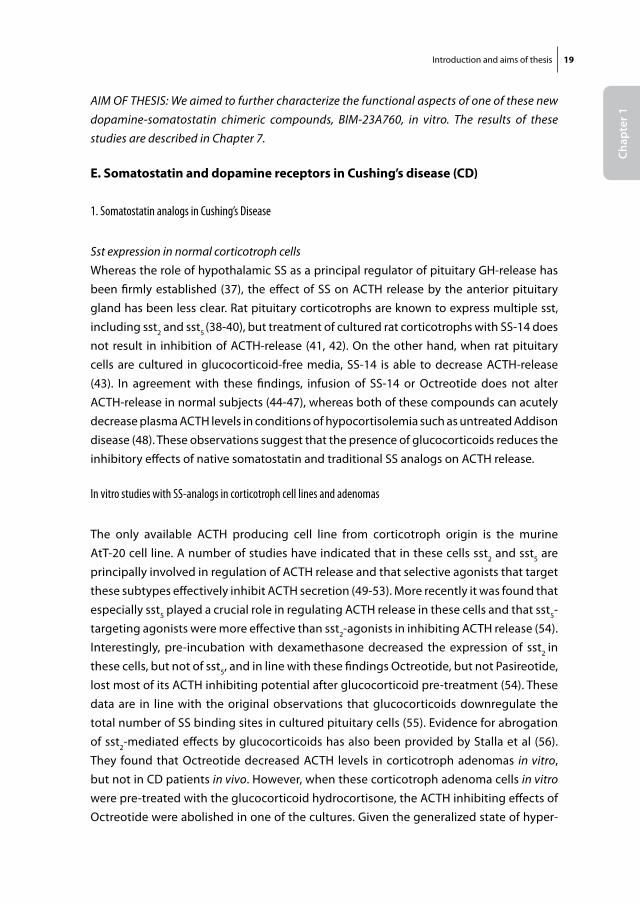
The binding affinities of dopamine (DA), bromocriptine and cabergoline to the D2 and D4 receptors, are also shown in table 1.
Chimeric somatostatin-dopamine compounds
The fact that many neuro-endocrine cells co-express both sst and DA receptors, has driven the hypothesis that these receptors may work synergistically. In 2000, Rocheville et al. published an important paper on the functional heterodimerization of sst5 and D2 receptors in stably transfected CHO-K1 cells, which resulted in overall enhanced biological potency (35). Based on these observations, new chimeric molecules have been synthesized that contain structural elements of both SS and DA compounds and therefore bind with high affinity to both sst and DA receptor subtypes. By binding to the two different receptors, these hybrid molecules were proposed to draw the recep-tors together in a spatial manner. This can lead to enhanced potency of the chimeric compound, compared to activation by two separate DA or SS analogs (36). At present it is not known, however, whether this enhanced potency is merely due to this proposed phenomenon of heterodimerization between the SS and DA receptors or whether other mechanisms may also be involved, such as superior activation at the level of individual receptors.
table 1 Binding affinities (IC50) of SS-analogs and DA-agonists (in nM)
Compound sst1 sst2 sst3 sst4 sst5 D2Short D2Long D4
SS-analogs
Somatostatin (SS-14) 0.93 0.15 0.56 1.50 0.29
Octreotide 280 0.38 7.10 >1000 6.30
Pasireotide (SOM230) 9.3 1.0 1.5 >1000 0.16
BIM-23244 >1000 0.3 133 >1000 0.7
Dopamine agonists
Dopamine 350 320 100
Bromocriptine 4.5 3.9 >1000
Cabergoline 0.53 0.41 81
Dopastatin chimera
BIM-23A760 622 0.03 160 >1000 42 15*
# References: SS-binding data (32, 117), DA-binding data (118), BIM-analog binding data (31, 119)* IC50 for D2 receptor (both short and long isoform)
Introduction and aims of thesis 19
Chap
ter 1
AIM OF THESIS: We aimed to further characterize the functional aspects of one of these new dopamine-somatostatin chimeric compounds, BIM-23A760, in vitro. The results of these studies are described in Chapter 7.
e. somatostatin and dopamine receptors in Cushing’s disease (CD)
1. Somatostatin analogs in Cushing’s Disease
Sst expression in normal corticotroph cellsWhereas the role of hypothalamic SS as a principal regulator of pituitary GH-release has been firmly established (37), the effect of SS on ACTH release by the anterior pituitary gland has been less clear. Rat pituitary corticotrophs are known to express multiple sst, including sst2 and sst5 (38-40), but treatment of cultured rat corticotrophs with SS-14 does not result in inhibition of ACTH-release (41, 42). On the other hand, when rat pituitary cells are cultured in glucocorticoid-free media, SS-14 is able to decrease ACTH-release (43). In agreement with these findings, infusion of SS-14 or Octreotide does not alter ACTH-release in normal subjects (44-47), whereas both of these compounds can acutely decrease plasma ACTH levels in conditions of hypocortisolemia such as untreated Addison disease (48). These observations suggest that the presence of glucocorticoids reduces the inhibitory effects of native somatostatin and traditional SS analogs on ACTH release.
In vitro studies with SS-analogs in corticotroph cell lines and adenomas
The only available ACTH producing cell line from corticotroph origin is the murine AtT-20 cell line. A number of studies have indicated that in these cells sst2 and sst5 are principally involved in regulation of ACTH release and that selective agonists that target these subtypes effectively inhibit ACTH secretion (49-53). More recently it was found that especially sst5 played a crucial role in regulating ACTH release in these cells and that sst5-targeting agonists were more effective than sst2-agonists in inhibiting ACTH release (54). Interestingly, pre-incubation with dexamethasone decreased the expression of sst2 in these cells, but not of sst5, and in line with these findings Octreotide, but not Pasireotide, lost most of its ACTH inhibiting potential after glucocorticoid pre-treatment (54). These data are in line with the original observations that glucocorticoids downregulate the total number of SS binding sites in cultured pituitary cells (55). Evidence for abrogation of sst2-mediated effects by glucocorticoids has also been provided by Stalla et al (56). They found that Octreotide decreased ACTH levels in corticotroph adenomas in vitro, but not in CD patients in vivo. However, when these corticotroph adenoma cells in vitro were pre-treated with the glucocorticoid hydrocortisone, the ACTH inhibiting effects of Octreotide were abolished in one of the cultures. Given the generalized state of hyper-
20 Chapter 1
cortisolism in CD patients and the relative resistance of sst5 to glucocorticoid-induced down-regulation compared to sst2, SS-analogs with high sst5 affinity are of great interest in the development of new medical therapies for CD.
AIM OF THESIS: To investigate more in detail the effects of glucocorticoids on the expression levels of not only sst2 and sst5, but also D2 in three human neuro-endocrine cell lines. These data could provide important insights into the biological processes that are responsible for the observed sst/DA receptor expression patterns in both pituitary-dependent and ectopic forms of Cushing’s syndrome. In addition, these data may also provide further directions for the type and timing of future sst/DA directed therapies in these patients. The results of these studies are described in Chapter 3.
In 2005 and 2006, two studies were published that independently investigated sst expression in human corticotroph adenoma tissues, obtained at the time of transsphe-noidal surgery. In the first study, Hofland et al. showed by quantitative PCR that sst5 was highly expressed in 6/6 adenomas, whereas sst1,2,3,4 were expressed at much lower levels (figure 5) (57). In concordance with this, functional studies in five additional adenomas demonstrated overall superior ACTH inhibition by Pasireotide (10nM) compared to Octreotide (10nM) after 72 hr.
In the second study, Batista et al. reported on a series of 13 corticotroph adenomas de-rived from both adult (n=7) and pediatric (n=6) CD patients (58). In this study, quantita-tive PCR demonstrated the expression of subtypes 1, 2, 4 and 5 in these adenomas, while at immunohistochemistry expression of all subtypes was found. Both of these methods showed the highest expression of the sst5 subtype. Six of the adenomas were cultured in vitro and treated with Pasireotide. In 6/6 adenomas Pasireotide significantly decreased cellular proliferation rates (range 10-70%) as measured by uptake of fluorescent vital stain and in 5/6 a significant decrease in ACTH production was observed (range 23-56%) at doses of 1 to 10 nM after 48-96 hr. Furthermore, a dissociation was seen in some of the adenomas between the anti-secretory and anti-proliferative effects of Pasireotide, simi-larly to what has been described previously for GH-producing adenomas in response to SS-analog treatment (59).
AIM OF THESIS: Since the above data were derived from relatively small patient series, we aimed to investigate somatostatin receptor subtype expression in a larger set of human corticotroph adenomas. Furthermore, we concomitantly investigated dopamine receptor subtype expression in these adenomas to assess the degree of co-expression of both receptor subtypes. In this way, we aimed to get a better estimate of the percentage of patients with CD that could benefit from DA or SS receptor targeted therapy. The results of these studies are described in Chapter 4.
Introduction and aims of thesis 21
Chap
ter 1
Clinical studies with SS-analogs in CD
Early studies showed that in patients with CD, Octreotide is not able to effectively re-duce ACTH secretion and hence cortisol levels (56, 60, 61). In contrast, several smaller studies and case reports found that patients with Nelson Syndrome, i.e. an expanding ACTH-producing pituitary adenoma after bilateral adrenalectomy, did respond to Oct-reotide with reductions in ACTH (60, 62-64). This difference is readily explained by the differences in average circulating cortisol levels in both disease states and the effects of glucocorticoid-induced down-regulation of sst2 receptors, as mentioned earlier (65).
Since then, few clinical studies have been reported that examined SS-analogue therapy in CD, until some important new insights developed. It was foremost the discovery that sst5 was highly expressed in the majority of human corticotroph adenomas, which made the novel multi-ligand SS-analog Pasireotide an interesting compound to evaluate in patients with CD, due to its subnanomolar sst5-affinity. A phase II multi-center clinical study has been performed in 29 patients with de novo or recurrent CD (66). Patients were treated with SOM230 600 μg twice daily over a 15-day period. Primary endpoint was normalization of 24-hour urinary free cortisol (UFC) levels. This study showed that out of 29 included patients, 5 (19%) obtained complete UFC normalization, while a total of 22 patients (76%) showed a decrease in UFC levels. Overall, Pasireotide was well toler-ated in the 2 x 600 μg dose, except for some mild gastro-intestinal side effects such as nausea, abdominal pain and loose stools or diarrhoea. A major side effect of Pasireotide, however, which was already known from previous studies in acromegalic patients, was an overall increase in blood glucose levels. Overt deterioration of glucose tolerance was observed in approximately one third of the patients in this study.
2. Dopamine agonists in Cushing’s Disease
DA receptor expression in normal corticotrophsIn humans, no firm data exist whether or not ACTH release is directly regulated by DA receptors in normal corticotroph cells. In rats it is known that the intermediate lobe in the pituitary is under tonic inhibitory control from dopaminergic neurons from the hy-pothalamus (67-69). The predominant cell type in the intermediate lobe is the melano-troph, which produces pro-opiomelanocortin (POMC). In the intermediate lobe POMC is processed into α-melanocyte stimulating hormone (α-MSH) and corticotropin-like intermediate lobe peptide (CLIP). This is different from the POMC-processing in anterior corticotroph cells, which mainly results in ACTH. The tonic inhibition by hypothalamic dopamine is thought to be exerted through D2 receptors. This is demonstrated by the fact that D2-deficient mice develop intermediate lobe hypertrophy with increased
22 Chapter 1
POMC expression, elevated ACTH and corticosterone levels, resulting in adrenal gland hypertrophy (70). In humans, the intermediate lobe in the pituitary is a rudimentary structure, but is still thought to contain important biological functions. Human cortico-troph adenomas arising from the intermediate lobe may have different characteristics than those arising from the anterior lobe, although some controversy exists around this subject (71, 72) .
In vitro studies with DA agonists in corticotroph cell lines and adenomas
Two reports have been published on the use of dopamine agonists in the murine corticotroph cell line AtT-20, but these have produced conflicting results. Farrell et al. found that treatment with the dopamine agonist bromocriptine did not reduce POMC mRNA expression in these cells, whereas Yin et al. did show that bromocriptine inhibited proliferation of these cells with induction of apoptosis (73, 74). The observed difference may be due to the fact that in the second study treatment with bromocriptine was significantly longer than in the first study (72 hr vs. 24 hr, respectively).
In 2004, Pivonello et al. investigated DA receptor expression in a series of 20 human corticotroph adenomas (75). They showed that the majority (80%) of these adenomas ex-press the D2 receptor as demonstrated by immunohistochemistry (IHC), receptor-ligand binding and RT-PCR. Of these D2-positive adenomas, approximately 40% expressed the D2 long isoform, 20% D2 short and 40% expressed both isoforms. D4 was expressed in 20% of cases, whereas D1, D3 and D5 expression was not observed. Functional studies in vitro correlated very well with the D2 expression data: adenomas high in D2 expression responded well to either bromocriptine or cabergoline therapy with inhibition of ACTH release by 43 to 60%, whereas D2-negative adenomas failed to respond. The D2- expres-sion data reported in this study are similar to those described by an earlier paper, where 11/16 (69%) of corticotroph adenomas, both functional and silent, expressed D2 recep-tors as demonstrated by in situ hybridisation and immunohistochemistry (76).
Clinical studies with DA agonists in CD
The DA agonist bromocriptine has been widely evaluated for its potential use in human corticotroph adenomas. Overall, results of these studies have been variable. Although initial reductions in ACTH levels are evident in almost half of CD patients in response to bromocriptine administration, these reductions are often minor and sustained responses to bromocriptine therapy occur only in a small percentage of patients (77). Some studies have suggested that corticotroph adenomas arising from the intermediate lobe may be more likely to respond to bromocriptine (78).
Introduction and aims of thesis 23
Chap
ter 1
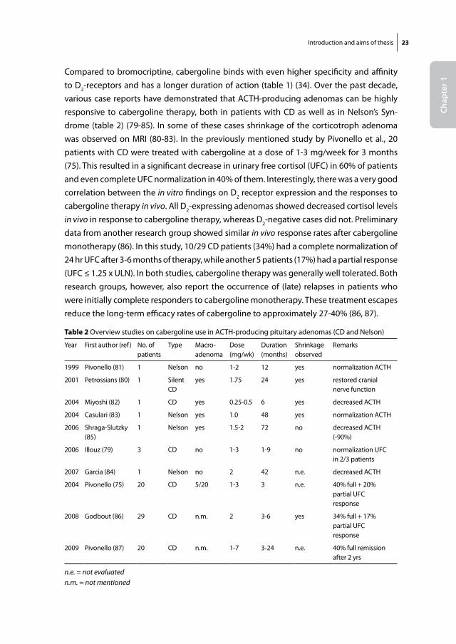
Compared to bromocriptine, cabergoline binds with even higher specificity and affinity to D2-receptors and has a longer duration of action (table 1) (34). Over the past decade, various case reports have demonstrated that ACTH-producing adenomas can be highly responsive to cabergoline therapy, both in patients with CD as well as in Nelson’s Syn-drome (table 2) (79-85). In some of these cases shrinkage of the corticotroph adenoma was observed on MRI (80-83). In the previously mentioned study by Pivonello et al., 20 patients with CD were treated with cabergoline at a dose of 1-3 mg/week for 3 months (75). This resulted in a significant decrease in urinary free cortisol (UFC) in 60% of patients and even complete UFC normalization in 40% of them. Interestingly, there was a very good correlation between the in vitro findings on D2 receptor expression and the responses to cabergoline therapy in vivo. All D2-expressing adenomas showed decreased cortisol levels in vivo in response to cabergoline therapy, whereas D2-negative cases did not. Preliminary data from another research group showed similar in vivo response rates after cabergoline monotherapy (86). In this study, 10/29 CD patients (34%) had a complete normalization of 24 hr UFC after 3-6 months of therapy, while another 5 patients (17%) had a partial response (UFC ≤ 1.25 x ULN). In both studies, cabergoline therapy was generally well tolerated. Both research groups, however, also report the occurrence of (late) relapses in patients who were initially complete responders to cabergoline monotherapy. These treatment escapes reduce the long-term efficacy rates of cabergoline to approximately 27-40% (86, 87).
table 2 Overview studies on cabergoline use in ACTH-producing pituitary adenomas (CD and Nelson)
Year First author (ref) No. of patients
Type Macro-adenoma
Dose (mg/wk)
Duration (months)
Shrinkageobserved
Remarks
1999 Pivonello (81) 1 Nelson no 1-2 12 yes normalization ACTH
2001 Petrossians (80) 1 Silent CD
yes 1.75 24 yes restored cranial nerve function
2004 Miyoshi (82) 1 CD yes 0.25-0.5 6 yes decreased ACTH
2004 Casulari (83) 1 Nelson yes 1.0 48 yes normalization ACTH
2006 Shraga-Slutzky (85)
1 Nelson yes 1.5-2 72 no decreased ACTH (-90%)
2006 Illouz (79) 3 CD no 1-3 1-9 no normalization UFC in 2/3 patients
2007 Garcia (84) 1 Nelson no 2 42 n.e. decreased ACTH
2004 Pivonello (75) 20 CD 5/20 1-3 3 n.e. 40% full + 20% partial UFC response
2008 Godbout (86) 29 CD n.m. 2 3-6 yes 34% full + 17% partial UFC response
2009 Pivonello (87) 20 CD n.m. 1-7 3-24 n.e. 40% full remission after 2 yrs
n.e. = not evaluatedn.m. = not mentioned
24 Chapter 1
One important issue that recently has dominated the field of medical therapy with DA-agonists has been the possible association between valvular heart disease and long-term therapy with the ergot-derived dopamine agonists (EDDA) pergolide and cabergoline. Two important papers were published in early 2007, which reported significantly in-creased risks (RR: 4.6-7.3) of valvular regurgitation in patients with idiopathic Parkinson’s disease that had received chronic treatment with either one of these drugs (88, 89). Other studies have recently confirmed these data (90). The pathogenetic mechanism behind this deleterious side effect is thought to be the binding of EDDA to 5-HT2 recep-tors expressed in the endocardial tissue of heart valves (89).
These findings have led to a number of important actions, including the withdrawal of pergolide from the US market. The impact of these studies on the (future) use of cabergoline in patients with CD cannot be fully determined yet, as one important issue needs to be emphasized. The maximum dose of cabergoline prescribed in CD is around 0.65 mg per day (4.5 mg/week), whereas the patients with Parkinson’s disease in the study by Zanettini et al. received an average daily dose of 3.6 mg/day (89). In the other study by Schade et al., an important risk difference was found between patients taking >3 mg cabergoline daily for more than 6 months (RR 50.3, 95% C.I: 6.6-381.4) compared to those who took less than 3 mg daily (RR 2.6; 95% C.I: 0.5-12.8) (88). Therefore, these observations in Parkinson’s disease patients can not be directly extrapolated towards lower-dose cabergoline therapy in CD. Recent studies have shown that patients who are on long-term cabergoline therapy for prolactinoma, do not have an increased incidence of heart valve abnormalities, as assessed by cardiac echocardiography (91-95). Most important, perhaps, is the notion that mild tricuspid regurgitation is a common finding, which can be present in up to 40% of the normal population (96). Therefore, the clinical relevance of finding mild, echocardiographic valve abnormalities in patients on long-term cabergoline use remains unclear (96, 97). Nonetheless, until definitive conclusions can be drawn on this subject, most clinicians will agree that periodical evaluation of cardiac function in any patient on long-term cabergoline therapy, especially those who are on higher doses, should be performed during follow-up (98).
3. Combined treatment with SS analogues and DA agonists in CD
Due to the reported presence of both sst and DA receptors in human corticotroph ad-enomas and the fact that both receptor types can inhibit ACTH production in vitro, the concept of a combination therapy with both SS-analogs and DA-agonists in CD seems to be a feasible approach (99). These studies could be performed by co-treatment with individual SS-analogs and DA-agonists (pasireotide + cabergoline) or perhaps, in the near future, by administration of SS-DA-chimeric compounds such as BIM-23A760, which
Introduction and aims of thesis 25
Chap
ter 1
displays high affinity for sst2, D2 and to a lesser extent sst5 (see table 1). If functional het-erodimerization of these receptor subtypes occurs in vivo, as has already been shown to occur in vitro by different groups, this type of treatment could result in greatly enhanced efficacy of these compounds (35). Also, as corticotroph adenomas can differ considerably in the total number of sst and D2 receptors they express (57, 58, 75), targeting of multiple receptors could increase the overall response rate in this group as a whole, compared to the use of individual SS or DA agonists. This has already been shown for GH-producing adenomas, where BIM-23A760 had overall superior efficacy compared to individual sst2, sst5 or D2-targeting agonists in terms of in vitro GH inhibition (100, 101). As it is known that also in CD only subsets of patients have responded to either cabergoline or pasir-eotide monotherapy in vivo, it may well be that similar phenomena occur in corticotroph adenomas and that combination therapy can increase overall response rates. Until now, no studies have been published that have investigated this hypothesis.
AIM OF THESIS: To investigate whether combined therapy with SS-analogues with sst5 affin-ity and D2 targeting agonists increases the percentage of CD patients that can be controlled biochemically during the standard pre-operative period of three months. We have investi-gated this in a multi-center clinical trial in patients with de novo or recurrent CD. Preliminary results of this ongoing study are presented in Chapter 5.
Theoretically, co-treatment with sst and DA agonists may have other advantages as well. As stated before, the inefficacy of sst2-preferring compounds in CD, is probably due to down-regulation of sst2-expression by high levels of circulating glucocorticoids (54, 56, 57). Inversely, if combined treatment with these analogs is effective and thus lowers cortisol levels in these patients, this could result in re-expression of sst2. The latter would result in enhanced efficacy of SS-analogs with sst2-affinity and hence strongly increase pharmacotherapeutical options in these patients (65, 102).
F. somAtostAtin AnD DoPAmine reCePtors in eCtoPiC ACtH-ProDuCing synDrome (eAs)
Ectopic ACTH-producing Syndrome (EAS)
Some peripheral neuro-endocrine tumours can secrete excessive amounts of ACTH, which can lead to the clinical entity known as the Ectopic ACTH-producing Syndrome (= Ectopic Cushing’s Syndrome). Primary therapy is preferably the surgical removal of the neuro-endocrine tumour, but in some cases this is not technically possible. Bilateral adrenalectomy can offer a definitive cure, but has the same disadvantages as described
26 Chapter 1
earlier for patients with persistent Cushing’s disease who undergo this procedure. For that reason, medical therapy could be an important secondary treatment option in selected cases of EAS.
Somatostatin analogs in EAS
For some decades it has been known that neuro-endocrine tumours that cause Ectopic ACTH-producing Syndrome (EAS), such as bronchial carcinoids or small cell lung cancer (SCLC), often express functional SS receptors. A number of smaller studies and case reports have been published on the use of Octreotide in patients with EAS. Interest-ingly, Octreotide was efficacious in lowering cortisol levels in a significant number of these patients, as opposed to the studies performed in patients with CD (103-106). This discrepancy is further confirmed by the fact that many patients with EAS have posi-tive lesions on 111In-pentreotide scan (OctreoScan), whereas most patients with CD do not (107). The observation that many of the EAS producing neuro-endocrine tumours have functional sst2 receptors, despite the chronic hypercortisolism they are exposed to, could be explained by aberrant glucocorticoid receptor signalling in these tumour cells. This has been investigated extensively by a number of research groups over the past twenty years. It was found that many of the cell lines, derived from EAS producing small-cell lung carcinomas, carry gross mutations in the genetic sequence of the gluco-corticoid receptor (GR) (108, 109). These can be located either in the DNA-binding or the ligand-binding domain, but can also involve a number of transcription factors. The loss of function of the GR has important impact on POMC production in these cells. Any form of negative feedback is generally lost in these cells, leading to excessive and uninhibited production of POMC and ultimately, the full clinical spectrum of Cushing’s Syndrome. Another result of aberrant GR functioning, may be that glucocorticoid-induced down-regulation of somatostatin receptor subtype 2 (sst2) does not occur in these tumours, as opposed to pituitary-derived corticotroph adenomas. This could well explain the relatively high degree of positive OctreoScans and reported efficacy of Octreotide in this group of neuro-endocrine tumours (110, 111). One main concern with the use of SS analogs in EAS, however, appears to be the long-term control of hypercortisolism. Although initial responses to Octreotide are frequent, these are not always sustained and treatment escapes are commonly encountered, due to a number of possible mecha-nisms of tachyphylaxis (112).
AIM OF THESIS: To illustrate the clinical relevance of glucocorticoid-induced changes in somatostatin receptor subtype expression in EAS, we have described our in vitro and in vivo findings on sst and DA receptor expression patterns in a patient with EAS, before and after glucocorticoid-lowering therapy. The results of these studies are described in Chapter 6.
Introduction and aims of thesis 27
Chap
ter 1
Dopamine agonists in EAS
Farrell et al. showed in 1992 that the dopamine agonist bromocriptine could effectively inhibit POMC mRNA and ACTH precursor secretion in a small cell lung cancer cell line (CORL103), that is known to cause EAS (73). After these initial observations, to our knowledge no (clinical) studies have been performed that investigated the potential use of DA compounds in EAS, until a recent study by Pivonello et al. (113). In this study, 6 patients with EAS-causing carcinoid tumours (4 lung, 1 thymic, 1 pancreatic) underwent surgery. Five out of these 6 resected EAS tumours expressed D2, as determined by IHC. Three patients had persistent EAS after surgery and were treated with cabergoline at 3.5 mg/week for 6 months. All three patients had measurable D2 mRNA and 2 out of 3 had D4 mRNA expression on RT-PCR. Two patients had complete normalization of UFC after 3 months of cabergoline treatment, although one of them had a treatment escape after-wards. Of note, the long-term responder had the strongest overall D2 expression, includ-ing the D2 short isoform, and was also D4-receptor positive. In other pituitary tumours this expression profile has been associated with a good response to cabergoline therapy (75, 114). Despite the small size of this study, it is probable that at least a subgroup of EAS patients could benefit from D2-targeted treatment, but obviously these results need to be confirmed in larger series.
Combined treatment with SS analogues and DA agonists in EAS
A recent case report suggests the potential synergism between sst and DA receptors in EAS. In this case, a man with EAS due to a lung carcinoid tumour was treated medi-cally after incomplete surgical removal. Cortisol levels normalized only temporarily with either a SS-analogue (Lanreotide) or a dopamine agonist (Cabergoline) alone. However, when both drugs were given simultaneously, based on co-expression of sst5 and D2 that was demonstrated by RT-PCR on the resected tumour specimen, the patient came into complete and prolonged remission (115).
28 Chapter 1
reFerenCes:
1. Bliss m 2005 Harvey Cushing: A life in surgery. New York: Oxford University Press 2. Cushing H 1932 The basophil adenomas of the pituitary body and their clinical manifestations.
Bull Johns Hopkins Hosp 50:137-195 3. orth Dn 1995 Cushing’s syndrome. N Engl J Med 332:791-803 4. lindholm J, Juul s, Jorgensen Jo, Astrup J, Bjerre P, Feldt-rasmussen u, Hagen C, Jorgensen
J, Kosteljanetz m, Kristensen l, laurberg P, schmidt K, Weeke J 2001 Incidence and late prog-nosis of cushing’s syndrome: a population-based study. J Clin Endocrinol Metab 86:117-123
5. Patil Cg, Prevedello Dm, lad sP, vance ml, thorner mo, Katznelson l, laws er, Jr. 2008 Late recurrences of Cushing’s disease after initial successful transsphenoidal surgery. J Clin Endocrinol Metab 93:358-362
6. Benveniste rJ, King WA, Walsh J, lee Js, Delman Bn, Post KD 2005 Repeated transsphenoidal surgery to treat recurrent or residual pituitary adenoma. J Neurosurg 102:1004-1012
7. locatelli m, vance ml, laws er 2005 Clinical review: the strategy of immediate reoperation for transsphenoidal surgery for Cushing’s disease. J Clin Endocrinol Metab 90:5478-5482
8. mahmoud-Ahmed As, suh JH 2002 Radiation therapy for Cushing’s disease: a review. Pituitary 5:175-180
9. vance ml 2005 Pituitary radiotherapy. Endocrinol Metab Clin North Am 34:479-487, xi 10. Jagannathan J, sheehan JP, Pouratian n, laws er, steiner l, vance ml 2007 Gamma Knife
surgery for Cushing’s disease. J Neurosurg 106:980-987 11. morris D, grossman A 2002 The Medical Management of Cushing’s Syndrome. Ann NY Acad Sci
970:119-133 12. Castinetti F, morange i, Jaquet P, Conte-Devolx B, Brue t 2008 Ketoconazole revisited: a
preoperative or postoperative treatment in Cushing’s disease. Eur J Endocrinol 158:91-99 13. nieman lK 2002 Medical therapy of Cushing’s disease. Pituitary 5:77-82 14. Chu JW, matthias DF, Belanoff J, schatzberg A, Hoffman Ar, Feldman D 2001 Successful
long-term treatment of refractory Cushing’s disease with high-dose mifepristone (RU 486). J Clin Endocrinol Metab 86:3568-3573
15. Heaney AP, melmed s 2004 Molecular targets in pituitary tumours. Nat Rev Cancer 4:285-295 16. etxabe J, vazquez JA 1994 Morbidity and mortality in Cushing’s disease: an epidemiological
approach. Clin Endocrinol (Oxf ) 40:479-484 17. Willeberg P PW 1982 Epidemiological aspects of clinical hyperadrenocorticism in dogs (Canine
Cushing’s Syndrome). J Am Anim Hosp Assoc 18:717-724 18. rijnberk A, der Kinderen PJ, thijssen JH 1968 Spontaneous hyperadrenocorticism in the dog. J
Endocrinol 41:397-406 19. Hanson Jm, teske e, voorhout g, galac s, Kooistra Hs, meij BP 2007 Prognostic factors for
outcome after transsphenoidal hypophysectomy in dogs with pituitary-dependent hyperadre-nocorticism. J Neurosurg 107:830-840
20. meij BP, voorhout g, van den ingh ts, Hazewinkel HA, teske e, rijnberk A 1998 Results of transsphenoidal hypophysectomy in 52 dogs with pituitary-dependent hyperadrenocorticism. Vet Surg 27:246-261
21. Barnett P 2003 Somatostatin and somatostatin receptor physiology. Endocrine 20:255-264 22. lamberts sW, Krenning eP, reubi JC 1991 The role of somatostatin and its analogs in the diag-
nosis and treatment of tumors. Endocr Rev 12:450-482 23. Patel yC 1999 Somatostatin and its receptor family. Front Neuroendocrinol 20:157-198
Introduction and aims of thesis 29
Chap
ter 1
24. missale C, nash sr, robinson sW, Jaber m, Caron mg 1998 Dopamine receptors: from structure to function. Physiol Rev 78:189-225
25. reubi JC, maurer r, von Werder K, torhorst J, Klijn Jg, lamberts sW 1987 Somatostatin recep-tors in human endocrine tumors. Cancer Res 47:551-558
26. Pivonello r FD, lombardi g, Colao A, lamberts sW, Hofland lJ 2007 Novel insights in dop-amine receptor physiology. Eur J Endocrinol 156:S13-S21
27. giustina g, Peracchi m, reschini e, Panerai e, Pinto m 1975 Dose-response study of the in-hibiting effect of somatostatin on growth hormone and insulin secretion in normal subjects and acromegalic patients. Metabolism 24:807-815
28. lamberts sW 1988 The role of somatostatin in the regulation of anterior pituitary hormone secretion and the use of its analogs in the treatment of human pituitary tumors. Endocr Rev 9:417-436
29. lamberts sW, oosterom r, neufeld m, del Pozo e 1985 The somatostatin analog SMS 201-995 induces long-acting inhibition of growth hormone secretion without rebound hypersecretion in acromegalic patients. J Clin Endocrinol Metab 60:1161-1165
30. lamberts sW, uitterlinden P, verschoor l, van Dongen KJ, del Pozo e 1985 Long-term treat-ment of acromegaly with the somatostatin analogue SMS 201-995. N Engl J Med 313:1576-1580
31. saveanu A, gunz g, Dufour H, Caron P, Fina F, ouafik l, Culler mD, moreau JP, enjalbert A, Jaquet P 2001 Bim-23244, a somatostatin receptor subtype 2- and 5-selective analog with enhanced efficacy in suppressing growth hormone (GH) from octreotide-resistant human GH-secreting adenomas. J Clin Endocrinol Metab 86:140-145
32. Bruns C, lewis i, Briner u, meno-tetang g, Weckbecker g 2002 SOM230: a novel somatostatin peptidomimetic with broad somatotropin release inhibiting factor (SRIF) receptor binding and a unique antisecretory profile. Eur J Endocrinol 146:707-716
33. molitch me, elton rl, Blackwell re, Caldwell B, Chang rJ, Jaffe r, Joplin g, robbins rJ, tyson J, thorner mo 1985 Bromocriptine as primary therapy for prolactin-secreting macroadenomas: results of a prospective multicenter study. J Clin Endocrinol Metab 60:698-705
34. Colao A, lombardi g, Annunziato l 2000 Cabergoline. Expert Opin Pharmacother 1:555-574 35. rocheville m, lange DC, Kumar u, Patel sC, Patel rC, Patel yC 2000 Receptors for dopamine
and somatostatin: formation of hetero-oligomers with enhanced functional activity. Science 288:154-157
36. Ferone D, Arvigo m, semino C, Jaquet P, saveanu A, taylor Je, moreau JP, Culler mD, Al-bertelli m, minuto F, Barreca A 2005 Somatostatin and dopamine receptor expression in lung carcinoma cells and effects of chimeric somatostatin-dopamine molecules on cell proliferation. Am J Physiol Endocrinol Metab 289:E1044-1050
37. Brazeau P, vale W, Burgus r, ling n, Butcher m, rivier J, guillemin r 1973 Hypothalamic polypeptide that inhibits the secretion of immunoreactive pituitary growth hormone. Science 179:77-79
38. o’Carroll Am, Krempels K 1995 Widespread distribution of somatostatin receptor messenger ribonucleic acids in rat pituitary. Endocrinology 136:5224-5227
39. Day r, Dong W, Panetta r, Kraicer J, greenwood mt, Patel yC 1995 Expression of mRNA for somatostatin receptor (sstr) types 2 and 5 in individual rat pituitary cells. A double labeling in situ hybridization analysis. Endocrinology 136:5232-5235
40. mezey e, Hunyady B, mitra s, Hayes e, liu Q, schaeffer J, schonbrunn A 1998 Cell specific expression of the sst2A and sst5 somatostatin receptors in the rat anterior pituitary. Endocrinol-ogy 139:414-419
30 Chapter 1
41. Brown mr, rivier C, vale W 1984 Central nervous system regulation of adrenocorticotropin secretion: role of somatostatins. Endocrinology 114:1546-1549
42. Kraicer J, gajewski tC, moor BC 1985 Release of pro-opiomelanocortin-derived peptides from the pars intermedia and pars distalis of the rat pituitary: effect of corticotrophin-releasing factor and somatostatin. Neuroendocrinology 41:363-373
43. lamberts sW, Zuyderwijk J, den Holder F, van Koetsveld P, Hofland l 1989 Studies on the conditions determining the inhibitory effect of somatostatin on adrenocorticotropin, prolactin and thyrotropin release by cultured rat pituitary cells. Neuroendocrinology 50:44-50
44. stafford PJ, Kopelman Pg, Davidson K, mcloughlin l, White A, rees lH, Besser gm, Coy DH, grossman A 1989 The pituitary-adrenal response to CRF-41 is unaltered by intravenous somatostatin in normal subjects. Clin Endocrinol (Oxf ) 30:661-666
45. Hall r, Besser gm, schally Av, Coy DH, evered D, goldie DJ, Kastin AJ, mcneilly As, mortimer CH, Phenekos C, tunbridge Wm, Weightman D 1973 Action of growth-hormone-release inhibi-tory hormone in healthy men and in acromegaly. Lancet 2:581-584
46. lightman sl, Fox P, Dunne mJ 1986 The effect of SMS 201-995, a long-acting somatostatin analogue, on anterior pituitary function in healthy male volunteers. Scand J Gastroenterol Suppl 119:84-95
47. invitti C, Pecori giraldi F, Dubini A, Piolini m, Cavagnini F 1991 Effect of sandostatin on CRF-stimulated secretion of ACTH, beta-lipotropin and beta-endorphin. Horm Metab Res 23:233-235
48. Fehm Hl, voigt KH, lang r, Beinert Ke, raptis s, Pfeiffer eF 1976 Somatostatin: a potent inhibitor of ACTH-hypersecretion in adrenal insufficiency. Klin Wochenschr 54:173-175
49. Cervia D, nunn C, Fehlmann D, langenegger D, schuepbach e, Hoyer D 2003 Pharmacologi-cal characterisation of native somatostatin receptors in AtT-20 mouse tumour corticotrophs. Br J Pharmacol 139:109-121
50. richardson ui, schonbrunn A 1981 Inhibition of adrenocorticotropin secretion by somatostatin in pituitary cells in culture. Endocrinology 108:281-290
51. tallent m, liapakis g, o’Carroll Am, lolait sJ, Dichter m, reisine t 1996 Somatostatin receptor subtypes SSTR2 and SSTR5 couple negatively to an L-type Ca2+ current in the pituitary cell line AtT-20. Neuroscience 71:1073-1081
52. strowski mZ, Dashkevicz mP, Parmar rm, Wilkinson H, Kohler m, schaeffer Jm, Blake AD 2002 Somatostatin receptor subtypes 2 and 5 inhibit corticotropin-releasing hormone-stimulated adrenocorticotropin secretion from AtT-20 cells. Neuroendocrinology 75:339-346
53. Cervia D, Fehlmann D, Hoyer D 2003 Native somatostatin sst2 and sst5 receptors functionally coupled to Gi/o-protein, but not to the serum response element in AtT-20 mouse tumour corti-cotrophs. Naunyn Schmiedebergs Arch Pharmacol 367:578-587
54. van der Hoek J, Waaijers m, van Koetsveld Pm, sprij-mooij D, Feelders rA, schmid HA, schoeffter P, Hoyer D, Cervia D, taylor Je, Culler mD, lamberts sW, Hofland lJ 2005 Distinct functional properties of native somatostatin receptor subtype 5 compared with subtype 2 in the regulation of ACTH release by corticotroph tumor cells. Am J Physiol Endocrinol Metab 289:E278-287
55. schonbrunn A 1982 Glucocorticoids down-regulate somatostatin receptors on pituitary cells in culture. Endocrinology 110:1147-1154
56. stalla gK, Brockmeier sJ, renner u, newton C, Buchfelder m, stalla J, muller oA 1994 Oct-reotide exerts different effects in vivo and in vitro in Cushing’s disease. Eur J Endocrinol 130:125-131
Introduction and aims of thesis 31
Chap
ter 1
57. Hofland lJ, van der Hoek J, Feelders r, van Aken mo, van Koetsveld Pm, Waaijers m, sprij-mooij D, Bruns C, Weckbecker g, de Herder WW, Beckers A, lamberts sW 2005 The multi-ligand somatostatin analogue SOM230 inhibits ACTH secretion by cultured human corticotroph adenomas via somatostatin receptor type 5. Eur J Endocrinol 152:645-654
58. Batista Dl, Zhang X, gejman r, Ansell PJ, Zhou y, Johnson sA, swearingen B, Hedley-Whyte et, stratakis CA, Klibanski A 2006 The effects of SOM230 on cell proliferation and adrenocorti-cotropin secretion in human corticotroph pituitary adenomas. J Clin Endocrinol Metab 91:4482-4488
59. Danila DC, Haidar Jns, Zhang X, Katznelson l, Culler mD, Klibanski A 2001 Somatostatin Receptor-Specific Analogs: Effects on Cell Proliferation and Growth Hormone Secretion in Human Somatotroph Tumors 10.1210/jc.86.7.2976. J Clin Endocrinol Metab 86:2976-2981
60. lamberts sW, uitterlinden P, Klijn Jm 1989 The effect of the long-acting somatostatin analogue SMS 201-995 on ACTH secretion in Nelson’s syndrome and Cushing’s disease. Acta Endocrinol (Copenh) 120:760-766
61. Ambrosi B, Bochicchio D, Fadin C, Colombo P, Faglia g 1990 Failure of somatostatin and octreotide to acutely affect the hypothalamic-pituitary-adrenal function in patients with corti-cotropin hypersecretion. J Endocrinol Invest 13:257-261
62. tyrrell JB, lorenzi m, gerich Je, Forsham PH 1975 Inhibition by somatostatin of ACTH secretion in Nelson’s syndrome. J Clin Endocrinol Metab 40:1125-1127
63. Petrini l, gasperi m, Pilosu r, marcello A, martino e 1994 Long-term treatment of Nelson’s syndrome by octreotide: a case report. J Endocrinol Invest 17:135-139
64. Kelestimur F, utas C, ozbakir o, selcuklu A, Kandemir o, ozcan n 1996 The effects of oct-reotide in a patient with Nelson’s syndrome. Postgrad Med J 72:53-54
65. van der Hoek J, lamberts sW, Hofland lJ 2004 The role of somatostatin analogs in Cushing’s disease. Pituitary 7:257-264
66. Boscaro m, ludlam WH, Atkinson B, glusman Je, Petersenn s, reincke m, snyder P, tabarin A, Biller Bm, Findling J, melmed s, Darby CH, Hu K, Wang y, Freda Pu, grossman AB, Frohman lA, Bertherat J 2009 Treatment of Pituitary-Dependent Cushing’s Disease with the Multireceptor Ligand Somatostatin Analog Pasireotide (SOM230): A Multicenter, Phase II Trial. J Clin Endocrinol Metab 94:115-122
67. Antakly t, sasaki A, liotta As, Palkovits m, Krieger Dt 1985 Induced expression of the gluco-corticoid receptor in the rat intermediate pituitary lobe. Science 229:277-279
68. stack J, surprenant A 1991 Dopamine actions on calcium currents, potassium currents and hormone release in rat melanotrophs. J Physiol 439:37-58
69. Farah Jm, Jr., malcolm Ds, mueller gP 1982 Dopaminergic inhibition of pituitary beta-endor-phin-like immunoreactivity secretion in the rat. Endocrinology 110:657-659
70. saiardi A, Borrelli e 1998 Absence of dopaminergic control on melanotrophs leads to Cushing’s-like syndrome in mice. Mol Endocrinol 12:1133-1139
71. lamberts sW, de lange sA, stefanko sZ 1982 Adrenocorticotropin-secreting pituitary ad-enomas originate from the anterior or the intermediate lobe in Cushing’s disease: differences in the regulation of hormone secretion. J Clin Endocrinol Metab 54:286-291
72. Croughs rJ, Koppeschaar HP, van’t verlaat JW, mcnicol Am 1989 Bromocriptine-responsive Cushing’s disease associated with anterior pituitary corticotroph hyperplasia or normal pituitary gland. J Clin Endocrinol Metab 68:495-498
32 Chapter 1
73. Farrell We, Clark AJ, stewart mF, Crosby sr, White A 1992 Bromocriptine inhibits pro-opiomel-anocortin mRNA and ACTH precursor secretion in small cell lung cancer cell lines. J Clin Invest 90:705-710
74. yin D, Kondo s, takeuchi J, morimura t 1994 Induction of apoptosis in murine ACTH-secreting pituitary adenoma cells by bromocriptine. FEBS Lett 339:73-75
75. Pivonello r, Ferone D, de Herder WW, Kros Jm, De Caro ml, Arvigo m, Annunziato l, lom-bardi g, Colao A, Hofland lJ, lamberts sW 2004 Dopamine receptor expression and function in corticotroph pituitary tumors. J Clin Endocrinol Metab 89:2452-2462
76. stefaneanu l, Kovacs K, Horvath e, Buchfelder m, Fahlbusch r, lancranjan l 2001 Dopamine D2 receptor gene expression in human adenohypophysial adenomas. Endocrine 14:329-336
77. miller JW, Crapo l 1993 The medical treatment of Cushing’s syndrome. Endocr Rev 14:443-458 78. lamberts sW, Klijn Jg, de Quijada m, timmermans HA, uitterlinden P, de Jong FH, Birkenha-
ger JC 1980 The mechanism of the suppressive action of bromocriptine on adrenocorticotropin secretion in patients with Cushing’s disease and Nelson’s syndrome. J Clin Endocrinol Metab 51:307-311
79. illouz F, Dubois-ginouves s, laboureau s, rohmer v, rodien P 2006 [Use of cabergoline in persisting Cushing’s disease]. Ann Endocrinol (Paris) 67:353-356
80. Petrossians P, ronci n, valdes socin H, Kalife A, stevenaert A, Bloch B, tabarin A, Beckers A 2001 ACTH silent adenoma shrinking under cabergoline. Eur J Endocrinol 144:51-57
81. Pivonello r, Faggiano A, Di salle F, Filippella m, lombardi g, Colao A 1999 Complete remission of Nelson’s syndrome after 1-year treatment with cabergoline. J Endocrinol Invest 22:860-865
82. miyoshi t, otsuka F, takeda m, inagaki K, suzuki J, ogura t, Date i, Hashimoto K, makino H 2004 Effect of cabergoline treatment on Cushing’s disease caused by aberrant adrenocorticotro-pin-secreting macroadenoma. J Endocrinol Invest 27:1055-1059
83. Casulari lA, naves lA, mello PA, Pereira neto A, Papadia C 2004 Nelson’s syndrome: complete remission with cabergoline but not with bromocriptine or cyproheptadine treatment. Horm Res 62:300-305
84. garcia C, Bordier l, garcia-Hejl C, Ceppa F, mayaudon H, Dupuy o, Bauduceau B 2007 [Nel-son’s syndrome management: current knowledge]. Rev Med Interne 28:766-769
85. shraga-slutzky i, shimon i, Weinshtein r 2006 Clinical and biochemical stabilization of Nelson’s syndrome with long-term low-dose cabergoline treatment. Pituitary 9:151-154
86. godbout A mm, Danilowicz K, Beauregard H, Bruno o, lacroix A 2008 Long-term therapy with cabergoline in Cushing’s disease. The Endocrine Society’s 90th annual meeting, June 15-18, San Francisco, U.S.A., 2008
87. Pivonello r, De martino mC, Cappabianca P, De leo m, Faggiano A, lombardi g, Hofland lJ, lamberts sW, Colao A 2009 The Medical Treatment of Cushing’s Disease: Effectiveness of Chronic Treatment with the Dopamine Agonist Cabergoline in Patients Unsuccessfully Treated by Surgery. J Clin Endocrinol Metab 94:223-230
88. schade r, Andersohn F, suissa s, Haverkamp W, garbe e 2007 Dopamine agonists and the risk of cardiac-valve regurgitation. N Engl J Med 356:29-38
89. Zanettini r, Antonini A, gatto g, gentile r, tesei s, Pezzoli g 2007 Valvular heart disease and the use of dopamine agonists for Parkinson’s disease. N Engl J Med 356:39-46
90. rasmussen vg, Poulsen sH, Dupont e, ostergaard K, safikhany g, egeblad H 2008 Heart valve disease associated with treatment with ergot-derived dopamine agonists: a clinical and echocardiographic study of patients with Parkinson’s disease. J Intern Med 263:90-98
Introduction and aims of thesis 33
Chap
ter 1
91. Herring n, szmigielski C, Becher H, Karavitaki n, Wass JA 2009 Valvular heart disease and the use of cabergoline for the treatment of prolactinoma. Clin Endocrinol (Oxf ) 70:104-108
92. Devin JK, lakhani vt, Byrd BF, 3rd, Blevins ls, Jr. 2008 Prevalence of valvular heart disease in a cohort of patients taking cabergoline for management of hyperprolactinemia. Endocr Pract 14:672-677
93. vallette s, serri K, rivera J, santagata P, Delorme s, garfield n, Kahtani n, Beauregard H, Aris-Jilwan n, Houde g, serri o 2008 Long-term cabergoline therapy is not associated with valvular heart disease in patients with prolactinomas. Pituitary
94. Wakil A, rigby As, Clark Al, Kallvikbacka-Bennett A, Atkin sl 2008 Low dose cabergoline for hyperprolactinaemia is not associated with clinically significant valvular heart disease. Eur J Endocrinol 159:R11-14
95. lancellotti P, livadariu e, markov m, Daly AF, Burlacu mC, Betea D, Pierard l, Beckers A 2008 Cabergoline and the risk of valvular lesions in endocrine disease. Eur J Endocrinol 159:1-5
96. sherlock m, toogood AA, steeds r 2009 Dopamine agonist therapy for hyperprolactinaemia and cardiac valve dysfunction; a lot done but much more to do. Heart Apr; 95(7):522-3
97. Kars m, Delgado v, Holman er, Feelders rA, smit JW, romijn JA, Bax JJ, Pereira Am 2008 Aortic valve calcification and mild tricuspid regurgitation but no clinical heart disease after 8 years of dopamine agonist therapy for prolactinoma. J Clin Endocrinol Metab 93:3348-3356
98. Kars m, Pereira Am, Bax JJ, romijn JA 2008 Cabergoline and cardiac valve disease in prolac-tinoma patients: additional studies during long-term treatment are required. Eur J Endocrinol 159:363-367
99. Colao A, Filippella m, Pivonello r, Di somma C, Faggiano A, lombardi g 2007 Combined therapy of somatostatin analogues and dopamine agonists in the treatment of pituitary tumours. Eur J Endocrinol 156 Suppl 1:S57-63
100. saveanu A, gunz g, guillen s, Dufour H, Culler mD, Jaquet P 2006 Somatostatin and dop-amine-somatostatin multiple ligands directed towards somatostatin and dopamine receptors in pituitary adenomas. Neuroendocrinology 83:258-263
101. Jaquet P, gunz g, saveanu A, Barlier A, Dufour H, taylor J, Dong J, Kim s, moreau JP, Culler mD 2005 BIM-23A760, a chimeric molecule directed towards somatostatin and dopamine recep-tors, vs universal somatostatin receptors ligands in GH-secreting pituitary adenomas partial responders to octreotide. J Endocrinol Invest 28:21-27
102. schmid HA 2008 Pasireotide (SOM230): Development, mechanism of action and potential ap-plications. Mol Cell Endocrinol 286:69-74
103. lamberts sW, de Herder WW, Krenning eP, reubi JC 1994 A role of (labeled) somatostatin ana-logs in the differential diagnosis and treatment of Cushing’s syndrome. J Clin Endocrinol Metab 78:17-19
104. Phlipponneau m, nocaudie m, epelbaum J, De Keyzer y, lalau JD, marchandise X, Bertagna X 1994 Somatostatin analogs for the localization and preoperative treatment of an adrenocorti-cotropin-secreting bronchial carcinoid tumor. J Clin Endocrinol Metab 78:20-24
105. Bertagna X, Favrod-Coune C, escourolle H, Beuzeboc P, Christoforov B, girard F, luton JP 1989 Suppression of ectopic adrenocorticotropin secretion by the long-acting somatostatin analog octreotide. J Clin Endocrinol Metab 68:988-991
106. vignati F, loli P 1996 Additive effect of ketoconazole and octreotide in the treatment of severe adrenocorticotropin-dependent hypercortisolism. J Clin Endocrinol Metab 81:2885-2890
107. de Herder WW, Krenning eP, malchoff CD, Hofland lJ, reubi JC, Kwekkeboom DJ, oei Hy, Pols HA, Bruining HA, nobels Fr, et al. 1994 Somatostatin receptor scintigraphy: its value
34 Chapter 1
in tumor localization in patients with Cushing’s syndrome caused by ectopic corticotropin or corticotropin-releasing hormone secretion. Am J Med 96:305-312
108. ray DW, littlewood AC, Clark AJ, Davis Jr, White A 1994 Human small cell lung cancer cell lines expressing the proopiomelanocortin gene have aberrant glucocorticoid receptor function. J Clin Invest 93:1625-1630
109. gaitan D, DeBold Cr, turney mK, Zhou P, orth Dn, Kovacs WJ 1995 Glucocorticoid receptor structure and function in an adrenocorticotropin-secreting small cell lung cancer. Mol Endocrinol 9:1193-1201
110. uwaifo gi, Koch CA, Hirshberg B, Chen CC, Hartzband P, nieman lK, Pacak K 2003 Is there a therapeutic role for octreotide in patients with ectopic Cushing’s syndrome? J Endocrinol Invest 26:710-717
111. lamberts sW, tilanus HW, Klooswijk Ai, Bruining HA, van der lely AJ, de Jong FH 1988 Suc-cessful treatment with SMS 201-995 of Cushing’s syndrome caused by ectopic adrenocorticotro-pin secretion from a metastatic gastrin-secreting pancreatic islet cell carcinoma. J Clin Endocrinol Metab 67:1080-1083
112. Hofland lJ, lamberts sW 2003 The pathophysiological consequences of somatostatin receptor internalization and resistance. Endocr Rev 24:28-47
113. Pivonello r, Ferone D, de Herder WW, Faggiano A, Bodei l, de Krijger rr, lombardi g, Colao A, lamberts sW, Hofland lJ 2007 Dopamine receptor expression and function in corticotroph ectopic tumors. J Clin Endocrinol Metab 92:65-69
114. Pivonello r, matrone C, Filippella m, Cavallo lm, Di somma C, Cappabianca P, Colao A, Annunziato l, lombardi g 2004 Dopamine receptor expression and function in clinically non-functioning pituitary tumors: comparison with the effectiveness of cabergoline treatment. J Clin Endocrinol Metab 89:1674-1683
115. Pivonello r, Ferone D, lamberts sW, Colao A 2005 Cabergoline plus lanreotide for ectopic Cushing’s syndrome. N Engl J Med 352:2457-2458
116. Hofland lJ, van der Hoek J, van Koetsveld Pm, de Herder WW, Waaijers m, sprij-mooij D, Bruns C, Weckbecker g, Feelders r, van der lely AJ, Beckers A, lamberts sW 2004 The novel somatostatin analog SOM230 is a potent inhibitor of hormone release by growth hormone- and prolactin-secreting pituitary adenomas in vitro. J Clin Endocrinol Metab 89:1577-1585
117. schmid HA, schoeffter P 2004 Functional activity of the multiligand analog SOM230 at human recombinant somatostatin receptor subtypes supports its usefulness in neuroendocrine tumors. Neuroendocrinology 80 Suppl 1:47-50
118. newman-tancredi A, Cussac D, Audinot v, nicolas JP, De Ceuninck F, Boutin JA, millan mJ 2002 Differential actions of antiparkinson agents at multiple classes of monoaminergic receptor. II. Agonist and antagonist properties at subtypes of dopamine D(2)-like receptor and alpha(1)/alpha(2)-adrenoceptor. J Pharmacol Exp Ther 303:805-814
119. Jaquet P, gunz g, saveanu A, Dufour H, taylor J, Dong J, Kim s, moreau JP, enjalbert A, Culler mD 2005 Efficacy of chimeric molecules directed towards multiple somatostatin and dopamine receptors on inhibition of GH and prolactin secretion from GH-secreting pituitary adenomas clas-sified as partially responsive to somatostatin analog therapy. Eur J Endocrinol 153:135-141
Chapter 2
Expression and functional analysis of dopamine receptor subtype 2
and somatostatin receptor subtypes in canine Cushing’s disease
de Bruin C, Hanson JM, Meij BP, Kooistra HS, Waaijers AM, Uitterlinden P, Lamberts SW, Hofland LJ.
Endocrinology. 2008 Sep;149(9):4357-66.
36 Chapter 2
ABstrACt
Cushing’s disease (CD) is a severe disorder characterized by chronic hypercortisolism due to an ACTH-secreting pituitary adenoma. Transsphenoidal adenomectomy is the treatment of choice in humans with CD but recurrences occur frequently. Finding an effective and safe medical treatment for CD may improve long-term clinical outcome. The recent demonstration of expression of somatostatin receptor subtypes (mainly sst5) and dopamine D2 receptors in human corticotroph adenomas offers the possibility for medical treatment of CD with novel somatostatin analogues and dopamine agonists. Investigation of the effects of these drugs is hampered by the low incidence of CD in humans. Interestingly, CD is a frequent disorder in dogs with striking clinical similarities with CD in humans. Therefore, we investigated the expression and functional role of D2 and sst receptors in corticotroph adenoma cells from 13 dogs with active CD that underwent therapeutic hypophysectomy and normal anterior pituitary (NAP) cells from 5 dogs. Quantitative RT-PCR and immunohistochemistry revealed that both in CD and NAP, sst2 was the predominant receptor subtype expressed, whereas D2 was modestly expressed and sst5 was expressed only at very low levels. In primary cultures of canine adenomas (n=7), the sst2-preferring agonist octreotide also showed the strongest ACTH-suppressive effects. In conclusion, canine corticotroph adenomas provide an interesting model to study CD, but differences in sst and dopamine receptor expression between humans and dogs should be taken into account when using dogs with CD as a model to evaluate efficacy of novel somatostatin analogues and dopamine agonists for human CD.
Dopamine and somatostatin receptors in canine Cushing 37
Chap
ter 2
introDuCtion
Cushing’s disease (CD) is a severe endocrinological disorder due to an ACTH-producing pituitary adenoma. The resulting chronic hypercortisolism causes significant morbidity and, if left untreated, mortality in these patients (1). Primary treatment of CD is trans-sphenoidal selective adenomectomy (2), but only results in long-term cure in 50-80% of patients (3). Secondary treatments such as radiotherapy or bilateral adrenalectomy are generally effective, but can cause permanent hypopituitarism or the necessity of life-long adrenal hormone replacement therapy, respectively.
For that reason, finding an effective and safe medical therapy for human CD can be of great importance for those CD patients that are not cured by neurosurgery alone. Various drugs have been used in patients with CD, but most of them have not been ef-ficacious in long-term treatment or are associated with an unfavorable safety profile (4). Novel drug targets have been identified, however, as it was found that the somatostatin receptor subtype 5 (sst5) and the dopamine (DA) receptor subtype 2 (D2) are expressed in the majority of human corticotroph adenomas (5-7). Compounds that target these receptor subtypes, such as the multiligand somatostatin analogue with high sst5-affinity pasireotide (SOM230) and the D2-agonist cabergoline, have already shown in some in vitro and in vivo studies to decrease ACTH release by corticotroph adenoma cells and thus lower cortisol levels (5, 6, 8).
For the development of new medical therapies in human CD, research on primary corticotroph adenoma tissue is crucial. The efficacy of new compounds in CD can only be genuinely tested in the cell type they are primarily directed at, i.e., the human corti-cotroph cell. This tissue can only be obtained at the time of transsphenoidal adenomec-tomy in a CD patient. Due to the low incidence of CD of approximately 1.2 to 2.4 cases/million/year (9, 10) and the fact that 80-90% of these cases are due to microadenomas with a diameter of less than 10 mm (11, 12), there is a severe shortage of human corti-cotroph tissue, which limits research options in human CD. For that reason, finding ways to increase the availability of primary corticotroph adenoma tissue is a major challenge in this research field.
In contrast to the situation in humans, CD is a frequent endocrinological disorder in dogs, with an estimated incidence of 1 to 2 cases/1000/yr (13-16). Canine CD, also re-ferred to as pituitary-dependent hyperadrenocorticism (PDH), has a remarkably similar pathophysiology and clinical presentation as CD in humans and can hence be regarded as a spontaneous animal model for human CD (17). In dogs with CD true microadenomas are rare and pituitaries are frequently enlarged (18). Medical treatment of dogs with CD
38 Chapter 2
involves the use of adrenolytic drugs such as mitotane or an inhibitor of steroidogenesis such as trilostane (19, 20). In the Netherlands, hypophysectomy has been performed in dogs with CD since 1993 and has proven to be a safe and effective treatment (18, 21, 22). The procedure consists of a complete hypophysectomy via a transsphenoidal approach as is described in detail elsewhere (22).
Given the high incidence of CD in dogs, the high degree of similarity with human CD and the availability of corticotroph adenoma tissue obtained at hypophysectomy, we hypothesized that evaluation of the efficacy of new compounds for treatment of human CD may be tested first in canine corticotroph adenoma tissue. Therefore, our main study aim was to characterize these canine corticotroph adenomas for the expression and functional role of those receptor subtypes that are of primary interest in the research of human CD: somatostatin receptor subtype 2 (sst2), somatostatin receptor subtype 5 (sst5) and dopamine receptor subtype 2 (D2), and to compare these results with the current knowledge on human corticotroph adenomas.
mAteriAls AnD metHoDs
Study population
Thirteen dogs (5 females (4 spayed) and 8 males (3 castrated)) with Cushing’s disease (i.e. pituitary-dependent hypercortisolism, PDH) from various breeds were included in the study (Table 1). The median age was 8 years (range 5-14 years) and the median body weight was 23.2 kg (range 6.7-48.0 kg). Hypercortisolism was diagnosed by clinical signs, routine laboratory investigation and determination of the urinary corticoid-to-creatinine ratio (UCCR) in two consecutive morning urine samples as described previously (23-26). The mean UCCR was 116.7 x 10⁻⁶ (range 26.5-302.5 x 10⁻⁶; normal <10 x 10-6) (18). After collection of the second urine sample, three oral doses of 0.1 mg dexamethasone per kg body weight were administered at 8 h intervals and the next morning a third urine sample was collected (high dose dexamethasone suppression test). In 10 dogs the UCCR in the third sample was less than 50% of the mean in the first 2 samples and PDH was diagnosed (18). In 2 cases with less than 50% suppression, dexamethasone-resistant PDH was confirmed by measurements of plasma ACTH concentrations and further supported by visualization of the adrenals by ultrasonography and pituitary imaging (27-30). Computed tomography (CT) of the pituitary gland revealed pituitary enlarge-ment in each case, except one (C8), with a mean pituitary height-to-brain area ratio (P/B) of 0.58 (range 0.30-1.00; pituitary enlarged when P/B > 0.31 x 10⁻² mm-1) (31). Plasma cortisol, ACTH and α-MSH concentrations were determined with assays that have been
Dopamine and somatostatin receptors in canine Cushing 39
Chap
ter 2
described previously (32). Pre-operative mean (+range) plasma values were: α-MSH 27.8 (<5-224) pg/ml, cortisol 196.9 (61-414) nmol/l and ACTH 21.5 (9.3-41.8) pmol/l (see Table 1 for reference values). Microsurgical transsphenoidal hypophysectomy was performed as published previously (22).
Unaffected pituitary tissue was obtained from five Beagle dogs, which had been eutha-nized for reasons unrelated to the present study and for which approval was obtained from the Ethical Committee of Utrecht University, the Netherlands. The pituitary gland was collected within 10 minutes after euthanasia. The anterior pituitary was separated from the neurointermediate lobe and the anterior pituitary was processed for analysis.
Surgical tissue and cell isolation
During transsphenoidal hypophysectomy, pituitary adenomatous tissue was identified macroscopically by the veterinary surgeon and resected. A representative part of the adenoma was fixated in 4% buffered paraformaldehyde and sent for histopathology for haematoxylin and eosin (HE) staining and immunohistochemistry to evaluate ACTH, α-MSH and GH expression (33). The surplus adenomatous tissue was immediately placed in a pre-chilled (4 ˚C) solution of Minimal Essential Medium (MEM) with Earle’s salts, supplemented with 10% fetal calf serum (FCS), L-glutamine (2 mmol/l), penicillin (105 U/l) and fungizone (0.25 mg/l). Media and supplements were obtained from Invit-rogen (Breda, the Netherlands). Upon arrival in the laboratory, the adenoma tissue was further divided into two parts: one part was snap-frozen on dry ice and stored at –80˚C for qPCR studies; the other part was kept overnight at 4˚C in MEM. Next day, the latter adenoma part was washed in HBSS/HSA, dispersed with dispase 103 U/l (Roche, Almere, the Netherlands) + collagenase 2 mg/ml (Sigma Aldrich, Zwijndrecht, the Netherlands) at 37ºC for 1 h and resuspended in MEM complete culture medium. Viable pituitary cells were counted in a standard haematocytometer.
Cell distribution and culture
The average yield per tumor in terms of viable canine pituitary cells was 2.4 x 10⁶ cells (range: 0.5-11.0 x 10⁶). Of these cells, 0.2 x 10⁶ were used for qPCR studies and 0.1 x 10⁶ for the preparation of cytospins for immunohistochemistry (IHC, see below). The remainder of the cells was cultured in 48 well plates (Corning, Cambridge, USA) at a den-sity of 10,000 cells/well for 4-6 days at 37 ˚C in a humidified incubator in 5% CO2. At that time, culture media were refreshed and incubations were started with the different DA agonists and SS analogues for 4-72 h. Both basal and corticotropin-releasing hormone (CRH)-induced ACTH-release were studied. At the end of the incubation period, media
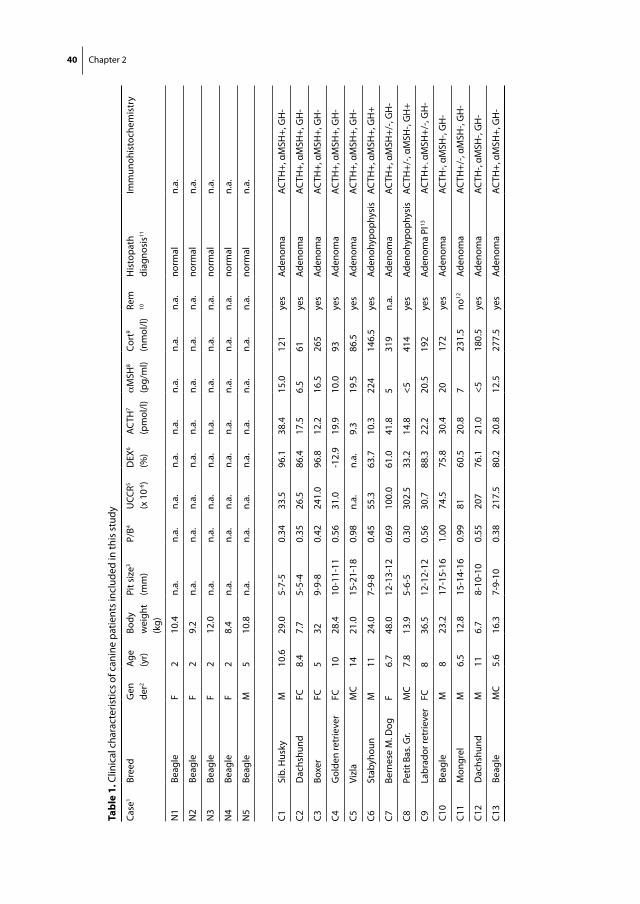
40 Chapter 2
tabl
e 1.
Clin
ical
cha
ract
eris
tics
of c
anin
e pa
tient
s in
clud
ed in
this
stu
dy
Case
1Br
eed
Gen
der2
Age
(yr)
Body
w
eigh
t (k
g)
Pit s
ize3
(mm
)P/
B4U
CCR5
(x 1
0-6)
DEX
6
(%)
ACTH
7
(pm
ol/l)
αM
SH8
(pg/
ml)
Cort
9
(nm
ol/l)
Rem
10
His
topa
th
diag
nosi
s11
Imm
unoh
isto
chem
istr
y
N1
Beag
leF
210
.4n.
a.n.
a.n.
a.n.
a.n.
a.n.
a.n.
a.n.
a.no
rmal
n.a.
N2
Beag
leF
29.
2n.
a.n.
a.n.
a.n.
a.n.
a.n.
a.n.
a.n.
a.no
rmal
n.
a.
N3
Beag
leF
212
.0n.
a.n.
a.n.
a.n.
a.n.
a.n.
a.n.
a.n.
a.no
rmal
n.a.
N4
Beag
leF
28.
4n.
a.n.
a.n.
a.n.
a.n.
a.n.
a.n.
a.n.
a.no
rmal
n.a.
N5
Beag
leM
510
.8n.
a.n.
a.n.
a.n.
a.n.
a.n.
a.n.
a.n.
a.no
rmal
n.
a.
C1Si
b. H
usky
M10
.629
.05-
7-5
0.34
33.5
96.1
38.4
15.0
121
yes
Aden
oma
ACTH
+, α
MSH
+, G
H-
C2D
achs
hund
FC8.
47.
75-
5-4
0.35
26.5
86.4
17.5
6.5
61ye
sAd
enom
aAC
TH+,
αM
SH+,
GH
-
C3Bo
xer
FC5
329-
9-8
0.42
241.
096
.812
.216
.526
5ye
sAd
enom
aAC
TH+,
αM
SH+,
GH
-
C4G
olde
n re
trie
ver
FC10
28.4
10-1
1-11
0.56
31.0
-12.
919
.910
.093
yes
Aden
oma
ACTH
+, α
MSH
+, G
H-
C5Vi
zla
MC
1421
.015
-21-
180.
98n.
a.n.
a.9.
319
.586
.5ye
sAd
enom
aAC
TH+,
αM
SH+,
GH
-
C6St
abyh
oun
M11
24.0
7-9-
80.
4555
.363
.710
.322
414
6.5
yes
Aden
ohyp
ophy
sis
ACTH
+, α
MSH
+, G
H+
C7Be
rnes
e M
. Dog
F6.
748
.012
-13-
120.
6910
0.0
61.0
41.8
531
9n.
a.Ad
enom
aAC
TH+,
αM
SH+/
-, G
H-
C8Pe
tit B
as. G
r.M
C7.
813
.95-
6-5
0.30
302.
533
.214
.8<5
414
yes
Aden
ohyp
ophy
sis
ACTH
+/-,
αMSH
-, G
H+
C9La
brad
or re
trie
ver
FC8
36.5
12-1
2-12
0.56
30.7
88.3
22.2
20.5
192
yes
Aden
oma
PI13
ACTH
+. α
MSH
+/-,
GH
-
C10
Beag
leM
823
.217
-15-
161.
0074
.575
.830
.420
172
yes
Aden
oma
ACTH
-, αM
SH-,
GH
-
C11
Mon
grel
M6.
512
.815
-14-
160.
9981
60.5
20.8
723
1.5
no12
Aden
oma
ACTH
+/-,
αMSH
-, G
H-
C12
Dac
hshu
ndM
116.
78-
10-1
00.
5520
776
.121
.0<5
180.
5ye
sAd
enom
a AC
TH-,
αMSH
-, G
H-
C13
Beag
leM
C5.
616
.37-
9-10
0.38
217.
580
.220
.812
.527
7.5
yes
Aden
oma
ACTH
+, α
MSH
+, G
H-
Dopamine and somatostatin receptors in canine Cushing 41
Chap
ter 2
¹ = c
ases
: N1-
N5:
nor
mal
dog
s; C
1-C1
3: p
atie
nts
with
Cus
hing
’s di
seas
e 2 =
gen
der:
F =
fem
ale
inta
ct, F
C =
fem
ale
cast
rate
d, M
= m
ale
inta
ct, M
C =
mal
e ca
stra
ted
3 =
pitu
itary
siz
e (in
mm
) as
mea
sure
d on
pre
-ope
rativ
e he
lical
CT
(hei
ght -
wid
th -
leng
th)
4 = p
ituita
ry h
eigh
t-to
-bra
in a
rea
ratio
x 1
0-2 m
m-1
(P/B
ratio
≤ 0
.31
= no
rmal
siz
ed p
ituita
ry, P
/B ra
tio >
0.3
1 =
enla
rged
pitu
itary
)5 =
pre
-ope
rativ
e ur
inar
y co
rtic
oid-
to-c
reat
inin
e ra
tio (U
CCR)
(ref
< 1
0 x
10-6
); va
lues
are
the
mea
n of
two
mor
ning
urin
e sa
mpl
es w
ith 1
day
inte
rval
6 = p
re-o
pera
tive
degr
ee o
f UCC
R su
ppre
ssio
n af
ter h
igh-
dose
dex
amet
haso
ne: 1
00 =
com
plet
e su
ppre
ssio
n, 0
= n
o su
ppre
ssio
n of
UCC
R 7 =
pre
-ope
rativ
e pl
asm
a AC
TH (r
ef 1
.1-1
8.7
pmol
/l); v
alue
s ar
e th
e m
ean
of tw
o sa
mpl
es w
ith a
n in
terv
al o
f 10-
15 m
in
8 = p
re-o
pera
tive
plas
ma
α-M
SH (r
ef <
36 p
g/m
l); v
alue
s ar
e th
e m
ean
of tw
o sa
mpl
es w
ith a
n in
terv
al o
f 10-
15 m
in
9 = p
re-o
pera
tive
plas
ma
cort
isol
(ref
11-
136
nmol
/l); v
alue
s ar
e th
e m
ean
of tw
o sa
mpl
es w
ith a
n in
terv
al o
f 10-
15 m
in10
= p
atie
nt p
ost-
oper
ativ
e in
rem
issi
on a
t tim
e of
writ
ing,
i.e.
, UCC
R <
5 x
10-6
(yes
/no)
11 =
dia
gnos
is a
s st
ated
by
vete
rinar
y pa
thol
ogis
t bas
ed o
n ha
emat
oxyl
in &
eos
in s
tain
ing
and
imm
unoh
isto
chem
istr
y fo
r AC
TH, α
-MSH
, and
GH
12 =
recu
rren
ce a
t 4 m
onth
s af
ter h
ypop
hyse
ctom
y, fo
llow
ing
initi
al re
mis
sion
13
= a
deno
ma
of p
ars
inte
rmed
ia (P
I)n.
a. =
not
ava
ilabl
e
42 Chapter 2
were collected and stored at –80˚C for hormone analysis after addition of aprotinin (4 x 10⁵ IU/ml medium; Bayer, Mijdrecht, the Netherlands) to prevent ACTH degradation. All experimental conditions were performed in quadruplicates.
Hormone analysis in vitro
ACTH production by the corticotroph cells in vitro was measured using a commercially available, non-isotopic, automatic, chemiluminescence immunoassay system (DPC Im-mulite, Los Angeles, USA). Intra- and interassay coefficients of variation were 5.6 and 7.8% respectively.
Design of canine sst2, sst5 and D2 primers
The sequences of the canine housekeeping gene hypoxanthine phosphoribosyltrans-ferase (hprt), sst2, sst5 and D2 genes are available at the NCBI website (www.ncbi.nlm.nih.gov) with the following accession numbers: AY283372 (hprt); AY702068 (sst2); XM_547202 (sst5); NM_001003110 (D2). Primers and probes were designed with Primer Express® software (Applied Biosystems, Foster City, USA) and ordered from Sigma Aldrich (Zwijndrecht, the Netherlands). Their sequences are depicted in Table 2.
Quantitative PCR
Expression analysis by quantitative PCR (qPCR) was performed both on the 2 x 10⁵ cells obtained via the isolation procedure, as well as on a representative part of adenoma tissue that had been stored at –80˚C directly post-operatively. For qPCR we used a previously
table 2. Primer-probe sequences
Primer/probe sequence 5’-3’ Bases
sst2 forward GCCATACTATGACCTGACCAGCA 23
sst2 reverse TGTTGCCACACAATCCAATGA 21
sst2 probe FAM-TGCAGTCCTCACATTCATATATTTTGTGGTCTGC-TAMRA 34
sst5 forward TGCTGGTCATCTGCCTCTGTT 21
sst5 reverse GCCGGACGCCTTCACC 16
sst5 probe FAM-CCTGCTCATCGTGGTC-TAMRA 16
D2 forward TGGCCACGCTCGTCATG 17
D2 reverse TGAATTTCCACTCACCTACCACC 23
D2 probe FAM-CCTGGGTTGTCTACCTG-TAMRA 17
hprt forward GCTTGCTGGTGAAAAGGACC 20
hprt reverse GAATTTCAAATCCAACAAGTCAGGT 25
hprt probe FAM-CTCGAAGTGTTGGCTATA-TAMRA 18
Dopamine and somatostatin receptors in canine Cushing 43
Chap
ter 2
described method (34). In short, poly(A+) mRNA was isolated from the corticotroph cells with the use of Dynabeads Oligo (dT)25 (Dynal AS, Oslo, Norway). The poly (A+) mRNA was eluted in H2O (65˚C) for 2 x 2 minutes and used for cDNA synthesis in a Tris buffer [50 mM Tris-HCl (pH 8.3), 100 mM KCl, 4 mM DTT and 10 mM MgCl2] with 10 units RNase inhibitor, 2 units avian myeloblastosis virus Super Reverse Transcriptase and 1 mM of each deoxy-nucleotide triphosphate in a final volume of 40 µL. This was incubated for 1 h at 42˚C and the resulting cDNA was diluted 5-fold in 160 µl sterile H20. One twentieth of the total cDNA library was used for quantification of hprt, sst2, sst5, and D2 mRNA levels. The total reaction volume (25 μl) consisted of 10 µl cDNA and 15 μl TaqMan Universal PCR Mastermix (Alphen a/d Rijn, the Netherlands). Primers and probes were used at final concentrations of 300nM (both primers) and 200 nM (probe). Real-time quantitative PCR was performed in 96-well optical plates with the TaqMan Gold nuclease assay (Applied Biosystems, Roche, USA) and the ABI Prism 7700 Sequence Detection System (Perkin Elmer, Foster City, USA). After two initial heating steps at 50˚C (2’) and 95˚C (10’), samples were subjected to 40 cycles of denaturation at 95˚C (15 sec) and annealing at 60˚C (60 sec). All samples were assayed in duplicate. Values were normalized against the expression of the housekeeping gene hprt. Dilution curves were constructed to calculate PCR efficiencies (E) for every primer-probe set (35). Efficiencies were: sst2 2.01, sst5 1.77, D2 1.96 and hprt 1.84. Estimated copy numbers were calculated using the comparative threshold method with efficiency cor-rection, as described previously (36). To exclude genomic DNA contamination in the RNA, the cDNA reactions were also performed without reverse transcriptase and amplified with each primer pair. To exclude contamination of the PCR reaction mixtures, the reactions were also performed in the absence of cDNA template, in parallel with cDNA samples.
Assessing purity of corticotroph cell population
Three steps were taken to secure the purity of the examined corticotroph adenoma tissue. First of all, the veterinary surgeon provided us only with pituitary tissue that was macroscopically adenomatous. When the surgeon assessed the pituitary tissue to be a mix of adenomatous and unaffected tissue, this was specifically noted. Secondly, a part of the isolated cells (1.0 x 10⁵) was used to check for ACTH-immunopositivity on freshly prepared cytospins (see below for methods). Only isolated cell populations with significant ACTH-immunopositivity were eligible for analysis. As a third and final step, the expression of growth hormone (GH) and pro-opiomelanocortin (POMC) mRNA was analyzed in all samples with Bio-Rad My-IQ detection system (IQ SYBR green Supermix and My-IQ Bio-Rad, Veenendaal, the Netherlands) with final primer concentrations of 400 nM according to previously published protocols (37). For GH and POMC, the ribo-somal protein S19 (rps-19) was used as a reference gene (38). Ratios of GH/POMC mRNA
44 Chapter 2
expression were established in normal anterior pituitary cells (N1-5) and compared to those in the corticotroph adenoma samples (C1-13).
Neuro-D1 expression
In order to investigate the possible origin of the corticotroph adenoma (anterior vs. intermediate lobe), we also assessed Neurogenic Differentiation factor D1 (Neuro D1) mRNA expression in all samples, using the same qPCR protocol as for GH and POMC and with rps-19 as reference gene. Neuro-D1 is a transcription factor that promotes POMC expression and is a corticotroph marker in mice, dogs and humans (39, 40). It is highly expressed in the normal canine anterior lobe but not in the intermediate lobe (41).
Dexamethasone and sst2 mRNA expression
To study the effects of glucocorticoids on sst2 expression, isolated corticotroph cells were plated at a density of 100,000 cells/well and cultured for 72 h in the presence or absence of the glucocorticoid dexamethasone (DEX) 10 nM, the glucocorticoid receptor antagonist RU-486 100 nM or their combination. After 72 h, cells were lysed and mRNA expression levels of sst2 and hprt were determined. All experimental conditions were performed in quadruplicates.
Immunohistochemistry (IHC): paraffin-embedded tissue and cytospins
The expression of ACTH and sst2 was assessed in representative adenoma tissue by means of IHC according to a previously published method (42). Formalin-fixed, paraffin-embed-ded corticotroph adenoma tissues were cut (5μm), deparaffinized, rehydrated, heated in citrate buffer (pH 6.0) for 20’ at 100˚C for antigen retrieval and incubated with the fol-lowing primary antibodies: anti-ACTH (Santa Cruz, mouse monoclonal, 1:100, 1 h RT) and anti-sst2 (Gramsch, rabbit polyclonal, 1:2000, o/n 4˚C). This was followed by 30’ incuba-tion at RT with Poly-AP-Goat anti Mouse/Rabbit IgG from PowerVision+ (ImmunoVision Technologies Co, Brisbane, CA, USA) and 30’ incubation in New Fuchsin solution. Slides were counterstained with HE and cover slipped. Negative controls included omission of the primary antibody and preabsorption with an immunizing receptor peptide (100 nM) for the sst2 polyclonal antibody. Three different commercially available antibodies against the human D2 and two against the human sst5 receptor were tested on canine normal an-terior pituitary tissues and on a number of canine corticotroph adenomas. Unfortunately, none of these antibodies resulted in specific immunohistochemical staining.
Dopamine and somatostatin receptors in canine Cushing 45
Chap
ter 2
To check for corticotroph purity of the adenoma specimen obtained at surgery (see above), cytospins of freshly isolated adenoma cells were made using a Cytospin 4 ma-chine (Thermo Shendon Limited, Astmoor, U.K.), in which 2 x 10⁴ cells were spun onto adhesive microscopic slides (Starfrost, Braunschweig, Germany). Subsequently they were air-dried, fixed in acetone for 10’ and next, a similar IHC protocol as described above was used with an anti-ACTH antibody dilution of 1:600. In these cytospins, we counted the percentage of ACTH-positive cells as a measure of the percentage of corticotrophs in our isolated cell population.
Test substances
Test substances were obtained from Novartis Pharma AG, Basel, Switzerland (octreotide, pasireotide), Sigma Aldrich (RU-486), Pharmacia, Milan, Italy (cabergoline) and the Eras-mus MC pharmacy (dexamethasone, CRH).
Statistical analyses
All data were analyzed with GraphPad Prism software (San Diego, CA, USA). Data on hor-mone release are expressed as mean ± SEM. All experiments were run in quadruplicate. Overall differences between treatment groups were determined by ANOVA. In case of significant differences found by ANOVA, a multiple comparison between groups was performed with a Newman-Keuls test. Correlation analyses were performed between the expression levels of NeuroD1, sst or D2 receptor subtypes and/or corresponding pre-operative hormone levels by determining Spearman’s correlation coefficients. P-values less than 0.05 were considered statistically significant.
results
Study population follow-up
Remission of hypercortisolism occurred in 12 of the 13 dogs and was confirmed by reso-lution of clinical signs and UCCR values < 5 x 10⁻⁶ within 8 weeks after hypophysectomy. In one dog (C11) hypercortisolism recurred 4 months post-operatively. One other dog was lost to follow-up (C7). Histopathology revealed pituitary adenoma in 11 of 13 cases, with an adenoma originating from the pars intermedia in one case (C9). Immunostain-ing was positive for ACTH in 11 of 13 cases (Table 1).
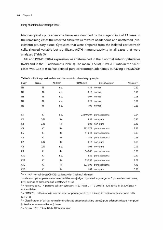
46 Chapter 2
Purity of obtained corticotroph tissue
Macroscopically pure adenoma tissue was identified by the surgeon in 9 of 13 cases. In the remaining cases the resected tissue was a mixture of adenoma and unaffected (pre-existent) pituitary tissue. Cytospins that were prepared from the isolated corticotroph cells, showed variable but significant ACTH-immunoreactivity in all cases that were analyzed (Table 3).
GH and POMC mRNA expression was determined in the 5 normal anterior pituitaries (NAP) and in the 13 adenomas (Table 3). The mean (± SEM) POMC/GH ratio in the 5 NAP cases was 0.36 ± 0.18. We defined pure corticotroph adenomas as having a POMC/GH
table 3. mRNA expression data and immunohistochemistry cytospins
Case1 Tissue2 ACTH+3 POMC/GH4 Classification5 NeuroD16
N1 N n.a. 0.35 normal 0.22
N2 N n.a. 0.10 normal 0.16
N3 N n.a. 0.07 normal 0.08
N4 N n.a. 0.22 normal 0.21
N5 N n.a. 1.05 normal 0.23
C1 C n.a. 231495.07 pure adenoma 0.04
C2 C/N 3+ 3.58 non-pure 0.43
C3 C/N 1+ 0.02 non-pure 0.10
C4 C 4+ 5920.75 pure adenoma 2.27
C5 C 3+ 149.43 pure adenoma 0.93
C6 C 3+ 11.45 pure adenoma 0.29
C7 C/N 3+ 0.17 non-pure 0.63
C8 C/N n.a. 0.03 non-pure 0.09
C9 C 4+ 548.86 pure adenoma 0.06
C10 C n.a. 12.82 pure adenoma 0.17
C11 C 3+ 856.95 pure adenoma 9.67
C12 C 1+ 6230.95 pure adenoma 4.43
C13 C 3+ 1.82 non-pure 0.50
¹ = N1-N5: normal dogs; C1-C13: patients with Cushing’s disease 2 = Macroscopic appearance of resected tissue as judged by veterinary surgeon. C: pure adenoma tissue; C/N: mixture of adenoma and unaffected tissue3 = Percentage ACTH-positive cells on cytospin: 1+ (0-10%); 2+ (10-20%); 3+ (20-30%); 4+ (>30%); n.a. = not available4 = POMC/GH mRNA ratio in normal anterior pituitary cells (N1-N5) and in corticotroph adenoma cells (C1-C13) 5 = Classification of tissue: normal (= unaffected anterior pituitary tissue); pure adenoma tissue; non-pure (mixed adenoma-unaffected) tissue6 = NeuroD1/rps-19 mRNA (x 10-2) expression
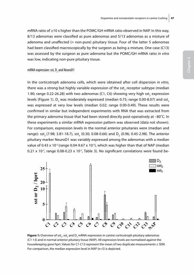
Dopamine and somatostatin receptors in canine Cushing 47
Chap
ter 2
mRNA ratio of ≥10 x higher than the POMC/GH mRNA ratio observed in NAP. In this way, 8/13 adenomas were classified as pure adenomas and 5/13 adenomas as a mixture of adenoma and unaffected (= non-pure) pituitary tissue. Four of the latter 5 adenomas had been classified macroscopically by the surgeon as being a mixture. One case (C13) was assessed by the surgeon as pure adenoma but the POMC/GH mRNA ratio in vitro was low, indicating non-pure pituitary tissue.
mRNA expression: sst, D2 and NeuroD1
In the corticotroph adenoma cells, which were obtained after cell dispersion in vitro, there was a strong but highly variable expression of the sst2 receptor subtype (median 1.90; range 0.22-26.28) with two adenomas (C1, C6) showing very high sst2 expression levels (Figure 1). D2 was moderately expressed (median 0.75; range 0.00-8.07) and sst5
was expressed at very low levels (median 0.02; range 0.00-0.49). These results were confirmed in similar but independent experiments with RNA that was extracted from the primary adenoma tissue that had been stored directly post-operatively at –80˚C. In these experiments a similar mRNA expression pattern was observed (data not shown). For comparison, expression levels in the normal anterior pituitaries were (median and range): sst2 (7.98; 3.81-18.7), sst5 (0.30; 0.08-0.66) and D2 (0.96; 0.45-2.98). The anterior pituitary marker NeuroD1 was variably expressed among the adenomas with a median value of 0.43 x 10⁻² (range 0.04-9.67 x 10⁻²), which was higher than that of NAP (median 0.21 x 10⁻², range 0.08-0.23 x 10⁻², Table 3). No significant correlations were found be-
Fig 1
D2sst2t25
10
sst5
20
25
D2
/ hpr
t
5sst o
r D
C1 C2 C3 C4 C5 C6 C7 C8 C9C10 C11 C12 C13
NAP0
Figure 1: Overview of sst2, sst5 and D2 mRNA expression in canine corticotroph pituitary adenomas (C1-13) and in normal anterior pituitary tissue (NAP). All expression levels are normalized against the housekeeping gene hprt. Values for C1-C13 represent the mean of two duplicate measurements ± SEM. For comparison, the median expression level in NAP (n=5) is depicted.
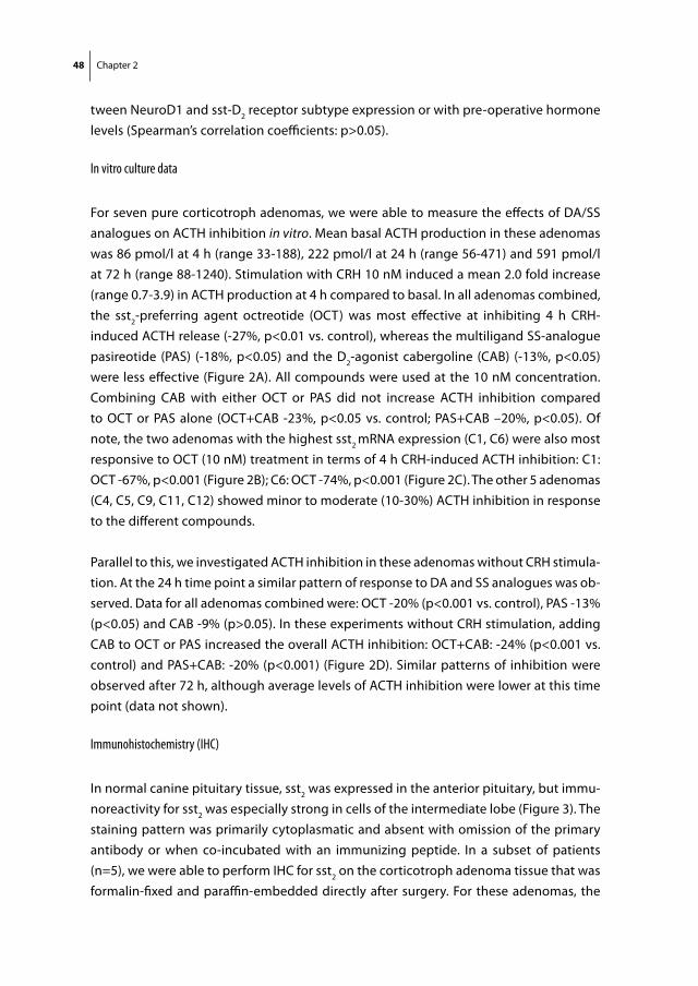
48 Chapter 2
tween NeuroD1 and sst-D2 receptor subtype expression or with pre-operative hormone levels (Spearman’s correlation coefficients: p>0.05).
In vitro culture data
For seven pure corticotroph adenomas, we were able to measure the effects of DA/SS analogues on ACTH inhibition in vitro. Mean basal ACTH production in these adenomas was 86 pmol/l at 4 h (range 33-188), 222 pmol/l at 24 h (range 56-471) and 591 pmol/l at 72 h (range 88-1240). Stimulation with CRH 10 nM induced a mean 2.0 fold increase (range 0.7-3.9) in ACTH production at 4 h compared to basal. In all adenomas combined, the sst2-preferring agent octreotide (OCT) was most effective at inhibiting 4 h CRH-induced ACTH release (-27%, p<0.01 vs. control), whereas the multiligand SS-analogue pasireotide (PAS) (-18%, p<0.05) and the D2-agonist cabergoline (CAB) (-13%, p<0.05) were less effective (Figure 2A). All compounds were used at the 10 nM concentration. Combining CAB with either OCT or PAS did not increase ACTH inhibition compared to OCT or PAS alone (OCT+CAB -23%, p<0.05 vs. control; PAS+CAB –20%, p<0.05). Of note, the two adenomas with the highest sst2 mRNA expression (C1, C6) were also most responsive to OCT (10 nM) treatment in terms of 4 h CRH-induced ACTH inhibition: C1: OCT -67%, p<0.001 (Figure 2B); C6: OCT -74%, p<0.001 (Figure 2C). The other 5 adenomas (C4, C5, C9, C11, C12) showed minor to moderate (10-30%) ACTH inhibition in response to the different compounds.
Parallel to this, we investigated ACTH inhibition in these adenomas without CRH stimula-tion. At the 24 h time point a similar pattern of response to DA and SS analogues was ob-served. Data for all adenomas combined were: OCT -20% (p<0.001 vs. control), PAS -13% (p<0.05) and CAB -9% (p>0.05). In these experiments without CRH stimulation, adding CAB to OCT or PAS increased the overall ACTH inhibition: OCT+CAB: -24% (p<0.001 vs. control) and PAS+CAB: -20% (p<0.001) (Figure 2D). Similar patterns of inhibition were observed after 72 h, although average levels of ACTH inhibition were lower at this time point (data not shown).
Immunohistochemistry (IHC)
In normal canine pituitary tissue, sst2 was expressed in the anterior pituitary, but immu-noreactivity for sst2 was especially strong in cells of the intermediate lobe (Figure 3). The staining pattern was primarily cytoplasmatic and absent with omission of the primary antibody or when co-incubated with an immunizing peptide. In a subset of patients (n=5), we were able to perform IHC for sst2 on the corticotroph adenoma tissue that was formalin-fixed and paraffin-embedded directly after surgery. For these adenomas, the
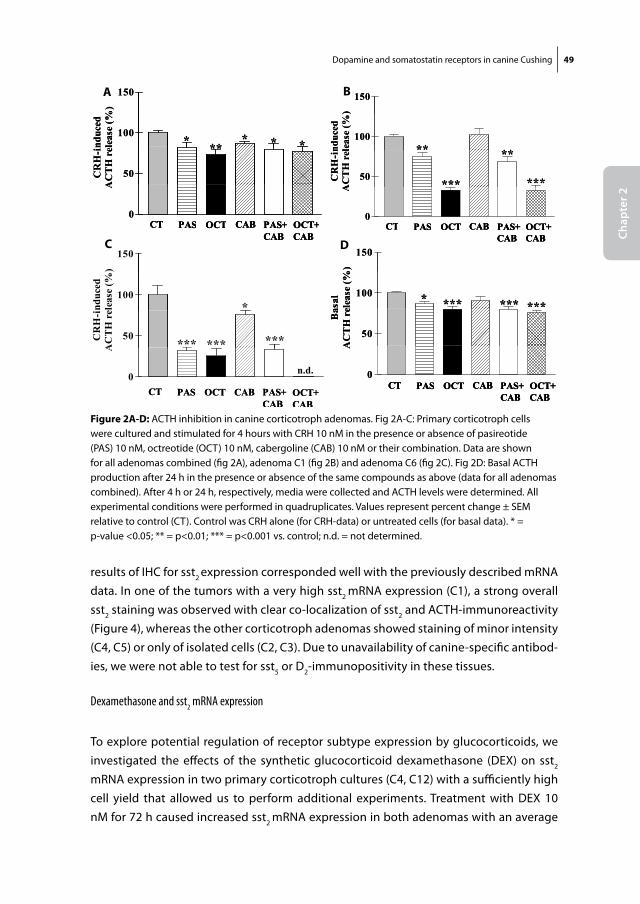
Dopamine and somatostatin receptors in canine Cushing 49
Chap
ter 2
results of IHC for sst2 expression corresponded well with the previously described mRNA data. In one of the tumors with a very high sst2 mRNA expression (C1), a strong overall sst2 staining was observed with clear co-localization of sst2 and ACTH-immunoreactivity (Figure 4), whereas the other corticotroph adenomas showed staining of minor intensity (C4, C5) or only of isolated cells (C2, C3). Due to unavailability of canine-specific antibod-ies, we were not able to test for sst5 or D2-immunopositivity in these tissues.
Dexamethasone and sst2 mRNA expression
To explore potential regulation of receptor subtype expression by glucocorticoids, we investigated the effects of the synthetic glucocorticoid dexamethasone (DEX) on sst2 mRNA expression in two primary corticotroph cultures (C4, C12) with a sufficiently high cell yield that allowed us to perform additional experiments. Treatment with DEX 10 nM for 72 h caused increased sst2 mRNA expression in both adenomas with an average
Fig 2AFig 2A
150)
150)
100
nduc
edle
ase
(%)
*** * **
100
nduc
edle
ase
(%)
*** * **
50CR
H-i
nA
CT
H r
el ***
50CR
H-i
nA
CT
H r
el ***
0
A
CT PAS CAB PAS+ OCT+OCT0
A
CT PAS CAB PAS+ OCT+OCTCT PAS CAB PAS+ OCT+OCTCT PAS CAB PAS+CAB
OCT+CAB
OCTCT PAS CAB PAS+CAB
OCT+CAB
OCTCT PAS CAB PAS+CAB
OCT+CAB
OCT
Fig 2BFig 2B
150
)
150
)
100
nduc
edle
ase
(%)
** **100
nduc
edle
ase
(%)
** **
50CR
H-i
nA
CT
H r
el
***
**
***50CR
H-i
nA
CT
H r
el
***
**
***
0
A
CT PAS CAB PAS OCTOCT0
A
CT PAS CAB PAS OCTOCTCT PAS CAB PAS OCTOCTCT PAS CAB PAS+CAB
OCT+CAB
OCTCT PAS CAB PAS+CAB
OCT+CAB
OCTCT PAS CAB PAS+CAB
OCT+CAB
OCTFig 2C
150
)
100
nduc
edel
ease
(%
*
50CR
H-i
AC
TH
re
*** ******
0
A
n.d.
CT PAS CAB PAS+CAB
OCT+CAB
OCTCT PAS CAB PAS+CAB
OCT+CAB
OCT
Fig 2DFig 2D
150
)
150
)
100
asal
elea
se (%
*** *******100
asal
elea
se (%
*** *******
50B
aA
CT
H r
e
*50
Ba
AC
TH
re
*
0
A
CT PAS CAB PAS OCTOCT0
A
CT PAS CAB PAS OCTOCTCT PAS CAB PAS OCTOCTCT PAS CAB PAS+CAB
OCT+CAB
OCTCT PAS CAB PAS+CAB
OCT+CAB
OCTCT PAS CAB PAS+CAB
OCT+CAB
OCT
Figure 2A-D: ACTH inhibition in canine corticotroph adenomas. Fig 2A-C: Primary corticotroph cells were cultured and stimulated for 4 hours with CRH 10 nM in the presence or absence of pasireotide (PAS) 10 nM, octreotide (OCT) 10 nM, cabergoline (CAB) 10 nM or their combination. Data are shown for all adenomas combined (fig 2A), adenoma C1 (fig 2B) and adenoma C6 (fig 2C). Fig 2D: Basal ACTH production after 24 h in the presence or absence of the same compounds as above (data for all adenomas combined). After 4 h or 24 h, respectively, media were collected and ACTH levels were determined. All experimental conditions were performed in quadruplicates. Values represent percent change ± SEM relative to control (CT). Control was CRH alone (for CRH-data) or untreated cells (for basal data). * = p-value <0.05; ** = p<0.01; *** = p<0.001 vs. control; n.d. = not determined.
A B
C D
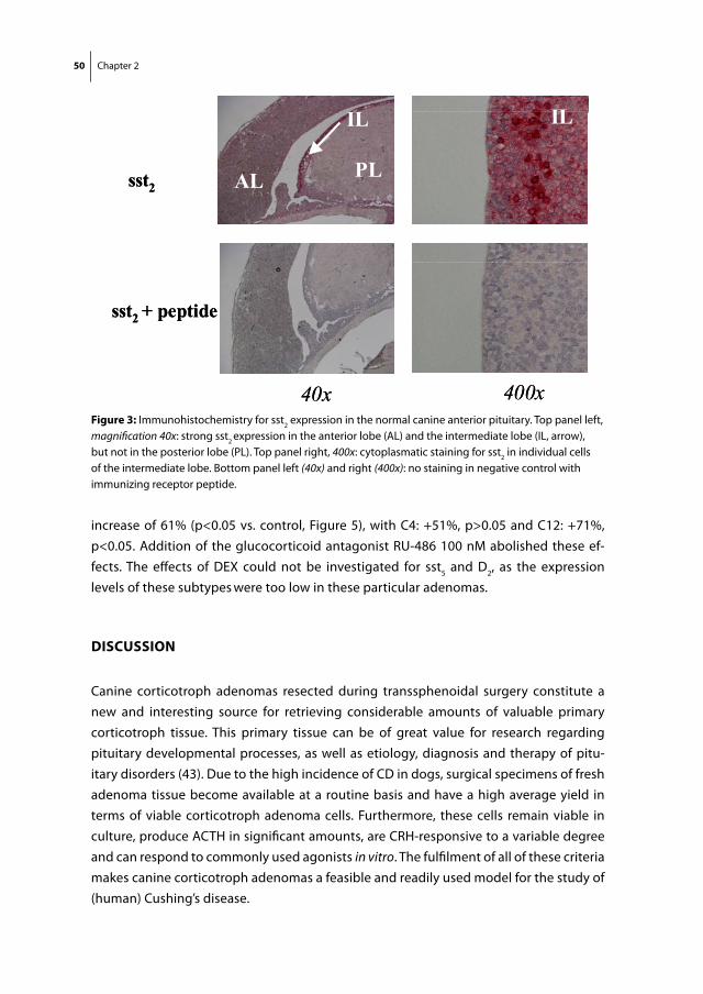
50 Chapter 2
increase of 61% (p<0.05 vs. control, Figure 5), with C4: +51%, p>0.05 and C12: +71%, p<0.05. Addition of the glucocorticoid antagonist RU-486 100 nM abolished these ef-fects. The effects of DEX could not be investigated for sst5 and D2, as the expression levels of these subtypes were too low in these particular adenomas.
DisCussion
Canine corticotroph adenomas resected during transsphenoidal surgery constitute a new and interesting source for retrieving considerable amounts of valuable primary corticotroph tissue. This primary tissue can be of great value for research regarding pituitary developmental processes, as well as etiology, diagnosis and therapy of pitu-itary disorders (43). Due to the high incidence of CD in dogs, surgical specimens of fresh adenoma tissue become available at a routine basis and have a high average yield in terms of viable corticotroph adenoma cells. Furthermore, these cells remain viable in culture, produce ACTH in significant amounts, are CRH-responsive to a variable degree and can respond to commonly used agonists in vitro. The fulfilment of all of these criteria makes canine corticotroph adenomas a feasible and readily used model for the study of (human) Cushing’s disease.
Fig 3
IL ILILIL ILIL
sst2 AL PL
IL ILIL
sst2 AL PL
IL ILIL
2 AL2 AL
sst2 + peptidesst2 + peptide
40x 400x40x 400xFigure 3: Immunohistochemistry for sst2 expression in the normal canine anterior pituitary. Top panel left, magnification 40x: strong sst2 expression in the anterior lobe (AL) and the intermediate lobe (IL, arrow), but not in the posterior lobe (PL). Top panel right, 400x: cytoplasmatic staining for sst2 in individual cells of the intermediate lobe. Bottom panel left (40x) and right (400x): no staining in negative control with immunizing receptor peptide.
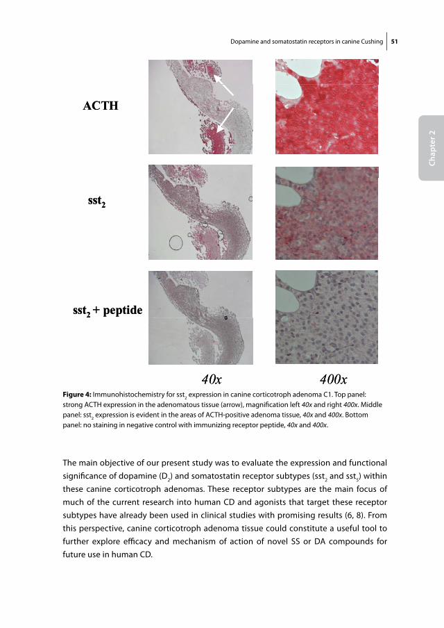
Dopamine and somatostatin receptors in canine Cushing 51
Chap
ter 2
The main objective of our present study was to evaluate the expression and functional significance of dopamine (D2) and somatostatin receptor subtypes (sst2 and sst5) within these canine corticotroph adenomas. These receptor subtypes are the main focus of much of the current research into human CD and agonists that target these receptor subtypes have already been used in clinical studies with promising results (6, 8). From this perspective, canine corticotroph adenoma tissue could constitute a useful tool to further explore efficacy and mechanism of action of novel SS or DA compounds for future use in human CD.
Fig 4
ACTHACTH
sst2sst2
sst2 + peptidesst2 + peptide
40x 400x40x 400xFigure 4: Immunohistochemistry for sst2 expression in canine corticotroph adenoma C1. Top panel: strong ACTH expression in the adenomatous tissue (arrow), magnification left 40x and right 400x. Middle panel: sst2 expression is evident in the areas of ACTH-positive adenoma tissue, 40x and 400x. Bottom panel: no staining in negative control with immunizing receptor peptide, 40x and 400x.
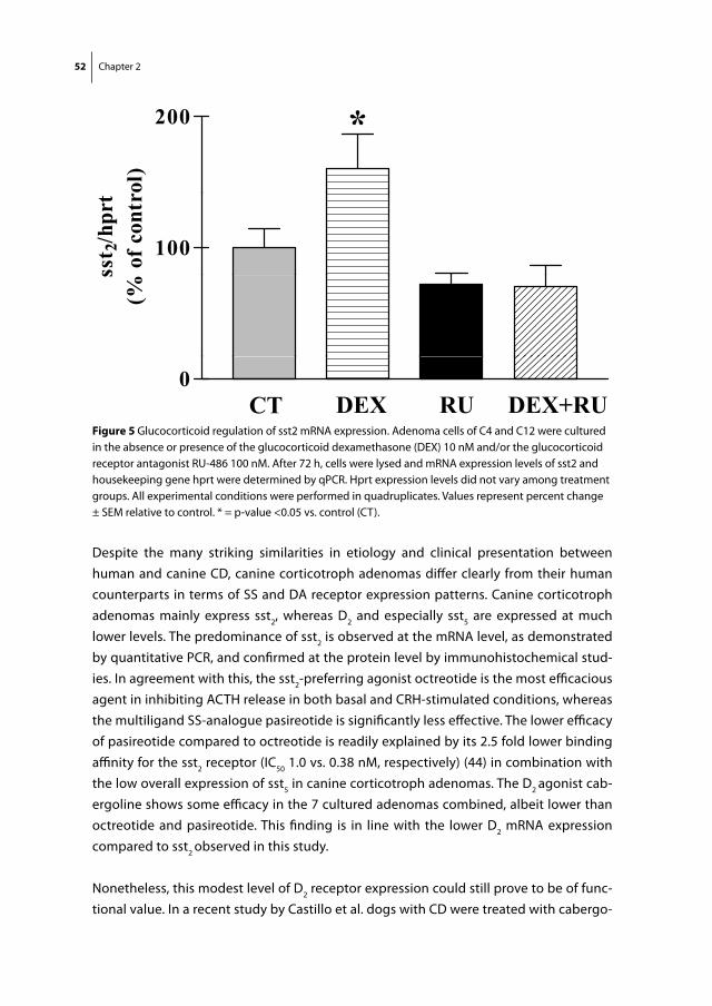
52 Chapter 2
Despite the many striking similarities in etiology and clinical presentation between human and canine CD, canine corticotroph adenomas differ clearly from their human counterparts in terms of SS and DA receptor expression patterns. Canine corticotroph adenomas mainly express sst2, whereas D2 and especially sst5 are expressed at much lower levels. The predominance of sst2 is observed at the mRNA level, as demonstrated by quantitative PCR, and confirmed at the protein level by immunohistochemical stud-ies. In agreement with this, the sst2-preferring agonist octreotide is the most efficacious agent in inhibiting ACTH release in both basal and CRH-stimulated conditions, whereas the multiligand SS-analogue pasireotide is significantly less effective. The lower efficacy of pasireotide compared to octreotide is readily explained by its 2.5 fold lower binding affinity for the sst2 receptor (IC50 1.0 vs. 0.38 nM, respectively) (44) in combination with the low overall expression of sst5 in canine corticotroph adenomas. The D2 agonist cab-ergoline shows some efficacy in the 7 cultured adenomas combined, albeit lower than octreotide and pasireotide. This finding is in line with the lower D2 mRNA expression compared to sst2 observed in this study.
Nonetheless, this modest level of D2 receptor expression could still prove to be of func-tional value. In a recent study by Castillo et al. dogs with CD were treated with cabergo-
Fig 5
200 *200ol
)*
100
sst 2
/hpr
t o
f con
trs
(%
0CT DEX RU DEX+RU
Figure 5 Glucocorticoid regulation of sst2 mRNA expression. Adenoma cells of C4 and C12 were cultured in the absence or presence of the glucocorticoid dexamethasone (DEX) 10 nM and/or the glucocorticoid receptor antagonist RU-486 100 nM. After 72 h, cells were lysed and mRNA expression levels of sst2 and housekeeping gene hprt were determined by qPCR. Hprt expression levels did not vary among treatment groups. All experimental conditions were performed in quadruplicates. Values represent percent change ± SEM relative to control. * = p-value <0.05 vs. control (CT).
Dopamine and somatostatin receptors in canine Cushing 53
Chap
ter 2
line (0.07 mg/kg/wk) for 1 year, which resulted in an overall response rate of 42.5% (45). One factor that could explain this observed difference between the in vitro and clinical efficacy of cabergoline could be the duration of treatment. It is known from studies in human patients with CD that it can take up to 3 months before the maximal cortisol-inhibiting effects of cabergoline are observed (6). In this respect, our in vitro data on ACTH inhibition after 4-72 h may not necessarily reflect the full potential of cabergoline as a drug in canine CD. On the other hand, the high levels of sst2 expression both on the mRNA and the protein level, in combination with the superior efficacy of octreotide in cultured canine corticotroph adenomas, suggest an even stronger role of this receptor subtype as a therapeutic target. Based on our findings, a clinical study to investigate the effects of an sst2-preferring compound such as octreotide on ACTH and cortisol levels in canine CD could be of great interest to see if superior response rates could be achieved with the use of such compounds, compared to those obtained with cabergoline. In ad-dition to this, it would be very interesting to study whether combined targeting of sst2 and D2 receptors, either by co-treatment with the individual SS/DA analogs or by the use of novel chimeric SS-DA molecules could result in even higher clinical efficacy.
To return to our original research question, the receptor expression pattern observed in canine adenomas is remarkably different from the one observed in human corti-cotroph adenomas, where sst5 and D2 are the predominant receptor subtypes and sst2
expression is generally low. The reasons for this dissimilarity between canine and hu-man corticotroph adenomas are yet unknown. One important factor, however, appears to be the difference in regulation by glucocorticoids of receptor subtype expression. Down-regulation of sst2 expression by glucocorticoids has been demonstrated in murine corticotroph AtT-20 tumor cells and is also thought to explain the low sst2 expression in human corticotroph adenomas (5, 46, 47). Striking, therefore, was the observation in our study that this glucocorticoid-induced down-regulation did not occur in canine corticotroph adenomas. In fact, treatment of the canine corticotroph cells in vitro with dexamethasone increased the expression of the sst2 receptor, as was observed in two different adenomas. From a future perspective, it would be interesting to see whether these differences can be ascribed to the 7% inhomology between the canine and the human sst2 genetic sequence, as it is possible that this genomic variation is also present in areas within the human sst2 gene that are known to contain glucocorticoid-responsive elements.
It is important to emphasize that sst and D2 are not the only receptors that have been linked to regulation of ACTH secretion in corticotroph cells. Receptors such as the retinoic acid receptor (RAR) and peroxisome-proliferator-activated receptor-γ (PPARγ) have also been shown to decrease ACTH regulation in different in vitro and rodent models and have therefore been implicated as potential new targets for medical therapy of CD in humans
54 Chapter 2
(48, 49). Most notably, retinoic acid was used in a recent clinical study in dogs with CD and showed significant clinical efficacy (50). In this respect, it would be very interesting to evaluate canine corticotroph adenomas for the presence and distribution of novel drug targets such as RAR and PPARγ, and to see whether correlation is higher between canine and human CD for these receptors than for SS and DA. These investigations could help to fully evaluate the potential of canine CD as a direct animal model for human CD.
In conclusion, canine corticotroph adenomas obtained after transsphenoidal surgery, provide a model to study corticotroph cell (patho)physiology, due to the high yield of viable, primary tissue that retains most of its corticotroph features in vitro. Some distinct differences do exist, however, between human and canine corticotroph adenomas in terms of sst and D2 receptor expression patterns and their responses to SS and DA ago-nists in vitro. These differences should be taken into account when using dogs with CD as a model to evaluate efficacy of novel somatostatin analogues and dopamine agonists for future use in human CD.
ACKnoWleDgements:
The authors would like to thank Michael P. Brugts, M.D., for his help in setting up the canine primers.
Dopamine and somatostatin receptors in canine Cushing 55
Chap
ter 2
reFerenCes 1. orth Dn 1995 Cushing’s syndrome. N Engl J Med 332:791-803 2. melby JC 1988 Therapy of Cushing disease: a consensus for pituitary microsurgery. Ann Intern
Med 109:445-446 3. Atkinson AB, Kennedy A, Wiggam mi, mcCance Dr, sheridan B 2005 Long-term remission
rates after pituitary surgery for Cushing’s disease: the need for long-term surveillance. Clin Endo-crinol (Oxf ) 63:549-559
4. morris D, grossman A 2002 The medical management of Cushing’s Syndrome. Ann NY Acad Sci 970:119-133
5. Hofland lJ, van der Hoek J, Feelders r, van Aken mo, van Koetsveld Pm, Waaijers m, sprij-mooij D, Bruns C, Weckbecker g, de Herder WW, Beckers A, lamberts sW 2005 The multi-ligand somatostatin analogue SOM230 inhibits ACTH secretion by cultured human corticotroph adenomas via somatostatin receptor type 5. Eur J Endocrinol 152:645-654
6. Pivonello r, Ferone D, de Herder WW, Kros Jm, De Caro ml, Arvigo m, Annunziato l, lom-bardi g, Colao A, Hofland lJ, lamberts sW 2004 Dopamine receptor expression and function in corticotroph pituitary tumors. J Clin Endocrinol Metab 89:2452-2462
7. Batista Dl, Zhang X, gejman r, Ansell PJ, Zhou y, Johnson sA, swearingen B, Hedley-Whyte et, stratakis CA, Klibanski A 2006 The effects of SOM230 on cell proliferation and adrenocorti-cotropin secretion in human corticotroph pituitary adenomas. J Clin Endocrinol Metab 91:4482-4488
8. Boscaro m, Petersenn, s., Atkinson, A.B., Bertherat, J., Findling J., snyder, P., mcBride, K., reincke, m., ludlam, W., gao, B., melmed, s., Freda, P., Frohman, l., grossman, A., Biller, B, glusman, J.e. 2006 Pasireotide (SOM230), the novel multi-ligand somatostatin analogue, is a promising medical therapy for patients with Cushing’s disease: preliminary safety and efficacy results of a phase II study. Presented at ENDO 2006, abstr OR9-1, Boston, U.S.A., 2006
9. etxabe J, vazquez JA 1994 Morbidity and mortality in Cushing’s disease: an epidemiological approach. Clin Endocrinol (Oxf ) 40:479-484
10. lindholm J, Juul s, Jorgensen Jo, Astrup J, Bjerre P, Feldt-rasmussen u, Hagen C, Jorgensen J, Kosteljanetz m, Kristensen l, laurberg P, schmidt K, Weeke J 2001 Incidence and late prog-nosis of cushing’s syndrome: a population-based study. J Clin Endocrinol Metab 86:117-123
11. Katznelson l, Bogan Js, trob Jr, schoenfeld DA, Hedley-Whyte et, Hsu DW, Zervas nt, swearingen B, sleeper m, Klibanski A 1998 Biochemical assessment of Cushing’s disease in patients with corticotroph macroadenomas. J Clin Endocrinol Metab 83:1619-1623
12. Bochicchio D, losa m, Buchfelder m 1995 Factors influencing the immediate and late outcome of Cushing’s disease treated by transsphenoidal surgery: a retrospective study by the European Cushing’s Disease Survey Group. J Clin Endocrinol Metab 80:3114-3120
13. rijnberk A, der Kinderen PJ, thijssen JH 1968 Spontaneous hyperadrenocorticism in the dog. J Endocrinol 41:397-406
14. Willeberg P, Priester WA 1982 Epidemiological aspects of clinical hyperadrenocorticism in dogs (Canine Cushing’s Syndrome). J Am Anim Hosp Assoc 18:717-724
15. mcnicol Am, thomson H, stewart CJ 1983 The corticotrophic cells of the canine pituitary gland in pituitary-dependent hyperadrenocorticism. J Endocrinol 96:303-309
16. el etreby mF, muller-Peddinghaus r, Bhargava As, trautwein g 1980 Functional morphol-ogy of spontaneous hyperplastic and neoplastic lesions in the canine pituitary gland. Vet Pathol 17:109-122
56 Chapter 2
17. Kemppainen rJ, Peterson me 1994 Animal models of Cushing’s disease. Trends Endocrinol Metab 5:21-28
18. Hanson Jm, teske e, voorhout g, galac s, Kooistra Hs, meij BP 2007 Prognostic factors for outcome after transsphenoidal hypophysectomy in dogs with pituitary-dependent hyperadre-nocorticism. J Neurosurg 107:830-840
19. den Hertog e, Braakman JC, teske e, Kooistra Hs, rijnberk A 1999 Results of non-selective adrenocorticolysis by o,p’-DDD in 129 dogs with pituitary-dependent hyperadrenocorticism. Vet Rec 144:12-17
20. Barker en, Campbell s, tebb AJ, neiger r, Herrtage me, reid sW, ramsey iK 2005 A com-parison of the survival times of dogs treated with mitotane or trilostane for pituitary-dependent hyperadrenocorticism. J Vet Intern Med 19:810-815
21. meij BP, voorhout g, van den ingh ts, Hazewinkel HA, teske e, rijnberk A 1998 Results of transsphenoidal hypophysectomy in 52 dogs with pituitary-dependent hyperadrenocorticism. Vet Surg 27:246-261
22. meij BP, voorhout g, van den ingh ts, Hazewinkel HA, van’t verlaat JW 1997 Transsphenoidal hypophysectomy in beagle dogs: evaluation of a microsurgical technique. Vet Surg 26:295-309
23. rijnberk A, van Wees A, mol JA 1988 Assessment of two tests for the diagnosis of canine hy-peradrenocorticism. Vet Rec 122:178-180
24. stolp r, rijnberk A, meijer JC, Croughs rJm 1983 Urinary corticoids in the diagnosis of canine hyperadrenocorticism. Res Vet Sci 34:141-144
25. meij B, voorhout g, rijnberk A 2002 Progress in transsphenoidal hypophysectomy for treatment of pituitary-dependent hyperadrenocorticism in dogs and cats. Mol Cell Endocrinol 197:89-96
26. Hanson Jm, van ‘t Hm, voorhout g, teske e, Kooistra Hs, meij BP 2005 Efficacy of transsphe-noidal hypophysectomy in treatment of dogs with pituitary-dependent hyperadrenocorticism. J Vet Intern Med 19:687-694
27. van der vlugt-meijer rH, voorhout g, meij BP 2002 Imaging of the pituitary gland in dogs with pituitary-dependent hyperadrenocorticism. Mol Cell Endocrinol 197:81-87
28. Bosje Jt, rijnberk A, mol JA, voorhout g, Kooistra Hs 2002 Plasma concentrations of ACTH precursors correlate with pituitary size and resistance to dexamethasone in dogs with pituitary-dependent hyperadrenocorticism. Domest Anim Endocrinol 22:201-210
29. rijnberk A, mol JA, rothuizen J, Bevers mm, middleton DJ 1987 Circulating pro-opiomelano-cortin-derived peptides in dogs with pituitary-dependent hyperadrenocorticism. Front Horm Res 17:48-60
30. voorhout g, rijnberk A, sjollema Be, van den ingh ts 1990 Nephrotomography and ultra-sonography for the localization of hyperfunctioning adrenocortical tumors in dogs. Am J Vet Res 51:1280-1285
31. Kooistra Hs, voorhout g, mol JA, rijnberk A 1997 Correlation between impairment of glucocorticoid feedback and the size of the pituitary gland in dogs with pituitary-dependent hyperadrenocorticism. J Endocrinol 152:387-394
32. Hanson Jm, Kooistra Hs, mol JA, teske e, meij BP 2006 Plasma profiles of adrenocorticotropic hormone, cortisol, alpha-melanocyte-stimulating hormone, and growth hormone in dogs with pituitary-dependent hyperadrenocorticism before and after hypophysectomy. J Endocrinol 190:601-609
33. meij BP, mol JA, van den ingh ts, Bevers mm, Hazewinkel HA, rijnberk A 1997 Assessment of pituitary function after transsphenoidal hypophysectomy in beagle dogs. Domest Anim Endo-crinol 14:81-97
Dopamine and somatostatin receptors in canine Cushing 57
Chap
ter 2
34. Hofland lJ, van der Hoek J, van Koetsveld Pm, de Herder WW, Waaijers m, sprij-mooij D, Bruns C, Weckbecker g, Feelders r, van der lely AJ, Beckers A, lamberts sW 2004 The novel somatostatin analog SOM230 is a potent inhibitor of hormone release by growth hormone- and prolactin-secreting pituitary adenomas in vitro. J Clin Endocrinol Metab 89:1577-1585
35. rasmussen r 2001 Quantification on the LightCycler. In: Meuer S, Wittwer C and Nakagawara K, eds. Rapid Cycle Real-time PCR, Methods and Applications. Heidelberg: Springer Press; 21-34
36. Pfaffl mW 2001 A new mathematical model for relative quantification in real-time RT-PCR. Nucleic Acids Res 29:e45
37. Bhatti sF, De vliegher sP, mol JA, van Ham lm, Kooistra Hs 2006 Ghrelin-stimulation test in the diagnosis of canine pituitary dwarfism. Res Vet Sci 81:24-30
38. Brinkhof B, spee B, rothuizen J, Penning lC 2006 Development and evaluation of canine refer-ence genes for accurate quantification of gene expression. Anal Biochem 356:36-43
39. oyama K, sanno n, teramoto A, osamura ry 2001 Expression of neuro D1 in human normal pituitaries and pituitary adenomas. Mod Pathol 14:892-899
40. Poulin g, turgeon B, Drouin J 1997 NeuroD1/beta2 contributes to cell-specific transcription of the proopiomelanocortin gene. Mol Cell Biol 17:6673-6682
41. Hanson Jm JmJ, meij BP. 2007 Differential expression of neurogenic differentiation 1 (NeuroD1) in the canine pituitary gland and corticotroph adenomas. In: Pathobiology and oncogenesis of pituitary corticotroph adenomas in dogs. Thesis. Atalanta, Houten, the Netherlands. Chapter 8: p. 161-171.
42. Hofland lJ, liu Q, van Koetsveld Pm, Zuijderwijk J, van Der Ham F, De Krijger rr, schonb-runn A, lamberts sW 1999 Immunohistochemical detection of somatostatin receptor subtypes sst1 and sst2A in human somatostatin receptor positive tumors. J Clin Endocrinol Metab 84:775-780
43. Bilodeau s, vallette-Kasic s, gauthier y, Figarella-Branger D, Brue t, Berthelet F, lacroix A, Batista D, stratakis C, Hanson J, meij B, Drouin J 2006 Role of Brg1 and HDAC2 in GR trans-repression of the pituitary POMC gene and misexpression in Cushing disease. Genes Dev 20:2871-2886
44. Bruns C, lewis i, Briner u, meno-tetang g, Weckbecker g 2002 SOM230: a novel somatostatin peptidomimetic with broad somatotropin release inhibiting factor (SRIF) receptor binding and a unique antisecretory profile. Eur J Endocrinol 146:707-716
45. Castillo vA, gomez nv, lalia JC, Cabrera Blatter mF, garcia JD 2007 Cushing’s disease in dogs: Cabergoline treatment. Res Vet Sci 2007 Sep 30 [Epub ahead of print], article in press
46. van der Hoek J, Waaijers m, van Koetsveld Pm, sprij-mooij D, Feelders rA, schmid HA, schoef-fter P, Hoyer D, Cervia D, taylor Je, Culler mD, lamberts sW, Hofland lJ 2005 Distinct functional properties of native somatostatin receptor subtype 5 compared with subtype 2 in the regulation of ACTH release by corticotroph tumor cells. Am J Physiol Endocrinol Metab 289:E278-287
47. schmid HA 2007 Pasireotide (SOM230): Development, mechanism of action and potential ap-plications. Mol Cell Endocrinol 2007 Sep 19 [Epub ahead of print], article in press
48. Heaney AP, melmed s 2004 Molecular targets in pituitary tumours. Nat Rev Cancer 4:285-295 49. Paez-Pereda m, Kovalovsky D, Hopfner u, theodoropoulou m, Pagotto u, uhl e, losa m,
stalla J, grubler y, missale C, Arzt e, stalla gK 2001 Retinoic acid prevents experimental Cush-ing syndrome. J Clin Invest 108:1123-1131
50. Castillo v, giacomini D, Paez-Pereda m, stalla J, labeur m, theodoropoulou m, Holsboer F, grossman AB, stalla gK, Arzt e 2006 Retinoic acid as a novel medical therapy for Cushing’s disease in dogs. Endocrinology 147:4438-4444

Chapter 3
Differential regulation of human dopamine D2 and somatostatin
receptor subtype expression by glucocorticoids in vitro
de Bruin C, Feelders RA, Waaijers AM, van Koetsveld PM, Sprij-Mooij DM, Lamberts SW, Hofland LJ.
J Mol Endocrinol. 2009 Jan;42(1):47-56.
60 Chapter 3
ABstrACt
Dopamine agonists and somatostatin analogues have been proposed in the treatment of ACTH-producing neuro-endocrine tumours that cause Cushing’s syndrome. Inversely, glucocorticoids (GC) can differentially influence dopamine receptor D2 or somatostatin receptor subtype (sst) expression in rodent models. If this also occurs in human neuro-endocrine cells, then cortisol-lowering therapy could directly affect the expression of these target receptors. In this study we investigated the effects of the GC dexametha-sone (DEX) on D2 and sst expression in three human neuro-endocrine cell lines: BON (carcinoid) and TT (medullary thyroid carcinoma) versus DMS (small cell lung cancer), which is severely GC-resistant. In BON and TT, sst2 mRNA was strongly down-regulated in a dose-dependent manner (IC50 0.84 nM and 0.16 nM), whereas sst5 and especially D2 were much more resistant to DEX-treatment. Sst2 down-regulation was abrogated by a GC receptor antagonist and reversible in time upon GC withdrawal. At the protein level, DEX also induced a decrease in the total number of SS (-52%) and sst2-specific (-42%) binding sites. Pre-treatment with DEX abrogated calcitonin-inhibition by sst2-preferring analogue octreotide in TT. In DMS, DEX did not cause significant changes in expression of these receptor subtypes. In conclusion, we show that GCs selectively down-regulate sst2, but not D2 and only to a minor degree sst5 in human neuro-endocrine BON and TT cells. This mechanism may also be responsible for the low expression of sst2 in corticotroph adenomas and underwrite the current interest in sst5 and D2 as possible therapeutic targets for a medical treatment of Cushing’s disease.
Glucocorticoid regulation of DA and SS receptors 61
Chap
ter 3
introDuCtion
Somatostatin (SS) and dopamine (DA) are small molecules with a variety of functions throughout the human body, including neurotransmission and inhibition of hormone release (1, 2). They bind to high-affinity receptors that belong to the family of G-protein coupled receptors. Both for SS and DA multiple receptor subtypes have been identified: sst1-5 and D1-5 (2, 3). The presence of these receptors has been demonstrated on a number of neuro-endocrine tumours and therefore, SS-analogues and DA-agonists could play an important role in the medical treatment of these tumours (4). Recent reports have shown significant efficacy of selective D2-receptor agonists, such as cabergoline, and of a multiligand SS-analogue with high sst5 affinity (pasireotide / SOM230) in the medi-cal management of pituitary-dependent Cushing’s syndrome (5-7). The use of sst2-or D2-targeting agonists has also been advocated in cases of Cushing’s Syndrome due to ectopic ACTH-production by neuro-endocrine tumours (8, 9).
The hallmark of Cushing’s syndrome, regardless of its cause, is a profound and sustained overproduction of glucocorticoids (GCs) by the adrenal glands. It is also known that GCs can have (in)direct effects on receptor expression patterns of different neurotransmit-ters and hormones in the human body. Therefore, when considering the clinical use of DA-agonists and/or SS-analogues to lower cortisol levels in a patient, it is important to investigate how alterations in glucocorticoid levels (i.e. response to treatment) can secondarily affect the expression of the target SS and DA receptors in ACTH-producing neuro-endocrine tumours. If such a (in)direct relationship exists, then this could first of all explain our current knowledge on SS and DA receptor expression patterns in human corticotroph adenomas as it has been described before by different groups (5, 10, 11). Secondly, perhaps more importantly, it could help us to predict what happens to the expression of the target SS and DA receptors, when medical therapy lowers circulating GC levels. This may have implications for the type and timing of additional SS-or DA-based medical treatment in these patients.
A considerable number of studies have investigated the effects of GCs on dopamine recep-tor expression, mainly within the central nervous system and associated neuro-psychiatric disorders, but not in human neuro-endocrine tumour cells (12-16). Most of these studies report no or only minor effects of GC exposure on D2 receptor expression. Several other studies have shown that GC’s can influence somatostatin receptor subtype expression in a differential manner in rat or murine pituitary cells and cell lines (17-19). No study thus far, however, has concomitantly evaluated D2 and SS receptor subtype expression and their regulation by GC’s within the same, human-derived neuro-endocrine cell system.
62 Chapter 3
For that reason we conducted the present study. We investigated the time- and dose-dependent effects of the synthetic glucocorticoid dexamethasone (DEX) on sst2, sst5 and D2 receptor expression in vitro in three different human neuro-endocrine cell lines (BON, TT and DMS-79), for which evidence of direct transcriptional regulation by GC’s has been reported (20-22). BON (pancreatic carcinoid) and TT (medullary thyroid carcinoma) cells are not known to have any defects in their GC regulation and signalling pathway. By using these two different neuro-endocrine cell lines we aimed to find general patterns in GC-mediated effects that are representative for the larger group of neuro-endocrine tumours as a whole. Since BON and TT cells do not necessarily reflect ectopic ACTH-producing tumours that display severe GC-resistance, we compared these results with a third and completely different neuro-endocrine cell line: the human ectopic ACTH-producing small cell lung carcinoma cell line DMS-79, which harbours distinct mutations in the glucocorticoid receptor gene and is known for its severe GC-resistant properties (22, 23).
mAteriAls AnD metHoDs
Cell culture
BON cells were routinely grown in 75 cm2 flasks containing DMEM-F12 Glutamax® me-dium supplemented with 10% Foetal Calf Serum, penicillin (1x10⁵ U/l) and fungizone (0.25 mg/L). Cells were cultured in a 5% CO2 incubator at 37ºC and routinely passaged by trypsinization (trypsine 0.05% -EDTA 0.02%). Medium was refreshed twice a week and cell viability always exceeded > 95% as measured by trypan blue staining. TT cells were cultured following the same protocol with F-12K Nutrient Mixture (Kaighn’s Modifica-tion). DMS cells were grown in suspension under the same conditions with 10% FCS, glutamine and penicillin in RPMI 1640 medium (ATCC 30-2001). All cell lines were con-firmed to be mycoplasma-free. Media and supplements were obtained from Invitrogen (Breda, the Netherlands) unless otherwise stated.
Cell treatment for mRNA expression studies
For the experiments, cells were trypsinized, counted in a standard haemocytometer and seeded at a density of 15,000 (BON) or 100,000 (TT, DMS) cells/well in 24 well plates (Corning, Cambridge, MA, U.S.A.) in 1 ml of medium. After 72 h media were refreshed and incubations started without or with different doses of DEX and/or the GC receptor antagonist RU-486. At the different time points, media were removed and cells were lysed on ice with a buffer containing 100 mM Tris-HCl (pH 8), 500 mM LiCl, 10 mM EDTA (pH
Glucocorticoid regulation of DA and SS receptors 63
Chap
ter 3
8), 5 mM DTT and 1% LiDS (HT Biotechnology Ltd, Cambridge, U.K.) and stored at –80˚C until further analysis. All experimental conditions were performed in quadruplicates.
Quantitative PCR
Quantitative PCR was performed according to a previously published method (24). In short, poly(A+) mRNA was isolated from the lysed cells with the use of Dynabeads Oligo (dT)25 (Dynal AS, Oslo, Norway). The poly (A+) mRNA was eluted in H2O (65˚C) for 2 x 2 minutes and used for cDNA synthesis in a Tris buffer [50 mM Tris-HCl (pH 8.3), 100 mM KCl, 4 mM DTT and 10 mM MgCl2] with 10 units RNase inhibitor, 2 units avian myeloblas-tosis virus Super Reverse Transcriptase and 1 mM of each deoxynucleotide triphosphate in a final volume of 40 µL. This was incubated for 1 h at 42˚C and the resulting cDNA was diluted 5-fold in 160 µl sterile H20.
One twentieth of the total cDNA library was used for quantification of hprt, sst2, sst5 and D2 mRNA levels. The total reaction volume (25 µl) consisted of 10 µl cDNA and 15 µl Taq-Man Universal PCR Mastermix (Applied Biosystems, Branchburg, NJ, U.S.A.) with primers-probes in the following concentrations: hprt, sst2 and sst5 500-500-100 nM and D2 300-300-200 nM of forward primer, reverse primer and probe, respectively. The primer and probe sequences that were used for hprt, sst2, and sst5 have been published previously (24). D2 primer-and-probe sequences were: D2 forward 5’-GCCACTCAGATGCTCGCC-3’, D2
reverse 5’-ATGTGTGTGATGAAGAAGGGCA-3’ and D2 probe 5’-FAM-TTGTTCTCGG CGTGT-TCATCATCTGC-TAMRA-3’. This primer-probe set measures total D2 expression (D2 long + short isoform). All primers and probes were purchased from Sigma Aldrich (Zwijndrecht, the Netherlands). Real-time quantitative PCR was performed in 96-well optical plates with the TaqMan Gold nuclease assay (Applied Biosystems, Roche, NJ, U.S.A.) and the ABI Prism 7700 Sequence Detection System (PerkinElmer, Foster City, CA, U.S.A.). After two initial heating steps at 50˚C (2 min) and 95˚C (10 min), samples were subjected to 40 cycles of denaturation at 95˚C (15 sec) and annealing at 60˚C (60 sec). All samples were assayed in duplicate. Values were normalized against the expression of the housekeeping gene hypoxanthine phosphoribosyltransferase (hprt). Dilution curves were constructed to calculate PCR efficiencies (E) for every primer-probe set (25). Efficiencies were: sst2 1.92, sst5 1.92, D2 1.94 and hprt 1.93. Estimated copy numbers were calculated using the comparative threshold method with efficiency correction, as described previously (26). To exclude genomic DNA contamination in the RNA, the cDNA reactions were also performed without reverse transcriptase and amplified with each primer pair. To exclude contamination of the PCR reaction mixtures, the reactions were also performed in the absence of cDNA template, in parallel with cDNA samples.
64 Chapter 3
Cell proliferation and DNA fragmentation assays
After trypsinization, the cells were counted, seeded and treated with DEX in similar fashion as described above in the experiments for qPCR analysis. At the different time points media were aspirated and cells were collected for DNA measurement. Measure-ment of total DNA contents, representative for the number of cells, was done using the bisbenzimide fluorescent dye (Hoechst 33258; Boehring Diagnostics, La Jolla, CA, U.S.A.) as previously described (27). Parallel to this, the induction of apoptosis was evaluated in these cells by DNA fragmentation analysis, using a commercially available ELISA kit (Cell Death Detection ELISAPlus, Roche Diagnostics GmbH, Almere, the Netherlands) according to the manufacturer’s protocol. Experiments were done in quadruplicates.
Membrane binding studies with [125I-Tyr11]-SS14 and [125I-Tyr3]-OCT
Membrane binding experiments were performed according to a previously published protocol (28). In brief, BON cells were grown to 40-50% confluency and subsequently treated with DEX 10 nM for 72 h. At 72 h cells were collected and homogenized with a Polytron Homogenizer (Kinematica). Membrane fractions were obtained by centrifuga-tion at 14000 rpm for 30’ and protein content was determined by Bradford analysis. 50μl of membrane homogenates were incubated for 45 minutes at room temperature with 25μl of increasing amounts of [125I-Tyr11]-SS14 (GE Healthcare, Brussels, Belgium) or [125I-Tyr3]-OCT (Novartis, Basel, Switzerland) tracer, with or without excess (1 µM) of unlabeled SS-14 or OCT, respectively, in 25 µl HEPES buffer (10 mM HEPES, 5 mM MgCl2, and 0.02 g/l bacitracin, pH 7.6) containing 0.2% BSA. Incubation was terminated by the addition of 1 ml of ice-cold HEPES-BSA buffer and membrane-bound radioactivity was separated from unbound by centrifugation for 2 min at 14,000 min-1. The remaining pellets were washed twice in HEPES buffer, air-dried and counted in a liquid scintillation γ-counter for 1 minute. Specific binding was regarded as total binding minus the binding in the pres-ence of excess (1μM) unlabeled SS-14 or OCT. Experimental conditions were in duplicate and experiments were performed at least twice.
Hormone release
After trypsinization, TT and DMS cells were pre-treated with DEX 10 nM or vehicle control for 72 h. Cells were then washed and subsequently treated with OCT 10 nM for 72 h. After that time, media were collected and stored at –20˚C. In DMS cells, aprotinin (4 x 10⁵ IU/ml medium; Bayer, Mijdrecht, the Netherlands) was added to the media prior to storage to prevent ACTH degradation. ACTH and calcitonin levels were measured by commercially available, non-isotopic, automatic, chemiluminescence immunoassay systems (DPC Im-
Glucocorticoid regulation of DA and SS receptors 65
Chap
ter 3
mulite, Los Angeles, CA, U.S.A.). Intra- and interassay coefficients of variation were 5.6 and 7.8% for ACTH, and 2.0 and 3.5% for calcitonin, respectively. Sensitivity thresholds were 2.0 pg/ml (calcitonin) and 5.0 pg/ml (ACTH).
Test substances
Dexamethasone and octreotide were obtained from the hospital pharmacy Erasmus Medical Center, aliquotted and stored at 4˚C. SS-14 and the glucocorticoid receptor antagonist RU-38486 (mifepristone) were obtained from Sigma Aldrich (Zwijndrecht, the Netherlands).
Statistical analysis
Each experimental condition was run in quadruplicate and experiments were performed at least twice, independently of each other. Statistical analysis was done using Graph-Pad Prism software Version 3.02 (San Diego, CA, U.S.A.). Average values per group were compared by ANOVA (Analysis of Variance). When significant differences were found, the Newman Keuls test was used to make comparisons between groups. IC50 values of dose-response curves were calculated by non-linear curve fitting. Data of membrane binding studies were analyzed by the method of Scatchard. Values of p<0.05 were considered statistically significant. Data are reported as mean ± SEM.
results
Baseline mRNA expression levels
The baseline expression levels of sst2, sst5 and D2 mRNA in the different cell lines are depicted in figure 1. All three cell lines expressed all receptors of interest, albeit in dif-ferent ratios. BON cells expressed relatively high amounts of sst5 (0.43±0.09, mean±se), followed by D2 (0.22±0.05) and sst2 (0.08±0.01); TT cells predominantly expressed D2 (1.26±0.13), followed by sst2 (0.40±0.06) and sst5 (0.34±0.03); DMS cells had overall lower expression levels: sst2 (0.15±0.03), D2 (0.12±0.01) and sst5 (0.04±0.01).
Cell proliferation and DNA fragmentation studies
We investigated whether DEX treatment (0.1-100 nM) caused significant changes at 24 h, 72 h and 168 h in total DNA contents, hprt expression per nanogram RNA and the induction of apoptosis compared to untreated cells (data not shown). In the BON and
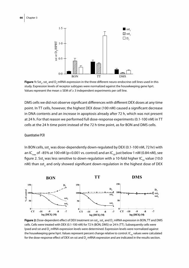
66 Chapter 3
DMS cells we did not observe significant differences with different DEX doses at any time point. In TT cells, however, the highest DEX dose (100 nM) caused a significant decrease in DNA contents and an increase in apoptosis already after 72 h, which was not present at 24 h. For that reason we performed full dose-response experiments (0.1-100 nM) in TT cells at the 24 h time point instead of the 72 h time point, as for BON and DMS cells.
Quantitative PCR
In BON cells, sst2 was dose-dependently down-regulated by DEX (0.1-100 nM, 72 hr) with an ICmax of –85% at 100 nM (p<0.001 vs. control) and an IC50 just below 1 nM (0.84 nM), see figure 2. Sst5 was less sensitive to down-regulation with a 10-fold higher IC50 value (10.0 nM) than sst2 and only showed significant down-regulation in the highest dose of DEX
different ratios. BON cells expressed relatively highamounts of sst5 (0.43G0.09, meanGS.E.M.), followed byD2 (0.22G0.05) and sst2 (0.08G0.01); TT cellspredominantly expressed D2 (1.26G0.13), followed bysst2 (0.40G0.06) and sst5 (0.34G0.03); DMS cells hadoverall lower expression levels: sst2 (0.15G0.03), D2
(0.12G0.01) and sst5 (0.04G0.01).
Cell proliferation and DNA fragmentation studies
We investigated whether DEX treatment (0.1–100 nM)caused significant changes at 24, 72 and 168 h in totalDNA contents, HPRT expression per nanogram RNAand the induction of apoptosis compared withuntreated cells (data not shown). In the BON andDMS cells, we did not observe significant differenceswith different DEX doses at any time point. In TT cells,however, the highest DEX dose (100 nM) caused asignificant decrease in DNA contents and an increase inapoptosis already after 72 h, which was not present at24 h. For that reason, we performed full dose–responseexperiments (0.1–100 nM) in TT cells at the 24 h time
point instead of the 72 h time point, as for BON andDMS cells.
Quantitative PCR
In BON cells, sst2 was dose-dependently down-regulatedby DEX (0.1–100 nM, 72 h) with an ICmax of K85% at100 nM (P!0.001 vs control) and an IC50 just below1 nM (0.84 nM; see Fig. 2). Sst5 was less sensitive todown-regulation with a tenfold higher IC50 value(10.0 nM) than sst2 and only showed significant down-regulation in the highest dose of DEX 100 nM (K50%,P!0.01). D2 did not show any down-regulation at any ofthe tested concentrations; in fact, at the highestconcentration (100 nM) D2 was slightly up-regulated(C33%, P!0.05). In TT cells (24 h), a similar dose-dependent down-regulation for sst2 was observed withan ICmax of K84% (P!0.001) and a subnanomolarIC50 (0.16 nM). In TTcells sst5, nor D2 showed any signsof down-regulation in any of the concentrations tested.In DMS cells (72 h), no significant effects were observedby DEX treatment.Based on the presence of DEX-induced effects in
BON cells and the absence of growth inhibition orapoptosis induction at any treatment duration or dose,we investigated in these cells the effects of differenttreatment periods (24 and 168 h) on DEX-inducedreceptor down-regulation. We found that at 24 and168 h, DEX treatment produced similar dose-depen-dent decreases in sst2 mRNA expression as thoseobserved at 72 h. ICmax and IC50 values were respect-ively K74% (P!0.001) and 0.60 nM at 24 h, andK80% (P!0.001) and 0.84 nM at 168 h (see Fig. 3).Also for sst5, comparable dose-dependent effects wereobserved at these time points: ICmax K45% (P!0.01)and IC50 4.0 nM at 24 h, and K52% (P!0.01) and5.4 nM at 168 h. For D2, no down-regulation wasdemonstrable at any DEX dose at these time points.The observed D2 up-regulation after 72 h with DEX100 nM was not demonstrable at 24 h or 168 h.
Figure 1 Sst2, sst5 and D2 mRNA expression in the three differentneuro-endocrine cell lines used in this study. Expression levels ofreceptor subtypes were normalized against the housekeeping geneHPRT. Values represent the meanGS.E.M. ofR3 independentexperiments per cell line. sst2&; sst5 G; D2,.
Figure 2 Dose-dependent effect of DEX treatment on sst2, sst5 and D2mRNA expression in BON, TT andDMS cells. Cells were treated withDEX (0.1–100 nM) for 72 h (BON and DMS) or 24 h (TT). Subsequently, cells were lysed and sst and D2 mRNA expression levels weredetermined. Expression levels were normalized against the housekeeping gene HPRT. Values represent per cent change relative to control.IC50 values were calculated for the dose–response effect of DEX on sst and D2 mRNA expression and are indicated in the results section.
C DE BRUIN and others . Glucocorticoid regulation of DA and SS receptors50
Journal of Molecular Endocrinology (2009) 42, 47–56 www.endocrinology-journals.org
sst5
D2
sst2
Figure 1: Sst2, sst5 and D2 mRNA expression in the three different neuro-endocrine cell lines used in this study. Expression levels of receptor subtypes were normalized against the housekeeping gene hprt. Values represent the mean ± SEM of ≥ 3 independent experiments per cell line.
BON TT
100
150
trol
D2
100
150
D2
50
100
% o
f con
t
sst550
100 sst5
0
sst2
CT 10 9 8 70
sst2
CT 10 9 8 7log [DEX] (M)
CT -10 -9 -8 -7log [DEX] (M)
CT -10 -9 -8 -7
DMS
150
D2
50
100D2
sst5sst2
0
50
log [DEX] (M)CT -10 -9 -8 -7
Figure 2: Dose-dependent effect of DEX treatment on sst2, sst5 and D2 mRNA expression in BON, TT and DMS cells. Cells were treated with DEX (0.1-100 nM) for 72 h (BON, DMS) or 24 h (TT). Subsequently cells were lysed and sst and D2 mRNA expression levels were determined. Expression levels were normalized against the housekeeping gene hprt. Values represent percent change relative to control. IC50 values were calculated for the dose-response effect of DEX on sst and D2 mRNA expression and are indicated in the results section.
DMS
150
D2
50
100D2
sst5sst2
0
50
log [DEX] (M)CT -10 -9 -8 -7
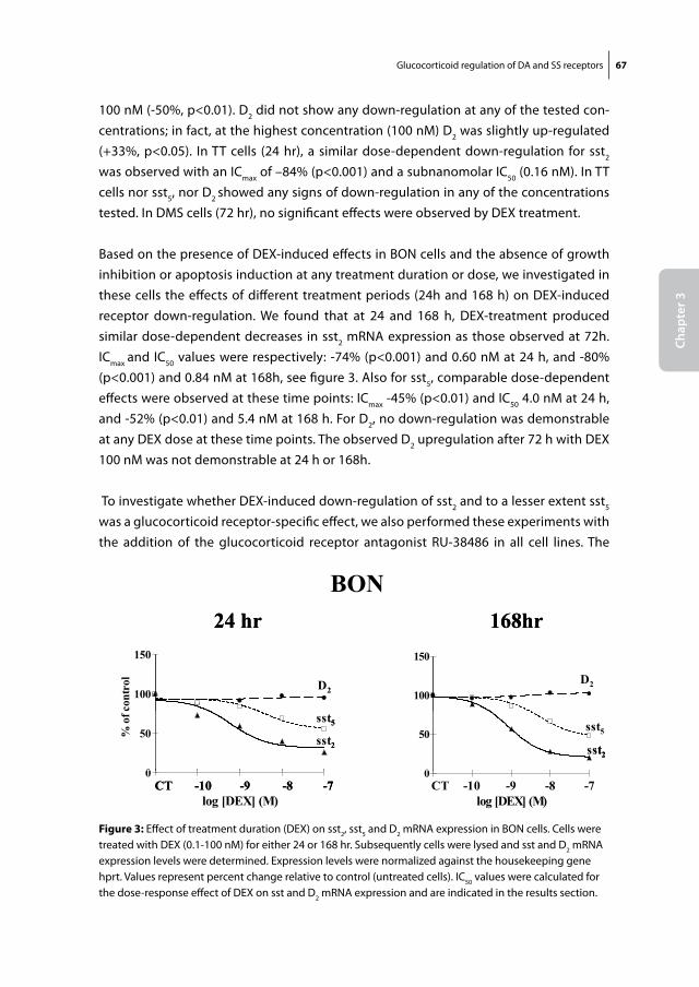
Glucocorticoid regulation of DA and SS receptors 67
Chap
ter 3
100 nM (-50%, p<0.01). D2 did not show any down-regulation at any of the tested con-centrations; in fact, at the highest concentration (100 nM) D2 was slightly up-regulated (+33%, p<0.05). In TT cells (24 hr), a similar dose-dependent down-regulation for sst2 was observed with an ICmax of –84% (p<0.001) and a subnanomolar IC50 (0.16 nM). In TT cells nor sst5, nor D2 showed any signs of down-regulation in any of the concentrations tested. In DMS cells (72 hr), no significant effects were observed by DEX treatment.
Based on the presence of DEX-induced effects in BON cells and the absence of growth inhibition or apoptosis induction at any treatment duration or dose, we investigated in these cells the effects of different treatment periods (24h and 168 h) on DEX-induced receptor down-regulation. We found that at 24 and 168 h, DEX-treatment produced similar dose-dependent decreases in sst2 mRNA expression as those observed at 72h. ICmax and IC50 values were respectively: -74% (p<0.001) and 0.60 nM at 24 h, and -80% (p<0.001) and 0.84 nM at 168h, see figure 3. Also for sst5, comparable dose-dependent effects were observed at these time points: ICmax -45% (p<0.01) and IC50 4.0 nM at 24 h, and -52% (p<0.01) and 5.4 nM at 168 h. For D2, no down-regulation was demonstrable at any DEX dose at these time points. The observed D2 upregulation after 72 h with DEX 100 nM was not demonstrable at 24 h or 168h.
To investigate whether DEX-induced down-regulation of sst2 and to a lesser extent sst5 was a glucocorticoid receptor-specific effect, we also performed these experiments with the addition of the glucocorticoid receptor antagonist RU-38486 in all cell lines. The
BON cells
168hr168hr24 hr 150
24 hr 150
100
150
D2100
150
D2100
150
cont
rol
D2100
150
cont
rol
D2
0
50sst2
0
50sst2
0
50% o
f c
sst2
sst5
0
50% o
f c
sst2
sst5 sst5
0
log [DEX] (M)CT -10 -9 -8 -7
0
log [DEX] (M)CT -10 -9 -8 -7
0
log [DEX] (M)CT -10 -9 -8 -7
0
log [DEX] (M)CT -10 -9 -8 -7CT -10 -9 -8 -7
Figure 3: Effect of treatment duration (DEX) on sst2, sst5 and D2 mRNA expression in BON cells. Cells were treated with DEX (0.1-100 nM) for either 24 or 168 hr. Subsequently cells were lysed and sst and D2 mRNA expression levels were determined. Expression levels were normalized against the housekeeping gene hprt. Values represent percent change relative to control (untreated cells). IC50 values were calculated for the dose-response effect of DEX on sst and D2 mRNA expression and are indicated in the results section.
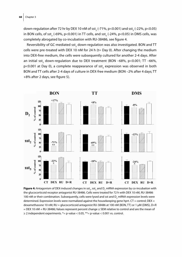
68 Chapter 3
down-regulation after 72 hr by DEX 10 nM of sst2 (-71%, p<0.001) and sst5 (-22%, p<0.05) in BON cells, of sst2 (-69%, p<0.001) in TT cells, and sst5 (-24%, p<0.05) in DMS cells, was completely abrogated by co-incubation with RU-38486, see figure 4.
Reversibility of GC-mediated sst2 down-regulation was also investigated. BON and TT cells were pre-treated with DEX 10 nM for 24 h (t= Day 0). After changing the medium into DEX-free medium, the cells were subsequently cultured for another 2-4 days. After an initial sst2 down-regulation due to DEX treatment (BON –68%, p<0.001; TT –66%, p<0.001 at Day 0), a complete reappearance of sst2 expression was observed in both BON and TT cells after 2-4 days of culture in DEX-free medium (BON –2% after 4 days; TT +8% after 2 days, see figure 5).
TT 17%
BON Fig 4
DMS
+8%
D275
100
125 +17%
cont
rol
-8%
D2
0
25
50
% o
f c
50
75
100
125
-69%sst2 -71%of c
ontr
ol -13%
0
25
50
125
69% **
2 71% **%
o
50
75
100 -10%
sst5
*
% o
f con
trol -22% -24%
*
0
25
CT DEX RU D+R
%
CT DEX RU D+R CT DEX RU D+R
Figure 4: Antagonism of DEX-induced changes in sst2, sst5 and D2 mRNA expression by co-incubation with the glucocorticoid receptor antagonist RU-38486. Cells were treated for 72 h with DEX 10 nM, RU-38486 100 nM or their combination. Subsequently, cells were lysed and sst and D2 mRNA expression levels were determined. Expression levels were normalized against the housekeeping gene hprt. CT = control; DEX = dexamethasone 10 nM; RU = glucocorticoid antagonist RU-38486 at 100 nM (BON, TT) or 1 μM (DMS), D+R = DEX 10 nM + RU-38486; Values represent percent change ± SEM relative to control and are the mean of ≥ 2 independent experiments. *= p-value < 0.05, **= p-value < 0.001 vs. control.
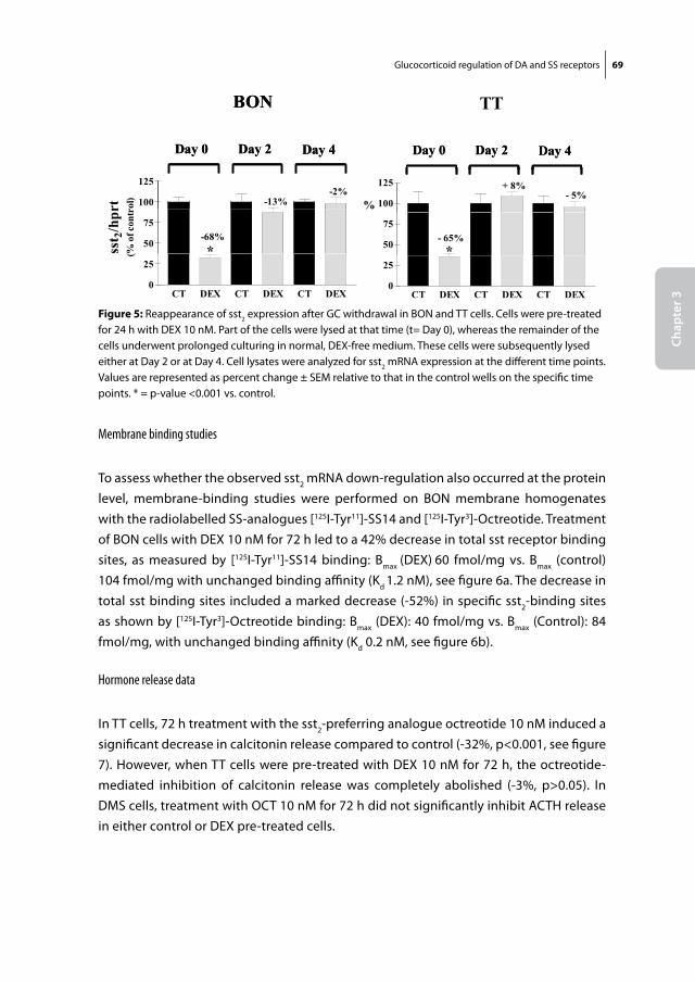
Glucocorticoid regulation of DA and SS receptors 69
Chap
ter 3
Membrane binding studies
To assess whether the observed sst2 mRNA down-regulation also occurred at the protein level, membrane-binding studies were performed on BON membrane homogenates with the radiolabelled SS-analogues [125I-Tyr11]-SS14 and [125I-Tyr3]-Octreotide. Treatment of BON cells with DEX 10 nM for 72 h led to a 42% decrease in total sst receptor binding sites, as measured by [125I-Tyr11]-SS14 binding: Bmax (DEX) 60 fmol/mg vs. Bmax (control) 104 fmol/mg with unchanged binding affinity (Kd 1.2 nM), see figure 6a. The decrease in total sst binding sites included a marked decrease (-52%) in specific sst2-binding sites as shown by [125I-Tyr3]-Octreotide binding: Bmax (DEX): 40 fmol/mg vs. Bmax (Control): 84 fmol/mg, with unchanged binding affinity (Kd 0.2 nM, see figure 6b).
Hormone release data
In TT cells, 72 h treatment with the sst2-preferring analogue octreotide 10 nM induced a significant decrease in calcitonin release compared to control (-32%, p<0.001, see figure 7). However, when TT cells were pre-treated with DEX 10 nM for 72 h, the octreotide-mediated inhibition of calcitonin release was completely abolished (-3%, p>0.05). In DMS cells, treatment with OCT 10 nM for 72 h did not significantly inhibit ACTH release in either control or DEX pre-treated cells.
BONBON
Day 4Day 2Day 0 Day 4Day 2Day 0
100
125
-13%-2%
rt rol) %100
125
-13%-2%
rt rol) %
50
75-68%
sst 2
/hpr
(% o
f con
tr
*50
75-68%
sst 2
/hpr
(% o
f con
tr
*
CT DEX CT DEX CT DEX0
25
(
CT DEX CT DEX CT DEX0
25
(
TT
Day 4Day 2Day 0 Day 4Day 2Day 0
100
125- 5%
+ 8%
%
50
75- 65%
*
CT DEX CT DEX CT DEX0
25
Figure 5: Reappearance of sst2 expression after GC withdrawal in BON and TT cells. Cells were pre-treated for 24 h with DEX 10 nM. Part of the cells were lysed at that time (t= Day 0), whereas the remainder of the cells underwent prolonged culturing in normal, DEX-free medium. These cells were subsequently lysed either at Day 2 or at Day 4. Cell lysates were analyzed for sst2 mRNA expression at the different time points. Values are represented as percent change ± SEM relative to that in the control wells on the specific time points. * = p-value <0.001 vs. control.
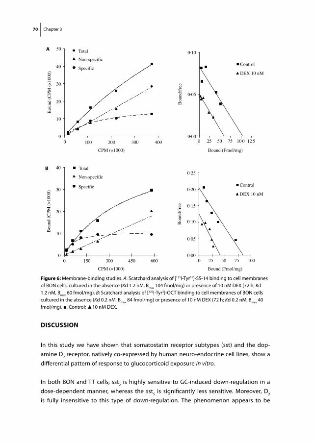
70 Chapter 3
DisCussion
In this study we have shown that somatostatin receptor subtypes (sst) and the dop-amine D2 receptor, natively co-expressed by human neuro-endocrine cell lines, show a differential pattern of response to glucocorticoid exposure in vitro.
In both BON and TT cells, sst2 is highly sensitive to GC-induced down-regulation in a dose-dependent manner, whereas the sst5 is significantly less sensitive. Moreover, D2 is fully insensitive to this type of down-regulation. The phenomenon appears to be
in vitro observations can model the clinical situation of aCD patient in whom long-term GC overexposure isrelieved through cortisol-lowering therapy. Clinicalevidence in favour of a return of sst2 expression in vivodoes exist uponGC withdrawal, however. In patients withNelson’s syndrome, inoperable or recurrent corticotrophadenomas (low sst2 expression) necessitate bilateraladrenalectomy, leading to chronic hypo-/or normocorti-solaemia. By removing most of the excessive negativefeedback loop on the pituitary, the ACTH-producingpituitary adenomas left in situmay expand with time andcan even show invasive growth in surrounding tissues.Some of these Nelson adenomas, however, are visible on111In-pentetreotide receptor scintigraphy (Octreoscan),whereasmostprimary corticotrophadenomasarenot (deHerder et al. 1994, de Herder & Lamberts 1996).
Moreover, octreotide has been effective in some of theseNelson patients by lowering ACTH levels and stabilizingtumour growth (Lamberts et al. 1989, Petrini et al. 1994,Kelestimur et al. 1996). Most likely, this reappearance offunctional sst2 receptor expression is a direct effect ofremoving chronic hypercortisolism in these patients.
In conclusion, we show that GCs selectively down-regulate sst2, but not D2 and only to a minor degree sst5in human neuro-endocrine BON and TT cells. If this isa common regulatory mechanism in human neuro-endocrine cells, then these data would support thehypothesis that chronically elevated GC levels inCushing’s disease may be directly responsible for thelow expression of sst2 in corticotroph adenomas. It alsosuggests that sst5 and D2 are interesting candidatereceptors in the search for a medical treatment of CD,
0 100 200 300 4000
10
20
30
40
50 Total
Non-specific
Specific
CPM (×1000)
Bou
nd (C
PM (×
1000
)
0 25 50 75 10 0 12 50·00
0·05
0·10
Bound (Fmol/mg)
Bou
nd/fr
ee
0 25 50 75 1000·00
0·05
0·15
0·20
0·25
Bound (Fmol/mg)
Bou
nd/fr
ee
0 150 300 450 6000
10
20
30
40 Total
Non-specific
Specific
CPM (×1000)
Bou
nd (C
PM (×
1000
)A
B
Control
DEX 10 nM
Control
DEX 10 nM
0·10
Figure 6 Membrane-binding studies. (A) Scatchard analysis of [125I-Tyr11]-SS-14 binding to cell membranes of BON cells, cultured in theabsence (Kd 1.2 nM, Bmax 104 fmol/mg) or the presence of 10 nM DEX (72 h; Kd 1.2 nM, Bmax 60 fmol/mg). (B) Scatchard analysis of[125I-Tyr3]-OCT binding to cell membranes of BON cells cultured in the absence (Kd 0.2 nM, Bmax 84 fmol/mg) or the presence of 10 nMDEX (72 h; Kd 0.2 nM, Bmax 40 fmol/mg).
C DE BRUIN and others . Glucocorticoid regulation of DA and SS receptors54
Journal of Molecular Endocrinology (2009) 42, 47–56 www.endocrinology-journals.org
Figure 6: Membrane-binding studies. A: Scatchard analysis of [125I-Tyr11]-SS-14 binding to cell membranes of BON cells, cultured in the absence (Kd 1.2 nM, Bmax 104 fmol/mg) or presence of 10 nM DEX (72 h; Kd 1.2 nM, Bmax 60 fmol/mg). B: Scatchard analysis of [125I-Tyr3]-OCT binding to cell membranes of BON cells cultured in the absence (Kd 0.2 nM, Bmax 84 fmol/mg) or presence of 10 nM DEX (72 h; Kd 0.2 nM, Bmax 40 fmol/mg). ■, Control; ▲10 nM DEX.
A
B
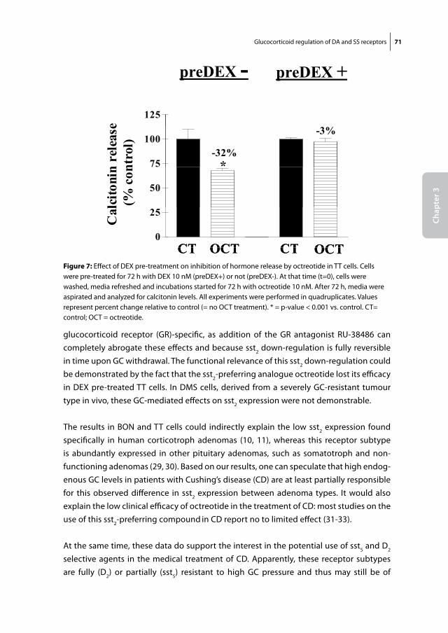
Glucocorticoid regulation of DA and SS receptors 71
Chap
ter 3
glucocorticoid receptor (GR)-specific, as addition of the GR antagonist RU-38486 can completely abrogate these effects and because sst2 down-regulation is fully reversible in time upon GC withdrawal. The functional relevance of this sst2 down-regulation could be demonstrated by the fact that the sst2-preferring analogue octreotide lost its efficacy in DEX pre-treated TT cells. In DMS cells, derived from a severely GC-resistant tumour type in vivo, these GC-mediated effects on sst2 expression were not demonstrable.
The results in BON and TT cells could indirectly explain the low sst2 expression found specifically in human corticotroph adenomas (10, 11), whereas this receptor subtype is abundantly expressed in other pituitary adenomas, such as somatotroph and non-functioning adenomas (29, 30). Based on our results, one can speculate that high endog-enous GC levels in patients with Cushing’s disease (CD) are at least partially responsible for this observed difference in sst2 expression between adenoma types. It would also explain the low clinical efficacy of octreotide in the treatment of CD: most studies on the use of this sst2-preferring compound in CD report no to limited effect (31-33).
At the same time, these data do support the interest in the potential use of sst5 and D2 selective agents in the medical treatment of CD. Apparently, these receptor subtypes are fully (D2) or partially (sst5) resistant to high GC pressure and thus may still be of
TT cells
preDEX - preDEX +
125e
75
100-32%
-3%
*n re
leas
ent
rol)
50
75
lcito
nin
(% c
on
0
25
CT OCT CT OCTCT OCT CT OCT
Cal
CT OCT CT OCT CT OCT CT OCT Figure 7: Effect of DEX pre-treatment on inhibition of hormone release by octreotide in TT cells. Cells were pre-treated for 72 h with DEX 10 nM (preDEX+) or not (preDEX-). At that time (t=0), cells were washed, media refreshed and incubations started for 72 h with octreotide 10 nM. After 72 h, media were aspirated and analyzed for calcitonin levels. All experiments were performed in quadruplicates. Values represent percent change relative to control (= no OCT treatment). * = p-value < 0.001 vs. control. CT= control; OCT = octreotide.
72 Chapter 3
functional value in the regulation of ACTH release in corticotroph adenomatous cells. Several clinical studies have been performed already with these agents and have shown promising results in subsets of patients. Pivonello et al. showed that 3-month treatment with the D2-specific agonist cabergoline could significantly reduce urinary free cortisol (UFC) levels in 60% of patients and even induce complete normalization of UFC in 40% of patients with CD (5). An early report on the use of the multi-ligand SS analogue pasireotide (SOM230) which has subnanomolar affinity for sst2, sst3 and sst5, showed equally interesting results: 15-day treatment with pasireotide 600μg twice daily, led to a complete normalization of UFC in 19% (4/21) and a ≥ 50% decrease in UFC in another 24% (5/21) of CD patients, probably mediated through sst5 receptor activation (6). Considering the reported efficacy of both cabergoline and pasireotide in subsets of CD patients, combination treatment with these agents is an interesting future option. For that reason, studying the effects of GC on a single cell system from a neuro-endocrine origin that expresses sst2, sst5 and D2, could help us understand some more of the biological backgrounds of these clinical observations.
This is the first study to describe a differential pattern of GC-responsiveness regarding both D2 and SS receptor subtypes co-expressed in human neuro-endocrine cell lines. The observed pattern closely resembles the one seen earlier in the murine corticotroph AtT-20 cells (19). In these cells, sst2 is sensitive whereas sst5 is largely insensitive to GC-induced down-regulation. Unfortunately, D2 was not expressed at sufficiently high levels in these cells to allow for investigation of this receptor subtype (unpublished observa-tions). Nevertheless, one could hypothesize that a similar pattern of GC-responsiveness exists in different human and murine neuro-endocrine cell lines.
Our findings are in line with earlier studies that describe the presence of distinct GC-responsive elements (GREs) in the murine sst2 gene, whereas the murine sst5 gene only contains multiple GRE-half sites (34-36). Moreover, in transfection experiments it was shown that the human sst2 gene promoter is under direct control of GC’s, whereas the sst5 promotor is not (37, 38). To our knowledge, comparable GC-responsive regulatory sequences have thus far not been identified within the D2 gene.
Previously, Petersenn et al. found in GH4 cells (rat pituitary adenoma cell line) that hydrocortisone 100 nM did not influence sst5 promoter activity whereas we do find moderate but significant down-regulation of sst5 at higher DEX doses (100 nM) in BON cells (37). We believe that this difference can be ascribed to the difference in relative in vitro potency of hydrocortisone compared to dexamethasone. In previous studies on differential regulatory mechanisms by glucocorticoids it was found that DEX has a 14-fold higher potency in vitro compared to hydrocortisone (GILZ EC50 of 4.1 and 56.7 nM,
Glucocorticoid regulation of DA and SS receptors 73
Chap
ter 3
respectively) (39). Therefore, the dose used by Petersenn et al. would be the equivalent of approximately 7 nM DEX in our study. At this dose we did not observe any significant effects on sst5 mRNA expression either.
Even though corticotroph adenoma cells are GC-resistant to a certain degree, GC regula-tion of SS receptors apparently remains intact in AtT-20 cells. Interestingly, BON and TT cells, derived from tumours which do not show GC-resistance in vivo, have indeed a higher degree of sst2 down-regulation when exposed to DEX 10 nM, compared with the partially GC-resistant corticotroph adenoma cell line AtT-20 (19). In the severely GC-resistant DMS cells, derived from an ectopic ACTH-producing small cell lung cancer, most of the GC-induced sst2 down-regulation is lost, as would be expected. The latter in vitro observation correlates with two interesting clinical observations. First of all, many ectopic ACTH-producing tumours are positive on 111In-pentreotide receptor scintigra-phy (OctreoScan) (40-43), despite the hypercortisolistic environment they are exposed to. And secondly, octreotide has been shown to be effective in controlling tumour size and cortisol production in some of these patients as opposed to patients with pituitary-derived CD (44, 45).
Another aspect we have observed in this study is the reappearance of sst2 expression within 2-4 days after withdrawing GC-exposure. This observation could be of clinical in-terest. When it is possible to lower GC levels in CD patients with the use of for instance sst5 or D2 selective agents, then this state of normocortisolemia could lead to a re-expression of sst2 in the corticotroph adenomas of these patients. Return of sst2 expression in vivo would result in a strong increase in efficacy of traditional SS analogs such as octreotide (sst2) as well as pasireotide (sst2+sst5) and thus expand the pharmacological options to maximally inhibit ACTH production. Obviously, the question remains to which extent these in vitro observations can model the clinical situation of a CD patient in whom long-term GC-overexposure is relieved through cortisol-lowering therapy. Clinical evidence in favour of a return of sst2 expression in vivo upon GC withdrawal does exist, however. In patients with Nelson’s Syndrome, inoperable or recurrent corticotroph adenomas (low sst2 expression) necessitate bilateral adrenalectomy, leading to chronic hypo-/ or normocortisolemia. By removing most of the excessive negative feedback loop on the pituitary, the ACTH-producing pituitary adenomas left in situ may expand with time and can even show invasive growth in surrounding tissues. Some of these Nelson adenomas, however, are visible on 111In-pentreotide receptor scintigraphy (OctreoScan), whereas most primary corticotroph adenomas are not (46, 47). Moreover, octreotide has been ef-fective in some of these Nelson patients by lowering ACTH levels and stabilizing tumour growth (31, 48, 49). Most likely, this reappearance of functional sst2 receptor expression is a direct effect of removing chronic hypercortisolism in these patients.
74 Chapter 3
In conclusion, we show that GCs selectively down-regulate sst2, but not D2 and only to a minor degree sst5 in human neuro-endocrine BON and TT cells. If this is a common regulatory mechanism in human neuro-endocrine cells, then these data would support the hypothesis that chronically elevated GC levels in Cushing’s disease may be directly responsible for the low expression of sst2 in corticotroph adenomas. It also suggests that sst5 and D2 are interesting candidate receptors in the search for a medical treat-ment of CD, due to their (relative) resistance to GC-induced down-regulation. It needs to be emphasized, however, that these data require confirmation, preferably in primary cultures of human corticotroph adenomas.
reFerenCes
1. Barnett P 2003 Somatostatin and somatostatin receptor physiology. Endocrine 20:255-264 2. missale C, nash sr, robinson sW, Jaber m, Caron mg 1998 Dopamine receptors: from structure
to function. Physiol Rev 78:189-225 3. Patel yC 1999 Somatostatin and its receptor family. Front Neuroendocrinol 20:157-198 4. Heaney AP, melmed s 2004 Molecular targets in pituitary tumours. Nat Rev Cancer 4:285-295 5. Pivonello r, Ferone D, de Herder WW, Kros Jm, De Caro ml, Arvigo m, Annunziato l, lom-
bardi g, Colao A, Hofland lJ, lamberts sW 2004 Dopamine receptor expression and function in corticotroph pituitary tumors. J Clin Endocrinol Metab 89:2452-2462
6. Boscaro m, Petersenn, s., Atkinson, A.B., Bertherat, J., Findling J., snyder, P., mcBride, K., reincke, m., ludlam, W., gao, B., melmed, s., Freda, P., Frohman, l., grossman, A., Biller, B, glusman, J.e. 2006 Pasireotide (SOM230), the novel multi-ligand somatostatin analogue, is a promising medical therapy for patients with Cushing’s disease: preliminary safety and efficacy results of a phase II study. Presented at ENDO 2006, abstr OR9-1, Boston, U.S.A., 2006, p 81
7. godbout A BH, Babin s, sabourin A, lacroix A 2007 Cabergoline in the long-term treatment of Cushing’s disease. The Endocrine Society’s 89th Annual Meeting, June 2-5, Toronto, Canada, 2007
8. von Werder K, muller oA, stalla gK 1996 Somatostatin analogs in ectopic corticotropin produc-tion. Metabolism 45:129-131
9. Pivonello r, Ferone D, de Herder WW, Faggiano A, Bodei l, de Krijger rr, lombardi g, Colao A, lamberts sW, Hofland lJ 2007 Dopamine receptor expression and function in corticotroph ectopic tumors. J Clin Endocrinol Metab 92:65-69
10. Hofland lJ, van der Hoek J, Feelders r, van Aken mo, van Koetsveld Pm, Waaijers m, sprij-mooij D, Bruns C, Weckbecker g, de Herder WW, Beckers A, lamberts sW 2005 The multi-ligand somatostatin analogue SOM230 inhibits ACTH secretion by cultured human corticotroph adenomas via somatostatin receptor type 5. Eur J Endocrinol 152:645-654
11. Batista Dl, Zhang X, gejman r, Ansell PJ, Zhou y, Johnson sA, swearingen B, Hedley-Whyte et, stratakis CA, Klibanski A 2006 The effects of SOM230 on cell proliferation and adrenocorti-cotropin secretion in human corticotroph pituitary adenomas. J Clin Endocrinol Metab 91:4482-4488
Glucocorticoid regulation of DA and SS receptors 75
Chap
ter 3
12. van Craenenbroeck K, De Bosscher K, vanden Berghe W, vanhoenacker P, Haegeman g 2005 Role of glucocorticoids in dopamine-related neuropsychiatric disorders. Mol Cell Endocrinol 245:10-22
13. lammers CH, D’souza um, Qin ZH, lee sH, yajima s, mouradian mm 1999 Regulation of striatal dopamine receptors by corticosterone: an in vivo and in vitro study. Brain Res Mol Brain Res 69:281-285
14. Czyrak A, mackowiak m, Chocyk A, Fijal K, Wedzony K 2003 Role of glucocorticoids in the regulation of dopaminergic neurotransmission. Pol J Pharmacol 55:667-674
15. sigala s, missale C, tognazzi n, spano P 2000 Differential gene expression of dopamine D-2 receptor subtypes in rat chromaffin cells and sympathetic neurons in culture. Neuroreport 11:2467-2471
16. lee D, Huang W, Copolov Dl, lim At 1995 Glucocorticoids inhibit D1B, but not D2, receptor-mediated effects on hypothalamic atrial natriuretic factor neurons. Endocrinology 136:5570-5576
17. Park s, Kamegai J, Kineman rD 2003 Role of glucocorticoids in the regulation of pituitary somatostatin receptor subtype (sst1-sst5) mRNA levels: evidence for direct and somatostatin-mediated effects. Neuroendocrinology 78:163-175
18. Xu y, Berelowitz m, Bruno JF 1995 Dexamethasone regulates somatostatin receptor subtype messenger ribonucleic acid expression in rat pituitary GH4C1 cells. Endocrinology 136:5070-5075
19. van der Hoek J, Waaijers m, van Koetsveld Pm, sprij-mooij D, Feelders rA, schmid HA, schoeffter P, Hoyer D, Cervia D, taylor Je, Culler mD, lamberts sW, Hofland lJ 2005 Distinct functional properties of native somatostatin receptor subtype 5 compared with subtype 2 in the regulation of ACTH release by corticotroph tumor cells. Am J Physiol Endocrinol Metab 289:E278-287
20. ikeda K, lu C, Weir eC, mangin m, Broadus Ae 1989 Transcriptional regulation of the parathy-roid hormone-related peptide gene by glucocorticoids and vitamin D in a human C-cell line. J Biol Chem 264:15743-15746
21. evers Bm, Wang X, Zhou Z, townsend Cm, Jr., mcneil gP, Dobner Pr 1995 Characterization of promoter elements required for cell-specific expression of the neurotensin/neuromedin N gene in a human endocrine cell line. Mol Cell Biol 15:3870-3881
22. gaitan D, DeBold Cr, turney mK, Zhou P, orth Dn, Kovacs WJ 1995 Glucocorticoid receptor structure and function in an adrenocorticotropin-secreting small cell lung cancer. Mol Endocrinol 9:1193-1201
23. ray DW, littlewood AC, Clark AJ, Davis Jr, White A 1994 Human small cell lung cancer cell lines expressing the proopiomelanocortin gene have aberrant glucocorticoid receptor function. J Clin Invest 93:1625-1630
24. Hofland lJ, van der Hoek J, van Koetsveld Pm, de Herder WW, Waaijers m, sprij-mooij D, Bruns C, Weckbecker g, Feelders r, van der lely AJ, Beckers A, lamberts sW 2004 The novel somatostatin analog SOM230 is a potent inhibitor of hormone release by growth hormone- and prolactin-secreting pituitary adenomas in vitro. J Clin Endocrinol Metab 89:1577-1585
25. rasmussen r 2001 Quantification on the LightCycler. Heidelberg: Springer Press 26. Pfaffl mW 2001 A new mathematical model for relative quantification in real-time RT-PCR. Nucleic
Acids Res 29:e45 27. Hofland lJ, van Koetsveld Pm, lamberts sW 1990 Percoll density gradient centrifugation of
rat pituitary tumor cells: a study of functional heterogeneity within and between tumors with
76 Chapter 3
respect to growth rates, prolactin production and responsiveness to the somatostatin analog SMS 201-995. Eur J Cancer 26:37-44
28. Hofland lJ, van Koetsveld Pm, Wouters n, Waaijers m, reubi JC, lamberts sW 1992 Dissocia-tion of antiproliferative and antihormonal effects of the somatostatin analog octreotide on 7315b pituitary tumor cells. Endocrinology 131:571-577
29. Jaquet P, saveanu A, gunz g, Fina F, Zamora AJ, grino m, Culler mD, moreau JP, enjalbert A, ouafik lH 2000 Human somatostatin receptor subtypes in acromegaly: distinct patterns of messenger ribonucleic acid expression and hormone suppression identify different tumoral phenotypes. J Clin Endocrinol Metab 85:781-792
30. taboada gF, luque rm, Bastos W, guimaraes rF, marcondes JB, Chimelli lm, Fontes r, mata PJ, Filho Pn, Carvalho DP, Kineman rD, gadelha mr 2007 Quantitative analysis of somatostatin receptor subtype (SSTR1-5) gene expression levels in somatotropinomas and non-functioning pituitary adenomas. Eur J Endocrinol 156:65-74
31. lamberts sW, uitterlinden P, Klijn Jm 1989 The effect of the long-acting somatostatin analogue SMS 201-995 on ACTH secretion in Nelson’s syndrome and Cushing’s disease. Acta Endocrinol (Copenh) 120:760-766
32. stalla gK, Brockmeier sJ, renner u, newton C, Buchfelder m, stalla J, muller oA 1994 Oct-reotide exerts different effects in vivo and in vitro in Cushing’s disease. Eur J Endocrinol 130:125-131
33. Ambrosi B, Bochicchio D, Fadin C, Colombo P, Faglia g 1990 Failure of somatostatin and octreotide to acutely affect the hypothalamic-pituitary-adrenal function in patients with corti-cotropin hypersecretion. J Endocrinol Invest 13:257-261
34. gordon DF, Woodmansee WW, lewis sr, James rA, Wood Wm, ridgway eC 1999 Cloning of the mouse somatostatin receptor subtype 5 gene: promoter structure and function. Endocrinol-ogy 140:5598-5608
35. Kraus J, Woltje m, Hollt v 1999 Regulation of mouse somatostatin receptor type 2 gene expres-sion by glucocorticoids. FEBS Lett 459:200-204
36. Kraus J, Woltje m, schonwetter n, Hollt v 1998 Alternative promoter usage and tissue specific expression of the mouse somatostatin receptor 2 gene. FEBS Lett 428:165-170
37. Petersenn s, rasch AC, Bohnke C, schulte Hm 2002 Identification of an upstream pituitary-active promoter of human somatostatin receptor subtype 5. Endocrinology 143:2626-2634
38. Petersenn s, rasch AC, Presch s, Beil Fu, schulte Hm 1999 Genomic structure and transcrip-tional regulation of the human somatostatin receptor type 2. Mol Cell Endocrinol 157:75-85
39. smit P, russcher H, de Jong FH, Brinkmann Ao, lamberts sW, Koper JW 2005 Differential regulation of synthetic glucocorticoids on gene expression levels of glucocorticoid-induced leucine zipper and interleukin-2. J Clin Endocrinol Metab 90:2994-3000
40. uwaifo gi, Koch CA, Hirshberg B, Chen CC, Hartzband P, nieman lK, Pacak K 2003 Is there a therapeutic role for octreotide in patients with ectopic Cushing’s syndrome? J Endocrinol Invest 26:710-717
41. ilias i, torpy DJ, Pacak K, mullen n, Wesley rA, nieman lK 2005 Cushing’s syndrome due to ectopic corticotropin secretion: twenty years’ experience at the National Institutes of Health. J Clin Endocrinol Metab 90:4955-4962
42. tsagarakis s, Christoforaki m, giannopoulou H, rondogianni F, Housianakou i, malagari C, rontogianni D, Bellenis i, thalassinos n 2003 A reappraisal of the utility of somatostatin receptor scintigraphy in patients with ectopic adrenocorticotropin Cushing’s syndrome. J Clin Endocrinol Metab 88:4754-4758
Glucocorticoid regulation of DA and SS receptors 77
Chap
ter 3
43. isidori Am, Kaltsas gA, Pozza C, Frajese v, newell-Price J, reznek rH, Jenkins PJ, monson JP, grossman AB, Besser gm 2006 The ectopic adrenocorticotropin syndrome: clinical features, diagnosis, management, and long-term follow-up. J Clin Endocrinol Metab 91:371-377
44. lamberts sW, tilanus HW, Klooswijk Ai, Bruining HA, van der lely AJ, de Jong FH 1988 Suc-cessful treatment with SMS 201-995 of Cushing’s syndrome caused by ectopic adrenocorticotro-pin secretion from a metastatic gastrin-secreting pancreatic islet cell carcinoma. J Clin Endocrinol Metab 67:1080-1083
45. Phlipponneau m, nocaudie m, epelbaum J, De Keyzer y, lalau JD, marchandise X, Bertagna X 1994 Somatostatin analogs for the localization and preoperative treatment of an adrenocorti-cotropin-secreting bronchial carcinoid tumor. J Clin Endocrinol Metab 78:20-24
46. de Herder WW, Krenning eP, malchoff CD, Hofland lJ, reubi JC, Kwekkeboom DJ, oei Hy, Pols HA, Bruining HA, nobels Fr, et al. 1994 Somatostatin receptor scintigraphy: its value in tumor localization in patients with Cushing’s syndrome caused by ectopic corticotropin or corticotropin-releasing hormone secretion. Am J Med 96:305-312
47. de Herder WW, lamberts sW 1996 Is there a role for somatostatin and its analogs in Cushing’s syndrome? Metabolism 45:83-85
48. Petrini l, gasperi m, Pilosu r, marcello A, martino e 1994 Long-term treatment of Nelson’s syndrome by octreotide: a case report. J Endocrinol Invest 17:135-139
49. Kelestimur F, utas C, ozbakir o, selcuklu A, Kandemir o, ozcan n 1996 The effects of oct-reotide in a patient with Nelson’s syndrome. Postgrad Med J 72:53-54

Chapter 4
Co-expression of dopamine and somatostatin receptor subtypes
in corticotroph adenomas
de Bruin C, Pereira AM, Feelders RA, Romijn JA, Roelfsema F, Sprij-Mooij DM, van Aken MO, van der Lelij AJ, de Herder WW, Lamberts SW, Hofland LJ.
J Clin Endocrinol Metab. 2009 Apr;94(4):1118-24.
80 Chapter 4
ABstrACt:
Context: Previous studies have demonstrated the expression of somatostatin receptor subtypes (mainly sst5) and dopamine (DA) receptor subtypes (mainly D2) in smaller se-ries of human corticotroph adenomas. In line with these findings, sst5 and D2-targeting agents have already been used clinically in patients with Cushing’s Disease (CD) and have shown promising results in subsets of patients. To what extent these receptor subtypes are co-expressed within individual adenomas, is not known however. objective: To investigate the (co-)expression of both sst and DA receptors in a large series of human corticotroph adenomas. Design: In vitro analysis of corticotroph adenoma tissue obtained via transsphenoidal adenomectomysetting: Two university medical centersPatients: 30 patients with Cushing’s disease results: Analyzed by quantitative RT-PCR, D2 and sst5 were significantly (co-)expressed in the majority (60%) of adenomas, whereas 23% of adenomas only expressed D2, but not sst5. The remaining 17% of adenomas did not significantly express either sst5 or D2. Overall, expression of sst1-4 and D4 was low to non-detectable. Corticotroph adenomas with invasive growth invariably showed loss of sst5 and D2 expression. Autoradiography revealed clear D2 and/or SS-14 binding in a subset of cases, which correlated well with their respective mRNA data. Conclusions: Sst5 and especially D2 are highly expressed in the majority of human cor-ticotroph adenomas with co-expression of sst5 and D2 being a common phenomenon. These findings support the current studies with sst5 and D2-targeting agents in patients with CD and highlight the rationale behind sst5-D2 combination therapy.
DA and SS receptors in corticotroph adenomas 81
Chap
ter 4
introDuCtion:
Cushing’s disease (CD) is a severe endocrine disorder, caused by an ACTH-producing pituitary adenoma. Patients suffer from the effects of chronic hypercortisolism, which can lead to hypertension, diabetes mellitus, osteoporosis and psychiatric disturbances among others (1). First line of treatment is transsphenoidal selective adenomectomy, but even in experienced hands long-term cure does not exceed 70% (2). In cases of per-sistent or recurrent CD, re-operation, radiotherapy and/or bilateral adrenalectomy can be performed, but these treatment options all have significant disadvantages, includ-ing the risk of permanent hypopituitarism and the lifelong dependence on hormonal replacement therapy. Until now, medical therapies with cortisol-lowering drugs such as ketoconazole or metyrapone, have not been shown to be both effective and safe for the long-term treatment of CD (3).
In recent years, however, some interesting new advances have been made in the field of receptor physiology in CD. It was found that the dopamine receptor subtype 2 (D2) is expressed in approximately 80% of human corticotroph adenomas and that these adenomas can be responsive to the ACTH-inhibiting actions of D2-agonists in vitro (4). Moreover, treatment for three months with the D2-selective agonist cabergoline induced complete normalization of 24 hr urinary free cortisol (UFC) in approximately 40% of CD patients (4). This percentage of complete responders appears to be maintained after two years of therapy (5). At the same time, different groups demonstrated that most human corticotroph adenomas strongly express somatostatin (SS) receptor subtype 5 (sst5) and that SS-analogues with high-binding affinity for sst5, such as the SS-multiligand pasireotide (SOM230), can inhibit ACTH-release and cellular proliferation in vitro (6, 7). The cortisol-lowering effects of pasireotide in vivo have recently been demonstrated in a phase II clinical trial in patients with CD (8).
Based on these findings, medical therapy with D2-agonists and/or SS-analogues with sst5-affinity appears to be an interesting treatment option in cases of persistent or recur-rent CD. To our knowledge, no study has investigated the expression of both SS and DA receptors within the same set of human corticotroph adenomas. A better understand-ing of the (co-)expression patterns of these two receptor subtypes could be of clinical importance. First of all this could help predict the overall percentage of corticotroph adenomas that possess a molecular target for SS and/or DA directed medical therapy. At present it is not known whether the D2–positive and sst5-positive adenomas in the previously mentioned studies largely overlap or whether they constitute separate groups that require different therapies. Secondly, since Rocheville et al. (9) published their report on heterodimerization of SS and DA receptors and resulting synergism in
82 Chapter 4
effect, combined treatment with DA agonists and SS analogues has been an important research topic in pituitary adenomas. It has also led to the pharmaceutical development of “dopastatin” chimeras: chemically engineered compounds that possess high binding affinity for both SS and DA receptor subtypes and which may display superagonistic properties compared with traditional SS analogues and DA agonists (10).
For that reason, our main study aim was to characterize the co-expression of dopamine and somatostatin receptor subtypes in a large series of human corticotroph adenomas to gain a better understanding of the distribution of the primary molecular targets for DA/SS based medical therapy in CD. In addition, we aimed to correlate the DA/SS receptor expression patterns in these adenomas with the main biochemical and clinical parameters of HPA-axis over-activation in the corresponding CD patients.
mAteriAls AnD metHoDs
Corticotroph adenoma tissue
Human corticotroph adenoma tissue was available from patients with CD who under-went transsphenoidal selective adenomectomy in the period between 1991 and 2007 in one of the two participating academic centers. Informed consent was obtained from all patients of whom we assessed receptor expression for investigational purposes. The diagnosis of CD was based on clinical signs, elevated 24 hr UFC excretion, incomplete suppression of serum cortisol by 1-mg dexamethasone and/or the absence of a diurnal cortisol rhythm. In all patients imaging (MRI with iv contrast) or inferior sinus petrosus sampling confirmed the pituitary source of ACTH overproduction. Normal anterior pituitary tissue was obtained at autopsy from two patients who had died from non-endocrine diseases.
Purity of corticotroph tissue
At the study start, the corticotroph adenoma specimens were transferred into Tissue Tek Compound (Sakura Finetek, Torrance, CA) at –80˚C for further handling. From these tissues, cryostat sections were cut at 5 μm. Sections from the center of the adenoma were used for an initial immunohistochemistry (IHC) step to check for ACTH expression (see below for protocol) and a sequential haematoxylin-and-eosin stain to demonstrate the histological presence of adenoma tissue. In samples where both of these criteria were sufficiently met, directly adjacent tissue sections were cut at 20μm, from which RNA was extracted for further quantitative PCR analysis of dopamine and somatostatin
DA and SS receptors in corticotroph adenomas 83
Chap
ter 4
receptor subtype expression (see below for qPCR methods). On average, 5-10 sequential sections (of 20μm each) per adenoma were needed to obtain sufficient amounts of RNA. Through this approach of sequential sections we aimed to obtain pure corticotroph adenoma RNA and limit the potential interference by normal anterior pituitary tissue surrounding the adenoma. As a secondary check for purity of corticotroph tissue, we assessed GH/POMC mRNA expression ratios in all corticotroph adenoma samples and compared these with the GH/POMC ratio in the normal human anterior pituitaries. GH/POMC ratios of less than 10% compared with normal pituitaries were regarded as pure corticotroph adenoma tissues; GH/POMC ratios of 10% or more were regarded as significantly contaminated with normal pituitary tissue and were excluded from further analysis in the pure corticotroph adenoma group (11).
Quantitative PCR
Expression analysis was performed by quantitative PCR, according to a previously described method (11, 12). The sequences and final concentrations of the hprt and sst1-5 primer-probe pairs have been described previously (6, 12). The sequence of the sst4 probe contains a single nucleotide polymorphism at one position. The other primer and probe sequences are depicted in Table 1. Samples were measured on an ABI Prism 7900 Sequence Detection System (Perkin Elmer, Foster City, CA) and normalized against the expression of the housekeeping gene hprt. PCR efficiencies (E) were calculated for every primer-probe set and varied between 1.91 and 2.00 (13). Estimated copy numbers
table 1 Primer-probe sequences for D2, D2L, D4, GH and POMC
Primer sequence 5’-3’ efficiency
D2 forw GCCACTCAGATGCTCGCC 1.94
D2 rev ATGTGTGTGATGAAGAAGGGCA
D2 pr FAM-TTGTTCTCGGCGTGTTCATCATCTGC-TAMRA
D2,L forw CCCACCTGAGGGCTCCA 1.95
D2,L rev TGATAACGGTGCAGAGTTTCATG
D2,L pr FAM-TAAAGGGCAACTGTACTCACCCCGAGG-TAMRA
D4 forw TGCACCGCCTCCATCTTC 2.00
D4 rev CCACGAACCTGTCCACGC
D4 pr FAM-ACCTGTGCGCCATC-TAMRA
GH forw GCCCATCGTCTGCACCAG 1.96
GH rev GGGATATAGGCTTCTTCAAACTCCT
GH pr FAM-TGGCCTTTGACACCTAC-TAMRA
POMC forw GAGCAGCCAGTGTCAGGACC 1.92
POMC rev CGGATGCACTCCAGCAGG
POMC pr FAM-CACCACGGAAAGCA-TAMRA
84 Chapter 4
were calculated using the comparative threshold method with efficiency correction, as described previously (14). Expression of the D2 short isoform was calculated as D2 total minus D2 long isoform.
Immunohistochemistry
The expression of ACTH in the corticotroph adenoma tissues was assessed by cutting cryostat sections (5μm) and performing immunohistochemistry according to a previ-ously published method (15). The anti-ACTH primary antibody (Santa Cruz, mouse monoclonal) was used at a dilution of 1:100.
Autoradiography
In a number of cases (n=9), sufficient adenoma tissue was available to perform addi-tional receptor autoradiography, according to previously published protocols (4, 15). Slides were incubated with either the D2 receptor antagonist 125I-sulpiride or the SS (sst1-5) analogue 11Tyr-SS-14 (both from GE Healthcare, Brussels, Belgium) in the absence or presence of unlabeled cabergoline 10-6 M (Pharmacia, Milan, Italy) or somatostatin-14 10-6 M (Sigma, Zwijndrecht, the Netherlands), respectively. The sections were exposed to phosphor imaging screens for 16 days in x-ray cassettes and analyzed using a Cyclone phosphor imager (Packard Instruments Co, Groningen, The Netherlands). The level of receptor binding was scored as: absent (-); weak or focal positivity (+); strong positivity (++).
Clinical data
Patient charts were retrieved to collect data on various clinical, biochemical and radiological parameters, see Table 2. Adenoma size and growth characteristics were categorized according to the modified Wilson classification system (16). Baseline mean 24 hr UFC values were documented at the time of initial diagnosis, before treatment with ketoconazole or metyrapone was initiated. For the determination of serum cortisol, plasma ACTH, serum prolactin and urinary free cortisol, different assays have been used in the two participating centers over the course of the study period (1991-2007). We expressed these parameters as a ratio of the upper limit of normal (ULN) of the specific assay.
DA and SS receptors in corticotroph adenomas 85
Chap
ter 4
table 2 Patient characteristics (n=30)
Parameters Value (% or mean±sd) Range
Clinical characteristics
Female sex (%) 83
Age (yrs) 42.1 14-68
BMI (kg/m2) 28.9 20.5-48.4
SBP (mm Hg) 154 120-240
DBP (mm Hg) 93 75-115
Pre-treatment received1 (%) 86
Duration pre-treatment (months) 3 1-7
Macroadenoma ≥10 mm (%) 50
Adenoma grade (modified Wilson):2
- Grade 1 43
- Grade 2 33
- Grade 3 7
- Grade 4 17
Pre-operative3
UFC 24 hr (xULN): 4.6 ± 4.1 0.60-18.7
Cortisol serum (xULN):
- mean diurnal4 1.04 ± 0.44 0.50-2.07
- evening/morning 0.97 ± 0.34 0.57-2.10
- 8 a.m. 1.15 ± 0.46 0.43-1.90
- after DEX 1 mg5 0.99 ± 0.74 0.09-2.63
- after CRH 100 μg.5 1.65 ± 0.77 0.68-3.80
- after DEX 7 mg5 0.51 ± 0.48 0.05-1.75
ACTH plasma (xULN):
- mean diurnal4 1.87 ± 2.06 0.33-9.13
- 8 a.m. 1.31 ± 1.00 0.40-3.41
- after CRH 100 μg5 2.11 ± 1.51 0.76-6.07
IGF-1 (nmol/l) 23.9 ± 10.5 5-46
PRL (xULN) 0.90 ± 1.27 0.18-5.77
Fasting blood glucose (mmol/l) 5.8 ± 2.3 3.7-14.4
Post-operative
% Remission 72
Cortisol serum 8 a.m. (xULN) 0.46 ± 0.39 0.02-1.32
Fasting blood glucose (mmol/l) 5.3 ± 1.2 3.6-8.0
Serum PRL (xULN) 0.55 ± 1.11 0.00-5.97
Follow-up (months) 55.2 0-150
Hydrocortisone dependence (months) 14.6 0-621 Pre-operative treatment consisted of either ketoconazole or metyrapone monotherapy.2 Modified Wilson classification: Grade 1 (noninvasive microadenoma), Grade 2 (noninvasive macroadenoma), Grade 3 (invasive microadenoma), Grade 4 (invasive macroadenoma)3 Laboratory parameters are expressed relative to the upper limit of normal (ULN) of the individual assay (1.0 = value at the level of ULN), except for fasting blood glucose (ref 3.5-5.6 mmol/l) and IGF-1 (reference values age- and gender-specific). 4 Mean diurnal serum cortisol and ACTH were calculated as the average of the 8 a.m, 5 p.m. and 10 p.m. values. 5 The resulting maximal (after CRH 100 μg iv) and minimal serum cortisol levels (after DEX 1 mg p.o. or 7 mg iv) are reported relative to the standard ULN of serum cortisol.
86 Chapter 4
Statistical analysis
SPSS for Windows 16.0 (SPSS Inc., Chicago, IL) was used for statistical analyses. Receptor expression levels are expressed as mean ± sd (or as median + range). Univariate analysis was performed between individual receptor expression levels and clinical parameters to calculate Spearman’s correlation coefficients, since the data were not normally distrib-uted. For categorical variables the Chi-Square test was performed.
results
Purity of corticotroph tissue and patient inclusion
Frozen corticotroph adenoma tissue was available from 48 different CD patients. Six of these 48 tissues did not display adequate ACTH+ staining on IHC or had an insufficient yield of mRNA and were therefore discarded. Twelve of the remaining 42 tissues had a GH/POMC mRNA expression ratio of >10% of the level found in normal human anterior pituitary (mean ± sd of GH/POMC mRNA copy number ratio in normal anterior pituitary: 410 ± 17). We regarded these 12 tissues as corticotroph adenomas with a significant degree of contamination with normal surrounding anterior pituitary tissue and these were also excluded. Therefore, a final group of 30 pure corticotroph adenomas remained for further analysis in this study.
The clinical characteristics of the 30 CD patients from whom we received pure corti-cotroph adenoma tissue, are depicted in Table 2. Mean age was 42 yrs (range 14-68) and 83% were female. The majority of patients (86%) had received pre-operative treatment with cortisol-lowering drugs (ketoconazole or metyrapone) for an average duration of 3 months. In our study population, 43% of cases were non-invasive microadenomas (Grade 1), whereas the remainder of the cases (57%) were adenomas with a Wilson clas-sification Grade 2 or higher. Overall remission rate was 72% in this series with a mean follow-up of 55.2 months.
Mean sst and DA expression levels
The mean mRNA expression levels of sst and DA receptor subtypes in the corticotroph adenomas are shown in Figure 1A and 1B. Among the different SS receptor subtypes, sst5 was most predominantly expressed with a mean of 0.136 ± 0.028 copy numbers relative to hprt (median 0.107), while sst2 was expressed at lower levels with a mean of 0.043 ± 0.012 (median: 0.020). Sst1 (mean 0.024 ± 0.009; median 0.004), sst3 (mean 0.004
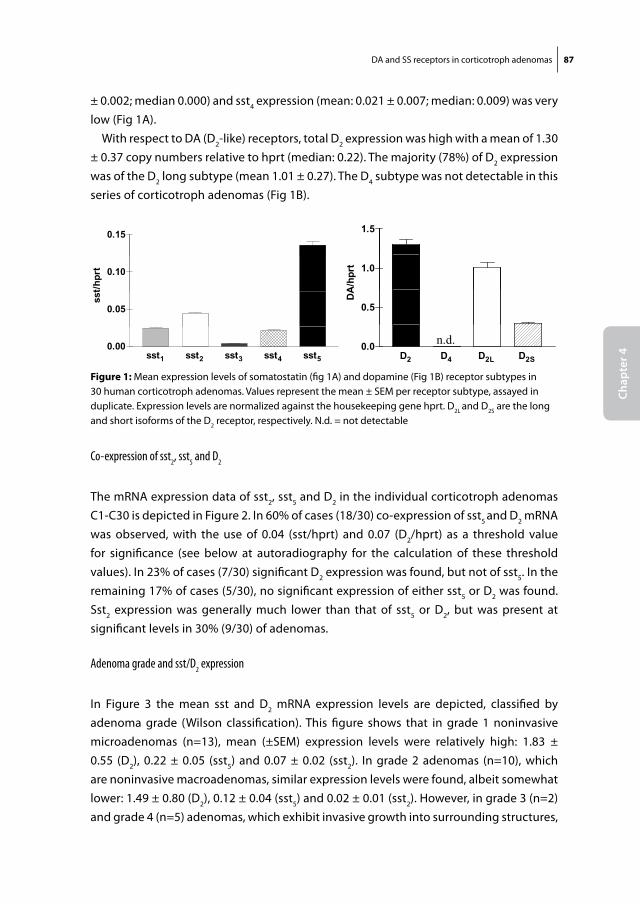
DA and SS receptors in corticotroph adenomas 87
Chap
ter 4
± 0.002; median 0.000) and sst4 expression (mean: 0.021 ± 0.007; median: 0.009) was very low (Fig 1A).
With respect to DA (D2-like) receptors, total D2 expression was high with a mean of 1.30 ± 0.37 copy numbers relative to hprt (median: 0.22). The majority (78%) of D2 expression was of the D2 long subtype (mean 1.01 ± 0.27). The D4 subtype was not detectable in this series of corticotroph adenomas (Fig 1B).
Co-expression of sst2, sst5 and D2
The mRNA expression data of sst2, sst5 and D2 in the individual corticotroph adenomas C1-C30 is depicted in Figure 2. In 60% of cases (18/30) co-expression of sst5 and D2 mRNA was observed, with the use of 0.04 (sst/hprt) and 0.07 (D2/hprt) as a threshold value for significance (see below at autoradiography for the calculation of these threshold values). In 23% of cases (7/30) significant D2 expression was found, but not of sst5. In the remaining 17% of cases (5/30), no significant expression of either sst5 or D2 was found. Sst2 expression was generally much lower than that of sst5 or D2, but was present at significant levels in 30% (9/30) of adenomas.
Adenoma grade and sst/D2 expression
In Figure 3 the mean sst and D2 mRNA expression levels are depicted, classified by adenoma grade (Wilson classification). This figure shows that in grade 1 noninvasive microadenomas (n=13), mean (±SEM) expression levels were relatively high: 1.83 ± 0.55 (D2), 0.22 ± 0.05 (sst5) and 0.07 ± 0.02 (sst2). In grade 2 adenomas (n=10), which are noninvasive macroadenomas, similar expression levels were found, albeit somewhat lower: 1.49 ± 0.80 (D2), 0.12 ± 0.04 (sst5) and 0.02 ± 0.01 (sst2). However, in grade 3 (n=2) and grade 4 (n=5) adenomas, which exhibit invasive growth into surrounding structures,
Fig 1A
0.15
0.10
t/hpr
t
0.05
sst
sst1 sst2 sst3 sst4 sst50.00
Fig 1B
1.5
1.0
A/h
prt
0.5
DA
n.d.D2 D4 D2L D2S
0.02 4 2L 2S
Figure 1: Mean expression levels of somatostatin (fig 1A) and dopamine (Fig 1B) receptor subtypes in 30 human corticotroph adenomas. Values represent the mean ± SEM per receptor subtype, assayed in duplicate. Expression levels are normalized against the housekeeping gene hprt. D2L and D2S are the long and short isoforms of the D2 receptor, respectively. N.d. = not detectable
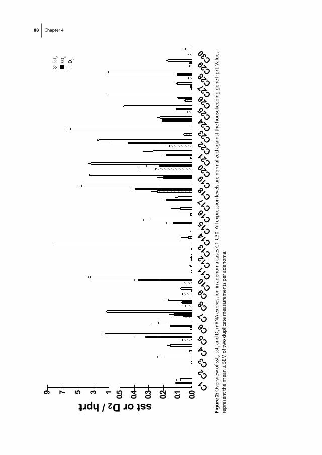
88 Chapter 4
Fig
2
9 57
prt 0.513
D2 / hp 0.30.405 sst or 000.10.2
C1C2C3C4C5C6C7C8C9 C10C11C12C13C14C15C16C17C18C19C20C21C22C23C24C25C26C27C28C29C30
0.0
sst 2
sst 5
D2
Figu
re 2
: Ove
rvie
w o
f sst
2, sst
5 and
D2 m
RNA
exp
ress
ion
in a
deno
ma
case
s C1
-C30
. All
expr
essi
on le
vels
are
nor
mal
ized
aga
inst
the
hous
ekee
ping
gen
e hp
rt. V
alue
s re
pres
ent t
he m
ean
± SE
M o
f tw
o du
plic
ate
mea
sure
men
ts p
er a
deno
ma.
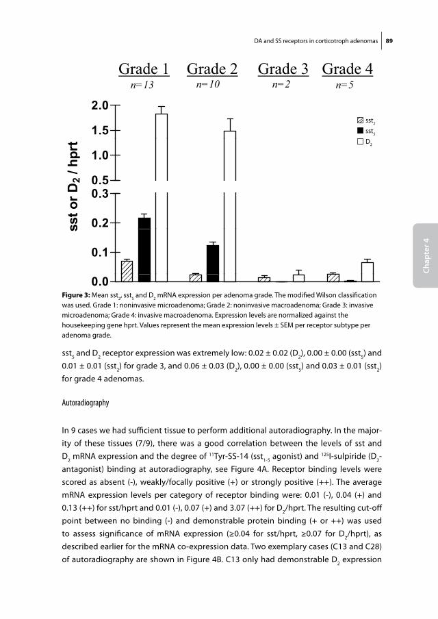
DA and SS receptors in corticotroph adenomas 89
Chap
ter 4
sst5 and D2 receptor expression was extremely low: 0.02 ± 0.02 (D2), 0.00 ± 0.00 (sst5) and 0.01 ± 0.01 (sst2) for grade 3, and 0.06 ± 0.03 (D2), 0.00 ± 0.00 (sst5) and 0.03 ± 0.01 (sst2) for grade 4 adenomas.
Autoradiography
In 9 cases we had sufficient tissue to perform additional autoradiography. In the major-ity of these tissues (7/9), there was a good correlation between the levels of sst and D2 mRNA expression and the degree of 11Tyr-SS-14 (sst1-5 agonist) and 125I-sulpiride (D2-antagonist) binding at autoradiography, see Figure 4A. Receptor binding levels were scored as absent (-), weakly/focally positive (+) or strongly positive (++). The average mRNA expression levels per category of receptor binding were: 0.01 (-), 0.04 (+) and 0.13 (++) for sst/hprt and 0.01 (-), 0.07 (+) and 3.07 (++) for D2/hprt. The resulting cut-off point between no binding (-) and demonstrable protein binding (+ or ++) was used to assess significance of mRNA expression (≥0.04 for sst/hprt, ≥0.07 for D2/hprt), as described earlier for the mRNA co-expression data. Two exemplary cases (C13 and C28) of autoradiography are shown in Figure 4B. C13 only had demonstrable D2 expression
Fig 3
Grade 1 Grade 2 Grade 3 Grade 4n=13 n=10 n=2 n=5
1.5
2.0
0.5
1.0
D2 /
hpr
t
0.2
0.30.5
sst o
r D
0.1
s
0.0
sst2
sst5
D2
Figure 3: Mean sst2, sst5 and D2 mRNA expression per adenoma grade. The modified Wilson classification was used. Grade 1: noninvasive microadenoma; Grade 2: noninvasive macroadenoma; Grade 3: invasive microadenoma; Grade 4: invasive macroadenoma. Expression levels are normalized against the housekeeping gene hprt. Values represent the mean expression levels ± SEM per receptor subtype per adenoma grade.
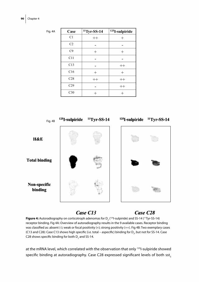
90 Chapter 4
at the mRNA level, which correlated with the observation that only 125I-sulpiride showed specific binding at autoradiography. Case C28 expressed significant levels of both sst5
Fig 4A
Case 11Tyr-SS-14 125I-sulpirideC1 ++ +C2 - -C9 + +C9 + +C11 - -C13 - ++C16 + +C28 ++ ++C29 ++C29 - ++C30 + +
Fig 4B11Tyr-SS-14 11Tyr-SS-14125I-sulpiride 125I-sulpiride11Tyr-SS-14 11Tyr-SS-14125I-sulpiride 125I-sulpiride
H&EH&E
Total bindingTotal binding
Non-specificbinding
Non-specificbinding
Case C13 Case C28Case C13 Case C28Figure 4: Autoradiography on corticotroph adenomas for D2 (125I-sulpiride) and SS-14 (11Tyr-SS-14) receptor binding. Fig 4A: Overview of autoradiography results in the 9 available cases. Receptor binding was classified as: absent (-); weak or focal positivity (+); strong positivity (++). Fig 4B: Two exemplary cases (C13 and C28): Case C13 shows high specific (i.e. total – aspecific) binding for D2, but not for SS-14. Case C28 shows specific binding for both D2 and SS-14.
Fig. 4A
Fig. 4B
DA and SS receptors in corticotroph adenomas 91
Chap
ter 4
and D2 mRNA, which was in line with the specific binding of both 11Tyr-SS-14 and 125I-sulpiride.
Correlation analyses
First, we performed univariate correlation analyses between the expression levels of the individual receptors (sst2, sst5 and D2). This revealed a positive correlation between the expression of all three receptor subtypes; the correlation was especially high for sst5 and D2. Spearman (non-parametric) correlation coefficients were: r = 0.384 (p=0.036) for sst2 and sst5; r = 0.377 (p=0.040) for sst2 and D2; r = 0.675 (p<0.001) for sst5 and D2.
Subsequently, we analyzed the correlation between the expression levels of the indi-vidual receptors and the principal biochemical parameters of hypercortisolism (i.e. 24 hr UFC, mean diurnal serum cortisol and cortisol diurnal rhythm) and responses to the CRH-test and low-and high-dose dexamethasone suppression tests. None of these vari-ables were significantly associated with the expression of any of the receptor subtypes. Moreover, after categorizing these nominal variables into tertiles to limit the effect of data heterogeneity, no significant associations could be identified.
DisCussion
This study investigated the (co-)expression of somatostatin and dopamine receptor subtypes in a series of 30 pure human corticotroph adenomas. We found that D2 and sst5 receptors are expressed in the majority of these adenomas and that co-expression of these receptor subtypes is a common phenomenon. Our data indicate that approxi-mately 80% of corticotroph adenomas obtained from CD patients express one or more of these molecular targets, which could make them suitable candidates for D2 and/or sst5-targeted medical therapy.
In this series, we find that approximately 60% of CD patients have adenomas that sig-nificantly express sst5 receptors. In a recent study, monotherapy with pasireotide (high sst5 affinity) decreased urinary free cortisol (UFC) levels in 76% (22/29) of patients after 15 days of treatment (8). Complete normalization of UFC occurred in only 17% (5/29) of these patients (8). It needs to be emphasized, however, that this was a short trial and that the maximal cortisol lowering effects of pasireotide may require a longer duration of treatment in some patients. From this perspective, the percentage of complete respond-ers found in this trial may still be an underestimation of the total number of responders with prolonged sst5-directed therapy. On the other hand, different mechanisms of tac-
92 Chapter 4
hyphylaxis have been described with the use of SS-analogues in the treatment of some, but not all neuro-endocrine tumours (17). Future studies will have to show whether this process of tachyphylaxis could affect the long-term efficacy of pasireotide therapy in hu-man CD. An important safety concern with the use of this drug in CD is the deterioration of glucose tolerance, which is observed in approximately one third of patients (8).
The vast majority of adenomas, more than 80% of cases, clearly expressed D2 recep-tors, which is similar to what has been found previously in another study by different analytical methods (4). These percentages are comparable, albeit somewhat higher than the percentage of initial responders (both complete and partial) observed in vivo with cabergoline monotherapy, which varies between 51 and 75% (4, 5, 18). In these studies, patients were treated considerably longer (3-6 months) than in the abovementioned 15-day pasireotide trial before response was assessed. Importantly, two of these studies also demonstrated that some patients on cabergoline monotherapy show a relapse after having been in full remission, which reduces long-term remission rates to 27-40% after approximately two years of treatment (5, 18). The mechanisms that cause these escapes from cabergoline monotherapy are not known. Receptor down-regulation or various post-receptor desensitization mechanisms may be involved in this process. Regardless of the cause, these treatment escapes pose a great challenge to physicians managing CD patients and limit the efficacy of cabergoline monotherapy as a long-term treatment option for human CD.
The observation that the majority of corticotroph adenomas in our study expressed both D2 and sst5 receptors suggests the possibility of combined targeting of these receptor subtypes. It has been shown that different sst and DA receptor subtypes can heterodi-merize at the plasma membrane when they are activated simultaneously, leading to enhanced cAMP inhibition and functional synergism (9, 19). Moreover, co-activation of sst and DA receptor subtypes by chimeric dopastatin molecules may further enhance inhibition of hormone release by pituitary adenomas in vitro (20, 21). These studies all suggest potentially beneficial effects of combined sst5 and D2 targeting therapy, but clinical studies are needed to show whether this concept also translates into increased normalization rates in patients with CD.
An important issue from a clinical perspective is that we found a very low level of expres-sion of sst5 and D2 in the corticotroph adenomas with invasive growth (Wilson grade III and IV). Whether this is due to loss of receptor expression in the course of dedifferentia-tion of these adenomas or whether low expression levels are causally related to extensive corticotroph adenoma growth, remains to be elucidated. Nonetheless, this finding could have clinical consequences, as not all subgroups of CD patients have the same likelihood
DA and SS receptors in corticotroph adenomas 93
Chap
ter 4
of receiving medical therapy at some point. It is well known that neurosurgical cure rates in patients with large corticotroph macroadenomas or those with invasive growth into structures surrounding the pituitary fossa, are decreased due to incomplete tumor removal (22). Therefore, these patients often require effective secondary therapies to achieve biochemical remission. Unfortunately, in the light of our findings, sst5 and/or D2-targeting agents are unlikely to be effective in this subgroup of patients.
It needs to be emphasized, however, that the group of corticotroph adenomas in-
cluded in the present study can not be regarded as a representative sample for the total group of corticotroph adenomas as they would occur in clinical practice. Due to the problem of limited tissue availability in the case of corticotroph microadenomas, our data set contains a relatively large number of macroadenomas for which adenomatous tissue was more readily available. Moreover, a significant number of microadenomas that were initially included in this study, turned out to be a mixture of adenoma and surrounding normal pituitary tissue, as expressed by a GH/POMC ratio similar to that in normal anterior pituitaries and had to be excluded from further analysis. For that reason, our data set contains an overrepresentation of corticotroph adenomas in the higher Wilson classification grades (≥2). In clinical practice, it is estimated that macroadenomas and/or invasive corticotroph adenomas represent less than 10-20% of the total num-ber of CD cases, whereas in our study 57% of cases belonged to this category (23, 24). Consequently, the true percentage of CD patients with grade 3 or 4 invasive adenomas and overall low sst/D2 expression levels, will certainly be lower than the 23% identified in this study. In addition, it is important to note that microadenomas and noninvasive macroadenomas (grade 1 and 2 adenomas, respectively), which represent the vast majority of corticotroph adenomas, were found to have a very similar sst and DA recep-tor expression pattern. Therefore, our main findings in this study could be regarded as representative for both grade 1 and grade 2 corticotroph adenomas alike.
Previous in vitro studies have shown that glucocorticoids may directly down-regulate sst2 expression in human and murine neuro-endocrine cell lines, whereas sst5 is sig-nificantly less sensitive and D2 is fully resistant to this type of glucocorticoid-induced down-regulation (25, 26). In this study we did not find a correlation between sst2, sst5 or D2 expression levels and the height of the main biochemical parameters of hypercorti-solism at the time of initial diagnosis, including 24-hour UFC and mean diurnal serum cortisol. The interpretation of the absence of these correlations is difficult, however, as most patients in our study were treated pre-operatively with cortisol-lowering drugs. Therefore, we do not know the exact effects of therapy-induced reductions in cortisol levels on the documented receptor expression levels in the adenomas. One can only hypothesize on the basis of the previous in vitro studies that reductions in cortisol are
94 Chapter 4
likely to have resulted in a relative increase in sst2 expression, rather than to have af-fected sst5 or D2 expression levels in these adenomas.
In conclusion, we demonstrate that the vast majority of human corticotroph adenomas express significant numbers of sst5 and/or D2 receptors, with co-expression of these receptor subtypes being a common phenomenon. These findings support the ongoing clinical studies with agents that target these receptor subtypes in patients with CD. Our results also suggest that combination therapy with both sst5 and D2 targeting agents could be a feasible next step, as this may increase the total number of CD patients that can be biochemically controlled by the use of these agents.
ACKnoWleDgements:
We would like to thank Erik de Blois from the department of Nuclear Medicine, Erasmus Medical Center, for his help with the autoradiography.
DA and SS receptors in corticotroph adenomas 95
Chap
ter 4
reFerenCes
1. newell-Price J, Bertagna X, grossman AB, nieman lK 2006 Cushing’s syndrome. Lancet 367:1605-1617
2. Patil Cg, Prevedello Dm, lad sP, vance ml, thorner mo, Katznelson l, laws er, Jr. 2008 Late recurrences of Cushing’s disease after initial successful transsphenoidal surgery. J Clin Endocrinol Metab 93:358-362
3. morris D, grossman A 2002 The Medical Management of Cushing’s Syndrome. Ann NY Acad Sci 970:119-133
4. Pivonello r, Ferone D, de Herder WW, Kros Jm, De Caro ml, Arvigo m, Annunziato l, lom-bardi g, Colao A, Hofland lJ, lamberts sW 2004 Dopamine receptor expression and function in corticotroph pituitary tumors. J Clin Endocrinol Metab 89:2452-2462
5. Pivonello r, De martino mC, Cappabianca P, de leo m, Faggiano A, lombardi g, Hofland lJ, lamberts sW, Colao A 2008 The medical treatment of Cushing’s disease: effectiveness of chronic treatment with the dopamine agonist cabergoline in patients unsuccessfully treated by surgery. J Clin Endocrinol Metab
6. Hofland lJ, van der Hoek J, Feelders r, van Aken mo, van Koetsveld Pm, Waaijers m, sprij-mooij D, Bruns C, Weckbecker g, de Herder WW, Beckers A, lamberts sW 2005 The multi-ligand somatostatin analogue SOM230 inhibits ACTH secretion by cultured human corticotroph adenomas via somatostatin receptor type 5. Eur J Endocrinol 152:645-654
7. Batista Dl, Zhang X, gejman r, Ansell PJ, Zhou y, Johnson sA, swearingen B, Hedley-Whyte et, stratakis CA, Klibanski A 2006 The effects of SOM230 on cell proliferation and adrenocorti-cotropin secretion in human corticotroph pituitary adenomas. J Clin Endocrinol Metab 91:4482-4488
8. Boscaro m, ludlam WH, Atkinson B, glusman Je, Petersenn s, reincke m, snyder P, tabarin A, Biller Bm, Findling J, melmed s, Darby CH, Hu K, Wang y, Freda Pu, grossman AB, Frohman lA, Bertherat J 2008 Treatment of pituitary dependent Cushing’s disease with the multi-receptor ligand somatostatin analog pasireotide (SOM230): A multicenter, phase II trial. J Clin Endocrinol Metab
9. rocheville m, lange DC, Kumar u, Patel sC, Patel rC, Patel yC 2000 Receptors for dopamine and somatostatin: formation of hetero-oligomers with enhanced functional activity. Science 288:154-157
10. saveanu A, Jaquet P 2008 Somatostatin-dopamine ligands in the treatment of pituitary ad-enomas. Rev Endocr Metab Disord
11. de Bruin C, Hanson Jm, meij BP, Kooistra Hs, Waaijers Am, uitterlinden P, lamberts sW, Hofland lJ 2008 Expression and functional analysis of dopamine receptor subtype 2 and soma-tostatin receptor subtypes in canine cushing’s disease. Endocrinology 149:4357-4366
12. Hofland lJ, van der Hoek J, van Koetsveld Pm, de Herder WW, Waaijers m, sprij-mooij D, Bruns C, Weckbecker g, Feelders r, van der lely AJ, Beckers A, lamberts sW 2004 The novel somatostatin analog SOM230 is a potent inhibitor of hormone release by growth hormone- and prolactin-secreting pituitary adenomas in vitro. J Clin Endocrinol Metab 89:1577-1585
13. rasmussen r 2001 Quantification on the LightCycler. In: Meuer S, Wittwer C and Nagakawara K, eds. Rapid cycle real-time PCR, methods and applications. Heidelberg: Springer Press; 21-34.
14. Pfaffl mW 2001 A new mathematical model for relative quantification in real-time RT-PCR. Nucleic Acids Res 29:e45
96 Chapter 4
15. Hofland lJ, liu Q, van Koetsveld Pm, Zuijderwijk J, van Der Ham F, De Krijger rr, schonb-runn A, lamberts sW 1999 Immunohistochemical detection of somatostatin receptor subtypes sst1 and sst2A in human somatostatin receptor positive tumors. J Clin Endocrinol Metab 84:775-780
16. greenman y, ouaknine g, veshchev i, reider g, ii, segev y, stern n 2003 Postoperative sur-veillance of clinically nonfunctioning pituitary macroadenomas: markers of tumour quiescence and regrowth. Clin Endocrinol (Oxf ) 58:763-769
17. Hofland lJ, lamberts sW 2003 The pathophysiological consequences of somatostatin receptor internalization and resistance. Endocr Rev 24:28-47
18. godbout A, manavela m, Danilowicz K, Beauregard H, Bruno o, lacroix A 2008 Long-term therapy with cabergoline in Cushing’s disease. The Endocrine Society’s 90th annual meeting, June 15-18, San Francisco, U.S.A., 2008
19. Baragli A, Alturaihi H, Watt Hl, Abdallah A, Kumar u 2007 Heterooligomerization of human dopamine receptor 2 and somatostatin receptor 2 Co-immunoprecipitation and fluorescence resonance energy transfer analysis. Cell Signal 19:2304-2316
20. saveanu A, lavaque e, gunz g, Barlier A, Kim s, taylor Je, Culler mD, enjalbert A, Jaquet P 2002 Demonstration of enhanced potency of a chimeric somatostatin-dopamine molecule, BIM-23A387, in suppressing growth hormone and prolactin secretion from human pituitary somatotroph adenoma cells. J Clin Endocrinol Metab 87:5545-5552
21. ren sg, Kim s, taylor J, Dong J, moreau JP, Culler mD, melmed s 2003 Suppression of rat and human growth hormone and prolactin secretion by a novel somatostatin/dopaminergic chimeric ligand. J Clin Endocrinol Metab 88:5414-5421
22. Blevins ls, Jr., Christy JH, Khajavi m, tindall gt 1998 Outcomes of Therapy for Cushing’s Dis-ease due to Adrenocorticotropin-Secreting Pituitary Macroadenomas 10.1210/jc.83.1.63. J Clin Endocrinol Metab 83:63-67
23. Katznelson l, Bogan Js, trob Jr, schoenfeld DA, Hedley-Whyte et, Hsu DW, Zervas nt, swearingen B, sleeper m, Klibanski A 1998 Biochemical assessment of Cushing’s disease in patients with corticotroph macroadenomas. J Clin Endocrinol Metab 83:1619-1623
24. Bochicchio D, losa m, Buchfelder m 1995 Factors influencing the immediate and late outcome of Cushing’s disease treated by transsphenoidal surgery: a retrospective study by the European Cushing’s Disease Survey Group. J Clin Endocrinol Metab 80:3114-3120
25. van der Hoek J, Waaijers m, van Koetsveld Pm, sprij-mooij D, Feelders rA, schmid HA, schoeffter P, Hoyer D, Cervia D, taylor Je, Culler mD, lamberts sW, Hofland lJ 2005 Distinct functional properties of native somatostatin receptor subtype 5 compared with subtype 2 in the regulation of ACTH release by corticotroph tumor cells. Am J Physiol Endocrinol Metab 289:E278-287
26. de Bruin C, Feelders r, Waaijers m, van Koetsveld P, sprij-mooij D, lamberts s, Hofland l 2008 Differential regulation of human dopamine D2 and somatostatin receptor subtype expres-sion by glucocorticoids in vitro. J Mol Endocrinol
Chapter 5
Early clinical experiences with combined somatostatin analogue,
dopamine agonist and/or low-dose ketoconazole therapy in
human Cushing’s diseaseManuscript in preparation

Early clinical experiences with sst/DA therapy 99
Chap
ter 5
introDuCtion
Recent studies show that complete normalization of urinary free cortisol levels occurs in approximately 40% of CD patients after 3-24 months of cabergoline monotherapy and in 17% of CD patients after 15 days of high-dose (2x600μg) pasireotide (SOM230) therapy (1-4). Whether or not the responders in these separate studies show a high degree of overlap or whether they constitute two different groups is not known. Either way, medical combination therapy with dopamine (DA) agonists and novel somatostatin (SS) analogues with high sst5 affinity appears to be a feasible approach (5). In theory, SS-DA combination therapy could increase the total number of (complete) respond-ers to this type of medical therapy. Hypothetically, it could also help in maintaining long-term remission rates by reducing the risk of tachyphylaxis, which can be observed under cabergoline monotherapy in human CD (2, 4). Moreover, in vitro studies using corticotroph tumour cells have shown that ketoconazole, which is used in many centers as monotherapy during the pre-operative phase to decrease cortisol levels, may display functional synergism with pasireotide in lowering ACTH secretion (Feelders et al., per-sonal communication). Whether this also occurs in vivo has not been investigated until now.
For the above reasons, we are currently conducting a phase II open-label multi-center clinical trial to assess the efficacy and safety of a novel stepwise medical treatment ap-proach with the use of the multiligand somatostatin analogue pasireotide (SOM230) with high sst5 affinity in either a low-dose (3x100μg) or a higher dose (3x250μg), the dopamine D2 agonist cabergoline and low-dose ketoconazole in patients with de novo or recurrent CD. The primary aim of this ongoing trial is to identify which percentage of CD patients can be biochemically normalized within a period of three months by the use of this treatment regimen. In this chapter we briefly describe four different patients that were included in this trial, who differ clearly in terms of disease presentation, adenoma characteristics and ultimate treatment outcome. The responses in these patients are discussed here to illustrate patient heterogeneity and as an extension of our in vitro findings in CD that were described in previous chapters. The complete details of this study and its results will be presented elsewhere.
CAse rePorts
Patient 1
Patient 1 is a 42-year-old female with de novo CD due to a pituitary microadenoma (9 mm). Main complaints consisted of severe obesity (135 kg, BMI 44.5 kg/m2), uncon-
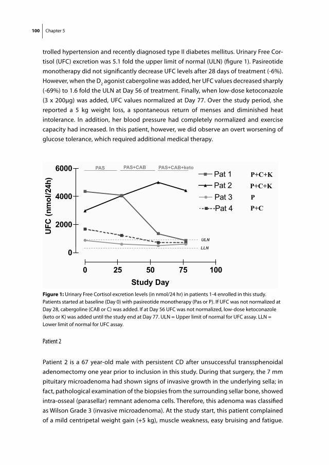
100 Chapter 5
trolled hypertension and recently diagnosed type II diabetes mellitus. Urinary Free Cor-tisol (UFC) excretion was 5.1 fold the upper limit of normal (ULN) (figure 1). Pasireotide monotherapy did not significantly decrease UFC levels after 28 days of treatment (-6%). However, when the D2 agonist cabergoline was added, her UFC values decreased sharply (-69%) to 1.6 fold the ULN at Day 56 of treatment. Finally, when low-dose ketoconazole (3 x 200μg) was added, UFC values normalized at Day 77. Over the study period, she reported a 5 kg weight loss, a spontaneous return of menses and diminished heat intolerance. In addition, her blood pressure had completely normalized and exercise capacity had increased. In this patient, however, we did observe an overt worsening of glucose tolerance, which required additional medical therapy.
Patient 2
Patient 2 is a 67 year-old male with persistent CD after unsuccessful transsphenoidal adenomectomy one year prior to inclusion in this study. During that surgery, the 7 mm pituitary microadenoma had shown signs of invasive growth in the underlying sella; in fact, pathological examination of the biopsies from the surrounding sellar bone, showed intra-osseal (parasellar) remnant adenoma cells. Therefore, this adenoma was classified as Wilson Grade 3 (invasive microadenoma). At the study start, this patient complained of a mild centripetal weight gain (+5 kg), muscle weakness, easy bruising and fatigue.
UFC 24 hr
6000 PAS PAS+CAB PAS+CAB+keto
4000
6000Pat 1Pat 2
/24h
)
PAS PAS+CAB PAS+CAB+ketoP+C+K
P+C+K4000 Pat 3
Pat 4
C (n
mol
/ PP+C
2000
UFC
ULN
LLN
0 25 50 75 100
0LLN
Study DayFigure 1: Urinary Free Cortisol excretion levels (in nmol/24 hr) in patients 1-4 enrolled in this study. Patients started at baseline (Day 0) with pasireotide monotherapy (Pas or P). If UFC was not normalized at Day 28, cabergoline (CAB or C) was added. If at Day 56 UFC was not normalized, low-dose ketoconazole (keto or K) was added until the study end at Day 77. ULN = Upper limit of normal for UFC assay. LLN = Lower limit of normal for UFC assay.
Early clinical experiences with sst/DA therapy 101
Chap
ter 5
At baseline, he was already receiving treatment for hypertension, osteoporosis and hy-percholesterolemia. On pasireotide low and high-dose monotherapy, his UFC excretion did not decrease, but rather increased over time (figure 1). Also after the addition of cabergoline, UFC levels continued to rise. Only when low-dose ketoconazole was added, UFC excretion decreased slightly, but did not normalize. At the study end, the patient did not report any symptomatic improvements; neither did his blood pressure or labora-tory values ameliorate.
Patient 3
Patient 3 is a 65-year old woman with de novo CD due to an invisible pituitary microad-enoma, which was suggested by inferior sinus petrosus sampling (IPSS). Biochemical parameters of hypercortisolism were mildly elevated, but over the years had gradually resulted in increased abdominal size, 20 kg weight gain and progressive fatigue. On low-dose pasireotide monotherapy her UFC levels decreased slightly but did not normalize. On high-dose pasireotide therapy, however, UFC levels completely normalized and remained below the ULN throughout the entire study period (figure 1). She lost 3 kg in weight and reported some minor symptom improvements.
Patient 4
Patient 4 is a 43-year old woman with progressive symptoms of central obesity, easy bruising, hirsutism and amenorrhea. Her de novo CD was due to an invisible pituitary adenoma, which was proven by IPSS. Low- and high dose pasireotide monotherapy decreased her 24 hr UFC excretion slightly, but not sufficient (figure 1). After addition of cabergoline, however, UFC values completely normalized and remained in the mid-normal range until the end of the study period. She reported a marked improvement of her emotional state and exercise capacity, a spontaneous return of menses and a decrease in symptoms of hirsutism.
DisCussion
We here describe four patients that were included in an ongoing phase II, open-label multi-center clinical study that investigates the feasibility of a stepwise medical treat-ment regimen that combines the SS-analogue pasireotide with high sst5 affinity, the D2 agonist cabergoline and/or low-dose ketoconazole in patients with de novo or recurrent CD. As this is an ongoing clinical trial, final results of this study will be presented in detail elsewhere. However, these four patients are illustrative for the wide spectrum of treat-
102 Chapter 5
ment responses, which can be observed with SS and/or DA-targeted medical therapy for this deleterious endocrine disease. Responses varied from partial or complete re-sponsiveness to SS-analogue monotherapy or combined SS/DA analogue treatment, to a total lack of response to these analogues or their combination.
The reasons behind this large variability in treatment responses are multiple, but the most important factor appears to be tumor heterogeneity. As already described in chapter 4 of this thesis and also in previous publications, significant differences exist in somatostatin and dopamine receptor subtype expression levels in series of human cor-ticotroph adenomas (1, 6, 7). Preferably, one would be able to correlate clinical outcome data with the corresponding in vitro data of adenomas of individual patients. In two out of four of the patients that we have described above, we were able to do so (patients 1 and 2).
Patient 1 had newly diagnosed CD when she entered the study and underwent trans-sphenoidal surgery approximately five months after completion of the study. In the time period between the study end and transsphenoidal surgery, she opted to continue the study drug regimen (extension study), which kept the UFC levels well within the normal range and further ameliorated her symptoms and signs of CD. When she underwent transsphenoidal surgery, we freshly isolated a part of the operative specimen and performed quantitative PCR for sst and DA receptor expression. The results are shown in figure 2. It shows she has clearly demonstrable sst2, sst5 and D2 receptor expression, which is partially in line with her clinical response observed in the trial. The data are supportive of a D2 mediated cabergoline effect, but do no fully explain why she did not respond better to sst-targeted monotherapy (pasireotide) in the first 28 days of the study.
Two issues need to be addressed in this respect. First of all, receptor status was assessed after 8 months of successful, glucocorticoid-lowering therapy in this patient. As has been described by many different studies and also in chapter 3 of this thesis, glucocorticoids can directly influence expression levels of sst in human neuro-endocrine cells, while for D2 such an effect can not be demonstrated to the same extent or may even be absent (8-12). Especially sst2 expression is sensitive for glucocorticoid-induced down-regulation and may be restored after correction of the hypercortisolism. Therefore, it is likely that the long-term normocortisolemia induced in this patient by the study drug regimen will have positively affected tumoral sst2 expression levels in this patient. Therefore, measured sst2 levels as shown in figure 1, may be an overestimation of tumoral sst2 levels when the patient first entered the study. This may explain the absence of a therapeutic effect after 28 days of treatment by SS-multiligand pasireotide in this patient, even though sst2 levels were well demonstrable after 8 months of therapy.
Another aspect could be the onset of rapid tachyphylaxis after the start of pasireotide monotherapy in patients with adenomas that are susceptible for such a phenomenon.
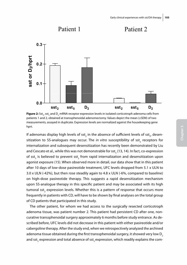
Early clinical experiences with sst/DA therapy 103
Chap
ter 5
If adenomas display high levels of sst2 in the absence of sufficient levels of sst5, desen-sitization to SS-analogues may occur. The in vitro susceptibility of sst2 receptors for internalization and subsequent desensitization has recently been demonstrated by Liu and Cescato et al., while this was not demonstrable for sst5 (13, 14). In fact, co-expression of sst5 is believed to prevent sst2 from rapid internalization and desensitization upon agonist exposure (15). When observed more in detail, our data show that in this patient after 10 days of low-dose pasireotide treatment, UFC levels dropped from 5.1 x ULN to 3.0 x ULN (-42%), but then rose steadily again to 4.8 x ULN (-6%, compared to baseline) on high-dose pasireotide therapy. This suggests a rapid desensitization mechanism upon SS-analogue therapy in this specific patient and may be associated with its high tumoral sst2 expression levels. Whether this is a pattern of response that occurs more frequently in patients with CD, will have to be shown by final analyses on the total group of CD patients that participated in this study.
The other patient, for whom we had access to the surgically resected corticotroph adenoma tissue, was patient number 2. This patient had persistent CD after one, non-curative transsphenoidal surgery approximately 6 months before study entrance. As de-scribed before, UFC levels did not decrease in this patient with either pasireotide and/or cabergoline therapy. After the study end, when we retrospectively analyzed the archived adenoma tissue obtained during the first transsphenoidal surgery, it showed very low D2 and sst2 expression and total absence of sst5 expression, which readily explains the com-
Patient 1 Patient 2
0.3
t
0.2
r D
2/hp
rt
0.1sst o
r
sst2 sst5 D2 sst2 sst5 D20.0
Figure 2: Sst2, sst5 and D2 mRNA receptor expression levels in isolated corticotroph adenoma cells from patients 1 and 2, obtained at transsphenoidal adenomectomy. Values depict the mean (±SEM) of two measurements, assayed in duplicate. Expression levels are normalized against the housekeeping gene hprt.
104 Chapter 5
plete lack of response in this patient to either SS-analogue and/or DA-agonist therapy. This specific expression pattern is unusual for corticotroph adenomas in general, but may be observed more frequently in advanced corticotroph adenoma stages (stages III and IV, see chapter 4 of this thesis (16)). In fact, the adenoma of this patient had been classified as a stage III adenoma, due to its invasive growth in the underlying sella and the presence of intra-osseal adenoma cells in the parasellar bones.
In summary, we have here described four patients in an ongoing clinical trial, which exhibit different responses to SS-analogue and/or dopamine agonist therapy, which could be correlated at least partially to their adenoma sst and D2 receptor status. Defini-tive results of this ongoing study, including correlation between treatment responses and in vitro receptor expression data, will be presented elsewhere.
Early clinical experiences with sst/DA therapy 105
Chap
ter 5
reFerenCes
1. Pivonello r, Ferone D, de Herder WW, Kros Jm, De Caro ml, Arvigo m, Annunziato l, lom-bardi g, Colao A, Hofland lJ, lamberts sW 2004 Dopamine receptor expression and function in corticotroph pituitary tumors. J Clin Endocrinol Metab 89:2452-2462
2. godbout A mm, Danilowicz K, Beauregard H, Bruno o, lacroix A 2008 Long-term therapy with cabergoline in Cushing’s disease. The Endocrine Society’s 90th annual meeting, June 15-18, San Francisco, U.S.A., 2008
3. Boscaro m, ludlam WH, Atkinson B, glusman Je, Petersenn s, reincke m, snyder P, tabarin A, Biller Bm, Findling J, melmed s, Darby CH, Hu K, Wang y, Freda Pu, grossman AB, Frohman lA, Bertherat J 2009 Treatment of Pituitary-Dependent Cushing’s Disease with the Multireceptor Ligand Somatostatin Analog Pasireotide (SOM230): A Multicenter, Phase II Trial. J Clin Endocrinol Metab 94:115-122
4. Pivonello r, De martino mC, Cappabianca P, De leo m, Faggiano A, lombardi g, Hofland lJ, lamberts sW, Colao A 2009 The Medical Treatment of Cushing’s Disease: Effectiveness of Chronic Treatment with the Dopamine Agonist Cabergoline in Patients Unsuccessfully Treated by Surgery. J Clin Endocrinol Metab 94:223-230
5. Colao A, Filippella m, Pivonello r, Di somma C, Faggiano A, lombardi g 2007 Combined therapy of somatostatin analogues and dopamine agonists in the treatment of pituitary tumours. Eur J Endocrinol 156 Suppl 1:S57-63
6. Hofland lJ, van der Hoek J, Feelders r, van Aken mo, van Koetsveld Pm, Waaijers m, sprij-mooij D, Bruns C, Weckbecker g, de Herder WW, Beckers A, lamberts sW 2005 The multi-ligand somatostatin analogue SOM230 inhibits ACTH secretion by cultured human corticotroph adenomas via somatostatin receptor type 5. Eur J Endocrinol 152:645-654
7. Batista Dl, Zhang X, gejman r, Ansell PJ, Zhou y, Johnson sA, swearingen B, Hedley-Whyte et, stratakis CA, Klibanski A 2006 The effects of SOM230 on cell proliferation and adrenocorti-cotropin secretion in human corticotroph pituitary adenomas. J Clin Endocrinol Metab 91:4482-4488
8. de Bruin C, Feelders rA, Waaijers Am, van Koetsveld Pm, sprij-mooij Dm, lamberts sW, Ho-fland lJ 2009 Differential regulation of human dopamine D2 and somatostatin receptor subtype expression by glucocorticoids in vitro. J Mol Endocrinol 42:47-56
9. van der Hoek J, Waaijers m, van Koetsveld Pm, sprij-mooij D, Feelders rA, schmid HA, schoeffter P, Hoyer D, Cervia D, taylor Je, Culler mD, lamberts sW, Hofland lJ 2005 Distinct functional properties of native somatostatin receptor subtype 5 compared with subtype 2 in the regulation of ACTH release by corticotroph tumor cells. Am J Physiol Endocrinol Metab 289:E278-287
10. Hofland lJ 2008 Somatostatin and somatostatin receptors in Cushing’s disease. Mol Cell Endo-crinol 286:199-205
11. Park s, Kamegai J, Kineman rD 2003 Role of glucocorticoids in the regulation of pituitary somatostatin receptor subtype (sst1-sst5) mRNA levels: evidence for direct and somatostatin-mediated effects. Neuroendocrinology 78:163-175
12. Xu y, Berelowitz m, Bruno JF 1995 Dexamethasone regulates somatostatin receptor subtype messenger ribonucleic acid expression in rat pituitary GH4C1 cells. Endocrinology 136:5070-5075

106 Chapter 5
13. Cescato r, schulz s, Waser B, eltschinger v, rivier Je, Wester HJ, Culler m, ginj m, liu Q, schonbrunn A, reubi JC 2006 Internalization of sst2, sst3, and sst5 receptors: effects of soma-tostatin agonists and antagonists. J Nucl Med 47:502-511
14. liu Q, Cescato r, Dewi DA, rivier J, reubi JC, schonbrunn A 2005 Receptor signaling and endocytosis are differentially regulated by somatostatin analogs. Mol Pharmacol 68:90-101
15. sharif n, gendron l, Wowchuk J, sarret P, mazella J, Beaudet A, stroh t 2007 Coexpression of somatostatin receptor subtype 5 affects internalization and trafficking of somatostatin receptor subtype 2. Endocrinology 148:2095-2105
16. de Bruin C, Pereira Am, Feelders rA, romijn JA, roelfsema F, sprij-mooij Dm, van Aken mo, van der lelij AJ, de Herder WW, lamberts sW, Hofland lJ 2009 Co-expression of dopamine and somatostatin receptor subtypes in corticotroph adenomas. J Clin Endocrinol Metab
Chapter 6
Somatostatin receptor expression in a patient with Cushing’s
syndrome due to ectopic adrenocorticotropin secretion after
successful mifepristone therapySubmitted
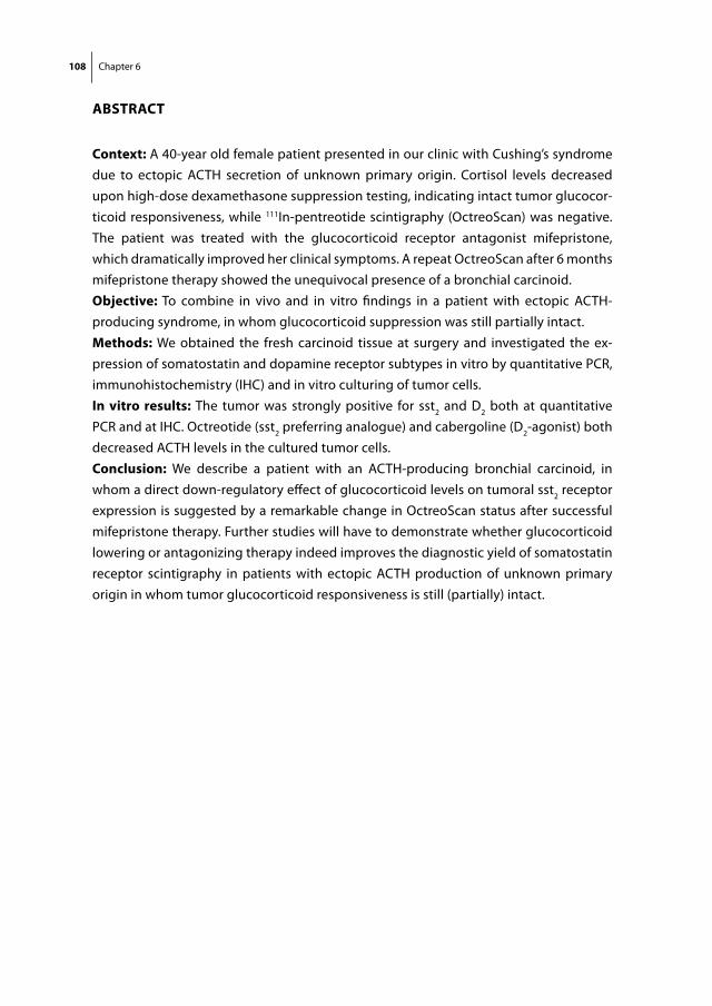
108 Chapter 6
ABstrACt
Context: A 40-year old female patient presented in our clinic with Cushing’s syndrome due to ectopic ACTH secretion of unknown primary origin. Cortisol levels decreased upon high-dose dexamethasone suppression testing, indicating intact tumor glucocor-ticoid responsiveness, while 111In-pentreotide scintigraphy (OctreoScan) was negative. The patient was treated with the glucocorticoid receptor antagonist mifepristone, which dramatically improved her clinical symptoms. A repeat OctreoScan after 6 months mifepristone therapy showed the unequivocal presence of a bronchial carcinoid. objective: To combine in vivo and in vitro findings in a patient with ectopic ACTH-producing syndrome, in whom glucocorticoid suppression was still partially intact.methods: We obtained the fresh carcinoid tissue at surgery and investigated the ex-pression of somatostatin and dopamine receptor subtypes in vitro by quantitative PCR, immunohistochemistry (IHC) and in vitro culturing of tumor cells.in vitro results: The tumor was strongly positive for sst2 and D2 both at quantitative PCR and at IHC. Octreotide (sst2 preferring analogue) and cabergoline (D2-agonist) both decreased ACTH levels in the cultured tumor cells.Conclusion: We describe a patient with an ACTH-producing bronchial carcinoid, in whom a direct down-regulatory effect of glucocorticoid levels on tumoral sst2 receptor expression is suggested by a remarkable change in OctreoScan status after successful mifepristone therapy. Further studies will have to demonstrate whether glucocorticoid lowering or antagonizing therapy indeed improves the diagnostic yield of somatostatin receptor scintigraphy in patients with ectopic ACTH production of unknown primary origin in whom tumor glucocorticoid responsiveness is still (partially) intact.
Sst expression in ectopic Cushing’s syndrome 109
Chap
ter 6
introDuCtion
Ectopic adrenocorticotropin (ACTH) secretion (EAS) by a non-pituitary tumor is a rare cause of ACTH-dependent Cushing’s syndrome (1). It is most frequently caused by either a bronchial carcinoid or a small-cell lung carcinoma, which accounts for approximately 50% of all cases, but also thymic carcinoids, gastro-entero-pancreatic neuro-endocrine tumors and medullary thyroid carcinoma are well-known causes of this syndrome (2, 3). The overall prognosis of the patient is largely determined by the nature of the underly-ing malignancy and the tumor stage at the time of diagnosis (1).
Primary therapy consists of surgical removal of the tumor, but in some EAS cases the pri-mary tumor can not be identified by routine imaging procedures, including ultrasound, CT and MRI. The fact that a significant proportion of these tumors express somatostatin receptors (sst) allows the use of sst-scintigraphy with 111In-pentreotide (OctreoScan) in the diagnostic work-up of these patients. The expression of sst is not only useful from a diagnostic perspective, it can also be used for therapeutic purposes because a signifi-cant number of EAS patients respond to traditional somatostatin (SS) analogues such as octreotide (1, 3-6). Apart from sst, dopamine receptors have also been proposed as potential targets in the medical treatment of these tumors (7).
However, a substantial number of tumors with EAS can not be detected with Octreo-Scan, although the exact percentage remains difficult to establish (6, 8-11). In addition, an important subset of EAS patients does not show any clinical or biochemical response to traditional SS-analogues that mainly target somatostatin receptor subtype 2 (sst2) (1, 3). The reason for this large variability in functional sst2 expression is not clear, but could be due to differences in glucocorticoid (GC) sensitivity of these tumors. It is well known that EAS tumors can differ strongly in their GC-responsiveness, which is reflected by the wide range of results obtained after high dose dexamethasone suppression testing in patients with EAS (12). At the same time, it also known that GCs can directly down-regulate sst expression in human neuro-endocrine tumor cells and more specifically sst2 (13-15). Therefore, in the subset of EAS patients, in whom regulatory feedback by GC is still partially intact, cortisol-lowering or antagonizing therapy could directly influence tumoral sst2 expression levels. If this occurs, one would expect to find evidence for such a mechanism both in vivo (serial OctreoScan imaging), as well as in vitro (analysis of the surgically resected tumor). We here present a case, for which we combined both investigations.
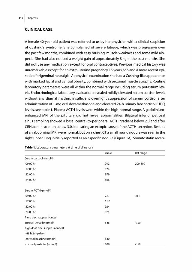
110 Chapter 6
CliniCAl CAse
A female 40-year old patient was referred to us by her physician with a clinical suspicion of Cushing’s syndrome. She complained of severe fatigue, which was progressive over the past few months, combined with easy bruising, muscle weakness and some mild alo-pecia. She had also noticed a weight gain of approximately 8 kg in the past months. She did not use any medication except for oral contraceptives. Previous medical history was unremarkable except for an extra-uterine pregnancy 15 years ago and a more recent epi-sode of trigeminal neuralgia. At physical examination she had a Cushing-like appearance with marked facial and central obesity, combined with proximal muscle atrophy. Routine laboratory parameters were all within the normal range including serum potassium lev-els. Endocrinological laboratory evaluation revealed mildly elevated serum cortisol levels without any diurnal rhythm, insufficient overnight suppression of serum cortisol after administration of 1-mg oral dexamethasone and elevated 24-h urinary free cortisol (UFC) levels, see table 1. Plasma ACTH levels were within the high-normal range. A gadolinium-enhanced MRI of the pituitary did not reveal abnormalities. Bilateral inferior petrosal sinus sampling showed a basal central-to-peripheral ACTH gradient below 2.0 and after CRH administration below 3.0, indicating an ectopic cause of the ACTH-secretion. Results of an abdominal MRI were normal, but on a chest CT a small round nodule was seen in the right upper lung initially reported as an aspecific nodule (Figure 1A). Somatostatin recep-
table 1. Laboratory parameters at time of diagnosis
Value Ref range
Serum cortisol (nmol/l)
09.00 hr 792 200-800
17.00 hr 924
22.00 hr 979
24.00 hr 866
Serum ACTH (pmol/l)
09.00 hr 7.4 <11
17.00 hr 11.0
22.00 hr 9.9
24.00 hr 9.9
1 mg dex. suppressiontest
cortisol 09.00 hr (nmol/l) 646 < 50
high dose dex. suppression test
(48 h 2mg/day)
cortisol baseline (nmol/l) 530
cortisol post-dex (nmol/l) 108 < 50
Sst expression in ectopic Cushing’s syndrome 111
Chap
ter 6
tor scintigraphy with 111In-pentreotide (OctreoScan) did not show pathological uptake at that time (Figure 1B), nor did additional 18F-DOPA-PET and 11C-5-hydroxytryptamin-PET scans. The patient was treated with the glucocorticoid receptor antagonist mifepristone (RU-486, obtained from HRA Pharma, Paris, France) at a dose of 400 to 600 mg/day, which clearly improved the condition of the patient with 23 kg weight loss and disappearance of Cushingoid features. At follow-up, six months after initiating mifepristone treatment, a repeat chest CT showed the same nodule in the right upper lung without signs of ana-tomic growth (Figure 1C). However, at this time a repeat OctreoScan showed a positive uptake at the site of this nodule (Figure 1D and 1E). Subsequently, the patient underwent resection of the right upper lobe of the lung, which revealed a small tumor of 5 mm in diameter. Immediately after resection, the fresh carcinoid tissue was obtained for further analysis in vitro (see methods section). Pathological examination showed a neuroendo-crine tumor with a positive staining for ACTH, synaptophysin and chromogranin A. The patient recovered well post-operatively without any major complications. She received hydrocortisone replacement therapy, which was gradually tapered. At present, 10 months after surgery, she is doing well without any evidence of a recurrence of hypercortisolism. Fig 1
Figure 1: 111In-pentreotide scintigraphy (OctreoScan) and CT imaging results in this patient before (A-B) and after 6 months of therapy with mifepristone (C-E). Before therapy was initiated, CT scan (A) shows a small round nodule in the right upper lung (white arrow), which is not visible at OctreoScan (B). After 6 months of therapy, the CT scan shows the same lesion (white arrow) within the upper lobe of the right lung (C). At that time a repeat OctreoScan shows pathological uptake at the site of the lesion (D and E; black arrows).
112 Chapter 6
in vitro stuDies
Informed consent
These studies were performed according to the rules of the ethics committee of our hospital. Informed consent was obtained from the patient.
Surgical tissue and cell isolation
After resection, the fresh carcinoid tissue was placed in 4˚C Minimal Essential Medium (MEM) with Earle’s salts, supplemented with 10% fetal calf serum (FCS), L-glutamine (2 mmol/l), penicillin (105 U/l) and fungizone (0.25 mg/l). The tissue was dispersed with dispase 103 U/l (Roche, Almere, the Netherlands) + collagenase 2 mg/ml (Sigma Aldrich, Zwijndrecht, the Netherlands) at 37ºC for 1 h to obtain a single cell population. Viable carcinoid cells were counted in a standard haematocytometer. 3 x 10⁵ of the cells were used for qPCR analysis. The remainder of the cells were cultured in 48 well plates (Corn-ing, Cambridge, USA) at a density of 40,000 cells/well for 4-6 days at 37 ˚C in a humidi-fied incubator in 5% CO2. At that time, culture media were refreshed and incubations were started with octreotide (Novartis, Basel, Switzerland), cabergoline (Pfizer, Capelle a/d Ijssel, the Netherlands) or their combination for 96 h. At the end of the incubation period, media were collected and stored at –20˚C for hormone analysis after addition of aprotinin (4 x 10⁵ IU/ml medium; Bayer, Mijdrecht, the Netherlands) to prevent ACTH degradation. All experimental conditions were performed in quadruplicates.
Quantitative PCR
Quantitative PCR for the different SS and DA receptor subtypes was performed accord-ing to a protocol that has been described previously (13, 16). All samples were assayed in duplicate and values were normalized against the expression of the housekeeping gene hprt. Dilution curves were constructed to calculate PCR efficiencies (E) for every primer-probe set (17). Estimated copy numbers were calculated using the comparative threshold method with efficiency correction, as described previously (18).
Immunohistochemistry
The expression of sst2 and D2 in the carcinoid tissue was assessed by cutting cryostat sec-tions (5μm) and performing immunohistochemistry according to a previously published method (19). The polyclonal anti-sst2 antibody (Gramsch laboratories, Schwabshausen,
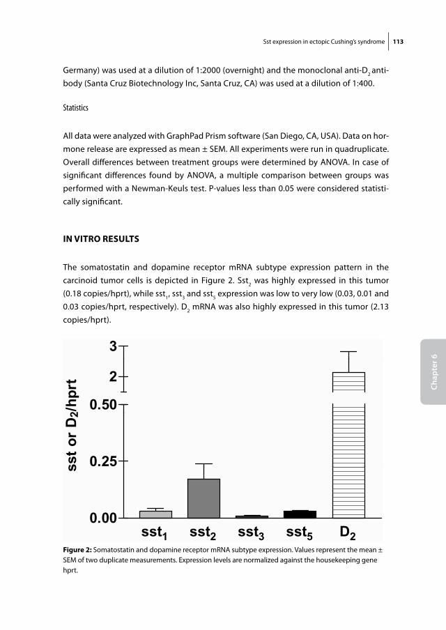
Sst expression in ectopic Cushing’s syndrome 113
Chap
ter 6
Germany) was used at a dilution of 1:2000 (overnight) and the monoclonal anti-D2 anti-body (Santa Cruz Biotechnology Inc, Santa Cruz, CA) was used at a dilution of 1:400.
Statistics
All data were analyzed with GraphPad Prism software (San Diego, CA, USA). Data on hor-mone release are expressed as mean ± SEM. All experiments were run in quadruplicate. Overall differences between treatment groups were determined by ANOVA. In case of significant differences found by ANOVA, a multiple comparison between groups was performed with a Newman-Keuls test. P-values less than 0.05 were considered statisti-cally significant.
in vitro results
The somatostatin and dopamine receptor mRNA subtype expression pattern in the carcinoid tumor cells is depicted in Figure 2. Sst2 was highly expressed in this tumor (0.18 copies/hprt), while sst1, sst3 and sst5 expression was low to very low (0.03, 0.01 and 0.03 copies/hprt, respectively). D2 mRNA was also highly expressed in this tumor (2.13 copies/hprt).
Fig 2
3
0.50
2
2/hp
rt
0.25sst o
r D
2
0.00
s
sst1 sst2 sst3 sst5 D20.00
Figure 2: Somatostatin and dopamine receptor mRNA subtype expression. Values represent the mean ± SEM of two duplicate measurements. Expression levels are normalized against the housekeeping gene hprt.
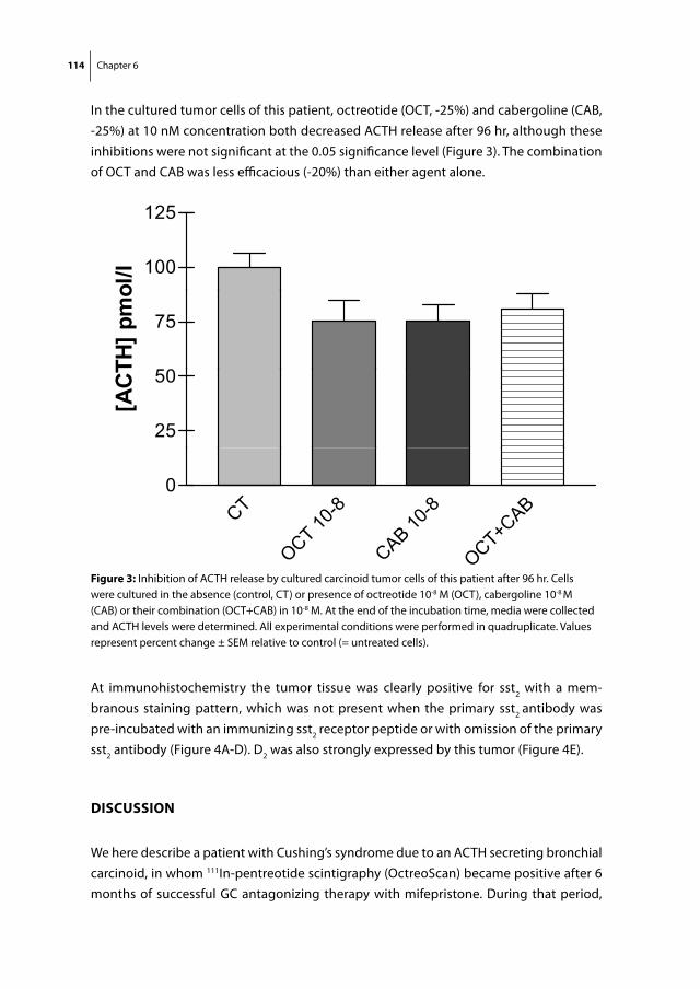
114 Chapter 6
In the cultured tumor cells of this patient, octreotide (OCT, -25%) and cabergoline (CAB, -25%) at 10 nM concentration both decreased ACTH release after 96 hr, although these inhibitions were not significant at the 0.05 significance level (Figure 3). The combination of OCT and CAB was less efficacious (-20%) than either agent alone.
At immunohistochemistry the tumor tissue was clearly positive for sst2 with a mem-branous staining pattern, which was not present when the primary sst2 antibody was pre-incubated with an immunizing sst2 receptor peptide or with omission of the primary sst2 antibody (Figure 4A-D). D2 was also strongly expressed by this tumor (Figure 4E).
DisCussion
We here describe a patient with Cushing’s syndrome due to an ACTH secreting bronchial carcinoid, in whom 111In-pentreotide scintigraphy (OctreoScan) became positive after 6 months of successful GC antagonizing therapy with mifepristone. During that period,
Fig 3
125
100
125
ol/l
50
75
TH] p
mo
25
50
[AC
T
CTT 10
-8
B10
-8+C
AB0
OCT 1CAB 1
OCT+C
Figure 3: Inhibition of ACTH release by cultured carcinoid tumor cells of this patient after 96 hr. Cells were cultured in the absence (control, CT) or presence of octreotide 10-8 M (OCT), cabergoline 10-8 M (CAB) or their combination (OCT+CAB) in 10-8 M. At the end of the incubation time, media were collected and ACTH levels were determined. All experimental conditions were performed in quadruplicate. Values represent percent change ± SEM relative to control (= untreated cells).
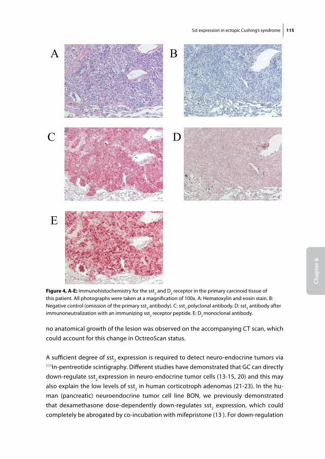
Sst expression in ectopic Cushing’s syndrome 115
Chap
ter 6
no anatomical growth of the lesion was observed on the accompanying CT scan, which could account for this change in OctreoScan status.
A sufficient degree of sst2 expression is required to detect neuro-endocrine tumors via 111In-pentreotide scintigraphy. Different studies have demonstrated that GC can directly down-regulate sst2 expression in neuro-endocrine tumor cells (13-15, 20) and this may also explain the low levels of sst2 in human corticotroph adenomas (21-23). In the hu-man (pancreatic) neuroendocrine tumor cell line BON, we previously demonstrated that dexamethasone dose-dependently down-regulates sst2 expression, which could completely be abrogated by co-incubation with mifepristone (13 ). For down-regulation
Fig 4 A B
C D
E
Figure 4, A-e: Immunohistochemistry for the sst2 and D2 receptor in the primary carcinoid tissue of this patient. All photographs were taken at a magnification of 100x. A: Hematoxylin and eosin stain. B: Negative control (omission of the primary sst2 antibody). C: sst2 polyclonal antibody. D: sst2 antibody after immunoneutralization with an immunizing sst2 receptor peptide. E: D2 monoclonal antibody.
116 Chapter 6
of sst2, however, an intact GC signaling pathway needs to be present within these cells, which is not the case in many of the neuro-endocrine tumors that cause ectopic Cush-ing’s syndrome (24, 25). In our patient, however, the glucocorticoid feedback system was still partially intact, as demonstrated by an absent suppression of serum cortisol on low-dose 1 mg DST, but a strong suppression on high-dose dexamethasone suppression testing (HDDST). This likely explains why after alleviation of the functional hypercorti-solism by mifepristone therapy, sst2 expression levels were restored.
The reported sensitivity of diagnostic OctreoScan in ectopic Cushing’s syndrome is 50% at best in most series (1, 8, 11, 26, 27). It is important to note, however, that most of these data were obtained in patients with untreated ectopic Cushing’s syndrome. As demonstrated by the results in our patient, the diagnostic accuracy of the OctreoScan may be increased after resolution of hypercortisolism in those EAS tumors, in which the GC signaling system is still (partially) intact. This is clinically relevant because in 12 to 18 % of patients the primary cause of EAS can not be localized initially (1), frequently neces-sitating a bilateral adrenalectomy. Future studies should prospectively investigate the diagnostic value of repeated OctreoScan after GC lowering or GC receptor antagonizing therapy.
The appearance of clearly demonstrable levels of sst2 in our patient following mifepris-tone treatment strongly suggests the presence of a dynamic and potentially reversible relationship between GC action and the expression of sst2 within neuro-endocrine tumors, including some cases of EAS. In this specific patient, we could not assess at what point of time the upregulation of sst2 occurred as we only had access to two serial OctreoScans in a 6-month interval. An interesting question would further be whether this visible return of sst2 expression also correlates with a response to treatment with SS analogues resulting in decreased ACTH production. If so, this could have implications for secondary treatment options in EAS patients that are not cured by surgery alone and in whom bilateral adrenalectomy is not feasible or desirable. Temporary pre-treatment with GC lowering (e.g. ketoconazole) or GC receptor antagonizing (mifepristone) therapy could result in a reappearance of sst2 expression and consequently increase the efficacy of sst2-preferring compounds such as Octreotide.
It is important to note that such a proposed mechanism of action only pertains to those cases of EAS, in which some form of GC regulation is still present. It is known that the majority of EAS cases will not suppress serum cortisol levels on HDDST (12). However, it is well conceivable that the minority of EAS cases, that do adequately suppress on HDDST, may constitute an important part of the group of EAS cases that are negative on OctreoScan.
Sst expression in ectopic Cushing’s syndrome 117
Chap
ter 6
In this tumor sst2 was highly expressed: 0.18 copies/hprt. For comparison, we have measured sst2 expression data recently in a large series of corticotroph adenomas with identical analytical methods. In these corticotroph adenomas the mean sst2 expression level was 0.04/hprt (22). EAS tumors can display substantial numbers of sst2 receptors, as demonstrated by the percentage of OctreoScan positive EAS tumors in several series (1, 3, 8, 11). These receptors can also be functional, as some EAS patients display a favour-able response to Octreotide therapy in contrast to patients with CD (4, 28).
The fact that sst2 expression levels tend to be higher in patients with EAS than in patients with CD is not surprising by itself. If EAS tumors are fully insensitive to gluco-corticoid feedback, as demonstrated by absent or insufficient suppression at HDDST, then GC-induced down-regulation of sst2 receptors is not likely to be present. We have investigated this phenomenon previously in vitro, in which the severely glucocorticoid-resistant EAS cell line DMS-79 did not show any signs of sst2 down-regulation, even at the highest dexamethasone doses of 100 nM (13). As stated previously, the majority of EAS tumors will belong to this category, because in approximately 90% of EAS patients serum cortisol levels are not adequately (>50%) suppressed after HDDST (3). This does not pertain, however, to our patient, in whom HDDST did decrease serum cortisol levels (>50%) and in whom a negative OctreoScan was observed at the time of initial diagno-sis. This indicates that some degree of intrinsic GC responsiveness was present in this tumor and that upon reversal of functional hypercortisolism by mifepristone therapy, sst2 expression was restored. It is therefore important to note that the tumoral sst2 levels we have measured in vitro in this individual patient after mifepristone therapy are not necessarily representative for the majority of fully GC resistant EAS tumors in clinical practice.
D2 was also well expressed in this tumor (2.13 copies/hprt), which is in line with the re-ports by Pivonello et al. that these tumors can respond to cabergoline therapy, depend-ing on their D2 receptor status (7). The level of expression in this tumor was in the same order of magnitude as what we observed earlier for corticotroph adenomas (22). The fact that, in contrast to the sst2 receptor, the expression levels between CD and EAS do not differ significantly for D2, can be explained by the fact that the D2 receptor gene does not appear to be as sensitive for GC regulation as sst2 and to a much lesser degree sst5 (13).
For both sst2 and D2, the mRNA expression levels were confirmed by the clear presence of sst2 and D2 at immunohistochemistry of sequential tumor tissue sections. Moreover, the expression data correlated with the inhibitory trend of Octreotide and Cabergoline in the primary cultured ACTH-producing carcinoid cells. The inhibition of ACTH release by cabergoline in vitro confirms earlier reports by Pivonello et al that these tumors can be responsive to D2 agonists. In a study of six patients with EAS, 5/6 of these primary tu-
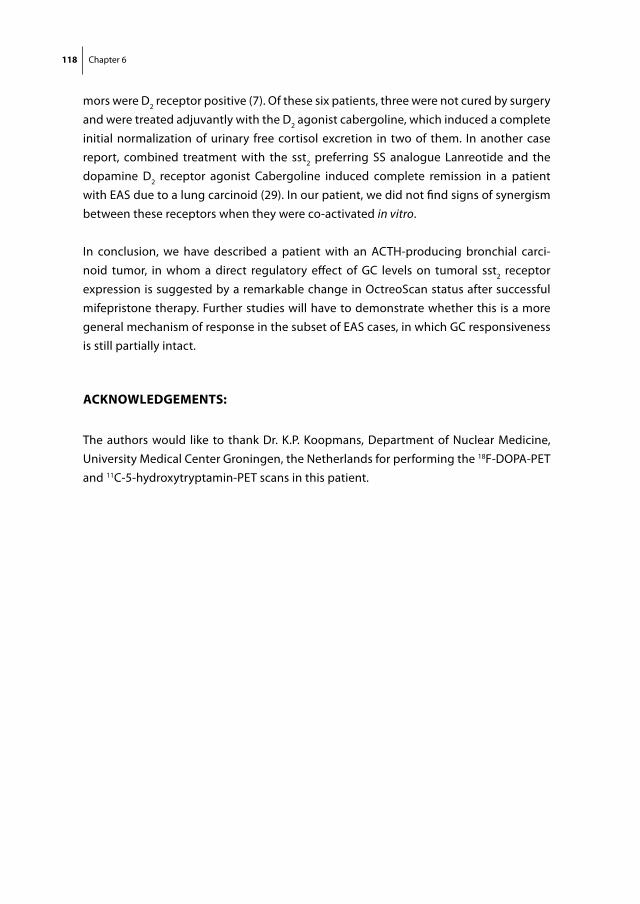
118 Chapter 6
mors were D2 receptor positive (7). Of these six patients, three were not cured by surgery and were treated adjuvantly with the D2 agonist cabergoline, which induced a complete initial normalization of urinary free cortisol excretion in two of them. In another case report, combined treatment with the sst2 preferring SS analogue Lanreotide and the dopamine D2 receptor agonist Cabergoline induced complete remission in a patient with EAS due to a lung carcinoid (29). In our patient, we did not find signs of synergism between these receptors when they were co-activated in vitro.
In conclusion, we have described a patient with an ACTH-producing bronchial carci-noid tumor, in whom a direct regulatory effect of GC levels on tumoral sst2 receptor expression is suggested by a remarkable change in OctreoScan status after successful mifepristone therapy. Further studies will have to demonstrate whether this is a more general mechanism of response in the subset of EAS cases, in which GC responsiveness is still partially intact.
ACKnoWleDgements:
The authors would like to thank Dr. K.P. Koopmans, Department of Nuclear Medicine, University Medical Center Groningen, the Netherlands for performing the 18F-DOPA-PET and 11C-5-hydroxytryptamin-PET scans in this patient.
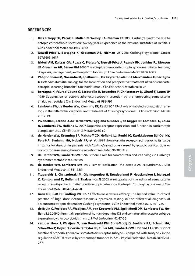
Sst expression in ectopic Cushing’s syndrome 119
Chap
ter 6
reFerenCes
1. ilias i, torpy DJ, Pacak K, mullen n, Wesley rA, nieman lK 2005 Cushing’s syndrome due to ectopic corticotropin secretion: twenty years’ experience at the National Institutes of Health. J Clin Endocrinol Metab 90:4955-4962
2. newell-Price J, Bertagna X, grossman AB, nieman lK 2006 Cushing’s syndrome. Lancet 367:1605-1617
3. isidori Am, Kaltsas gA, Pozza C, Frajese v, newell-Price J, reznek rH, Jenkins PJ, monson JP, grossman AB, Besser gm 2006 The ectopic adrenocorticotropin syndrome: clinical features, diagnosis, management, and long-term follow-up. J Clin Endocrinol Metab 91:371-377
4. Phlipponneau m, nocaudie m, epelbaum J, De Keyzer y, lalau JD, marchandise X, Bertagna X 1994 Somatostatin analogs for the localization and preoperative treatment of an adrenocorti-cotropin-secreting bronchial carcinoid tumor. J Clin Endocrinol Metab 78:20-24
5. Bertagna X, Favrod-Coune C, escourolle H, Beuzeboc P, Christoforov B, girard F, luton JP 1989 Suppression of ectopic adrenocorticotropin secretion by the long-acting somatostatin analog octreotide. J Clin Endocrinol Metab 68:988-991
6. lamberts sW, de Herder WW, Krenning eP, reubi JC 1994 A role of (labeled) somatostatin ana-logs in the differential diagnosis and treatment of Cushing’s syndrome. J Clin Endocrinol Metab 78:17-19
7. Pivonello r, Ferone D, de Herder WW, Faggiano A, Bodei l, de Krijger rr, lombardi g, Colao A, lamberts sW, Hofland lJ 2007 Dopamine receptor expression and function in corticotroph ectopic tumors. J Clin Endocrinol Metab 92:65-69
8. de Herder WW, Krenning eP, malchoff CD, Hofland lJ, reubi JC, Kwekkeboom DJ, oei Hy, Pols HA, Bruining HA, nobels Fr, et al. 1994 Somatostatin receptor scintigraphy: its value in tumor localization in patients with Cushing’s syndrome caused by ectopic corticotropin or corticotropin-releasing hormone secretion. Am J Med 96:305-312
9. de Herder WW, lamberts sW 1996 Is there a role for somatostatin and its analogs in Cushing’s syndrome? Metabolism 45:83-85
10. de Herder WW, lamberts sW 1999 Tumor localization--the ectopic ACTH syndrome. J Clin Endocrinol Metab 84:1184-1185
11. tsagarakis s, Christoforaki m, giannopoulou H, rondogianni F, Housianakou i, malagari C, rontogianni D, Bellenis i, thalassinos n 2003 A reappraisal of the utility of somatostatin receptor scintigraphy in patients with ectopic adrenocorticotropin Cushing’s syndrome. J Clin Endocrinol Metab 88:4754-4758
12. Aron DC, raff H, Findling JW 1997 Effectiveness versus efficacy: the limited value in clinical practice of high dose dexamethasone suppression testing in the differential diagnosis of adrenocorticotropin-dependent Cushing’s syndrome. J Clin Endocrinol Metab 82:1780-1785
13. de Bruin C, Feelders rA, Waaijers Am, van Koetsveld Pm, sprij-mooij Dm, lamberts sW, Ho-fland lJ 2009 Differential regulation of human dopamine D2 and somatostatin receptor subtype expression by glucocorticoids in vitro. J Mol Endocrinol 42:47-56
14. van der Hoek J, Waaijers m, van Koetsveld Pm, sprij-mooij D, Feelders rA, schmid HA, schoeffter P, Hoyer D, Cervia D, taylor Je, Culler mD, lamberts sW, Hofland lJ 2005 Distinct functional properties of native somatostatin receptor subtype 5 compared with subtype 2 in the regulation of ACTH release by corticotroph tumor cells. Am J Physiol Endocrinol Metab 289:E278-287
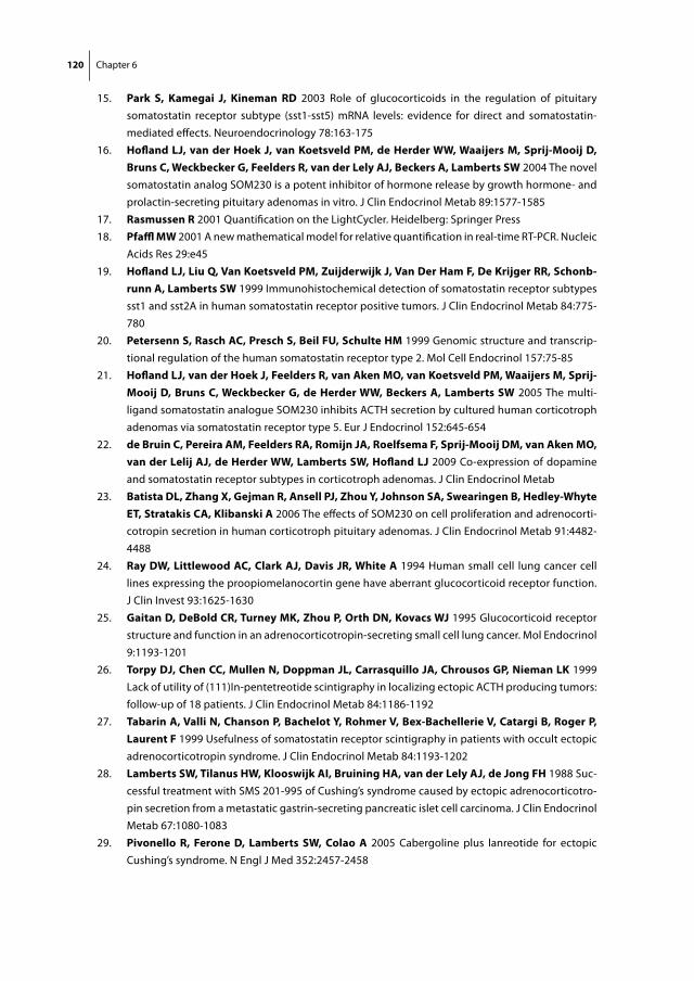
120 Chapter 6
15. Park s, Kamegai J, Kineman rD 2003 Role of glucocorticoids in the regulation of pituitary somatostatin receptor subtype (sst1-sst5) mRNA levels: evidence for direct and somatostatin-mediated effects. Neuroendocrinology 78:163-175
16. Hofland lJ, van der Hoek J, van Koetsveld Pm, de Herder WW, Waaijers m, sprij-mooij D, Bruns C, Weckbecker g, Feelders r, van der lely AJ, Beckers A, lamberts sW 2004 The novel somatostatin analog SOM230 is a potent inhibitor of hormone release by growth hormone- and prolactin-secreting pituitary adenomas in vitro. J Clin Endocrinol Metab 89:1577-1585
17. rasmussen r 2001 Quantification on the LightCycler. Heidelberg: Springer Press 18. Pfaffl mW 2001 A new mathematical model for relative quantification in real-time RT-PCR. Nucleic
Acids Res 29:e45 19. Hofland lJ, liu Q, van Koetsveld Pm, Zuijderwijk J, van Der Ham F, De Krijger rr, schonb-
runn A, lamberts sW 1999 Immunohistochemical detection of somatostatin receptor subtypes sst1 and sst2A in human somatostatin receptor positive tumors. J Clin Endocrinol Metab 84:775-780
20. Petersenn s, rasch AC, Presch s, Beil Fu, schulte Hm 1999 Genomic structure and transcrip-tional regulation of the human somatostatin receptor type 2. Mol Cell Endocrinol 157:75-85
21. Hofland lJ, van der Hoek J, Feelders r, van Aken mo, van Koetsveld Pm, Waaijers m, sprij-mooij D, Bruns C, Weckbecker g, de Herder WW, Beckers A, lamberts sW 2005 The multi-ligand somatostatin analogue SOM230 inhibits ACTH secretion by cultured human corticotroph adenomas via somatostatin receptor type 5. Eur J Endocrinol 152:645-654
22. de Bruin C, Pereira Am, Feelders rA, romijn JA, roelfsema F, sprij-mooij Dm, van Aken mo, van der lelij AJ, de Herder WW, lamberts sW, Hofland lJ 2009 Co-expression of dopamine and somatostatin receptor subtypes in corticotroph adenomas. J Clin Endocrinol Metab
23. Batista Dl, Zhang X, gejman r, Ansell PJ, Zhou y, Johnson sA, swearingen B, Hedley-Whyte et, stratakis CA, Klibanski A 2006 The effects of SOM230 on cell proliferation and adrenocorti-cotropin secretion in human corticotroph pituitary adenomas. J Clin Endocrinol Metab 91:4482-4488
24. ray DW, littlewood AC, Clark AJ, Davis Jr, White A 1994 Human small cell lung cancer cell lines expressing the proopiomelanocortin gene have aberrant glucocorticoid receptor function. J Clin Invest 93:1625-1630
25. gaitan D, DeBold Cr, turney mK, Zhou P, orth Dn, Kovacs WJ 1995 Glucocorticoid receptor structure and function in an adrenocorticotropin-secreting small cell lung cancer. Mol Endocrinol 9:1193-1201
26. torpy DJ, Chen CC, mullen n, Doppman Jl, Carrasquillo JA, Chrousos gP, nieman lK 1999 Lack of utility of (111)In-pentetreotide scintigraphy in localizing ectopic ACTH producing tumors: follow-up of 18 patients. J Clin Endocrinol Metab 84:1186-1192
27. tabarin A, valli n, Chanson P, Bachelot y, rohmer v, Bex-Bachellerie v, Catargi B, roger P, laurent F 1999 Usefulness of somatostatin receptor scintigraphy in patients with occult ectopic adrenocorticotropin syndrome. J Clin Endocrinol Metab 84:1193-1202
28. lamberts sW, tilanus HW, Klooswijk Ai, Bruining HA, van der lely AJ, de Jong FH 1988 Suc-cessful treatment with SMS 201-995 of Cushing’s syndrome caused by ectopic adrenocorticotro-pin secretion from a metastatic gastrin-secreting pancreatic islet cell carcinoma. J Clin Endocrinol Metab 67:1080-1083
29. Pivonello r, Ferone D, lamberts sW, Colao A 2005 Cabergoline plus lanreotide for ectopic Cushing’s syndrome. N Engl J Med 352:2457-2458

Chapter 7
Functional characterization of the dopamine somatostatin
chimeric molecule BIM-23A760Submitted
122 Chapter 7
ABstrACt
Somatostatin (sst) and dopamine (D2) receptor subtypes are co-expressed in various neuro-endocrine tumours and may show functional synergism. Novel dopamine-somatostatin chimeric molecules that bind to both receptor subtypes, have displayed superagonistic properties in earlier studies. Whether this is due to heterodimerization of target receptors or to enhanced activation of individual receptors, remains unknown. In this study we investigated the functional role of the different components of the novel dopamine-somatostatin chimera BIM-23A760 (high sst2-D2 affinity). We transiently transfected HEK-293 cells with D2 and/or sst2 cDNA and measured inhibition of forskolin-stimulated cAMP release by BIM-23A760 in comparison to its internal controls BIM-53097 (D2) and BIM-23023 (sst2). In D2-monotransfected cells, BIM-23A760 was 15-fold more potent than BIM-53097 (EC50 0.02 vs. 0.3 nM, p<0.01), even though both compounds have a similar D2 receptor binding affinity (IC50 15.9 and 22.1 nM, respectively). In contrast, in sst2-monotransfected cells BIM-23A760 was 4-fold less potent than BIM-23023 (EC50 0.04 vs. 0.01 nM, p<0.01), while BIM-23A760 sst2 receptor binding affinity is 14-fold higher (IC50 0.03 and 0.42 nM, respectively). In sst2-D2 co-transfected cells cAMP inhibition by BIM-23A760 was identical to that in D2-monotransfected cells (EC50 0.02 nM). Both the D2-antagonist sulpiride and the sst2-antagonist BIM-23454 were required to fully abrogate the effects of BIM-23A760. We conclude that in this transfected cell system, BIM-23A760 is a strong activator of sst2, but also a remarkably potent activator of D2 receptors, which cannot be explained by its binding affinity profile alone. Co-expression of sst2 and D2 does not enhance cAMP-inhibition by BIM-23A760 via either of these receptors. This suggests that the superagonistic properties of this compound in vivo, may at least partially be due to superior activation at the level of the individual (D2) receptors.
Functional characterization of BIM-23A760 123
Chap
ter 7
introDuCtion
Dopamine (DA) and somatostatin (SS) receptors are (co-)expressed in many different neuro-endocrine tumours, including pituitary adenomas, gastro-entero-pancreatic tumours and carcinoids (1-3). Agents that target these receptors have been shown to be effective in inhibiting hormone release and/or tumour growth (4). Several years ago it was shown that dopamine receptor subtype 2 (D2) and somatostatin receptor subtype 5 (sst5), both members of the family of G-protein coupled receptors (GPCRs), could form heterodimers in a transfected cell system, leading to enhanced functionality of these receptors and synergism of action (5). Recently, similar phenomena have been described for the D2 and sst2 receptor (6). The discovery that these receptors can heterodimerize with a resulting increase in efficacy has opened up the way for potential combination therapy of both DA and SS-targeting agents in patients with neuro-endocrine tumours. Along with these developments, new compounds have been designed, which combine high-affinity binding to both DA and SS receptors within the same molecule, the so-called dopastatin chimeric molecules. In addition to the activation of both receptors individually, these compounds could in theory also draw both receptors together in a spatial manner, and thereby increase the degree of synergism between these recep-tors.
One of such chimeric compounds of interest is BIM-23A760. Saveanu and Jaquet have shown previously that this compound has superior efficacy in terms of GH-inhibition compared with traditional SS-analogues in some GH-secreting adenomas (7, 8). In a re-cent multicenter study, it was found that this compound also inhibited the proliferation rate of non-functioning pituitary adenomas (Florio et al. 2008 End. Rel. Ca.). Due to its binding affinity profile, with superior affinity for sst2 and D2, combined with a good affin-ity for sst5,, this compound could in theory also be of interest for the medical treatment of Cushing’s disease (9), as well as the Ectopic-ACTH producing Syndrome (10, 11).
The mechanism behind the reported superpotency of these chimeric dopastatins has not been fully elucidated (1). In view of the clinical interest in this molecule and the on-going debate on whether or not its superpotency can be attributed to heterodimeriza-tion of its target receptors, we performed the present study. With the use of an in vitro reporter gene assay, we aimed to study some of the functional characteristics of this interesting new dopastatin compound BIM-23A760, with the main research question being: can we ascribe the observed superpotency of this compound to heterodimeriza-tion or could it be due to superior activation of the individual receptors?
124 Chapter 7
metHoDs
Transfection studies and reporter gene assay
Since sst2 and D2 are both GPCR’s that signal through the activation of cAMP, we investi-gated the functionality of BIM-23A760 in a reporter gene system, which indirectly mea-sures intracellular cAMP levels. HEK293 cells (kind gift from Dr. A.P.N. Themmen, Erasmus MC) that natively do not express sst2 and D2, were transiently transfected with either human sst2 cDNA (kind gift from Dr. Bell, Howard Hughes Medical Institute, Chicago, IL) or human D2 cDNA (commercially available at UMR cDNA resource center; www.cDNA.com). For transfection, the calcium chloride precipitation method was chosen as this method has been used previously for this particular assay (12, 13). HEK-293 cells seeded at 20% confluency were transiently transfected with 2 μg of the cAMP-reporter plasmid pCRE6Lux (14), 2 μg pRSVlacZ (15) as a control for transfection efficiency, 2μg of human sst2 cDNA and/or 0.1 μg of human D2 cDNA and 14 μg carrier DNA in 75 cm2 culture flasks (Costar, Cambridge, MA). Two days after transfection the cells were trypsinized and plated in 48-well tissue culture plates (Costar, Cambridge, MA). On the third day, medium was changed to serum-free medium supplemented with 0.1% bovine serum albumin. Cells were then stimulated with forskolin (10-6 M) for 6 hr in the presence or absence of the different BIM-agonists and/or antagonists. After 6 hr stimulation, cells were lysed with 25 mM Tris phosphate, pH 7.8, 8 mM MgCl, 1 mM dithiothreitol, 15% glycerol, 1% Triton X-100 and analyzed for luciferase (cAMP-responsive element driven) and Renilla luciferase (transfection control) activities using the Dual-Luciferase Reporter Assay System according to the manufacturer’s instructions (Promega Corporation, Madi-son, WI) (16).
Quantitative PCR
To check for adequate sst2 and D2 transfection efficiency, additional transfected cells were lysed for quantitative PCR (qPCR) in every experiment. This was performed via a previously described method (17). The sequences and final concentrations of the hprt, sst2 and D2 primer-probe pairs have been described previously (17, 18). The D2 primer-probe set measures total D2 expression (D2 long + short isoform). All primers and probes were purchased from Sigma Aldrich (Zwijndrecht, the Netherlands). Samples were measured on an ABI Prism 7900 Sequence Detection System (Perkin Elmer, Foster City, CA) and normalized against the expression of the housekeeping gene hprt. All samples were assayed in duplicate.
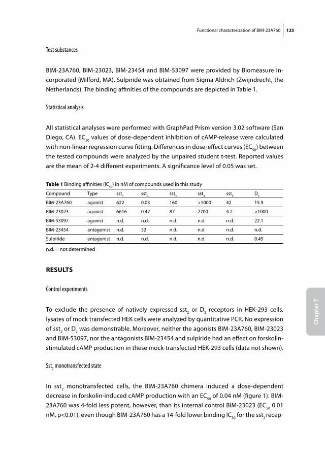
Functional characterization of BIM-23A760 125
Chap
ter 7
Test substances
BIM-23A760, BIM-23023, BIM-23454 and BIM-53097 were provided by Biomeasure In-corporated (Milford, MA). Sulpiride was obtained from Sigma Aldrich (Zwijndrecht, the Netherlands). The binding affinities of the compounds are depicted in Table 1.
Statistical analysis
All statistical analyses were performed with GraphPad Prism version 3.02 software (San Diego, CA). EC50 values of dose-dependent inhibition of cAMP-release were calculated with non-linear regression curve fitting. Differences in dose-effect curves (EC50) between the tested compounds were analyzed by the unpaired student t-test. Reported values are the mean of 2-4 different experiments. A significance level of 0.05 was set.
results
Control experiments
To exclude the presence of natively expressed sst2 or D2 receptors in HEK-293 cells, lysates of mock transfected HEK cells were analyzed by quantitative PCR. No expression of sst2 or D2 was demonstrable. Moreover, neither the agonists BIM-23A760, BIM-23023 and BIM-53097, nor the antagonists BIM-23454 and sulpiride had an effect on forskolin-stimulated cAMP production in these mock-transfected HEK-293 cells (data not shown).
Sst2 monotransfected state
In sst2 monotransfected cells, the BIM-23A760 chimera induced a dose-dependent decrease in forskolin-induced cAMP production with an EC50 of 0.04 nM (figure 1). BIM-23A760 was 4-fold less potent, however, than its internal control BIM-23023 (EC50 0.01 nM, p<0.01), even though BIM-23A760 has a 14-fold lower binding IC50 for the sst2 recep-
table 1 Binding affinities (IC50) in nM of compounds used in this study
Compound Type sst1 sst2 sst3 sst4 sst5 D2
BIM-23A760 agonist 622 0.03 160 >1000 42 15.9
BIM-23023 agonist 6616 0.42 87 2700 4.2 >1000
BIM-53097 agonist n.d. n.d. n.d. n.d. n.d. 22.1
BIM-23454 antagonist n.d. 32 n.d. n.d. n.d. n.d.
Sulpiride antagonist n.d. n.d. n.d. n.d. n.d. 0.45
n.d. = not determined
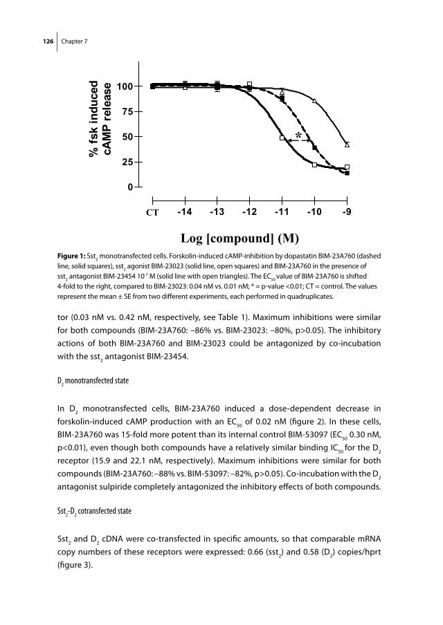
126 Chapter 7
tor (0.03 nM vs. 0.42 nM, respectively, see Table 1). Maximum inhibitions were similar for both compounds (BIM-23A760: –86% vs. BIM-23023: –80%, p>0.05). The inhibitory actions of both BIM-23A760 and BIM-23023 could be antagonized by co-incubation with the sst2 antagonist BIM-23454.
D2 monotransfected state
In D2 monotransfected cells, BIM-23A760 induced a dose-dependent decrease in forskolin-induced cAMP production with an EC50 of 0.02 nM (figure 2). In these cells, BIM-23A760 was 15-fold more potent than its internal control BIM-53097 (EC50 0.30 nM, p<0.01), even though both compounds have a relatively similar binding IC50 for the D2
receptor (15.9 and 22.1 nM, respectively). Maximum inhibitions were similar for both compounds (BIM-23A760: –88% vs. BIM-53097: –82%, p>0.05). Co-incubation with the D2 antagonist sulpiride completely antagonized the inhibitory effects of both compounds.
Sst2-D2 cotransfected state
Sst2 and D2 cDNA were co-transfected in specific amounts, so that comparable mRNA copy numbers of these receptors were expressed: 0.66 (sst2) and 0.58 (D2) copies/hprt (figure 3).
Fig 1
75
100nd
uced
elea
se
25
50
% fs
k in
cAM
P re
*
0
25
-15 -14 -13 -12 -11 -10 -9
Log [compound] (M)
CT
Figure 1: Sst2 monotransfected cells. Forskolin-induced cAMP-inhibition by dopastatin BIM-23A760 (dashed line, solid squares), sst2 agonist BIM-23023 (solid line, open squares) and BIM-23A760 in the presence of sst2 antagonist BIM-23454 10-7 M (solid line with open triangles). The EC50 value of BIM-23A760 is shifted 4-fold to the right, compared to BIM-23023: 0.04 nM vs. 0.01 nM; * = p-value <0.01; CT = control. The values represent the mean ± SE from two different experiments, each performed in quadruplicates.
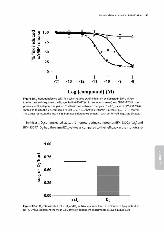
Functional characterization of BIM-23A760 127
Chap
ter 7
In this sst2-D2 cotransfected state, the monotargeting compounds BIM-23023 (sst2) and BIM-53097 (D2) had the same EC50 values as compared to their efficacy in the monotrans-
Fig 2
75
100nd
uced
elea
se
25
50
% fs
k in
cAM
P re
*
0
25
-14 -13 -12 -11 -10 -9 -8
Log [compound] (M)
CT
Figure 2: D2 monotransfected cells. Forskolin-induced cAMP-inhibition by dopastatin BIM-23A760 (dashed line, solid squares), the D2 agonist BIM-53097 (solid line, open squares) and BIM-23A760 in the presence of D2 antagonist sulpiride 10-4M (solid line with open triangles). The EC50 value of BIM-23A760 is shifted 15-fold to the left, compared to BIM-53097: 0.02 nM vs. 0.30 nM; * = p-value <0.01; CT = control. The values represent the mean ± SE from two different experiments, each performed in quadruplicates.
Fig 3
1 00
0.75
1.00
hprt
0.50
2 or
D2/
h
0.25sst 2
sst2 D20.00
Figure 3: Sst2-D2 cotransfected cells. Sst2 and D2 mRNA expression levels as determined by quantitative RT-PCR. Values represent the mean ± SE of two independent experiments, assayed in duplicate.
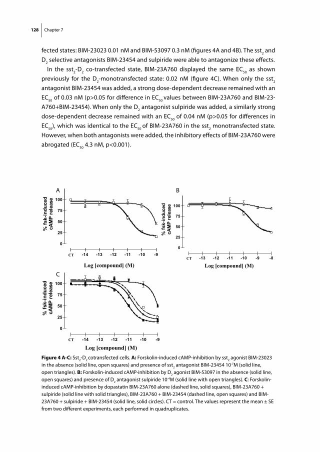
128 Chapter 7
fected states: BIM-23023 0.01 nM and BIM-53097 0.3 nM (figures 4A and 4B). The sst2 and D2 selective antagonists BIM-23454 and sulpiride were able to antagonize these effects.
In the sst2-D2 co-transfected state, BIM-23A760 displayed the same EC50 as shown previously for the D2-monotransfected state: 0.02 nM (figure 4C). When only the sst2 antagonist BIM-23454 was added, a strong dose-dependent decrease remained with an EC50 of 0.03 nM (p>0.05 for difference in EC50 values between BIM-23A760 and BIM-23-A760+BIM-23454). When only the D2 antagonist sulpiride was added, a similarly strong dose-dependent decrease remained with an EC50 of 0.04 nM (p>0.05 for differences in EC50), which was identical to the EC50 of BIM-23A760 in the sst2 monotransfected state. However, when both antagonists were added, the inhibitory effects of BIM-23A760 were abrogated (EC50 4.3 nM, p<0.001).
Fig 4A
75
100
nduc
edre
leas
e
25
50
% fs
k-in
cAM
P r
0
25
-15 -14 -13 -12 -11 -10 -9
Log [compound] (M)
CT
Fig 4B
100
uced ase
50
75
fsk-
indu
AM
P re
le
0
25
% cA
-14 -13 -12 -11 -10 -9 -8
0
Log [compound] (M)CT
Log [compound] (M)
Fig 4C
100
uced
ease
50
75
% fs
k-in
dcA
MP
rele
0
25
% c
-15 -14 -13 -12 -11 -10 -9
Log [compound] (M) CT
Figure 4 A-C: Sst2-D2 cotransfected cells. A: Forskolin-induced cAMP-inhibition by sst2 agonist BIM-23023 in the absence (solid line, open squares) and presence of sst2 antagonist BIM-23454 10-7M (solid line, open triangles). B: Forskolin-induced cAMP-inhibition by D2 agonist BIM-53097 in the absence (solid line, open squares) and presence of D2 antagonist sulpiride 10-4M (solid line with open triangles). C: Forskolin-induced cAMP-inhibition by dopastatin BIM-23A760 alone (dashed line, solid squares), BIM-23A760 + sulpiride (solid line with solid triangles), BIM-23A760 + BIM-23454 (dashed line, open squares) and BIM-23A760 + sulpiride + BIM-23454 (solid line, solid circles). CT = control. The values represent the mean ± SE from two different experiments, each performed in quadruplicates.
A B
C
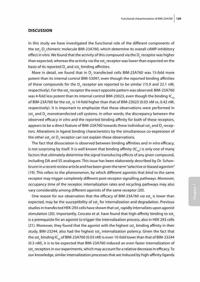
Functional characterization of BIM-23A760 129
Chap
ter 7
DisCussion
In this study we have investigated the functional role of the different components of the sst2-D2 chimeric molecule BIM-23A760, which determine its overall cAMP-inhibitory effect in vitro. We found that the activity of this compound via the D2 receptor was higher than expected, whereas the activity via the sst2 receptor was lower than expected on the basis of its reported D2 and sst2 binding affinities.
More in detail, we found that in D2 transfected cells BIM-23A760 was 15-fold more potent than its internal control BIM-53097, even though the reported binding affinities of these compounds for the D2 receptor are reported to be similar (15.9 and 22.1 nM, respectively). For the sst2 receptor the exact opposite pattern was observed: BIM-23A760 was 4-fold less potent than its internal control BIM-23023, even though the binding IC50 of BIM-23A760 for the sst2 is 14-fold higher than that of BIM-23023 (0.03 nM vs. 0.42 nM, respectively). It is important to emphasize that these observations were performed in sst2 and D2 monotransfected cell systems. In other words, the discrepancy between the observed efficacy in vitro and the reported binding affinity for both of these receptors, appears to be a direct feature of BIM-23A760 towards these individual sst2 and D2 recep-tors. Alterations in ligand binding characteristics by the simultaneous co-expression of the other sst2 or D2 receptor can not explain these observations.
The fact that dissociation is observed between binding affinities and in vitro efficacy, is not surprising by itself. It is well known that binding affinity (IC50) is only one of many factors that ultimately determine the signal transducing effects of any given compound, including DA and SS analogues. This issue has been elaborately described by Dr. Schon-brunn in a recent review article and has been given the term “selective or biased agonism” (19). This refers to the phenomenon, by which different agonists that bind to the same receptor may trigger completely different post-receptor signalling pathways. Moreover, occupancy time of the receptor, internalization rates and recycling pathways may also vary considerably among different agonists of the same receptor (20).
One reason for our observation that the efficacy of BIM-23A760 via sst2 is lower than expected, may be the susceptibility of sst2 for internalization and degradation. Previous studies in transfected HEK-293 cells have shown that sst2 rapidly internalizes upon agonist stimulation (20). Importantly, Cescato et al. have found that high-affinity binding to sst2
is a prerequisite for an agonist to trigger the internalization process, also in HEK 293 cells (21). Moreover, they found that the agonist with the highest sst2 binding affinity in their study, BIM-23244, also had the highest sst2 internalization potency. Given the fact that the sst2 binding IC50 of BIM-23A760 (0.03 nM) is even 10-fold lower than that of BIM-23244 (0.3 nM), it is to be expected that BIM-23A760 induced an even faster internalization of sst2 receptors in our experiments, which may account for a relative decrease in efficacy. To our knowledge, similar internalization processes that are induced by high-affinity ligands
130 Chapter 7
have not been described for the D2 receptor. The question remains of course whether these processes, observed in (mono)transfected cell systems, occur in a similar fashion in vivo, where many different receptors are expressed in the close vicinity of these sst2 and D2 receptors. It has been shown for instance that sst5 may directly decrease the internal-ization rate of sst2 (22). This may prove to be a favourable determinant of the efficacy of BIM-23A760 in human neuro-endocrine tumors, which often co-express sst2 and sst5.
In the sst2-D2 co-transfected state, we had hypothesized that enhanced potency might occur because of synergism between the sst2 and D2 receptor. This has previously been shown to occur in a transfected cell system between the sst5 and D2 receptor (5). In this study by Rocheville et al. hetero-oligomerization of these receptor subtypes was proposed to lead to a sst5-D2 dimer with increased affinity for both sst5 and D2-targeting agonists and enhanced receptor activation. Recently, another group also showed that in co-transfected HEK-293 cells, sst2 and D2 receptors form heterodimers upon activation by selective ligands, leading to enhanced D2 signalling and also prolonged sst2 inter-nalization (6). In our study, however, BIM-23A760 displayed the same EC50 in the sst2-D2
co-transfected state as compared to the D2-monotransfected state (0.02 nM), without any increase or decrease in IC50 due to the presence of the extra sst2 receptors.
As said, the mono-targeting compounds had the same EC50 and ECmax values in the monotransfected compared to the co-transfected states. In other words, the abundant presence of additional sst2 or D2 receptors did not stimulate nor decrease the signalling via these receptors. This is different from what has been reported previously by some but not all research groups in the same field of study. Both Ren et al. and Saveanu et al. found that the chimeric molecule BIM-23A387 (high sst2-high D2 affinity) showed enhanced inhibitory effects in human GH-producing adenomas compared to incubation with the individual targeting compounds (23, 24). Ren et al., however, found that by the use of the D2 antagonist sulpiride, all inhibitory properties of BIM-23A387 were lost, suggest-ing a functional interaction between sst2 and D2, whereas Saveanu et al. showed only partial loss of inhibition by BIM-23A387 in this setting. Saveanu et al. therefore conclude that the potent effects of BIM-23A387 can not be explained by the process of receptor heterodimerization alone, but may also depend on specific interactions between the chimeric molecule and the individual sst2 and D2 receptors (24).
In summary, we find that in this transfected cell system BIM-23A760 is a strong activator of sst2, but also a remarkably potent activator of D2 receptors, which can not be explained by its binding affinity profile alone. In our study, co-expression of sst2 and D2 does not enhance the cAMP-inhibiting capacities of BIM-23A760 via either of these receptors. This suggests that the reported superagonistic properties of this compound in vivo, may at least partially be due to superior activation at the level of the individual (D2) receptors.
Functional characterization of BIM-23A760 131
Chap
ter 7
ACKnoWleDgements:
The authors would like to thank Anke van Kerkwijk and Miriam Verhoef-Post for their technical assistance in this study.
reFerenCes
1. saveanu A, Jaquet P 2008 Somatostatin-dopamine ligands in the treatment of pituitary ad-enomas. Rev Endocr Metab Disord
2. Patel yC 1999 Somatostatin and its receptor family. Front Neuroendocrinol 20:157-198 3. stefaneanu l, Kovacs K, Horvath e, Buchfelder m, Fahlbusch r, lancranjan l 2001 Dopamine
D2 receptor gene expression in human adenohypophysial adenomas. Endocrine 14:329-336 4. Barnett P 2003 Somatostatin and somatostatin receptor physiology. Endocrine 20:255-264 5. rocheville m, lange DC, Kumar u, Patel sC, Patel rC, Patel yC 2000 Receptors for dopamine
and somatostatin: formation of hetero-oligomers with enhanced functional activity. Science 288:154-157
6. Baragli A, Alturaihi H, Watt Hl, Abdallah A, Kumar u 2007 Heterooligomerization of human dopamine receptor 2 and somatostatin receptor 2 Co-immunoprecipitation and fluorescence resonance energy transfer analysis. Cell Signal 19:2304-2316
7. saveanu A, gunz g, guillen s, Dufour H, Culler mD, Jaquet P 2006 Somatostatin and dop-amine-somatostatin multiple ligands directed towards somatostatin and dopamine receptors in pituitary adenomas. Neuroendocrinology 83:258-263
8. Jaquet P, gunz g, saveanu A, Dufour H, taylor J, Dong J, Kim s, moreau JP, enjalbert A, Culler mD 2005 Efficacy of chimeric molecules directed towards multiple somatostatin and dopamine receptors on inhibition of GH and prolactin secretion from GH-secreting pituitary adenomas clas-sified as partially responsive to somatostatin analog therapy. Eur J Endocrinol 153:135-141
9. Pivonello r, Ferone D, de Herder WW, Kros Jm, De Caro ml, Arvigo m, Annunziato l, lom-bardi g, Colao A, Hofland lJ, lamberts sW 2004 Dopamine receptor expression and function in corticotroph pituitary tumors. J Clin Endocrinol Metab 89:2452-2462
10. Pivonello r, Ferone D, de Herder WW, Faggiano A, Bodei l, de Krijger rr, lombardi g, Colao A, lamberts sW, Hofland lJ 2007 Dopamine receptor expression and function in corticotroph ectopic tumors. J Clin Endocrinol Metab 92:65-69
11. Pivonello r, Ferone D, lamberts sW, Colao A 2005 Cabergoline plus lanreotide for ectopic Cushing’s syndrome. N Engl J Med 352:2457-2458
12. richter-unruh A, verhoef-Post m, malak s, Homoki J, Hauffa BP, themmen AP 2004 Leydig cell hypoplasia: absent luteinizing hormone receptor cell surface expression caused by a novel homozygous mutation in the extracellular domain. J Clin Endocrinol Metab 89:5161-5167
13. martens JW, verhoef-Post m, Abelin n, ezabella m, toledo sP, Brunner Hg, themmen AP 1998 A homozygous mutation in the luteinizing hormone receptor causes partial Leydig cell hypoplasia: correlation between receptor activity and phenotype. Mol Endocrinol 12:775-784
14. Himmler A, stratowa C, Czernilofsky AP 1993 Functional testing of human dopamine D1 and D5 receptors expressed in stable cAMP-responsive luciferase reporter cell lines. J Recept Res 13:79-94
132 Chapter 7
15. Hall Cv, Jacob Pe, ringold gm, lee F 1983 Expression and regulation of Escherichia coli lacZ gene fusions in mammalian cells. J Mol Appl Genet 2:101-109
16. Bruysters m, verhoef-Post m, themmen AP 2008 Asp330 and Tyr331 in the C-terminal cysteine-rich region of the luteinizing hormone receptor are key residues in hormone-induced receptor activation. J Biol Chem 283:25821-25828
17. de Bruin C, Feelders rA, Waaijers Am, van Koetsveld Pm, sprij-mooij Dm, lamberts sW, Ho-fland lJ 2009 Differential regulation of human dopamine D2 and somatostatin receptor subtype expression by glucocorticoids in vitro. J Mol Endocrinol 42:47-56
18. Hofland lJ, van der Hoek J, van Koetsveld Pm, de Herder WW, Waaijers m, sprij-mooij D, Bruns C, Weckbecker g, Feelders r, van der lely AJ, Beckers A, lamberts sW 2004 The novel somatostatin analog SOM230 is a potent inhibitor of hormone release by growth hormone- and prolactin-secreting pituitary adenomas in vitro. J Clin Endocrinol Metab 89:1577-1585
19. schonbrunn A 2008 Selective agonism in somatostatin receptor signaling and regulation. Mol Cell Endocrinol 286:35-39
20. liu Q, Cescato r, Dewi DA, rivier J, reubi JC, schonbrunn A 2005 Receptor signaling and endocytosis are differentially regulated by somatostatin analogs. Mol Pharmacol 68:90-101
21. Cescato r, schulz s, Waser B, eltschinger v, rivier Je, Wester HJ, Culler m, ginj m, liu Q, schonbrunn A, reubi JC 2006 Internalization of sst2, sst3, and sst5 receptors: effects of soma-tostatin agonists and antagonists. J Nucl Med 47:502-511
22. sharif n, gendron l, Wowchuk J, sarret P, mazella J, Beaudet A, stroh t 2007 Coexpression of somatostatin receptor subtype 5 affects internalization and trafficking of somatostatin receptor subtype 2. Endocrinology 148:2095-2105
23. ren sg, Kim s, taylor J, Dong J, moreau JP, Culler mD, melmed s 2003 Suppression of rat and human growth hormone and prolactin secretion by a novel somatostatin/dopaminergic chimeric ligand. J Clin Endocrinol Metab 88:5414-5421
24. saveanu A, lavaque e, gunz g, Barlier A, Kim s, taylor Je, Culler mD, enjalbert A, Jaquet P 2002 Demonstration of enhanced potency of a chimeric somatostatin-dopamine molecule, BIM-23A387, in suppressing growth hormone and prolactin secretion from human pituitary somatotroph ad-enoma cells. J Clin Endocrinol Metab 87:5545-5552

Chapter 8
General discussion

General discussion 135
Chap
ter 8
The principal aim of this thesis was to further explore the possibilities for a medical treat-ment of human Cushing’s disease based on the use of somatostatin analogues and/or dopamine agonists.
Despite all advancements in surgical technique, selective transsphenoidal adenomec-tomy still fails to induce long-term cure in approximately 30% of patients with CD, even in most experienced hands (1). The group of patients with persistent or recurrent CD is known to suffer from increased morbidity and mortality and represents one of the most difficult clinical challenges for endocrinologists (2). Until now, medical therapy has only played a minor role in the treatment of these refractory patients (3-5). Antiserotin-ergic drugs have been employed in the past without much success (5). Retinoic acid derivatives, despite promising results in vitro and in some animal models, have failed to make the transition to clinical studies in human subjects (6, 7). Moreover, peroxisome-proliferator-activated-receptor-gamma agonists have not shown any significant efficacy in several recent clinical studies in humans with CD (8-10). The use of dopamine agonists including bromocriptine and cabergoline has been widely studied in CD, but its success is limited by the occurrence of treatment escapes in a subset of patients (3, 11). Therefore, the search for an effective and safe medical treatment for human CD continues. In this thesis we have focussed on the role of dopamine and somatostatin receptor subtypes as potential targets for such a medical therapy.
AnimAl moDels For CD
One of the most important rate limiting factors in the advancements of research into the origins and treatment of human CD, has been the rarity of the disease. Interestingly, in dogs this disease is much more common, perhaps over 1000-fold more (12-14). The fact that at Utrecht University dogs with CD can be treated by a therapeutic hypophysectomy, provided a more than welcome opportunity to increase the availability of corticotroph adenoma tissue for in vitro functional studies. In chapter 2 we have described the char-acterization of 13 of these adenomas for the presence of the receptor subtypes, in which we were primarily interested from a human point of view: sst2, sst5 and D2. We found that these adenomas do express all of these receptor subtypes, but in very different ratios than anticipated beforehand. Whereas in human CD sst2 only plays a minor role, it appears to be the dominant receptor subtype in canine CD, both in terms of expression levels, as well as functional responses in vitro. At the same time, sst5 only has a minor role in canine CD, while D2 also has lower expression levels than what is observed in humans. Inter-species differences in glucocorticoid regulation of the expression of these receptor subtypes are likely to contribute to these differences between human and canine CD:
136 Chapter 8
whereas glucocorticoids decrease sst2 expression levels in humans, they remarkably increase the expression of sst2 in canine corticotroph adenomas.
We do believe, however, that valuable research hypotheses can still be generated from the use of canine CD as a model for human CD. Due to the predominance of sst2 and D2 in canine CD, the use of analogues that target these specific receptor subtypes could be of interest. This could be performed by co-administration of an sst2 (octreotide or lanreotide) and a D2 (cabergoline) targeting drug. The advantage of the use of these compounds is that both have been proven to be effective in dogs for the treatment of insulinomas and reproductive disorders, respectively (15, 16) . In other words, they are known to bind to canine sst2 and D2 receptors with sufficient affinity and can induce clinical effects. Their combined in vivo use in canine CD could provide firsthand evidence for the possible functional synergism between sst2 and D2 receptors, which has been suggested by recent in vitro studies (17). Moreover, the use of the novel dopamine-somatostatin chimera BIM-23A760, which is also discussed in chapter 7, could be of great interest here. This compound has a very high sst2 and D2 binding affinity and is therefore likely to induce profound ACTH suppression in canine corticotroph adenomas, provid-ing that the reported binding affinity profile of this compound is similar in humans and dogs.
Finally, it is necessary to emphasize that canine corticotroph adenoma tissue can be valuable for various research questions that address the different etiological aspects of CD. For example, one study used canine corticotroph adenomas to investigate novel molecular aspects of glucocorticoid receptor function in CD (18). This study would have been much more difficult to perform if only human corticotroph tissue was available.
HumAn CortiCotroPH ADenomAs
An important aim of this thesis was to increase our understanding of how many patients with CD have a reasonable chance of ultimately responding to somatostatin and/or dopamine targeted medical therapy. Data on this topic were still scarce at the start of this study, being derived from relatively small series of patients. Moreover, it was not known to which extent sst5 and D2 were co-expressed within the same corticotroph tumors. We aimed to reinvestigate the distribution of these target receptor subtypes in a larger population of CD patients, as the presence of demonstrable receptors is likely to be the first and foremost predictor of a successful response to this type of therapy. The importance of sufficient receptor numbers has been demonstrated by several studies, in which a direct relationship was found between the expression levels of sst and DA recep-tor subtypes in GH and mixed GH/PRL-secreting pituitary adenomas and specific agonist
General discussion 137
Chap
ter 8
action, both in vitro and in vivo (19-22). The in vitro correlation has been demonstrated both at the mRNA level (21) as well as at the protein level by immunohistochemistry (19, 20).
The main results of our study in 30 human corticotroph adenomas are described in chapter 4. We found a large variation in expression levels of sst2, sst5 and D2 in our series of adenomas, similarly to what has been described before in GH-producing and non-functioning pituitary adenomas (21, 23). Overall, we believe that the results from this study are encouraging, as we found that over 80% of patients express at least one of these target receptors at a level, where protein expression can be visualized by means of receptor autoradiography. Of course, demonstrable protein expression at recep-tor autoradiography does not necessarily equal biological response, but at least the molecular substrate can be demonstrated on the cell membrane of the corticotroph adenoma cells. Various other factors, apart from the number of receptors, will ultimately determine how many patients will show a clinical response to sst2/sst5/D2 targeted therapy. In prolactinomas, for instance, loss of the Gi2 alpha protein can cause resistance to the dopamine agonist bromocriptine, even though sufficient D2 receptors are present on the cell membrane (24). In different neuro-endocrine tumors, especially carcinoids, various mechanisms of tachyphylaxis have been described (25). New studies will have to investigate whether similar phenomena also affect response rates in corticotroph adenomas.
tHe role oF gluCoCortiCoiDs
One aspect that could play a decisive role in the efficacy of sst/DA-targeting agents at any given time during therapy, is the level of circulating glucocorticoids. Before the start of this thesis it had already been clearly demonstrated that murine sst2 gene expression was under direct control by glucocorticoids, while sst5 appeared to be less sensitive for this type of regulation (26, 27). For D2, not much was known on glucocorticoid regulation in human neuro-endocrine tissues.
In chapter 3 we have described the results of our study on glucocorticoid regulation of sst and DA receptors in three different human neuro-endocrine cell lines. Our results confirm the previous studies in murine corticotroph cells that sst2 is highly sensitive, while sst5 is far less sensitive for this type of down-regulation. We also found that the D2 gene is completely insensitive for down-regulation by high levels of glucocorticoids. This could be an interesting finding for future medical therapies of CD. It may well explain why D2 receptors are so highly expressed in the majority (80%) of corticotroph
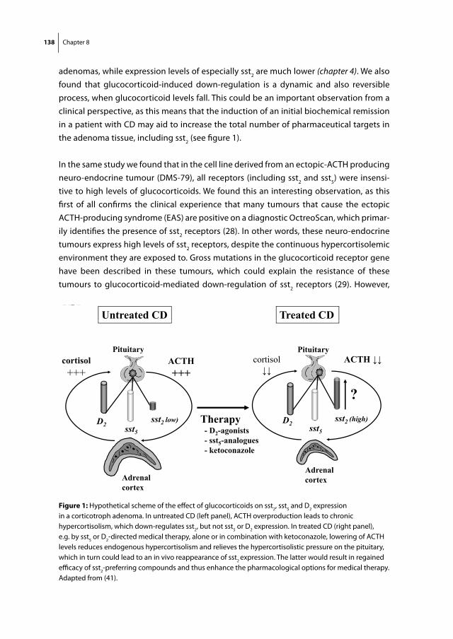
138 Chapter 8
adenomas, while expression levels of especially sst2 are much lower (chapter 4). We also found that glucocorticoid-induced down-regulation is a dynamic and also reversible process, when glucocorticoid levels fall. This could be an important observation from a clinical perspective, as this means that the induction of an initial biochemical remission in a patient with CD may aid to increase the total number of pharmaceutical targets in the adenoma tissue, including sst2 (see figure 1).
In the same study we found that in the cell line derived from an ectopic-ACTH producing neuro-endocrine tumour (DMS-79), all receptors (including sst2 and sst5) were insensi-tive to high levels of glucocorticoids. We found this an interesting observation, as this first of all confirms the clinical experience that many tumours that cause the ectopic ACTH-producing syndrome (EAS) are positive on a diagnostic OctreoScan, which primar-ily identifies the presence of sst2 receptors (28). In other words, these neuro-endocrine tumours express high levels of sst2 receptors, despite the continuous hypercortisolemic environment they are exposed to. Gross mutations in the glucocorticoid receptor gene have been described in these tumours, which could explain the resistance of these tumours to glucocorticoid-mediated down-regulation of sst2 receptors (29). However,
Untreated CD Treated CDFig 1
Pituitary PituitaryACTH+++
cortisol +++
PituitaryACTH cortisol
Pituitary
?Therapy- D2-agonists- sst5-analogues
D2 sst5
sst2 low) D2 sst5
sst2 (high)
5 g- ketoconazole
AAdrenal
Adrenal cortex
cortex
Figure 1: Hypothetical scheme of the effect of glucocorticoids on sst2, sst5 and D2 expression in a corticotroph adenoma. In untreated CD (left panel), ACTH overproduction leads to chronic hypercortisolism, which down-regulates sst2, but not sst5 or D2 expression. In treated CD (right panel), e.g. by sst5 or D2-directed medical therapy, alone or in combination with ketoconazole, lowering of ACTH levels reduces endogenous hypercortisolism and relieves the hypercortisolistic pressure on the pituitary, which in turn could lead to an in vivo reappearance of sst2 expression. The latter would result in regained efficacy of sst2-preferring compounds and thus enhance the pharmacological options for medical therapy. Adapted from (41).
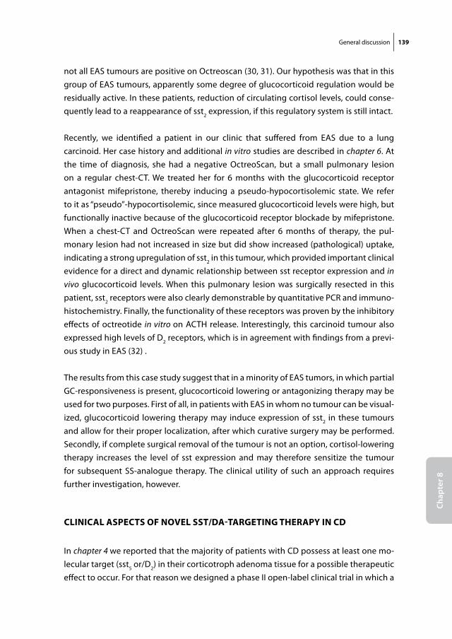
General discussion 139
Chap
ter 8
not all EAS tumours are positive on Octreoscan (30, 31). Our hypothesis was that in this group of EAS tumours, apparently some degree of glucocorticoid regulation would be residually active. In these patients, reduction of circulating cortisol levels, could conse-quently lead to a reappearance of sst2 expression, if this regulatory system is still intact.
Recently, we identified a patient in our clinic that suffered from EAS due to a lung carcinoid. Her case history and additional in vitro studies are described in chapter 6. At the time of diagnosis, she had a negative OctreoScan, but a small pulmonary lesion on a regular chest-CT. We treated her for 6 months with the glucocorticoid receptor antagonist mifepristone, thereby inducing a pseudo-hypocortisolemic state. We refer to it as “pseudo”-hypocortisolemic, since measured glucocorticoid levels were high, but functionally inactive because of the glucocorticoid receptor blockade by mifepristone. When a chest-CT and OctreoScan were repeated after 6 months of therapy, the pul-monary lesion had not increased in size but did show increased (pathological) uptake, indicating a strong upregulation of sst2 in this tumour, which provided important clinical evidence for a direct and dynamic relationship between sst receptor expression and in vivo glucocorticoid levels. When this pulmonary lesion was surgically resected in this patient, sst2 receptors were also clearly demonstrable by quantitative PCR and immuno-histochemistry. Finally, the functionality of these receptors was proven by the inhibitory effects of octreotide in vitro on ACTH release. Interestingly, this carcinoid tumour also expressed high levels of D2 receptors, which is in agreement with findings from a previ-ous study in EAS (32) .
The results from this case study suggest that in a minority of EAS tumors, in which partial GC-responsiveness is present, glucocorticoid lowering or antagonizing therapy may be used for two purposes. First of all, in patients with EAS in whom no tumour can be visual-ized, glucocorticoid lowering therapy may induce expression of sst2 in these tumours and allow for their proper localization, after which curative surgery may be performed. Secondly, if complete surgical removal of the tumour is not an option, cortisol-lowering therapy increases the level of sst expression and may therefore sensitize the tumour for subsequent SS-analogue therapy. The clinical utility of such an approach requires further investigation, however.
CliniCAl AsPeCts oF novel sst/DA-tArgeting tHerAPy in CD
In chapter 4 we reported that the majority of patients with CD possess at least one mo-lecular target (sst5 or/D2) in their corticotroph adenoma tissue for a possible therapeutic effect to occur. For that reason we designed a phase II open-label clinical trial in which a
140 Chapter 8
stepwise medical therapy, consisting of pasireotide ± cabergoline ± low-dose ketocon-azole, was evaluated for its efficacy in patients with de novo or recurrent CD. At present, patient enrolment for this trial has just been completed and final data analyses are cur-rently ongoing. As the definitive results of this trial will be published elsewhere, it is not possible to discuss the results of this trial in full detail within the context of this thesis. In chapter 5, however, we present and discuss four illustrative cases for the different types of clinical responses we have observed during this trial. On a general note, we can state that the study has produced firm clinical evidence for the efficacy of combined sst5 and/or D2-targeted medical therapy in human CD. A few important issues, however, require further discussion as they could prove to be of substantial clinical importance.
Dose
In this trial we observed that already on low-dose pasireotide (3 x 100 μg), selected patients can respond with a dramatic reduction in serum and urinary cortisol levels. These reductions can be observed within days after the first administration of the study drug. This is an interesting observation, as we initiated treatment in these patients with only a quarter of the pasireotide dose that Boscaro et al. used in their study (total daily dose: 300 μg vs. 1200 μg)(33). The significant response on these low doses may be ex-plained by the subnanomolar affinity of pasireotide for sst5 (0.38 nM). For comparison, octreotide can induce significant clinical responses in the 100μg dose in acromegalic patients, while its affinity for sst2 is around 1.0 nM. Therefore, from a pharmacological perspective, the 100 µg pasireotide dose should be able to induce similar biological ef-fects in corticotroph cells, provided that the adenoma expresses relatively high levels of sst5 receptors, which some patients clearly do (chapter 4). The occurrence of these rapid reductions in serum cortisol after the initiation of pasireotide therapy also has clinical implications. The occurrence of acute glucocorticoid withdrawal symptoms or even an overt Addisonian crisis is a realistic possibility during this type of treatment. Therefore, close follow-up of these patients needs to be provided upon treatment initiation.
Effects on glucose metabolism
Another important observation in this trial was that glucose homeostasis deteriorated significantly in two of the patients that we describe in chapter 5. This problem has also been clearly reported in the first study on pasireotide use in patients with CD by Boscaro et al., where over one third of patients had significant worsening of glycaemic control (33). Different biological mechanisms could be responsible for this phenomenon. First of all, it could be due to a direct effect of pasireotide on pancreatic insulin release. Sst1, sst2 and sst5 receptors, which are all targeted by pasireotide, are well expressed in human
General discussion 141
Chap
ter 8
pancreatic islet cells and activation of these receptors, especially sst2, has been shown to strongly decrease pancreatic insulin secretion in vitro (34). One argument that strongly rules against such a direct effect via inhibition of insulin release, however, are the observa-tions by Van der Hoek et al. in acromegalic patients, in whom pasireotide administration acutely increased blood glucose levels without a concomitant major decrease in insulin levels (35, 36). The latter would rather suggest that the observed hyperglycemia is due to acute peripheral insulin resistance, presumably at the level of the liver, skeletal muscle or adipose tissue. Another possible cause for the acute hyperglycemia after pasireotide exposure could be located at a more central level, for instance in the arcuate nucleus. It is known that this structure in the central nervous system is important for glucose homeostasis and that somatostatin receptors have been demonstrated here, including sst1 and sst2 (37, 38). Since pasireotide has nanomolar binding affinity for both of these receptor subtypes, this may be a potential mechanism of action, which has been un-derinvestigated thus far. In conclusion, the mechanism of action of pasireotide-induced hyperglycemia remains poorly understood and strongly demands further investigation.
Effects on GH/IGF-1 axis
Another potential problem might be any direct effects of pasireotide therapy on GH and IGF-1 levels in CD patients. It is well known that pasireotide, via sst2/sst5 receptor activa-tion, directly decreases GH release and hence IGF-1 levels in patients with acromegaly. Whether this also occurs in normal subjects without GH/IGF-1 axis overactivation, has not been reported. Normal somatotrophs, however, express sst5 in significant numbers and in normal rats, primates and dogs pasireotide has been shown to significantly decrease GH and IGF-1 levels (39). In patients with CD, sustained hypercortisolism by itself causes a state of relative growth-hormone deficiency and therefore these patients may be at greater risk to become GH-deficient. Current and future clinical studies with pasireotide in CD patients should therefore include careful investigation of the effects on the GH/IGF-1 axis.
Future DireCtions: novel DoPAstAtin CHimeriC moleCules
Finally, in chapter 7 we have investigated some functional aspects of the novel dopasta-tin chimera BIM-23A760. This compound has been postulated as a promising new com-pound for the future treatment of neuro-endocrine tumours, due to its unique binding affinity profile (IC50): high D2 (15.9 nM) and extremely high sst2 (0.03 nM) affinity. Given the abundant (co-)expression of both of these receptors in various neuro-endocrine tumours and the fact that these receptors can heterodimerize in vitro (40), which may

142 Chapter 8
result in enhanced combined potency, this compound is of great interest from a clinical perspective. Some earlier studies have already shown superagonistic properties of this drug in selected cases (23).
In our in vitro study, in which we transiently (co-)transfected sst2 and D2 cDNA into HEK293 cells and used a luciferase reporter gene system to determine efficacy of the different SS/DA compounds, we found that BIM-23A760 is an unexpectedly strong activator of D2 receptors in vitro, while sst2 activation is less pronounced than would be expected on the basis of its extremely high sst2 binding affinity. Both receptors are functionally active, however, in the low nanomolar range. We did not find direct evidence for an enhanced effect by combined targeting of both sst2 and D2 receptors by BIM-23A760. Moreover, in the presence of selective sst2 and D2 antagonists, BIM-23A760 potently inhibits cAMP formation by either one of the receptors.
In our view, these results indicate that the superior effects of BIM-23A760 are at least partially due to individual receptor-ligand interactions, as opposed to mere heterodi-merization. This enhanced activation of individual receptors can be due to many differ-ent factors that are involved in the process of ligand binding, receptor occupancy time, internalization rates and post-receptor processing of the ligand-receptor complex. At present, clinical studies in different phases (I-III) are ongoing with BIM-23A760 in hu-man subjects and results of these studies are eagerly awaited. One crucial aspect for the future applicability of this drug will be whether BIM-23A760 will provide superior efficacy, compared to combined SS-analogue and DA-agonist therapy.
In conclusion, we find in this thesis that the majority of patients with CD possess a molecular target for sst5 or D2 directed medical therapy, which makes them candidates for therapy with either one or both of these agents. Combination therapy by co-administration of individual sst2/sst5 or D2-targeting compounds or by novel dopastatin chimeras, appears to be a feasible approach, which is supported by different in vitro studies. The decrease in circulating cortisol levels may induce a secondary beneficial effect of upregulation of sst2 expression in these adenomas, which further enhances pharmacotherapeutical options in these patients. Preliminary data from an ongoing clinical trial, which uses a stepwise approach of pasireotide (sst2/sst5), cabergoline (D2) and low-dose ketoconazole, are supportive for the efficacy of combining these partially independent medical therapies in the treatment of human Cushing’s disease.
General discussion 143
Chap
ter 8
reFerenCes
1. Patil Cg, Prevedello Dm, lad sP, vance ml, thorner mo, Katznelson l, laws er, Jr. 2008 Late recurrences of Cushing’s disease after initial successful transsphenoidal surgery. J Clin Endocrinol Metab 93:358-362
2. newell-Price J, Bertagna X, grossman AB, nieman lK 2006 Cushing’s syndrome. Lancet 367:1605-1617
3. miller JW, Crapo l 1993 The medical treatment of Cushing’s syndrome. Endocr Rev 14:443-458 4. nieman lK 2002 Medical therapy of Cushing’s disease. Pituitary 5:77-82 5. morris D, grossman A 2002 The Medical Management of Cushing’s Syndrome. Ann NY Acad Sci
970:119-133 6. Heaney AP, melmed s 2004 Molecular targets in pituitary tumours. Nat Rev Cancer 4:285-295 7. Paez-Pereda m, Kovalovsky D, Hopfner u, theodoropoulou m, Pagotto u, uhl e, losa m,
stalla J, grubler y, missale C, Arzt e, stalla gK 2001 Retinoic acid prevents experimental Cush-ing syndrome. J Clin Invest 108:1123-1131
8. morcos m, Fohr B, tafel J, Pfisterer F, Hamann A, Humpert P, Bode H, schwenger v, Zeier m, Becker C, Kasperk C, schilling t, Hammes HP, Bierhaus A, nawroth PP 2007 Long-term treat-ment of central Cushing’s syndrome with rosiglitazone. Exp Clin Endocrinol Diabetes 115:292-297
9. Pecori giraldi F, scaroni C, Arvat e, martin m, giordano r, Albiger n, leao AA, Picu A, mantero F, Cavagnini F 2006 Effect of protracted treatment with rosiglitazone, a PPARgamma agonist, in patients with Cushing’s disease. Clin Endocrinol (Oxf ) 64:219-224
10. Ambrosi B, Dall’Asta C, Cannavo s, libe r, vigo t, epaminonda P, Chiodini i, Ferrero s, trimarchi F, Arosio m, Beck-Peccoz P 2004 Effects of chronic administration of PPAR-gamma ligand rosiglitazone in Cushing’s disease. Eur J Endocrinol 151:173-178
11. Pivonello r, De martino mC, Cappabianca P, De leo m, Faggiano A, lombardi g, Hofland lJ, lamberts sW, Colao A 2009 The Medical Treatment of Cushing’s Disease: Effectiveness of Chronic Treatment with the Dopamine Agonist Cabergoline in Patients Unsuccessfully Treated by Surgery. J Clin Endocrinol Metab 94:223-230
12. Willeberg P PW 1982 Epidemiological aspects of clinical hyperadrenocorticism in dogs (Canine Cushing’s Syndrome). J Am Anim Hosp Assoc 18:717-724
13. etxabe J, vazquez JA 1994 Morbidity and mortality in Cushing’s disease: an epidemiological approach. Clin Endocrinol (Oxf ) 40:479-484
14. lindholm J, Juul s, Jorgensen Jo, Astrup J, Bjerre P, Feldt-rasmussen u, Hagen C, Jorgensen J, Kosteljanetz m, Kristensen l, laurberg P, schmidt K, Weeke J 2001 Incidence and late prog-nosis of cushing’s syndrome: a population-based study. J Clin Endocrinol Metab 86:117-123
15. gobello C, Castex g, Corrada y 2002 Use of cabergoline to treat primary and secondary anestrus in dogs. J Am Vet Med Assoc 220:1653-1654
16. robben JH, van den Brom We, mol JA, van Haeften tW, rijnberk A 2006 Effect of octreotide on plasma concentrations of glucose, insulin, glucagon, growth hormone, and cortisol in healthy dogs and dogs with insulinoma. Res Vet Sci 80:25-32
17. Baragli A, Alturaihi H, Watt Hl, Abdallah A, Kumar u 2007 Heterooligomerization of human dopamine receptor 2 and somatostatin receptor 2 Co-immunoprecipitation and fluorescence resonance energy transfer analysis. Cell Signal 19:2304-2316
18. Bilodeau s, vallette-Kasic s, gauthier y, Figarella-Branger D, Brue t, Berthelet F, lacroix A, Batista D, stratakis C, Hanson J, meij B, Drouin J 2006 Role of Brg1 and HDAC2 in GR trans-
144 Chapter 8
repression of the pituitary POMC gene and misexpression in Cushing disease. Genes Dev 20:2871-2886
19. Ferone D, de Herder WW, Pivonello r, Kros Jm, van Koetsveld Pm, de Jong t, minuto F, Colao A, lamberts sW, Hofland lJ 2008 Correlation of in vitro and in vivo somatotropic adenoma responsiveness to somatostatin analogs and dopamine agonists with immunohistochemical evaluation of somatostatin and dopamine receptors and electron microscopy. J Clin Endocrinol Metab 93:1412-1417
20. Plockinger u, Albrecht s, mawrin C, saeger W, Buchfelder m, Petersenn s, schulz s 2008 Selective loss of somatostatin receptor 2 in octreotide-resistant growth hormone-secreting adenomas. J Clin Endocrinol Metab 93:1203-1210
21. taboada gF, luque rm, Bastos W, guimaraes rF, marcondes JB, Chimelli lm, Fontes r, mata PJ, Filho Pn, Carvalho DP, Kineman rD, gadelha mr 2007 Quantitative analysis of somatostatin receptor subtype (SSTR1-5) gene expression levels in somatotropinomas and non-functioning pituitary adenomas. Eur J Endocrinol 156:65-74
22. saveanu A, gunz g, guillen s, Dufour H, Culler mD, Jaquet P 2006 Somatostatin and dop-amine-somatostatin multiple ligands directed towards somatostatin and dopamine receptors in pituitary adenomas. Neuroendocrinology 83:258-263
23. Jaquet P, gunz g, saveanu A, Dufour H, taylor J, Dong J, Kim s, moreau JP, enjalbert A, Culler mD 2005 Efficacy of chimeric molecules directed towards multiple somatostatin and dopamine receptors on inhibition of GH and prolactin secretion from GH-secreting pituitary adenomas clas-sified as partially responsive to somatostatin analog therapy. Eur J Endocrinol 153:135-141
24. Barlier A, Pellegrini-Bouiller i, Caccavelli l, gunz g, morange-ramos i, Jaquet P, enjalbert A 1997 Abnormal transduction mechanisms in pituitary adenomas. Horm Res 47:227-234
25. Hofland lJ, lamberts sW 2003 The pathophysiological consequences of somatostatin receptor internalization and resistance. Endocr Rev 24:28-47
26. van der Hoek J, Waaijers m, van Koetsveld Pm, sprij-mooij D, Feelders rA, schmid HA, schoef-fter P, Hoyer D, Cervia D, taylor Je, Culler mD, lamberts sW, Hofland lJ 2005 Distinct functional properties of native somatostatin receptor subtype 5 compared with subtype 2 in the regulation of ACTH release by corticotroph tumor cells. Am J Physiol Endocrinol Metab 289:E278-287
27. Petersenn s, rasch AC, Presch s, Beil Fu, schulte Hm 1999 Genomic structure and transcrip-tional regulation of the human somatostatin receptor type 2. Mol Cell Endocrinol 157:75-85
28. de Herder WW, lamberts sW 1996 Is there a role for somatostatin and its analogs in Cushing’s syndrome? Metabolism 45:83-85
29. ray DW, Davis Jr, White A, Clark AJ 1996 Glucocorticoid receptor structure and function in glucocorticoid-resistant small cell lung carcinoma cells. Cancer Res 56:3276-3280
30. isidori Am, Kaltsas gA, Pozza C, Frajese v, newell-Price J, reznek rH, Jenkins PJ, monson JP, grossman AB, Besser gm 2006 The ectopic adrenocorticotropin syndrome: clinical features, diagnosis, management, and long-term follow-up. J Clin Endocrinol Metab 91:371-377
31. ilias i, torpy DJ, Pacak K, mullen n, Wesley rA, nieman lK 2005 Cushing’s syndrome due to ectopic corticotropin secretion: twenty years’ experience at the National Institutes of Health. J Clin Endocrinol Metab 90:4955-4962
32. Pivonello r, Ferone D, de Herder WW, Faggiano A, Bodei l, de Krijger rr, lombardi g, Colao A, lamberts sW, Hofland lJ 2007 Dopamine receptor expression and function in corticotroph ectopic tumors. J Clin Endocrinol Metab 92:65-69
33. Boscaro m, ludlam WH, Atkinson B, glusman Je, Petersenn s, reincke m, snyder P, tabarin A, Biller Bm, Findling J, melmed s, Darby CH, Hu K, Wang y, Freda Pu, grossman AB, Frohman
General discussion 145
Chap
ter 8
lA, Bertherat J 2009 Treatment of Pituitary-Dependent Cushing’s Disease with the Multireceptor Ligand Somatostatin Analog Pasireotide (SOM230): A Multicenter, Phase II Trial. J Clin Endocrinol Metab 94:115-122
34. singh v, Brendel mD, Zacharias s, mergler s, Jahr H, Wiedenmann B, Bretzel rg, Plockinger u, strowski mZ 2007 Characterization of somatostatin receptor subtype-specific regulation of insulin and glucagon secretion: an in vitro study on isolated human pancreatic islets. J Clin Endocrinol Metab 92:673-680
35. van der Hoek J, de Herder WW, Feelders rA, van der lely AJ, uitterlinden P, Boerlin v, Bruns C, Poon KW, lewis i, Weckbecker g, Krahnke t, Hofland lJ, lamberts sW 2004 A single-dose comparison of the acute effects between the new somatostatin analog SOM230 and octreotide in acromegalic patients. J Clin Endocrinol Metab 89:638-645
36. van der Hoek J, van der lelij AJ, Feelders rA, de Herder WW, uitterlinden P, Poon KW, Boerlin v, lewis i, Krahnke t, Hofland lJ, lamberts sW 2005 The somatostatin analogue SOM230, com-pared with octreotide, induces differential effects in several metabolic pathways in acromegalic patients. Clin Endocrinol (Oxf ) 63:176-184
37. stroh t, sarret P, tannenbaum gs, Beaudet A 2006 Immunohistochemical distribution and subcellular localization of the somatostatin receptor subtype 1 (sst1) in the rat hypothalamus. Neurochem Res 31:247-257
38. tannenbaum gs, turner J, guo F, videau C, epelbaum J, Beaudet A 2001 Homologous upregulation of sst2 somatostatin receptor expression in the rat arcuate nucleus in vivo. Neu-roendocrinology 74:33-42
39. Weckbecker g, Briner u, lewis i, Bruns C 2002 SOM230: a new somatostatin peptidomimetic with potent inhibitory effects on the growth hormone/insulin-like growth factor-I axis in rats, primates, and dogs. Endocrinology 143:4123-4130
40. rocheville m, lange DC, Kumar u, Patel sC, Patel rC, Patel yC 2000 Receptors for dopamine and somatostatin: formation of hetero-oligomers with enhanced functional activity. Science 288:154-157
41. van der Hoek J, lamberts sW, Hofland lJ 2004 The role of somatostatin analogs in Cushing’s disease. Pituitary 7:257-264


Chapter 9
Summary
Samenvatting

Samenvatting 149
Chap
ter 9
summAry
Cushing’s disease (CD) is a rare endocrinological disorder due to an ACTH-producing adenoma in the pituitary gland. Although the clinical presentation is highly variable in terms of type and severity of symptoms, the disease invariably has grave consequences for the physical and emotional well being of patients. Despite the fact that neurosurgical removal of the adenoma is the first choice of treatment, only 70% of patients with CD can obtain long-term cure by surgery alone. Adjuvant radiotherapy and bilateral adrena-lectomy are both effective secondary treatments, but have considerable disadvantages and side effects. Therefore, an effective and safe medical treatment for patients with persistent or recurrent CD could provide an important addition to the current manage-ment of these patients. At present, the role of medical therapy for CD is limited, mainly due to a lack of treatment efficacy or unfavourable safety profiles, but new approaches have been sought. Dopamine and somatostatin receptors have been identified as novel targets for a medical therapy of CD, but data on their clinical applicability are still limited. The primary aim of the present thesis was to further investigate the future potential of medical therapies that target dopamine and/or somatostatin receptor subtypes in patients with CD.
Chapter 1 provides an overview of the pathophysiology and current treatment of Cush-ing’s disease. It also describes the current knowledge on somatostatin and dopamine receptors in the human neuro-endocrine system and especially in Cushing’s disease.
In Chapter 2 we investigate the possibility of using dogs with Cushing’s disease (canine Cushing) as a model to study human CD. In contrast to humans, CD is a very common disorder in dogs, but only a few veterinary centers worldwide can perform transsphenoi-dal surgery for this indication. The abundance of pure corticotroph adenoma tissue that is surgically removed during this type of surgery could be of great benefit for the study of human CD. It could help in determining the efficacy of various novel compounds that are considered for use in human CD. In this study we investigated the functional expres-sion of the somatostatin (sst2 and sst5) and dopamine (D2) receptor subtypes in 13 canine corticotroph adenomas. We found that canine and human corticotroph adenomas both express these receptor subtypes, but that distinct differences do exist, so that direct comparisons between humans and canine CD can not be made. In spite of these dif-ferences, however, several future research questions in human CD could very well be addressed by the use of canine corticotroph adenoma tissue.
Chapter 3 describes the studies we have performed to investigate the role of glucocorti-coids on expression levels of sst2, sst5 and D2 in human neuro-endocrine tumor cell lines.
150 Chapter 9
We found that glucocorticoids strongly down-regulate sst2, while sst5 and especially D2 are relatively resistant to this type of down-regulation. These results may explain the observed sst and D2 expression levels in corticotroph adenomas of patients with CD who suffer from sustained hypercortisolism (e.g. low sst2, high sst5 and D2) and are also in line with earlier clinical studies that have shown marked inefficacy of sst2-preferring compounds such as octreotide in patients with CD, while sst5 and D2 targeting drugs are generally more effective. We also observed that this glucocorticoid-mediated down-regulation of sst2 is a reversible phenomenon in vitro. This means that initial glucocorti-coid lowering therapy in patients with CD may increase the expression of sst2 receptors within the adenoma and consequently increase the pharmacological options in these patients.
Although some studies had already investigated the individual expression patterns of SS and DA receptor subtypes in patients with CD, these data were derived from relatively small patient series and co-expression data were not available. In chapter 4 we have investigated the expression of both receptor types in a relatively large number of hu-man corticotroph adenomas (n=30). We found that more than 80% of these adenomas express sst5 and/or D2 at significant levels, which makes them potential candidates for sst5/D2-directed medical therapy. An important finding in this study was that large, invasively growing macroadenomas appear to be less suitable for this type of therapy, due to a general absence of sst5/D2 expression in these specific adenomas. Apart from adenoma stage, we were not able to identify pre-operative clinical correlates, which could predict sst5 or D2 status in patients diagnosed with de novo or recurrent CD.
In chapter 5 we show some early results from an ongoing multicenter clinical trial, which investigates the efficacy of combined sst2/sst5 and/or D2 targeted medical therapy in human patients with CD. This study uses a stepwise medical approach with the use of the novel multiligand SS receptor analogue pasireotide, the D2 agonist cabergoline and/or low-dose ketoconazole in de novo or recurrent CD patients. In this chapter we describe four patients who participated in this study and who showed a large variation in treatment responses.
Apart from pituitary-derived hypercortisolism (CD), patients with ectopic ACTH-produc-ing Cushing’s syndrome (EAS) could benefit from SS or DA receptor targeted therapies as well. In chapter 6, we describe our clinical and in vitro findings in a patient who presented in our clinic with EAS due to an ACTH-secreting bronchial carcinoid tumor. We found that six months of glucocorticoid receptor antagonizing therapy by mifepristone, caused a reappearance of sst2 receptors in this tumour, which was previously invisible on 111In-Pentreotide scintigraphy (Octreoscan). We hypothesize that the subset of EAS
Samenvatting 151
Chap
ter 9
tumors, which still display partial glucocorticoid responsiveness in vivo, could benefit from initial glucocorticoid lowering or antagonizing therapy to improve diagnostic ac-curacy (111 In-Pentreotide scintigraphy) and to enhance potential treatment response to sst2 targeting therapies (Octreotide).
The interest in combined targeting of sst and DA receptors in the treatment of human neuro-endocrine tumours has led to the development of novel dopamine-somatostatin chimeric compounds, which combine high binding affinities for both receptors. In chapter 7 we have investigated the in vitro efficacy of one these chimeric compounds, BIM-23A760, which has displayed superagonistic properties in earlier preclinical studies. We found that in comparison to its reported binding affinities, this compound was a remarkably potent activator of D2, while its efficacy via sst2 was lower than expected. Combined targeting of both receptors in vitro did not result in enhanced potency, sug-gesting that at least some of the superagonistic properties of BIM-23A760 are likely to be due to increased activation of individual receptors.
In chapter 8, the general discussion, the results of these studies are further discussed. Special emphasis is given to those issues, which will ultimately determine the clinical applicability of future medical therapies that target sst2, sst5 and D2 receptors in patients with human CD.

Samenvatting 153
Chap
ter 9
sAmenvAtting
De ziekte van Cushing is een zeldzame endocrinologische aandoening, die veroorzaakt wordt door een ACTH-producerend adenoom in de hypofyse. Hoewel de klinische pre-sentatie en de ernst van de symptomen sterk per patiënt kan variëren, heeft de ziekte doorgaans ernstige consequenties voor de lichamelijke en geestelijke gezondheid voor patiënten die lijden aan deze aandoening. Hoewel neurochirurgische verwijdering van het adenoom via de transsphenoidale route de behandeling van eerste keus is, kan slechts bij 70% van de patienten blijvende genezing bereikt worden door middel van deze operatie alleen. Het geven van aanvullende radiotherapie en/of het chirurgisch verwijderen van de bijnieren, zijn beide effectieve secundaire behandelingsvormen, maar worden tevens gekenmerkt door aanzienlijke nadelen en bijwerkingen. Om die reden zou de ontwikkeling van een effectieve en veilige medische therapie een belangrijke aanvulling betekenen in het huidige arsenaal van behandelingsmogelijkheden, met name in de subgroep van Cushing patiënten, bij wie de ziekte persisteert of recidiveert na een eerste operatie. Op dit moment is de rol van medische behandeling voor de ziekte van Cushing nog zeer beperkt, met name door een gebrek aan behandelingseffect en ongunstige bijwerkingsprofielen, maar nieuwe invalshoeken zijn in ontwikkeling. Inmiddels kunnen dopamine en somatostatine receptoren gezien worden als mogelijke nieuwe aangrijpingspunten voor een medische behandeling voor de ziekte van Cushing, maar data over hun klinische toepasbaarheid is vooralsnog beperkt. Het hoofddoel van de huidige dissertatie was om verder te onderzoe-ken wat het toekomstig potentieel is van medische therapieën die gericht zijn op dopamine en/of somatostatine receptor subtypen bij patiënten met de ziekte van Cushing.
Hoofdstuk 1 geeft een overzicht van de pathofysiologie en de huidige behandeling van de ziekte van Cushing. Het beschrijft ook de reeds aanwezige kennis over de rol van somatostatine en dopamine receptoren in het humane neuro-endocriene systeem en met name bij de ziekte van Cushing.
In Hoofdstuk 2 onderzoeken we of honden die aan de ziekte van Cushing lijden (“ca-nine Cushing”), model kunnen staan voor humane patiënten met de ziekte van Cushing. In tegenstelling tot bij mensen is de ziekte van Cushing een frequent voorkomende aandoening bij honden. In een zeer beperkt aantal klinieken ter wereld, waaronder de Universiteit van Utrecht, kunnen honden door middel van transsphenoidale chirurgie behandeld worden voor deze aandoening. Het pure corticotrofe adenoomweefsel dat tijdens een dergelijke operatie in aanzienlijke hoeveelheden verwijderd wordt, zou van grote waarde kunnen zijn voor het onderzoek naar de humane ziekte van Cushing. Zo zou het verwijderde weefsel in vitro gebruikt kunnen worden om de effectiviteit te bepa-len van diverse nieuwe medicamenten, die overwogen worden voor gebruik bij mensen
154 Chapter 9
met de ziekte van Cushing. In deze studie hebben we de functionele expressie onder-zocht van somatostatine receptor subtypen 2 en 5 (sst2 en sst5) en dopamine receptor subtype 2 (D2) in 13 gevallen van canine (=hond) Cushing. We vonden dat zowel canine als humane corticotrofe adenomen deze receptor subtypen tot expressie brengen, maar dat er specifieke verschillen bestaan, waardoor directe vergelijkingen tussen de humane en canine vorm van de ziekte van Cushing niet gemaakt kunnen worden. Ondanks deze distincte verschillen, zal het corticotrofe adenoom weefsel van honden zeer goed ge-bruikt kunnen worden voor het beantwoorden van diverse andere onderzoeksvragen in het onderzoek naar de humane vorm van de ziekte van Cushing.
Hoofdstuk 3 beschrijft de studies die we verricht hebben naar de effecten van glucocorti-coiden (GC’s) op de expressie van sst2, sst5 en D2 in drie humane neuro-endocriene cellijnen. We hebben gevonden dat GC’s in sterke mate de expressie van sst2 down-reguleren, terwijl sst5 en met name D2 veel resistenter zijn tegen dit type down-regulatie. Deze resultaten zouden de sst en D2 expressie niveaus kunnen verklaren (laag sst2, hoog sst5 en D2), die in de corticotrofe adenomen gevonden worden van patienten met de ziekte van Cushing, aangezien deze blootgesteld worden aan langdurig hypercortisolisme. Daarnaast zijn deze resultaten in overeenstemming met eerdere klinische studies, die een opmerkelijk lage effectiviteit hebben aangetoond van sst2-gerichte medicamenten zoals Octreotide, terwijl sst5 en D2-gerichte therapieën doorgaans effectiever zijn. Verder vonden we dat deze glucocorticoid-gemedieerde down-regulatie een reversibel proces in vitro is. Dit betekent dat initiële glucocorticoid-verlagende therapie in patiënten met de ziekte van Cushing de expressie niveaus van sst2 in het adenoom zouden kunnen verhogen, waar-mee de pharmacotherapeutische opties in deze patiënten aanzienlijk vergroot worden.
Hoewel enkele andere studies reeds de individuele expressie patronen van somatostatine en dopamine receptor subtypen in patiënten met de ziekte van Cushing hadden onder-zocht, waren deze data afkomstig van relatief kleine series patiënten en waren co-expressie data niet beschikbaar. In Hoofdstuk 4 hebben we de simultane expressie van beide typen receptoren onderzocht in een relatief grote serie humane corticotrofe adenomen (n=30). Hieruit bleek dat meer dan 80% van deze adenomen sst5 en/of D2 receptoren significant tot expressie brengt, waarmee dit potentiële kandidaten zijn voor sst5/D2 gerichte medische therapie. Een belangrijke waarneming in deze studie was dat grote, invasief groeiende macroadenomen minder geschikt lijken te zijn voor dergelijke therapie, als gevolg van een algehele afwezigheid van sst5/D2 receptor expressie in deze specifieke adenomen. Afgezien van het stadium van het adenoom, konden we geen andere pre-operatieve kli-nische variabelen identificeren, die voorspellend zijn voor de sst5 of D2 status in patiënten die gediagnosticeerd worden met de novo of recidief ziekte van Cushing.
Samenvatting 155
Chap
ter 9
In Hoofdstuk 5 hebben we een aantal eerste resultaten laten zien van een lopende mul-ticenter klinische studie, die de effectiviteit onderzoekt van gecombineerde sst2/sst5 en/of D2 gerichte medische therapie in humane patiënten met de ziekte van Cushing. Deze studie gebruikt een stapsgewijze medische benadering met het nieuwe multiligand somatostatine receptor analogon pasireotide, de D2 agonist cabergoline en lage dosis ketoconazol in patiënten met de novo of recidief ziekte van Cushing. In dit hoofdstuk staan vier patiënten beschreven die deel hebben genomen aan deze studie en die een grote variatie in respons op deze therapie hebben laten zien.
Naast de hypofyse-afhankelijke vorm van hypercortisolisme (de ziekte van Cushing), zouden patiënten met het ectopisch ACTH-producerend syndroom (EAS, ook wel Ectopisch Cushing syndroom genoemd) kunnen profiteren van somatostatine of do-pamine receptor gerichte therapieën. In Hoofstuk 6 hebben we onze klinische en in vitro bevindingen beschreven in een patiënt die zich in onze kliniek presenteerde met EAS, welke veroorzaakt werd door een ACTH-secernerend bronchus carcinoid. Wij zagen dat 6 maanden behandeling met glucocorticoid receptor antagonerende therapie in de vorm van mifepristone, leidde tot een terugkeer van sst2 receptoren in deze tumor, die voordien onzichtbaar was op 111In-Pentreotide scintigraphie (OctreoScan). Wij den-ken dat de subgroep van EAS tumoren, die in vivo nog enige mate van glucocorticoid responsiviteit toont, zou kunnen profiteren van initiële glucocorticoid verlagende of antagonerende therapie door enerzijds het vergroten van de diagnostische gevoelig-heid van 111In-Pentreotide scintigraphie (OctreoScan) en anderzijds het versterken van de mogelijke respons op sst2 gerichte therapieën (Octreotide).
De interesse in het gelijktijdig activeren van somatostatine en dopamine receptoren in de behandeling van humane neuro-endocriene tumoren heeft geleid tot de ontwikke-ling van nieuwe dopamine-somatostatine chimere moleculen, die een hoge affiniteit voor beide receptoren bevatten. In Hoofdstuk 7 hebben we de in vitro effectiviteit van één van deze chimere moleculen onderzocht, BIM-23A760, welke in eerdere preklinische studies superagonistische eigenschappen heeft laten zien. We hebben gevonden dat in vergelijking met de opgegeven bindingsaffiniteiten van dit molecuul, BIM-23A760 een opmerkelijk krachtige activator van de D2 receptor is, terwijl dat voor sst2 juist minder is dan verwacht. De simultane activatie van beide receptoren in vitro resulteerde niet in toegenomen effectiviteit, hetgeen suggereert dat in ieder geval een deel van de superagonistische eigenschappen van BIM-23A760 verklaard kunnen worden door een verhoogde activatie van individuele receptoren.
In Hoofdstuk 8, de algemene discussie, worden de resultaten van bovengenoemde stu-dies verder besproken. Hierbij wordt de nadruk gelegd op die aspecten, die uiteindelijk

156 Chapter 9
de klinische toepasbaarheid zullen gaan bepalen van toekomstige medische therapieën die gericht zijn op sst2, sst5 en D2 receptor activatie in patiënten met de ziekte van Cu-shing.

Appendix
Cushing’s disease in dogs and humans
de Bruin C, Meij BP, Kooistra HS, Hanson JM, Lamberts SW, Hofland LJ
Horm Res. 2009 Jan;71 Suppl 1:140-3.
158 Appendix
ABstrACt
Background: Cushing’s disease (CD) is a common endocrinological disorder in dogs with an estimated incidence of 1-2 cases/l000 dogs/year. This is in contrast to humans in whom CD is rare. The clinical presentation of CD, however, is highly similar between dogs and humans, with characteristic signs, such as abdominal obesity, weight gain, fatigue, muscle atrophy and skin changes. Canine CD may therefore serve as an animal model for human CD, especially since therapeutic canine hypophysectomy can generate substantial amounts of primary corticotroph adenoma tissue for in vitro research pur-poses. In a recent study, we found that dopamine (DA) D2 and somatostatin (SS) recep-tor subtypes are well expressed in canine corticotroph adenomas, but there are some distinct differences compared with the expression profile observed in human CD. These differences need to be considered when using canine CD as a model to evaluate the ef-ficacy of novel DA/SS compounds for potential use in human CD. Case report: This case involves an 8-year-old female dog that developed signs of exercise intolerance, muscle weakness and polyuria/polydipsia due to an adrenocorticotropic hormone-secreting pituitary adenoma. The dog underwent curative transsphenoidal hypophysectomy and has remained in complete remission in the 3.5 years since surgery.
Cushing’s disease in dogs and humans 159
App
endi
x
introDuCtion
Cushing’s disease (CD) is a severe endocrinological disorder due to an adrenocorticotro-pic hormone (ACTH)-secreting adenoma in the pituitary gland. Patients with CD suffer from the effects of sustained hypercortisolism, such as truncal obesity, diabetes mellitus, osteoporosis, hypertension and psychiatric disturbances [1]. In humans, CD is a rare disorder with an estimated incidence of 1.2 -2.4 new cases/million/year [2, 3].
CD is also known to occur in several animal species, including horses and dogs. In fact, canine CD is one of the most common endocrinological disorders encountered in gen-eral veterinary practice. Exact incidence figures are lacking because there is no formal registration system, but some epidemiological studies have estimated its incidence to be 1-2 cases/1000 dogs/year [4]. Standard treatment of canine CD consists of medical therapy with drugs such as mitotane or trilostane. Another option is to perform transs-phenoidal pituitary surgery. At present this option is only routinely available at a limited numbers of centers worldwide. We now present such a case.
CAse PresentAtion
The canine patient is an 8-year-old female Belgian shepherd dog. The owner had wit-nessed a gradual decline in the dog’s condition over the course of the previous year, especially in terms of decreased physical activity. In addition, the owner had noticed a remarkable change in drinking pattern, which was estimated to be around 10 L/day (normal water intake corrected for breed, age and body weight: 2-4 L daily). On physi-cal examination, the dog had mild abdominal obesity and pronounced atrophy of the leg and lumbar muscles. There was no alopecia or skin atrophy. Furthermore, the dog was panting while at rest, which is abnormal for dogs not participating in any physical activity.
Based on these clinical signs, a preliminary diagnosis of hypercortisolism (Cushing’s syndrome) was considered and further laboratory investigation was undertaken. Morning urine samples collected on 2 consecutive days showed elevated urinary corticoid:creatinine ratios (UCCRs) of 69 and 72 × 10-6, respectively (normal range: < 10 × 10-6). To distinguish between a pituitary versus adrenal cause of the hypercortisolism, high doses of dexamethasone (DEX) were administered after collection of the second urine sample (three doses of 0.1 mg/kg at 8-h intervals). The next morning, a third urine sample showed DEX-induced suppression of the UCCR to 6.7 × l0-6 (> 50% suppression), which strongly suggested a functional corticotroph pituitary adenoma. Random blood
160 Appendix
samples (average of two independent measurements drawn 15 min apart) confirmed the diagnosis of pituitary-dependent hypercortisolism. The mean plasma concentration of cortisol was 141 nmol/l (normal range: 11–136 nmol/l), ACTH was 17.9 pmol/l (normal range: 1.1–18.7 pmol/l) and α-melanocyte-stimulating hormone (α-MSH) was 23.4 pmol/l (normal range: < 22 pmol/l). As a final diagnostic step, a computed tomography scan of the pituitary fossa was performed under general anaesthesia. This showed an adenoma within an enlarged pituitary of 7.3 mm (height) × 6.9 mm (width) × 8.0 mm (length) (see figure 1).
The dog underwent transsphenoidal hypophysectomy with complete removal of the pituitary gland including the adenoma. The postoperative phase was unremarkable with rapid clinical improvement and discharge from the hospital on the second day after surgery. Post operative replacement therapy consisted of desmopressin for 4 weeks and lifelong treatment with cortisone acetate and levothyroxine. Histological examination of the resected pituitary tissue revealed a basophilic adenoma, which stained positive for ACTH and α-MSH on immunohistochemistry. Two weeks after surgery, UCCRs were measured after temporary withdrawal (24 h) of the cortisone acetate. These values were very low (0.8-1.0 × 10⁻⁶), which strongly suggested complete remission of the hypercor-
Pituitary adenoma
Figure 1
Cushing’s disease in dogs and humans 161
App
endi
x
tisolism. In the months thereafter, a gradual return of full physical activity and muscular strength occurred and the drinking pattern also normalized. At present, 42 months after surgery, the dog is in very good condition and UCCR values during follow-up have remained within the normal range.
DisCussion
As illustrated by this case, many similarities exist between the clinical presentation of CD in dogs and humans (see table 1). Fatigue, weight gain, truncal obesity and muscle atrophy are cardinal signs in both species [1, 5]. In both humans and dogs, approximately 70% of CD cases are due to a pituitary adenoma, while the remaining cases are due to adrenal or ectopic causes. Subtle differences are present as well. Human CD patients often develop osteoporosis and hirsutism while dogs do not, and the main clinical sign of canine CD is polyuria/polydipsia, which is not observed in humans. The major dif-ference, however, between dogs and humans is the large difference in incidence rates: 1-2 cases/1000 dogs/year compared with 1.2-2.4 cases/million humans/year. The causes of the remarkable susceptibility of dogs to develop this endocrine disorder are still unknown.
The high natural incidence of canine CD and its many clinical similarities to human CD raises the question whether canine CD could serve as a spontaneous animal model for human CD. This question is particularly important because at present there is no accepted animal model for this deleterious disease and there are no human ACTH-producing cell lines to facilitate in vitro research into the pathogenetic mechanisms of CD. Moreover,
table 1 Characteristics of human and canine Cushing’s disease
Clinical sign Human Canine
Fatigue + +
Weight gain + +
Truncal obesity + +
Muscle atrophy + +
Osteoporosis + -
Lordosis - +
Hypertension + +
Polyuria / polydipsia - +
Thinning of skin + +
Easy bruising + -
Hirsutism + -
Alopecia +/- +
162 Appendix
due to the low incidence of the disease, human primary corticotroph tissue obtained at transsphenoidal surgery is difficult to obtain, which can further slow advancements in research. From this perspective, the establishment of transsphenoidal hypophysectomy as an accepted treatment for canine CD may have opened up a new source of valuable primary corticotroph tissue, which could overcome some of the these obstacles [6, 7]. Van Wijk et al. have shown previously that canine corticotroph adenoma cells cultured in vitro retain important biological properties, such as the production of ACTH, and a variable responsiveness to both corticotropin-releasing hormone and glucocorticoids, just like human corticotroph adenoma cells in vitro [8]. Canine corticotroph adenomas also have been used to study fundamental aspects of the impaired feedback regulation of the glucocorticoid receptor in CD [9].
To fully evaluate the feasibility of using canine CD as a direct model for human CD, it is necessary to understand the molecular make-up of these canine corticotroph adenomas and to compare this with what is known about human corticotroph adenomas. To this end, we recently performed a study in which we investigated the expression of dop-amine (DA) and somatostatin (SS) receptor subtypes in canine corticotroph adenomas [10]. We specifically investigated these receptor subtypes because compounds that tar-get them have been shown to reduce ACTH and cortisol levels in subsets of human CD patients [11, 12]. Our study showed that DA and SS receptor subtypes were functionally expressed in these canine adenomas, but with some distinct differences compared with human adenomas. In particular, SS receptor subtype 2 (sst2) was highly expressed in canine adenomas, whereas its expression in human adenomas is very low, probably due to glucocorticoid-induced downregulation [13]. In the canine adenomas, D2 was also moderately well expressed, while sst5 expression was remarkably low, which is different from human adenomas [14, 15]. The expression pattern in canines was confirmed by quantitative polymerase chain reaction, immunohistochemistry and functional studies measuring ACTH release under the influence of specific DA/SS analogues.
imPliCAtions
These findings have implications for the use of canine corticotroph adenomas as a direct model for human CD. With respect to SS and DA receptors, direct extrapolations cannot be made regarding the efficacy of novel SS/DA compounds for future use in human CD patients. Based on our findings, it is likely that canine corticotroph adenomas will respond differently in vivo to specific SS/DA analogues compared with what we have ob-served previously for humans. However, canine CD could provide a unique opportunity to directly test the in vitro and in vivo ACTH-inhibiting capacities of novel compounds
Cushing’s disease in dogs and humans 163
App
endi
x
such as the DA-SS chimeric molecules, for which data from primary corticotroph cell sys-tems are still limited. The canine corticotroph model also would enable more thorough investigation of secondary or auxiliary factors that determine the level of ACTH inhibi-tion induced by treatment with SS/DA-targeting compounds in primary corticotroph cells. Furthermore, it would be interesting to characterize canine corticotroph adenomas for the functional expression of other receptors known to regulate ACTH release, such as retinoic acid receptor and peroxisome proliferator-activated receptor-γ [16]. In this way, we might gain a better understanding of the full spectrum of molecular differences and similarities between canine and human corticotroph adenomas and consequently increase our knowledge of the possibilities and limitations of using canine CD as a model for human CD.
164 Appendix
reFerenCes
1. Orth DN: Cushing’s syndrome. N Engl J Med 1995;332:791–803. 2. Etxabe J, Vazquez JA: Morbidity and mortality in Cushing’s disease: an epidemiological approach.
Clin Endocrinol (Oxf ) 1994;40:479–484. 3. Lindholm J, Juul S, Jorgensen JO, Astrup J, Bjerre P, Feldt-Rasmussen U, Hagen C, Jorgensen J,
Kosteljanetz M, Kristensen L, Laurberg P, Schmidt K, Weeke J: Incidence and late prognosis of Cushing’s syndrome: a population- based study. J Clin Endocrinol Metab 2001:86:117–123.
4. Willeberg P, Priester WA: Epidemiological aspects of clinical hyperadrenocorticism in dogs (canine Cushing’s syndrome). J Am Anim Hosp Assoc 1982;18:717-724.
5. Rijnberk A, der Kinderen PJ, Thijssen JH: Spontaneous hyperadrenocorticism in the dog. J Endo-crinol 1968;41:397–406.
6. Hanson JM, Teske E, Voorhout G, Galac S, Kooistra HS, Meij BP: Prognostic factors for outcome after transsphenoidal hypophysectomy in dogs with pituitary-dependent hyperadrenocorticism. J Neurosurg 2007;107:830–840.
7. Hanson JM, van’t HM, Voorhout G, Teske E, Kooistra HS, Meij BP: Efficacy of transsphenoidal hypo-physectomy in treatment of dogs with pituitary- dependent hyperadrenocorticism. J Vet Intern Med 2005;19:687–694.
8. van Wijk PA, Rijnberk A, Croughs RJ, Meij BP, Mol JA: Effects of corticotrophin-releasing hormone, vasopressin and insulin-like growth factor-I on proliferation of and adrenocorticotrophic hormone secretion by canine corticotrophic adenoma cells in vitro. Eur J Endocrinol 1998;138:309–315.
9. Bilodeau S, Vallette-Kasic S, Gauthier Y, Figarella-Branger D, Brue T, Berthelet F, Lacroix A, Batista D, Stratakis C, Hanson J, Meij B, Drouin J: Role of Brgl and HDAC2 in GR trans-repression of the pituitary POMC gene and misexpression in Cushing disease. Genes Dev 2006;20:2871–2886.
10. de Bruin C, Hanson JM, Meij BP, Kooistra HS, Waaijers AM, Uitterlinden P, Lamberts SWJ, Hofland LJ: Expression and functional analysis of dopamine D2 and somatostatin receptor subtypes in canine Cushing’s disease. Endocrinology 2008 May 15; [E-Pub ahead of print] doi:l0.1210/en.2008–0244.
11. Pivonello R, Ferone D, de Herder WW, Kros JM, De Caro ML, Arvigo M, Annunziato L, Lombardi G, Colao A, Hofland LJ, Lamberts SW: Dopamine receptor expression and function in corticotroph pituitary tumors. J Clin Endocrinol Metab 2004;89:2452–2462.
12. Boscaro M, Petersenn S, Atkinson AB, Bertherat J, Findling J, Snyder P, McBride K, Reincke M, Ludlam W, Gao B, Melmed S, Freda P, Frohman L, Grossman A, Biller B, Glusman JE: Pasireotide (SOM230), the novel multi-ligand somatostatin analogue, is a promising medical therapy for pa-tients with Cushing’s disease: preliminary safety and efficacy results of a phase II study. Presented at ENDO 2006, abstract OR9-1, Boston, USA.
13. van der Hoek J, Waaijers M, van Koetsveld PM, Sprij-Mooij D, Feelders RA, Schmid HA, Schoeffler P, Hoyer D, Cervia D, Taylor JE, Culler MD, Lamberts SW, Hofland LJ: Distinct functional properties of native somatostatin receptor subtype 5 compared with subtype 2 in the regulation of ACTH release by corticotroph tumor cells. Am J Physiol Endocrinol Metab 2005;289:E278–287.
14. Batista DL, Zhang X, Gejman R, Ansell PJ, Zhou Y, Johnson SA, Swearingen B, Hedley-Whyte ET, Stratakis CA, Klibanski A: The effects of SOM230 on cell proliferation and adrenocorticotropin se-cretion in human corticotroph pituitary adenomas. J Clin Endocrinol Metab 2006:91:4482–4488.
15. Hofland LJ, van der Hoek J, Feelders A, van Aken MO, van Koetsveld PM, Waaijers M, Sprij-Mooij D, Bruns C, Weckbecker G, de Herder WW, Beckers A, Lamberts SW: The multi-ligand somatostatin analogue SOM230 inhibits ACTH secretion by cultured human corticotroph adenomas via soma-tostatin receptor type 5. Eur J Endocrinol 2005;152:645–654.

Cushing’s disease in dogs and humans 165
App
endi
x
16. Heaney AP, Melmed S: Molecular targets in pituitary tumours. Nat Rev Cancer 2004;4:285–295.


Dankwoord

Dankwoord 169
Dan
kwoo
rd
DAnKWoorD
Aan de studies die in dit proefschrift beschreven worden hebben vele mensen bijgedra-gen, die ik graag zou willen bedanken.
Professor Lamberts, hartelijk dank voor de kans die u mij destijds geboden heeft om onderzoek te kunnen verrichten op uw afdeling. Uw enthousiasme, betrokkenheid en heldere visie op de grote lijnen alsmede de details van het onderzoek, zijn van zeer grote waarde geweest bij het tot stand komen van dit proefschrift.
Dr. Hofland, beste Leo, vanaf het allereerste begin tot aan het einde aan toe heb jij je onmisbare input, kennis en morele steun aan alle hierboven beschreven studies ver-leend. Je maakte altijd tijd vrij voor eventuele vragen en overleg, ook als je zeer druk was. Hartelijk dank voor je geweldige begeleiding gedurende het gehele traject. Heel veel succes met al het onderzoek dat jullie in de komende jaren gaan verrichten.
Dr. Feelders, beste Richard, hartelijk dank voor de prima samenwerking bij de diverse studies en in het bijzonder bij onze gezamenlijke activiteiten voor de klinische studie.
Professor van der Lely, Professor Visser en Professor Romijn, hartelijk dank dat u plaats wilt nemen in de kleine commissie van mijn promotie.
Dr. Meij, beste Björn, het was een genoegen om zo nauw samen te werken bij onze ge-zamenlijke studie op jouw aandachtsgebied, canine Cushing. Dr. Hanson en Dr. Kooistra, beste Jeanette en Hans, hartelijk dank ook voor jullie uitgebreide hulp bij deze studie.
Dr. Pereira, beste Alberto, dank voor de altijd hartelijke en welkome ontvangst in het LUMC en je uitgebreide input bij onze gezamenlijke studies.
Beste Michel en Rogier, allereerst dank dat jullie mijn paranimfen willen zijn. We doorlo-pen alle drie een vergelijkbaar traject en hebben in de afgelopen jaren veel met elkaar gedeeld en meegemaakt. Het was en is me een zeer groot genoegen. Ik wens jullie veel succes met het afronden van jullie eigen proefschriften en we zien elkaar snel weer op een vergelijkbare gelegenheid.
Beste Marlijn en Diane, jullie hulp op het lab is onmisbaar geweest. Ik wil jullie allebei heel hartelijk danken voor jullie grote bijdragen aan de diverse studies en de gezellig-heid die jullie met je meebrengen op de werkvloer.

170 Dankwoord
Beste Peter, in de afgelopen jaren ben je een geweldige kamergenoot geweest en had je altijd tijd voor vragen of advies, dank daarvoor. Dr. Koper, beste Jan-Willem, bedankt voor je uitleg en hulp waar nodig. Daarnaast wil ik graag de overige mensen van het neuro-endocrinologie lab bedanken voor de fijne tijd: Joost, Giovanni, Kati, Bedette, Fa-dime, Cristina, Davine, Roxane, Rob en Aimee. Veel succes met al jullie mooie plannen.
Daarnaast wil ik graag de diverse stafleden, fellows en arts-assistenten van de afdeling Endocrinologie van het Erasmus MC bedanken voor hun medewerking en betrokken-heid bij het verrichte onderzoek en met name bij de klinische studie.
Verder wil ik alle werknemers van de onderzoeksafdeling interne geneeskunde bedan-ken voor de gezellige jaren. In het bijzonder wil ik de medewerkers van het diagnostisch lab bedanken en met name Ramona, Jeanine, Piet, Hans en Professor de Jong. Jullie zijn onmisbaar geweest voor het goede verloop van een aantal van de studies in dit proefschrift.
Graag wil ik de verpleegkundigen van afdeling 5N en 5M zeer hartelijk danken voor hun uitgebreide hulp en zorg voor de patiënten die in het kader van de Cushing studie opgenomen werden in het Erasmus MC. Daarnaast wil ik de patiënten zelf bedanken en complimenteren voor hun inzet en doorzettingsvermogen tijdens deelname aan deze studie.
Tot slot wil ik graag Margaux, mijn ouders, Eveline en overige familie en vrienden bedan-ken voor jullie niet aflatende geduld en steun bij de voltooiing van dit proefschrift.

About the author

CurriCulum vitAe
Personal information:
Name: Christiaan de BruinDate of birth: August 11th, 1977 Place of birth: Zevenaar, the Netherlands
Education and work experience:
1988-1994 Secondary school: Stedelijk Gymnasium, Leiden
1994-1995 Foreign exchange student (high school), Raleigh, NC, U.S.A.
1995-1996 Biomedical Sciences, Leiden University
1996-2004 Medical School, Erasmus University Rotterdam
1998-2001 Master of Science degree in Clinical Epidemiology, NIHES, Rotterdam (Supervisors: Professor Stricker, Professor Coebergh)
1998-1999 Research elective tumorangiogenesis (8 months), Georgetown University, Washington DC, U.S.A. (Supervisor: Professor Wellstein): Nijbakker-Morra student research prize.
2000 Summer Course Epidemiology (1 month), Boston, U.S.A.
2001 Clinical clerkships at Triemli Stadtspital Zurich, Switzerland and Universita degli studi di Parma, Italy (5 months)
2004 Medical degree (cum laude)
2004-2005 Resident (ANIOS) in pediatrics (MCRZ, Rotterdam) and internal medicine (GHZ, Gouda)
2005 USMLE certification (Step 1: 227; Step 2: 230; Step 2 CS passed)
2005-present Research fellow (PhD program) at Department of Endocrinology, Erasmus MC, Rotterdam (Supervisors: Professor Lamberts and Dr. Hofland)
2009-present Pediatric residency training (AIOS) at Rijnstate Hospital Arnhem and UMC St. Radboud Nijmegen


![Relevance of somatostatin receptors and other peptide ... · homeostasis of an organ, whereas quanti- ... subtypes ss h and sst 4 [3,4]. In Vitro Detection of Somatostatin Receptors](https://img.pdfslide.net/doc/110x75/5f32aff17c45c5451b2e5a86/relevance-of-somatostatin-receptors-and-other-peptide-homeostasis-of-an-organ.jpg)


![Aldosterone and dopamine receptors in the kidney: Sites for ...Aldosterone and dopamine receptors 625 with the aldosterone receptor, when measured in vitro [21, 23] (Funder and Adam,](https://img.pdfslide.net/doc/110x75/608977add019a330f10765d3/aldosterone-and-dopamine-receptors-in-the-kidney-sites-for-aldosterone-and.jpg)