Embed Size (px)
Citation preview
1 J
BULLETIN DE L'INSTITUT ROYAL DES SCIENCES NATURELLES DE BELGIQUE, BIOLOGIE, 57: 133-148, 1987 BULLETIN VAN HET KONINKLIJK BELGISCH INSTITUUT VOOR NATUURWETENSCHAPPEN, BIOLOGIE, 57: 133-148, 1987
Some morphological observations on the Neobradyidae Olofsson, 1917 (Copepoda, Harpacticoida) including the redescription of Antarcticobradya tenuis (Brady, 1910) comb. nov.
by Rony HUYS
Summary
Neobradya pectinifera T. SCOTI, 1892 is thoroughly redescribed and figured. Parastenhelia (?) tenuis BRADY, 1910 collected during the "Deutsche Siidpolar Expedition 1901-1903" and placed species incerta sedis in the Parastenheliidae by LANG (1948) is also recognized as a Neobradyidae and assigned to a new genus Antarcticobradya gen. nov. The Neobradyidae are considered remnants of a formerly widespread group and brief reference is made to the phylogenetic relationships of the family. Key-words: Neobrady pectinifera, Antarcticobradya, gen. nov., copepods, phylogeny, distribution.
Resume
Redescription approfondie et illustree de Neobradya pectinifera T. SCOTI, 1892. Parastenhelia (?) tenuis BRADY, 1910, recoltee !ors de la «Deutsche Siidpolar Expedition 1901-1903» et consideree par LANG (1948) comme species incerta sedis dans la famille des Parastenheliidae est transferee dans un nouveau genre Antarcticobradya gen. nov. de la famille des Neobradyidae. Cette famille est consideree comme relicte d'un groupe ii large distribution anterieure. La position systematique de la famille est discutee brievement. Mots-clefs: Neobradya pectinifera, Antarcticobradya gen. nov., copepodes, phylogenie, distribution.
Introduction
In 1892 Thomas SCOTT reported a new species, Neobradya pectinifer, which he considered to be nearly allied to the genus Bradya BOECK, 1872 (cfr. generic name). Similarly, T. & A. SCOTT (1896) subsequently stated "... N. pectinifer is somewhat intermediate between Longipedia and Brady a (or Ectinosoma)."; however, they failed in assigning a distinct place within the Harpacticoida. The systematic position of the genus Neobradya remained doubtful until SARS (1911), who altered the species name into pectinifera, placed it tentatively in the family Ectinosomatidae, and further, recognised a certain affinity with the Cylindropsyllidae with which the general appearance agreed better. However, in the same paper, SARS (1911) recommended the removal of the taxon from both these families and its recognition as the type species of a separate family.
Based primarily on SARS' suggestion, OLOFSSON (1917) separated Neobradya from the other ectinosomatid genera and finally established a new family Neobradyidae. LANG (1935, 1936) supported this view and by comparison of the maxillipeds and the fifth leg, concluded that the Neobradyidae are more closely related to the Darcythompsoniidae than to the Cylindropsyllidae. Conversely, in his "Synopsis universalis generum harpacticoidarum" MONARD (1927) assigned N. pectinifera to the Ectinosomatidae emphasizing however the questionable taxonomic status of the genus. SCHAFER (1936), probably ignorant of OLOFSSON's contribution, also considered Neobradya as an ectinosomatid and used the fused character of the fifth leg as an argument to remove Sigmatidium GIESBRECHT, 1881 from the Cerviniidae. In his monograph, LANG (1948) established a new superfamily Neobradyidimorpha (= Neobradyoidea in BOWMAN & ABELE (1982), however tliis nomenclatural change is not necessary: see Art. 28A ICZN, 3d ed.) to accomodate the Neobradyidae, the Phyllognathopodidae GURNEY, 1932, the Darcythompsoniidae LANG, 1936 and the Chappuisiidae CHAPPUIS, 1940. Following WELLS' (1976) keys N. pectinifera is still the sole species in the family. The species has been seldom collected and little studied since the original description based on material from St. Monans in the Firth of Forth (T. SCOTT, 1892). Since SARS' (1911) description, full and valid by contemporary standards, only LANG (1935) has provided a little additional information. Parastenhelia (?) tenuis BRADY, 1910, collected at a depth of 385 m at Gauss Station during the "Deutsche Siidpolar Expedition 1901-1903" and placed species incerta sedis in the family Parasteriheliidae by LANG (1948) is also recognized as belonging to the Neobradyidae.
Material and methods
Before dissection (the drawings of A. tenuis comb. nov. were made from BRADY's original slides) the

134 Rony HUYS
habitus was drawn in lactophenol and body length measurements were made. The specimens were dissected in lactic acid and the dissected parts were placed in polyvinyl lactophenol mounting medium, between two coverslips, and individually positioned on Cobb aluminium slide frames. This mounting procedure allows the slide to be placed on either of its surfaces so that both anterior and posterior aspects of the appendages can be observed. All figures have been prepared using a camera lucida. Abbreviations used throughout the next and figures are: Pl-P6 = first to sixth leg. The terminology and presentation of the setal formulae are adopted from LANG (1948, 196S). The terms pars incisiva, pars molaris and lacinia mobilis are omitted in the description of the mandibular coxa (MIELKE, 1984). BOXSHALL's (198S, pp. 341-344) terminology for the maxillipeds and that of HUYS (in press, a) for the caudal ramus structure are followed.
Systematics
Family Neobradyidae
Diagnosis: Body slender, cylindrical, no distinct separation between anterior and posterior body. Pl-bearing somite fused with cephalothorax. Genital doublesomite without any trace of subdivision. Anal somite small, markedly notched in middle of posterior border; anal operculum absent. Ventral abdominal muscles inserting at the anterior margin of the anal somite. Caudal rami short, furnished with 6 setae; inner terminal seta (V) strongly developed, anterolateral accessory seta absent (I). Rostrum well developed, fused with cephalosoma. Antennula with numerous plumose setae, first segment shorter than second one; 9-segmented in female, furnished with aesthetasc on fourth and ninth segments; 10-segmented and subchirocer in male. Antennal basis with setae; endopodite 2-segmented, first one unarmed; exopodite 4-segmented, segments 1 and 4 with 2 setae. Mandibular praecoxa with two setae at cutting edge; palp strongly developed with unisegmented endopodite, 4-segmented exopodite and 3 setae on the elongated coxa-basis. Maxillula with trisetose coxal epipodite; exo-· and endopodite defined at base and unisegmented. Maxilla with 4 endites on syncoxa; endopodite 3-segmented and furnished with geniculate setae. Maxilliped not prehensile, phyllopodial; syncoxa with 1 strongly developed and 4 blunt setae; basis with 1 seta; unisegmented endopodite with 4 setae. Swimming legs with small intercoxal plates. Leg 1 with both rami 3-segmented; proximal exopodite segment without inner seta; distal exopodite segment
with S setae. P2-P4 with 3-segmented exopodites and 2-segmented endopodites. Baseoendopodite and exopodite PS at least partially confluent in both sexes; baseoendopodite with 2 setae, exopodite with 4-S setae and a remarkable tube pore. Sixth pair of legs symmetrical and partially fused medially in the male. Sexual dimorphism in antennula, endopodite P3 (occurrence of a remarkable tube pore on the anterior surface), fifth and sixth legs and in genital segmentation. One egg sac. Marine. Type genus: Neobradya T. SCOTT, 1892. Other genera: Antarcticobradya gen. nov.
Neobradya T. SCOTT, 1892
Diagnosis: Neobradyidae. Second and third segments of antennula prolonged. First antennal endopodite segment distinctly longer than second one. Proximal endopodite segment P2-P4 strongly prolonged, at least 3 times as long as average width. Distal endopodite segment of P4 without inner seta. Baseoendopodite and exopodite PS partially confluent in the female; exopodite with S setae, the second innermost of which is long and bare. Penultimate somite nearly 3 times as long as anal somite. Type and sole species: Neobradya pectinifera T. SCOTT, 1892.
Neobradya pectinifera T. SCOTT, 1892
Material: (1) Scotland, Firth of Clyde (collected by T. SCOTT): 1 female ( = "cotype") dissected on 8 slides and deposited in the British Museum (Natural History), London, under no. 44496 as part of the Canon A. M. NORMAN collection (no. 1911.11.8). (2) Sweden, Skagerak, BoshusH:in, Bonden (collected by H. KUNZ): 1 male dissected on 6 slides and deposited in the collection of the Recent Invertebrates Section of the "Koninklijk Belgisch Instituut voor Natuurwetenschappen", Brussels, under no. IG 27219.
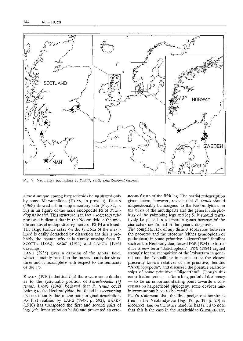
Distribution (Fig. 7). Norway: Korshavn, Lindesnes (SARS, 1911); Korsfjorden (DRZYCIMSKI, 1969). Sweden: Gullmar Fjord (LANG, 1948); Bonden, Bohuslan (POR, 1964; KUNZ, pers. comm.). Scotland: lnchkeith (T. SCOTT, 1903, 1906) and St. Monans (T. SCOTT, 1892, 1903, 1906) in the Firth of Forth; Ballantrae Bank in the Firth of Clyde (T. & A. SCOTT, 1896); Firth of Clyde (T. SCOTT, 1903). England: Isle of Man (by l.C. THOMPSON: in T. SCOTT, 1906).
A
~(t(';
~~
~I y/'.\.i\..
11<·\f n~
.·,~:;·,:.://~·";-:./;:.:( ~ .
' ~~·>'"'':':>i:.<•:',:.
r w:1~t':l~~.G~t
50 µm
B
20µm
c
135
136 Rony HUYS
REDESCRIPTION
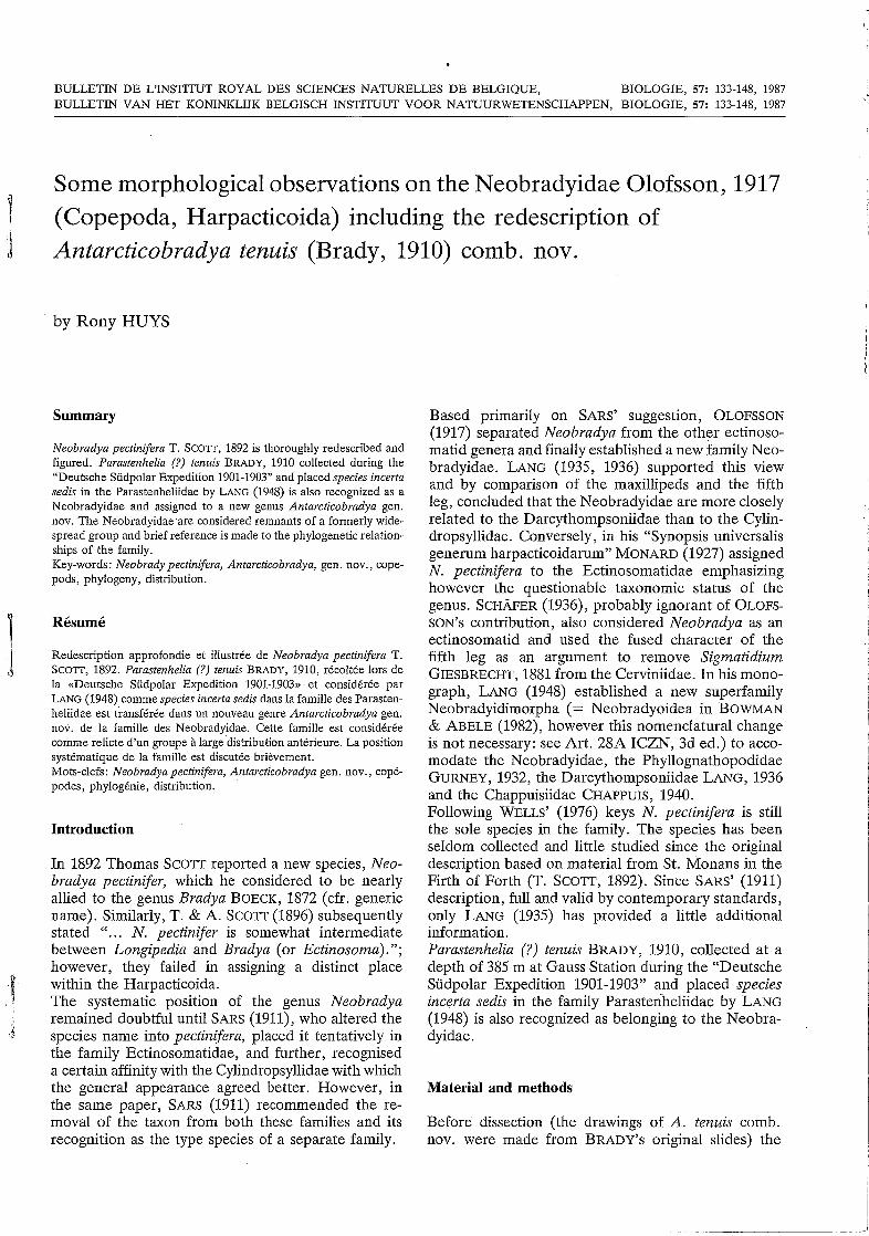
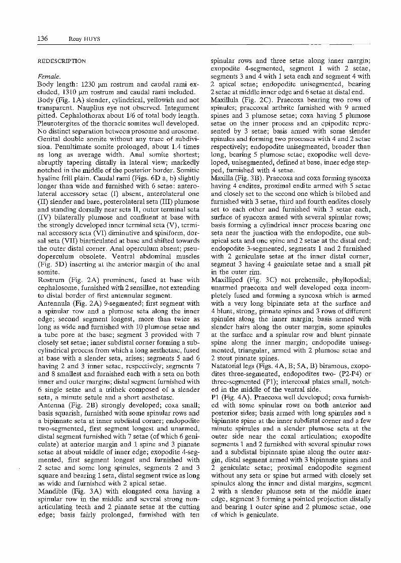
Female. Body length: 1230 µm rostrum and caudal rami excluded, 1310 µm rostrum and caudal rami included. Body (Fig. lA) slender, cylindrical, yellowish and not transparent. Nauplius eye not observed. Integument pitted. Cephalothorax about 1/6 of total body length. Pleurotergites of the thoracic somites well developed. No distinct separation between prosome and urosome. Genital double somite without any trace of subdivision. Penultimate somite prolonged, about 1.4 times as long as average width. Anal somite shortest; abruptly tapering distally in lateral view; markedly notched in the middle of the posterior border. Somitic hyaline frill plain. Caudal rami (Figs. 6D a, b) slightly longer than wide and furnished with 6 setae: anterolateral accessory setae (I) absent, anterolateral one (II) slender and bare, posterolateral seta (III) plumose and standing dorsally near seta II, outer terminal seta (IV) bilaterally plumose and confluent at base with the strongly developed inner terminal seta (V), terminal accessory seta (VI) diminutive and spiniform, dorsal seta (VII) biarticulated at base and shifted towards the outer distal corner. Anal operculum absent; pseudoperculum obsolete. Ventral abdominal muscles (Fig. SD) inserting at the anterior margin of the anal somite. Rostrum (Fig. 2A) prominent, fused at base with cephalosome, furnished with 2 sensillae, not extending to distal border of first antennular segment. Antennula (Fig. 2A) 9-segmented; first segment with a spinular row and a plumose seta along the inner edge; second segment longest, more than twice as long as wide and furnished with 10 plumose setae and a tube pore at the base; segment 3 provided with 7 closely set setae; inner subdistal corner forming a subcylindrical process from which a long aesthetasc, fused at base with a slender seta, arises; segments 5 and 6 having 2 and 3 inner setae, respectively; segments 7 and 8 smallest and furnished each with a seta on both inner and outer margins; distal segment furnished with 6 single setae and a trithek composed of a slender seta, a minute setule and a short aesthetasc. Antenna (Fig. 2B) strongly developed; coxa small; basis squarish, furnished with some spinular rows and a bipinnate seta at inner subdistal corner; endopodite two-segmented, first segment longest and unarmed, distal segment furnished with 7 setae (of which 6 geniculate) at anterior margin and 1 spine and 3 pinnate setae at about middle of inner edge; exopodite 4-segmented, first segment longest and furnished with 2 setae and some long spinules, segments 2 and 3 square and bearing 1 seta, distal segment twice as long as wide and furnished with 2 apical setae. Mandible (Fig. 3A) with elongated coxa having a spinular row in the middle and several strong nonarticulating teeth and 2 pinnate setae at the cutting edge; basis fairly prolonged, furnished with ten
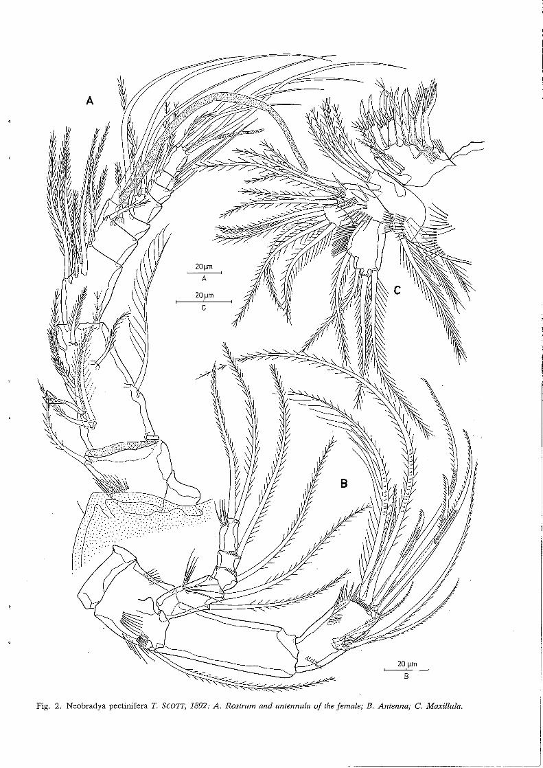
spinular rows and three setae along inner margin; exopodite 4-segmented, segment 1 with 2 setae, segments 3 and 4 with 1 seta each and segment 4 with 2 apical setae; endopodite unisegmented, bearing 2 setae at middle inner edge and 6 setae at distal end. Maxillula (Fig. 2C). Praecoxa bearing two rows of spinules; praecoxal arthrite furnished with 9 armed spines and 3 plumose setae; coxa having 5 plumose setae on the inner process and an epipodite represented by 3 setae; basis armed with some slender spinules and forming two processes with 4 and 2 setae respectively; endopodite unisegmented, broader than long, bearing 5 plumose setae; exopodite well developed, unisegmented, defined at base, inner edge stepped, furnished with 4 setae. Maxilla (Fig. 3B). Praecoxa and coxa forming syncoxa having 4 endites, proximal endite armed with 5 setae and closely set to the second one which is bilobed and furnished with 3 setae, third and fourth endites closely set to each other and furnished with 3 setae each, surface of syncoxa armed with several spinular rows; basis forming a cylindrical inner process bearing one seta near the junction with the endopodite, one subapical seta and one spine and 2 setae at the distal end; endopodite 3-segmented, segments 1 and 2 furnished with 2 geniculate setae at the inner distal corner, segment 3 having 4 geniculate setae and a small pit in the outer rim. Maxilliped (Fig. 3C) not prehensile, phyllopodial; unarmed praecoxa and well developed coxa incompletely fused and forming a syncoxa which is armed with a very long bipinriate seta at the surface and 4 blunt, strong, pinnate spines and 3 rows of different spinules along the inner margin; basis armed with slender hairs along the outer margin, some spinules at the surface and a spinular row and blunt pinnate spine along the inner margin; endopodite unisegmented, triangular, armed with 2 plumose setae and 2 stout pinnate spines. Natatorial legs (Figs. 4A, B; SA, B) biramous, exopodites three-segmented, endopodites two- (P2-P4) or three-segmented (Pl); intercoxal plates small, notched in the middle of the ventral side. Pl (Fig. 4A). Praecoxa well developed; coxa furnished with some spinular rows on both anterior and posterior sides; basis armed with long spinules and a bipinnate spine at the inner subdistal corner and a few minute spinules and a slender plumose seta at the outer side near the coxal articulation; exopodite segments 1 and 2 furnished with several spinular rows and a subdistal bipinnate spine along the outer margin, distal segment armed with 3 bipinnate spines and 2 geniculate setae; proximal endopodite segment without any seta or spine but armed with closely set spinules along the inner and distal margins, segment 2 with a slender plumose seta at the middle inner edge, segment 3 forming a pointed projection distally and bearing 1 outer spine and 2 plumose setae, one of which is geniculate.
A
20µm
8
Fig. 2. Neobradya pectinifera T. SCOTT, 1892: A. Rostrum and antennula of the female; B. Antenna; C. Maxillula.

138 Rony HUYS
Fig. 3. Neobradya pectinifera T. SCOTT, 1892: A. Mandible; B. Maxilla (arrow indicating a small pit in the outer rim of the distal endopodite segment); C. Maxilliped.
25 µm
AB
25 µm
D
20 µm
c
Morpholo gy of Neobrad "d y1 ae 139
--··· j

140 Rony HUYS
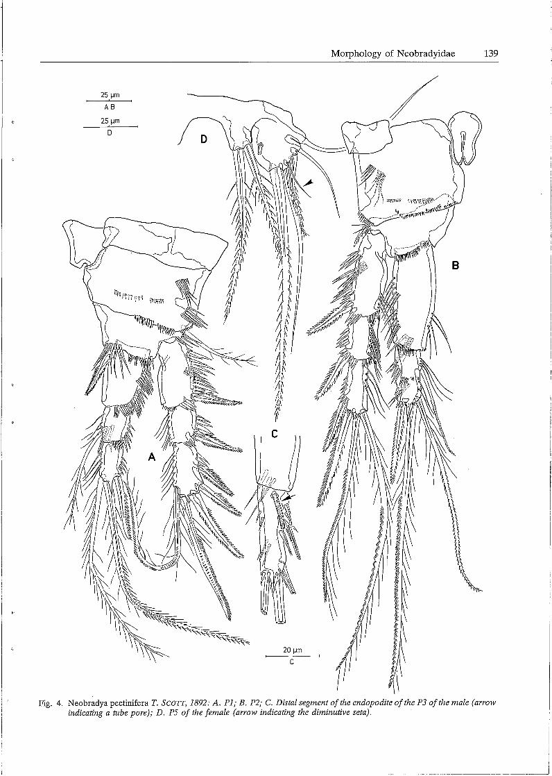
P2-P4 (Figs. 4B; SA, B). Praecoxa represented as a small rectangular plate at the outer subproximal corner; coxa square, well developed; basis forming a sharp process (in all probability with a secretary pore) at the inner subdistal corner and armed with a pinnate spine (P2) or a plumose seta (P3-P4) at the outer edge; first endopodite segment strongly prolonged, without any seta or spine; distal endopodite segment forming a sharp projection distally; seta- and spine formulae of the swimming legs are given in table 1.
Table 1 Spine- and seta formulae of the swimming legs in Neobradya pectinifera T. SCOTT, 1892.
exopodite endopodite
Pl 0.0.122 0.1.111 P2 0.0.112 0.211 P3 0.0.112 0.211 P4 0.0.122 0.111
PS (Fig. 4D). Fifth pair of legs fused medially; baseoendopodite and exopodite partially confluent; inner process of baseoendopodite armed with 2 bipinnate setae, outer process with a smooth slender seta; exopodite almost circular, furnished with a remarkable tube pore at the inner part of the surface and armed with S setae, the innermost one longest and bipinnate, the following one bare and slender, the middle one diminutive.
Genital complex (Fig. 1 C) situated in anterior half of genital double somite, furnished with a short, bipinnate spine and a long slender seta (which are non articulating and confluent at base) at both sides; genital aperture clearly visible.
Male. Body length: lOSO µm rostrum and caudal rami excluded, 1110 µm rostrum and caudal rami included. Habitus as in the female except for the free genital somites (Figs. 6A, B). Sexual dimorphism in the antennula, endopodite P3, fifth and sixth legs. Antennula (Fig. lB) 10-segmented, subchirocer; first segment furnished with an inner seta and some spinular rows; second segment longest and provided with a tube pore and 11 plumose setae; third segment relatively short and having 9 setae; fourth one well-developed and bearing 10 setae, one of which is fused at base with a long aethetasc; specialized joint between segments 3 and 4 allowing considerable lateral flexion of the distal portion; segment S having 2 setae; segments 6 and 7 forming subchirocer apparatus and furnished with a slender seta and several spinous structures each; segments 8 and 9 smallest and provided each with a seta on both inner and outer subdistal
corners; distal segment forming a long spinous process and armed with 7 setae and a short aesthetasc. Leg 3 (Fig. 4C). Protopodite, exopodite and proximal endopodite segment exactly as in the female; distal endopodite segment with the same setal arrangement as in the female, however, more slender and provided with a remarkable tube pore at the anterior surface and near the articulation with segment l. PS (Fig. 6C). Fifth pair of legs fused medially; baseoendopodite and exopodite partially confluent; endopodital part of baseoendopodite partially free and furnished with 2 bipinnate setae; exopodite almost circular and provided with a tube pore at the surface and armed with S setae: the outermost one bare and slender, the middle one small and bi-articulated at base, the others bipinnate and of different lengths. P6 (Fig. SC). Sixth pair of legs symmetrical, partially fused in the middle; each represented by an oval plate and furnished at the outer corner with 3 setae: outer seta longest and plumose, middle one smooth and slender, inner one shortest and bi pinnate. Spermatophore (Figs. 6A, B) elongate, relatively small.
Antarcticobradya gen. nov.
Diagnosis: Neobradyidae. Second and third segments of antennula almost as long as wide. Both antenna! endopodite segments equal in length. Proximal endopodite segment of P2-P4 squarish. Distal segment of endopodite P4 with inner seta. Baseoendopodite and exopodite PS completely confluent in the female; exopodite with 4 setae, the second innermost of which is thick, stout and bipinnate. Penultimate somite less than 2 times as long as anal somite. Type and sole species: Antarcticobradya tenuis (BRADY, 1910) comb. nov. Gender: feminine.
Antarcticobradya tenuis (BRADY, 1910) comb. nov.
Synonymy: Parastenhelia (?) tenuis BRADY, 1910.
Material: Antarctica, Kaiser Wilhelm II Land, Posadowsky Bay, Gauss Station: 1 female dissected on slide and deposited in the Hancock Museum, Newcastle upon Tyne, under no. 2.44.10.
Distribution (Fig. 8A). Antarctica, Kaiser Wilhelm II Land, Posadowsky Bay, Gauss Station (BRADY, 1910).
REDESCRIPTION
Female. Body length: according to the original description by BRADY (1910) about 770 µm; length of the urosoma
Morphology of Neobradyidae 141
··.... ... .... \ /
c
8
25 µm
A-C
20 µm
D
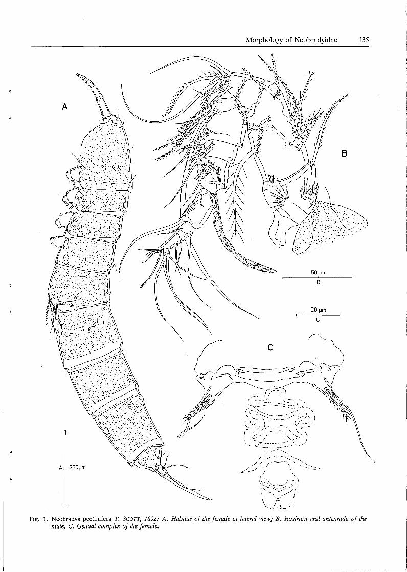
Fig. 5. Neobradya pectinifera T. SCOTT, 1892: A. P3; B. P4; C. P6 of the male. D. Ventral abdominal muscles in the penultimate somite.
.. _J
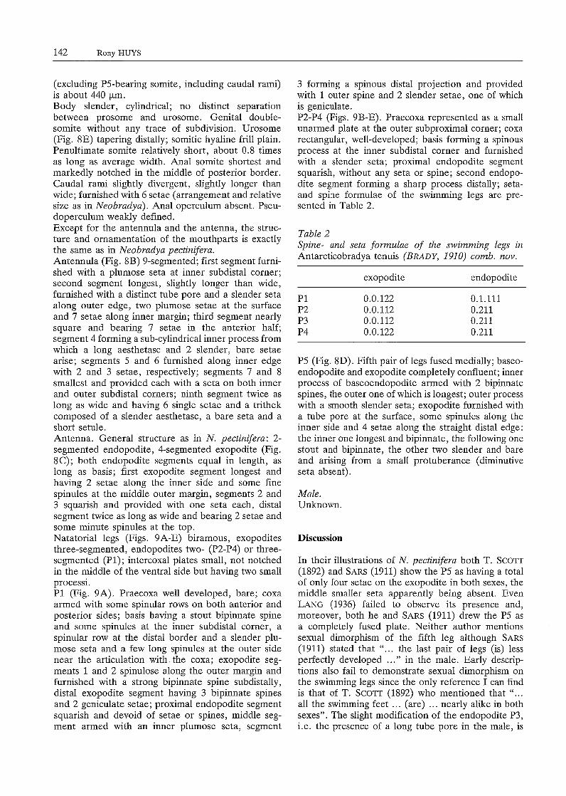
142 Rony HUYS
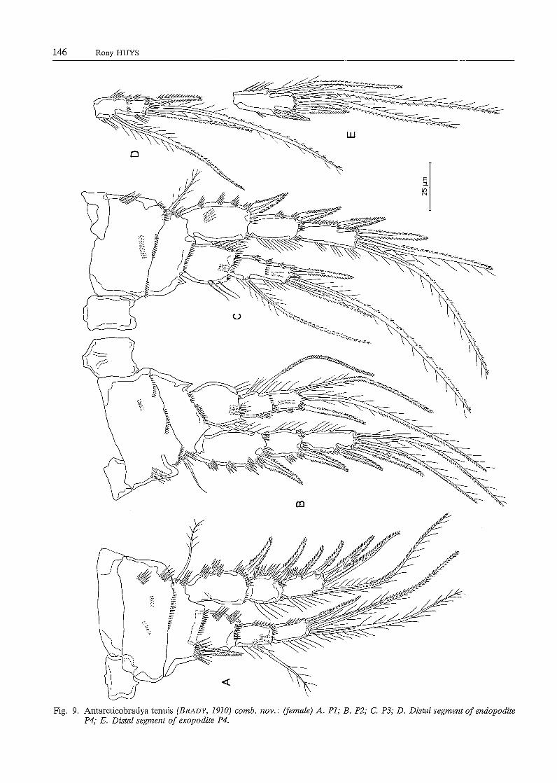
(excluding PS-bearing somite, including caudal rami) is about 440 µm. Body slender, cylindrical; no distinct separation between prosome and urosome. Genital doublesomite without any trace of subdivision. Urosome (Fig. 8E) tapering distally; somitic hyaline frill plain. Penultimate somite relatively short, about 0.8 times as long as average width. Anal somite shortest and markedly notched in the middle of posterior border. Caudal rami slightly divergent, slightly longer than wide; furnished with 6 setae (arrangement and relative size as in Neobradya). Anal operculum absent. Pseudoperculum weakly defined. Except for the antennula and the antenna, the structure and ornamentation of the mouthparts is exactly the same as in Neobradya pectinifera. Antennula (Fig. 8B) 9-segmented; first segment furnished with a plumose seta at inner subdistal corner; second segment longest, slightly longer than wide, furnished with a distinct tube pore and a slender seta along outer edge, two plumose setae at the surface and 7 setae along inner margin; third segment nearly square and bearing 7 setae in the anterior half; segment 4 forming a sub-cylindrical inner process from which a long aesthetasc and 2 slender, bare setae arise; segments S and 6 furnished along inner edge with 2 and 3 setae, respectively; segments 7 and 8 smallest and provided each with a seta on both inner and outer subdistal corners; ninth segment twice as long as wide and having 6 single setae and a trithek composed of a slender aesthetasc, a bare seta and a short setule. Antenna. General structure as in N. pectinifera: 2-segmented endopodite, 4-segmented exopodite (Fig. 8C); both endopodite segments equal in length, as long as basis; first exopodite segment longest and having 2 setae along the inner side and some fine spinules at the middle outer margin, segments 2 and 3 squarish and provided with one seta each, distal segment twice as long as wide and bearing 2 setae and some minute spinules at the top. Natatorial legs (Figs. 9A-E) biramous, exopodites three-segmented, endopodites two- (P2-P4) or threesegmented (Pl); intercoxal plates small, not notched in the middle of the ventral side but having two small processi. Pl (Fig. 9A). Praecoxa well developed, bare; coxa armed with some spinular rows on both anterior and posterior sides; basis having a stout bi pinnate spine and some spinules at the inner subdistal corner, a spinular row at the distal border and a slender plumose seta and a few long spinules at the outer side near the articulation with the coxa; exopodite segments 1 and 2 spinulose along the outer margin and furnished with a strong bipinnate spine subdistally, distal exopodite segment having 3 bipinnate spines and 2 geniculate setae; proximal endopodite segment squarish and devoid of setae or spines, middle segment armed with an inner plumose seta, segment
3 forming a spinous distal projection and provided with 1 outer spine and 2 slender setae, one of which is geniculate. P2-P4 (Figs. 9B-E). Praecoxa represented as a small unarmed plate at the outer subproximal corner; coxa rectangular, well-developed; basis forming a spinous process at the inner subdistal corner and furnished with a slender seta; proximal endopodite segment squarish, without any seta or spine; second endopodite segment forming a sharp process distally; setaand spine formulae of the swimming legs are presented in Table 2.
Table 2 Spine- and seta formulae of the swimming legs in Antarcticobradya tenuis (BRADY, 1910) comb. nov.
exopodite endopodite
Pl 0.0.122 0.1.111 P2 0.0.112 0.211 P3 0.0.112 0.211 P4 0.0.122 0.211
PS (Fig. 8D). Fifth pair of legs fused medially; baseoendopodite and exopodite completely confluent; inner process of baseoendopodite armed with 2 bipinnate spines, the outer one of which is longest; outer process with a smooth slender seta; exopodite furnished with a tube pore at the surface, some spinules along the inner side and 4 setae along the straight distal edge: the inner one longest and bipinnate, the following one stout and bipinnate, the other two slender and bare and arising from a small protuberance (diminutive seta absent).
Male. Unknown.
Discussion
In their illustrations of N. pectinifera both T. Scorr (1892) and SARS (1911) show the PS as having a total of only four setae on the exopodite in both sexes, the middle smaller seta apparently being absent. Even LANG (1936) failed to observe its presence and, moreover, both he and SARS (1911) drew the PS as a completely fused plate. Neither author mentions sexual dimorphism of the fifth leg although SARS (1911) stated that " ... the last pair of legs (is) less perfectly developed ... " in the male. Early descrip-tions also fail to demonstrate sexual dimorphism on the swimming legs since the only reference I can find is that of T. Scorr (1892) who mentioned that " ... all the swimming feet ... (are) ... nearly alike in both sexes". The slight modification of the endopodite P3, i.e. the presence of a long tube pore in the male, is
Morphology of Neobradyidae 143
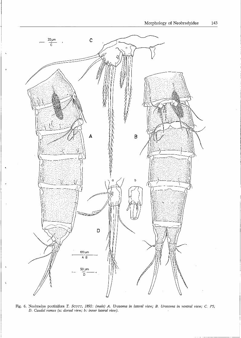
Fig. 6. Neobradya pectinifera T. SCOTT, 1892: (male) A. Urosoma in lateral view; B. Urosoma in ventral view; C. P5; D. Caudal ramus (a: dorsal view; b: inner lateral view).
144 Rony HUYS
NORWAY
~AN
I
i i
( .... ;
" i _;
.i
Fig. 7. Neobradya pectinifera T. SCOTT, 1892: Distributional records.
almost unique among harpacticoids being shared only by some Marsteiniidae (HUYS, in press b). BODIN (1968) showed a thin supplementary seta (Fig. 32, p. 56) in his figure of the male endopodite P3 of Tachidiopsis bozici. This structure is in fact a secretary tube pore and indicates that in the Neobradyidae the middle and distal endopodite segments of P2-P4 are fused. The large surface setae on the syncoxa of the maxilliped is easily detatched by dissection ant this is probably the reason why it is simply missing from T. SCOTT'S (1892), SARS' (1911) and LANG'S (1936) drawings. LANG (1935) gives a drawing of the genital field, which is mainly based on the internal cuticular structures and is incomplete with respect to the remnants of the P6.
BRADY (1910) admitted that there were some doubts as to the systematic position of Parastenhelia (?) tenuis. LANG (1948) believed that P. tenuis could belong to the Neobradyidae, but failed in ascertaining its true identity due to the poor original description. As first realised by LANG (1948, p. 592), BRADY (1910) has transposed the first and second pairs of legs ( cfr. inner spine on basis) and presented an erro-
neous figure of the fifth leg. The partial redescription given above, however, reveals that P. tenuis should unquestionably be assigned to the Neobradyidae on the basis of the mouthparts and the general morphology of the swimming legs and leg 5. It should tentatively be placed in a separate genus because of the characters mentioned in the generic diagnosis. The complete lack of any distinct separation between the prosome and the urosome (either gymnoplean or podoplean) in some primitive "oligoarthran" families such as the Neobradyidae, forced POR (1984) to introduce a new term "dolichoplean". POR (1984) argued strongly for the recognition of the Polyarthra in general and the Canuellidae in particular as the closest presently known relatives of the primitive, benthic "Archicopepoda", and discussed the possible relationships of some primitive "Oligoarthra". Though this contribution seems - after a long period of dormancy - to be an important starting point towards a concensus on harpacticoid phylogeny, some obvious misinterpretations have to be rectified. POR's statement that the first pedigerous somite is free in the Neobradyidae (Fig. 19, p. 19; p. 20) is incorrect, and on the other hand, he has failed to note that this is the case in the Aegisthidae GIESBRECHT,
E ...........
i:t.r:•:::":::::.'..·:\ ,::: ·.;.;· : .. · ·.·.· .>x:~1 .... ·.:. :·
:· . ...... :: ·.· .:· .. · .. ·: . ... : .·. . ........ ·.-: I ~,·~t2:/:·:·.:'.:.:: ... ·::.:::;:.:.:t·>,) \
E 100µm
. . . . ....... . ·.···:: ::·.·:::i:.::::·.·· .. · ' ··· .. ·· .. ·.· .. ·:·:_.,-._· ...
. . .. . .. '. .. .'· ~-:.:::: :: ·. ·.··· ·.' . ' .. : ............... ·
I~'
B
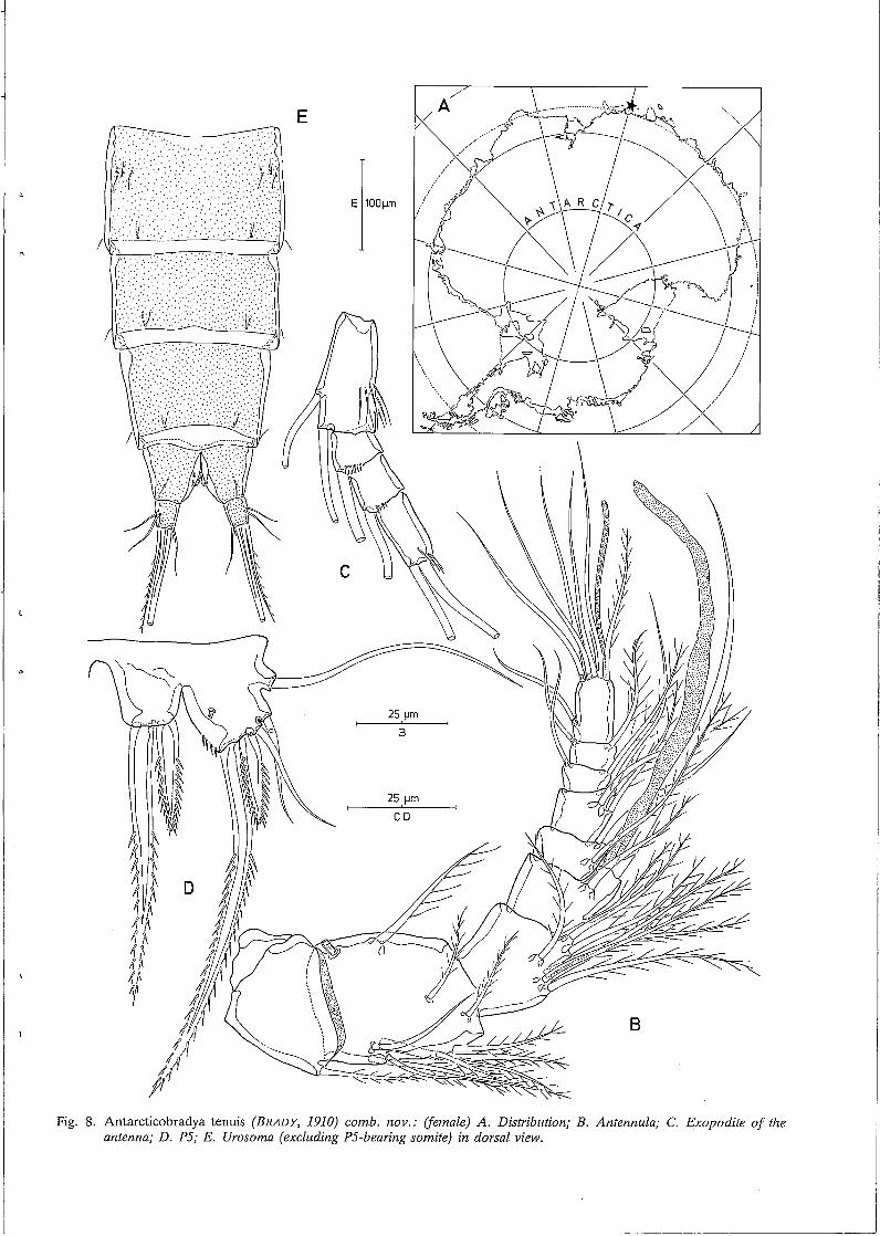
Fig. 8. Antarcticobradya tenuis (BRADY, 1910) comb. nov.: (female) A. Distribution; B. Antennula; C. Exopodite of the antenna; D. P5; E. Urosoma (excluding P5-bearing somite) in dorsal view.
___ j
146 Rony HUYS
Fig. 9. Antarcticobradya tenuis (BRADY, 1910) comb. nov.: (female) A. PI; B. P2; C. P3; D. Distal segment of endopodite P4; E. Distal segment of exopodite P4.
1892, in some Latiremidae BOZIC, 1969 and in Cubanocleta PETKOVSKI, 1977. According to PoR (1984, p. 13) (probably) the most important character which unites the Canuellidae and the Longipediidae is the foliaceous maxilliped. However, this character is also shared by the Neobradyidae and the Marsteiniidae DRZYCIMSKI, 1969 (HUYS, in press b), and, consequently, his statement that " ... in all other Copepoda the maxilliped is stenopodial", cannot be supported. Finally, his identification of N. pectinifera is probably incorrect since the specimen in his photograph (Fig. 1, p. 3) has long caudal rami and a habitus quite different from the real N. pectinifera. It is even possible that this specimen belongs to another family (Paranannopidae ?) since it shows a remarkable resemblance with Cylindronannopus.
The origin and evolution of the Neobradyidae can be regarded as an early attempt at the exploitation of the interstitial environment in coarse sediments. However, they have obviously been superseded by the more succesfull (although primitive) Paramesochridae and the advanced Cylindropsyllidae. They form without doubt a phylogenetically ancient taxon and their current zoogeographical pattern can be interpreted as being that of a relict group which was formerly widespread. In this context it is surprising that the Neobradyidae have retained their morphological integrity over such long distances, Neobradya
References
BODIN, P., 1968. Copepodes harpacticoides des etages bathyal et abyssal du Golfe de Gascogne. Memoires du Museum National d' Histoire Naturelle, Nouvelle Serie, Serie A, Zoologie, 55 (1): 1-107.
BOWMAN, T.E. & ABELE, L.G., 1982. Classification of the recent Crustacea. In: ABELE, L.G. (Editor), The Biology of the Crustacea. Vol. 1 Systematics, the Fossil Record and Biogeography, pp. 1-29.
BOXSHALL, G.A., 1985. The comparative anatomy of two copepods, a predatory calanoid and a particle-feeding mormonilloid. Philosophical Transactions of the Royal Society of London, B311: 303-377.
BRADY, G.S., 1910. Die marinen Copepoden der deutschen Siidpolar-Expedition. Deutsche Sudpolar-Expedition 1901-1903, ll, Zoologie 3: 499-593.
DRZYCIMSKI, I., 1969. Rarpacticoida (Copepoda; wad morkich okolich Bergen (zachodnie wybrzese norwegii) i ich ekologia. Rozprawy Wyzsza Szkola Rolnicza w Szczecinie, 17: 1-72.
RUYS, R., (in press, a). A redescription of the presumed associated Caligopsyllus primus Kunz, 1975 (Rarpacticoida, Paramesochridae) with emphasis on its phylogenetic affinity with Apodopsyllus Kunz, 1962. Hydrobiologia, in press.
Morphology of Neobradyidae 147
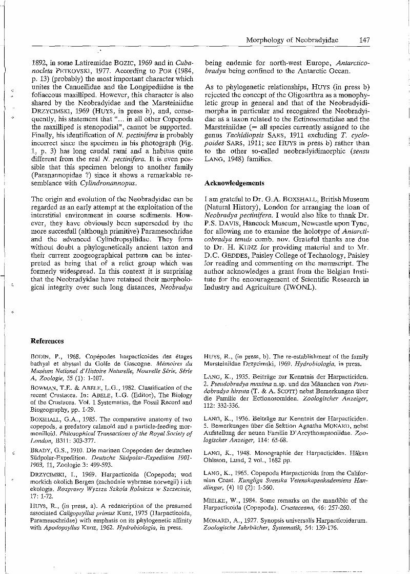
being endemic for north-west Europe, Antarcticobradya being confined to the Antarctic Ocean.
As to phylogenetic relationships, RUYS (in press b) rejected the concept of the Oligoarthra as a monophyletic group in general and that of the Neobradyidimorpha in particular and recognized the Neobradyidae as a taxon related to the Ectinosomatidae and the Marsteiniidae ( = all species currently assigned to the genus Tachidiopsis SARS, 1911 excluding T. cyclopoides SARS, 1911; see HUYS in press b) rather than to the other so-called neobradyidimorphic (sensu LANG, 1948) families.
Acknowledgements
I am grateful to Dr. G.A. BOXSHALL, British Museum (Natural History), London for arranging the loan of Neobradya pectinifera. I would also like to thank Dr. P .S. DAVIS, Hancock Museum, Newcastle upon Tyne, for allowing me to examine the holotype of Antarcticobradya tenuis comb. nov. Grateful thanks are due to Dr. H. KUNZ for providing material and to Mr. D.C. GEDDES, Paisley College of Technology, Paisley for reading and commenting on the manuscript. The author acknowledges a grant from the Belgian Institute for the encouragement of Scientific Research in Industry and Agriculture (IWONL).
RUYS, R., (in press, b). The re-establishment of the family Marsteiniidae Drzycimski, 1969. Hydrobiologia, in press.
LANG, K., 1935. Beitrage zur Kenntnis der Rarpacticiden. 2. Pseudobradya maxima n.sp. und des Mannchen von Pseudobradya hirsuta (T. & A. SCOTT) nebst Bemerkungen iiber die Familie der Ectionosomiden. Zoologischer Anzeiger, 112: 332-336.
LANG, K., 1936. Beitrage zur Kenntnis der Rarpacticiden. 5. Bemerkungen iiber die Sektion Agnatha MONARD, nebst Aufstellung der neuen Familie D'Arcythompsoniidae. Zoologischer Anzeiger, 114: 65-68.
LANG, K., 1948. Monographie der Rarpacticiden. Rakan Ohlsson, Lund, 2 vol., 1682 pp.
LANG, K., 1965. Copepoda Rarpacticoida from the Californian Coast. Kungliga Svenska Vetenskapsakademiens Handlingar, (4) 10 (2): 1-560.
MIELKE, W., 1984. Some remarks on the mandible of the Rarpacticoida (Copepoda). Crustaceana, 46: 257-260.
MONARD, A., 1927. Synopsis universalis Rarpacticoidarum. Zoologische Jahrbucher, Systematik, 54: 139-176.
148 Rony HUYS
OLOFFSON, 0., 1917. Beitrag zur Kenntnis der Harpacticiden-Familien Ectinosomidae, Canthocamptidae (Gen. Maraenobiotus) und Tachidiidae nebst Beschreibungen einiger neuen und wenig bekannten arktischen Brackwasserund Siisswasser-Arten. Zoologiska Bidrag fran Uppsala, 6: 1-39.
POR, F.D., 1964. Les harpacticoides (Copepoda Crustacea) des fonds meubles du Skagerak. Cahiers de Biologie marine, 5 (3): 233-270.
POR, F.D., 1984. Canuellidae LANG (Harpacticoida, Polyarthra) and the ancestor of the Copepoda. Crustaceana, Suppl. 7: 1-24.
SARS, G.O., 1911. An Account of the Crustacea of Norway. V. Copepoda, Harpacticoida. Bergen Museum, pp. 369-449.
SCHAFER, H.W., 1936. Harpacticoiden aus dem Brackwasser der Insel Hiddensee. Zoologische Jahrbiicher, Abteilung fur Systematik, Okologie und Geographie der Tiere, 68 (6): 445-588.
SCOTT, T., 1892. Additions to the fauna of the Firth of Forth. Part IV. Tenth Annual Report of the Fishery Board for Scotland for the year 1891, Part III. Scientific Investigations: 244-272.
SCOTT, T., 1903. On some new and rare Crustacea collected at various times in connection with the investigations of the
Note added in proof
Since this paper was submitted for publication, SCHMINKE (pers. comm.) found several males and females of A. tenuis in the Antarctic. In addition to the peculiar tube pore on the endopodite P3, the male
Fishery Boards for Scotland. Twenty-first Annual Report of the Fishery Board for Scotland for the year 1902, Part III. Scientific Investigations: 109-135.
SCOTT, T., 1906. A catalogue of land, fresh-water, and marine Crustacea found in the bassin of the river Forth and its estuary. Part II. Proceedings of the Royal Physical Society of Edinburgh, 16: 267-386.
SCOTT, T. & A., 1896. On some new and rare Copepoda from the Clyde. Annals of Scottish Natural History, 1896: 224-230.
WELLS, J.B.J., 1976. Keys to aid in the identification of marine harpacticoid copepods. Department of Zoology, University of Aberdeen, U.K., pp. 215.
WELLS, J.B.J., 1986. Biogeography of benthic harpacticoid copepods of the marine littoral and continental shelf. Syllogeus, 58: 126-135.
Rony HUYS Marine Biology Section
Zoology Institute State University of Gent K.L. Ledeganckstraat 35 B-9000 Gent (Belgium)
of A tenuis exhibits slight sexual dimorphism in the outer seta of the distal exopodite segment P3, a character notably absent in N. pectinifera, and supporting the separate generic status of A. tenuis.