Embed Size (px)
Citation preview
Some Surgical aspects of Vascular Access
Chris Russell
Timing of referral and placement
Ideal time is 3-6 months prior to starting dialysis! 6 weeks probably min time for maturity – dilation
of vein but also arterialisation of wall Always ‘soft’ on first needling, often ‘blow’
? Is this any different if AVF left for longer
Timing of referral and placement
ANZDATA 2009 – Aus - 38% start with adequate access NZ - 23% start with adequate access
KP today – unit variation 25 to 80 % start with CVC- surgeons not the problem
Does late access matter?
5924 incident haemodialysis patients Early access creation (≥ 4 months before
dialysis) lower risk of death, with a relative risk of 0.76
(95% CI 0.58 – 1.00) lower risk of sepsis 0.57 (95% CI 0.41 – 0.79)
Oliver et al JASN 2004
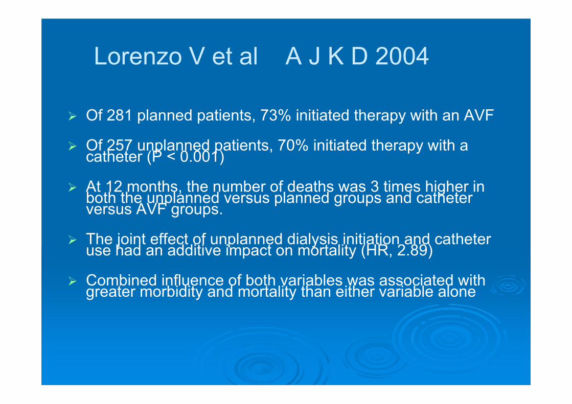
Lorenzo V et al A J K D 2004
Of 281 planned patients, 73% initiated therapy with an AVF
Of 257 unplanned patients, 70% initiated therapy with a catheter (P < 0.001)
At 12 months, the number of deaths was 3 times higher in both the unplanned versus planned groups and catheter versus AVF groups.
The joint effect of unplanned dialysis initiation and catheter use had an additive impact on mortality (HR, 2.89)
Combined influence of both variables was associated with greater morbidity and mortality than either variable alone
Problems associated with too early placement
Primary patency rate variably reported Patient may never use access
Waste of theatre time Waste of venous site
Problems associated with too early placement
Development of neo-intimal hyperplasia Occurs at sites of curve, valves, junctions etc No surviellance of access if not on dialysis Pts not often aware they have stopped and _ -
unsalvageable fistula
??? PD catheter and AVF‘CAPD first’ and not ‘fistula first’
Problems associated with too late placement
Fistula not ready at time patient needs to start dialysis
Cannulation of access before it is matured can damage the access
Problems associated with too late placement
Usually try R-C or B-C first – but be prepared to try any combination
Failure to mature due to stenosis, small vessels
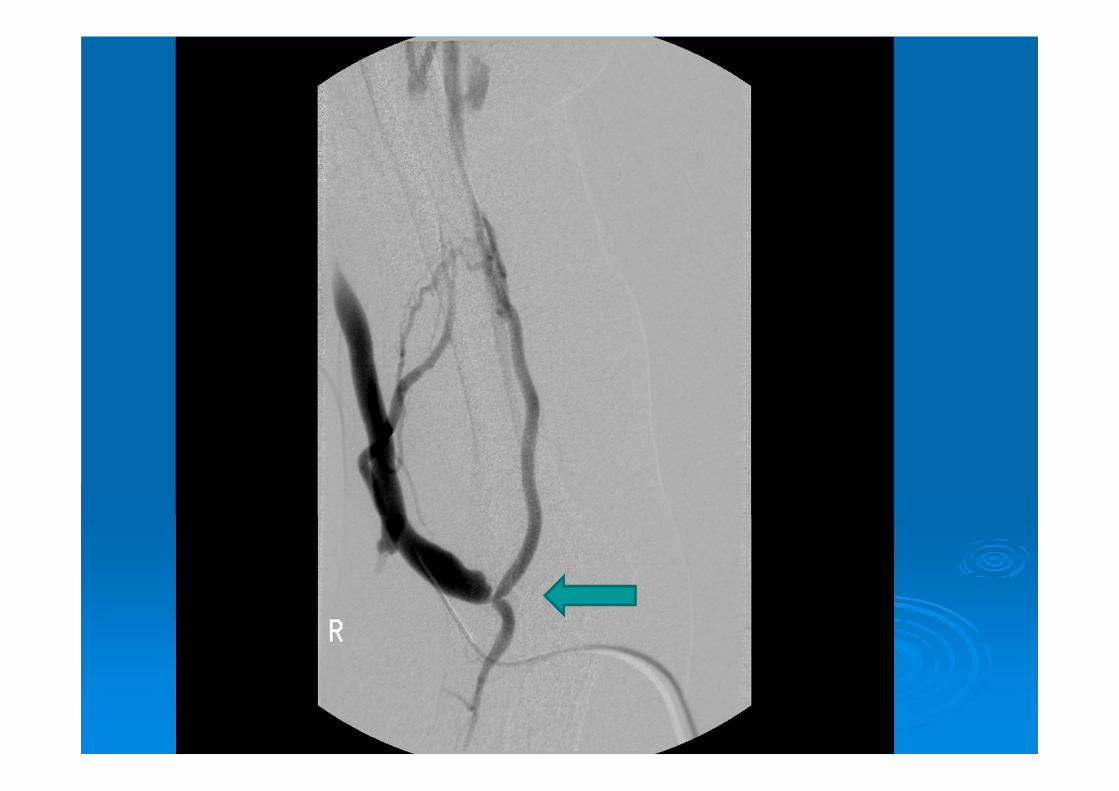
May be undiagnosed arterial inflow problems which need fixing prior to surgery
‘synthetic VAG last’
Timing of placement
Patients unpredicatable!Creat 450 for 4 years!Creat slow creep to 300, then sudden
deterioration to 650 Early referral good, so veins can be saved
Timing of referral
Waiting time for surgical OPD Waiting time to theatre Likelihood of requiring multiple procedures Likelihood of patients turning up!
Make allowance for local factors rather using them as an excuse
Upper arm AVF or graft Brachiocephalic best
Simpler procedure Basilic vein can be saved for later
primary patency for brachio-cephalic and brachio-basilic fistulas are fairly high 87% and 81% for brachio-cephalic fistulas at 1 and 3 years 86% and 73% for brachio-basilic fistulas at 1 and 3 years
Brachiobasilic versus brachiocephalic arteriovenousfistula: A prospective randomized study. Journal of Vascular Surgery 2009; 49(1):171-177.
BB AVF – allow 4 to 6 months before initial use
/////////
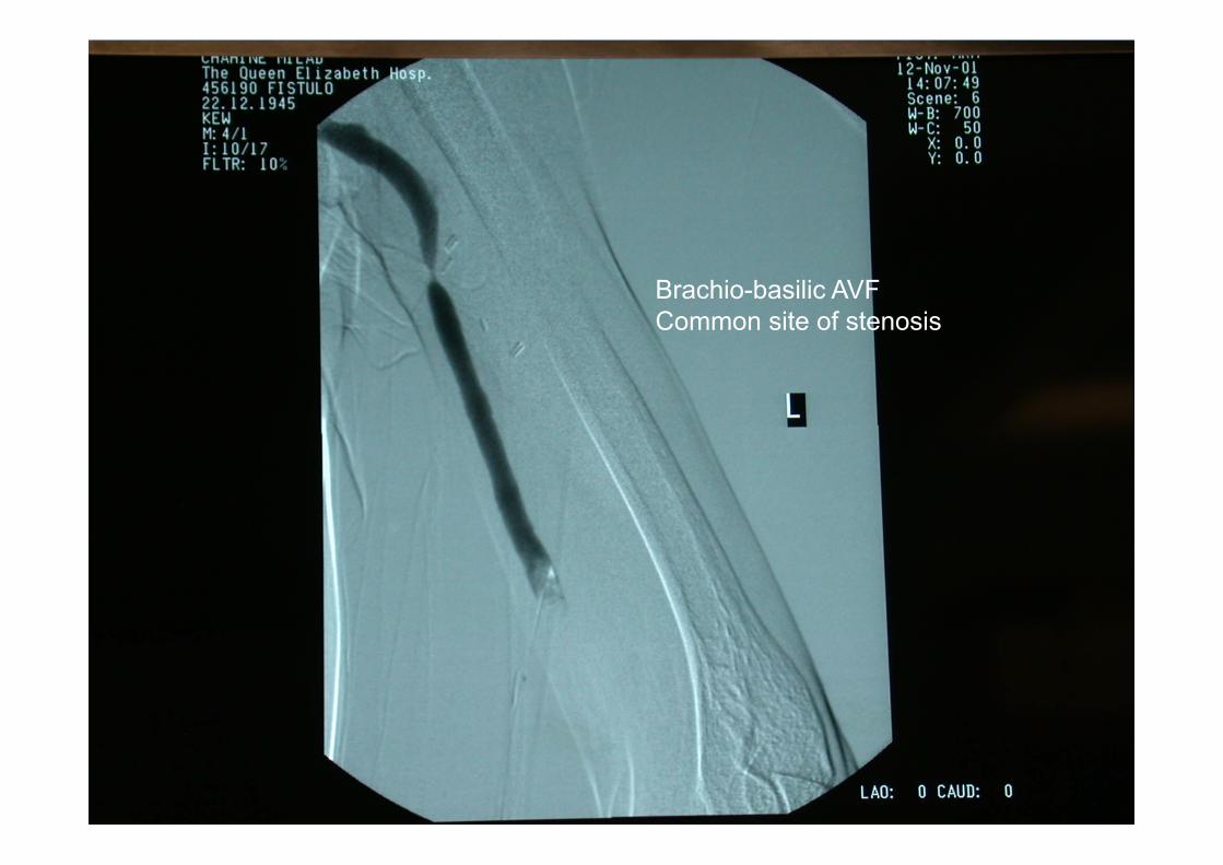
Brachio-basilic AVFCommon site of stenosis
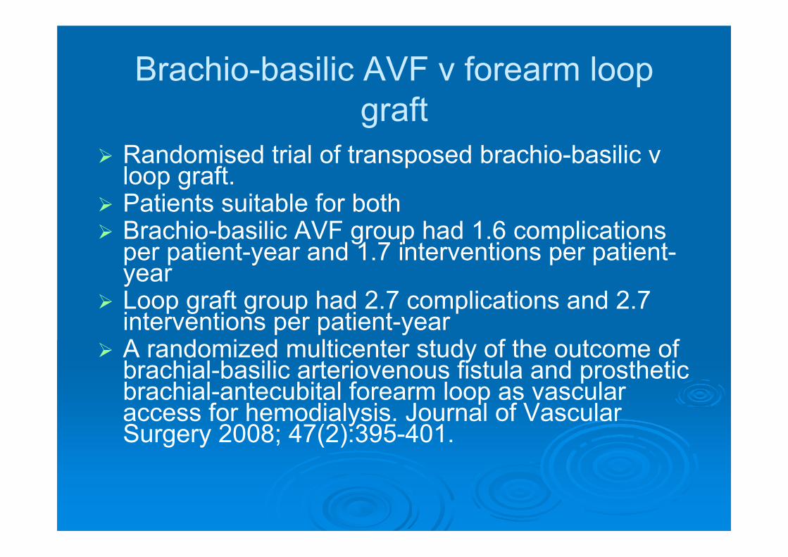
Brachio-basilic AVF v forearm loop graft
Randomised trial of transposed brachio-basilic v loop graft.
Patients suitable for both Brachio-basilic AVF group had 1.6 complications
per patient-year and 1.7 interventions per patient-year
Loop graft group had 2.7 complications and 2.7 interventions per patient-year
A randomized multicenter study of the outcome of brachial-basilic arteriovenous fistula and prosthetic brachial-antecubital forearm loop as vascular access for hemodialysis. Journal of Vascular Surgery 2008; 47(2):395-401.
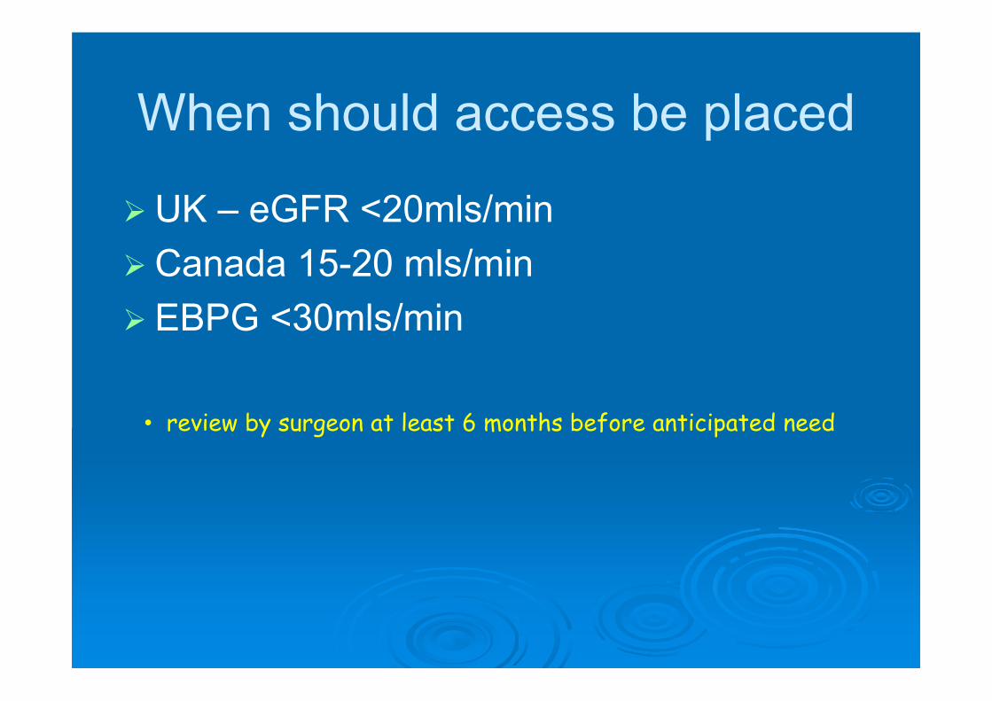
When should access be placed
UK – eGFR <20mls/minCanada 15-20 mls/min EBPG <30mls/min
• review by surgeon at least 6 months before anticipated need
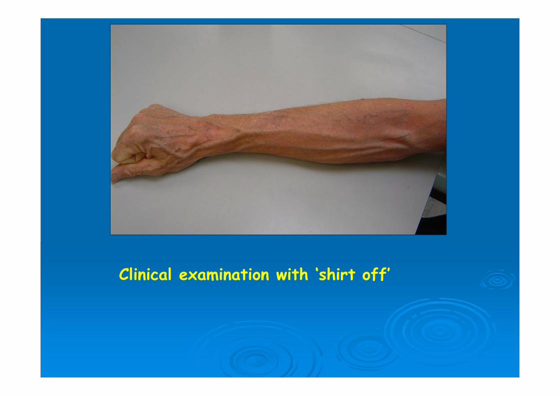
Clinical examination with ‘shirt off’
Patients likely to have difficult access
Female Peripheral vascular diseaseObese > 65yrs old – ‘mobile veins’ Fragile skin
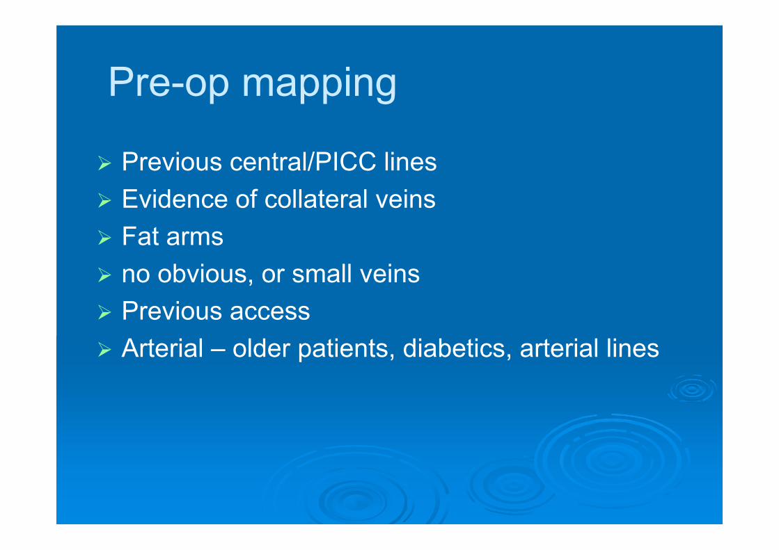
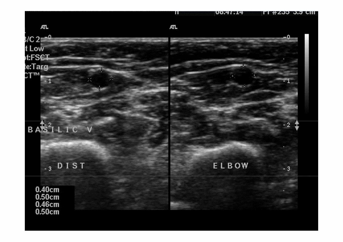
Pre-op mapping
Previous central/PICC lines Evidence of collateral veins Fat arms no obvious, or small veins Previous access Arterial – older patients, diabetics, arterial lines
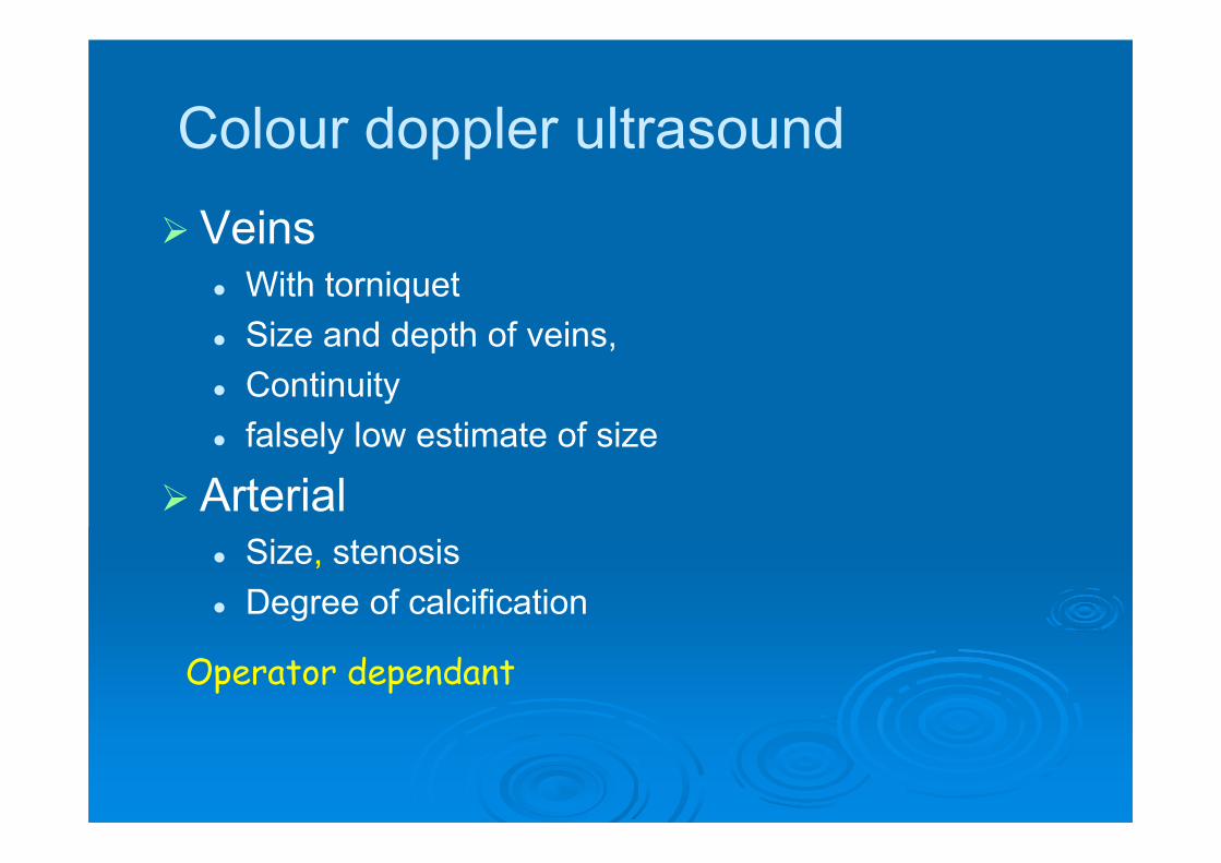
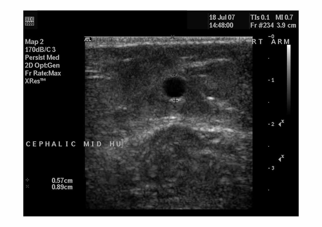
Colour doppler ultrasound Veins
With torniquet Size and depth of veins, Continuity falsely low estimate of size
Arterial Size, stenosis Degree of calcification
Operator dependant
dddddddddddddddddddddddddddddddddddddddd
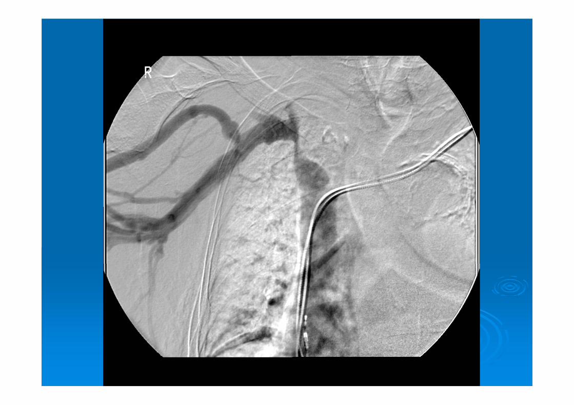
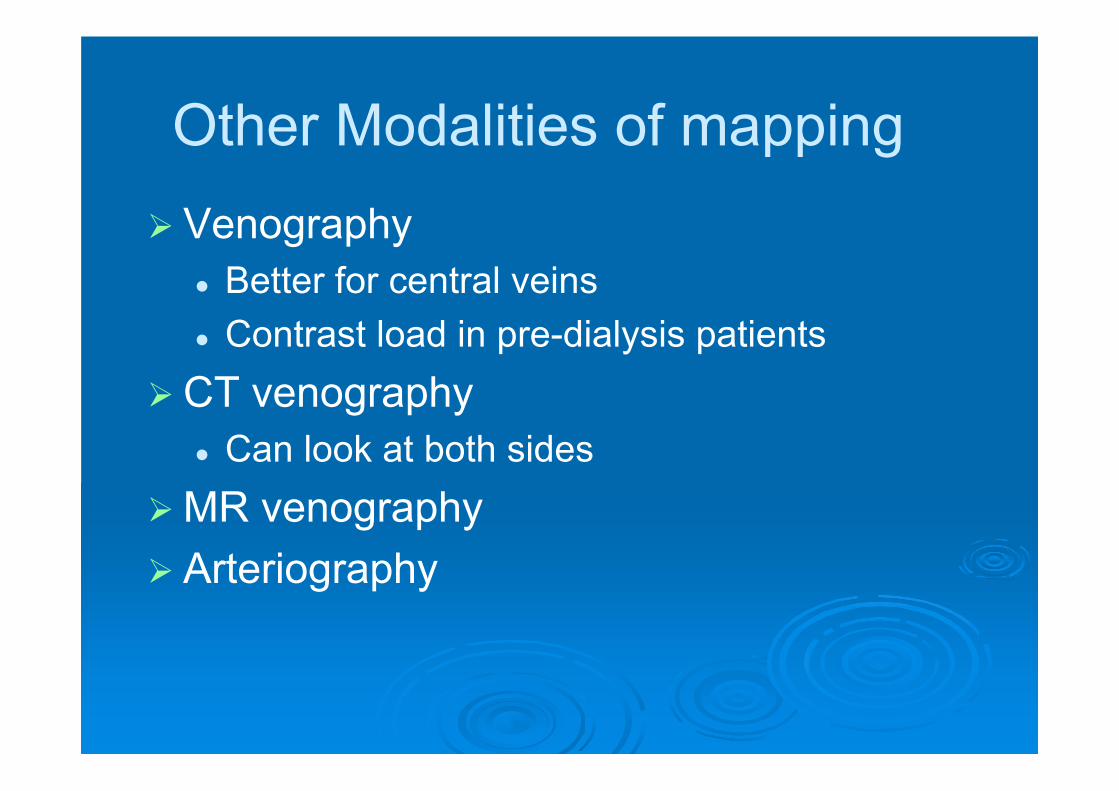
Other Modalities of mapping Venography
Better for central veins Contrast load in pre-dialysis patients
CT venography Can look at both sides
MR venography Arteriography
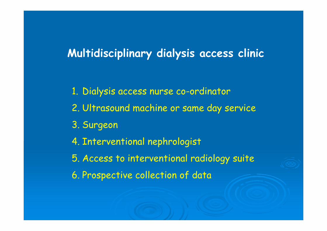
1. Dialysis access nurse co-ordinator
2. Ultrasound machine or same day service
3. Surgeon
4. Interventional nephrologist
5. Access to interventional radiology suite
6. Prospective collection of data
Multidisciplinary dialysis access clinic


![Unusual Complication of Hemodialysis Cuffed Catheter Tunnel ... · 2019. 7. 30. · hemodialysis patients with vascular access central venous catheter [2, 5]. Infection is the second](https://img.pdfslide.net/doc/110x75/6112f543c4e8093a88485054/unusual-complication-of-hemodialysis-cuffed-catheter-tunnel-2019-7-30-hemodialysis.jpg)