Embed Size (px)
Citation preview

Sonography of Cat Scratch Disease
David M. Melville, MD, Jon A. Jacobson, MD, Brian Downie, PA-C, MS, J. Sybil Biermann, MD, Sung Moon Kim, MD, Corrie M. Yablon, MD
at scratch disease is typically a self-limiting infectiouscondition, often presenting in children and adolescents as abenign regional lymphadenitis that results from a cat scratch
or bite involving the distal upper extremity.1 The initial traumaresults in inoculation with Bartonella henselae and subsequentregional lymph node enlargement, most commonly the medialepitrochlear region of the elbow.2–5 The development of a palpablemass may raise clinical concern for a soft tissue neoplasm, such assarcoma, lymphoma, or metastatic disease; therefore, imaging eval-uation is often considered.6–9
Received April 8, 2014, from the Departments ofRadiology (D.M.M., J.A.J., B.D., S.M.K., C.M.Y.)and Orthopedic Surgery (J.S.B.), University ofMichigan, Ann Arbor, Michigan USA; andDepartment of Medical Imaging, University ofArizona Health Sciences Center, Tucson, ArizonaUSA (D.M.M.). Revision requested June 4, 2014.Revised manuscript accepted for publication June16, 2014.
We thank Elizabeth Krupinksi, PhD, forassistance.
Address correspondence to David M.Melville, MD, Department of Medical Imaging,University of Arizona Health Network, 1501 NCampbell Ave, PO Box 245067, Tucson, AZ85724 USA.
E-mail: [email protected]
AbbreviationsMRI, magnetic resonance imaging
C
©2015 by the American Institute of Ultrasound in Medicine | J Ultrasound Med 2015; 34:387–394 | 0278-4297 | www.aium.org
ORIGINAL RESEARCH
Objectives—To characterize the sonographic features of cat scratch disease and to iden-tify features that allow differentiation from other causes of medial epitrochlear masses.
Methods—After Institutional Review Board approval was obtained, patients who under-went sonography for a medial epitrochlear mass or lymph node were identified via theradiology information system. Patients were divided into 2 groups: cat scratch disease andnon–cat scratch disease, based on pathologic results and clinical information. Sonogramswere retrospectively reviewed and characterized with respect to dimension, shape(round, oval, or lobular), symmetry, location (subcutaneous or intramuscular), multi-plicity, echogenicity (anechoic, hypoechoic, isoechoic, hyperechoic, or mixed), hyper-echoic hilum (present or absent), adjacent anechoic or hypoechoic area, hyperemia(present or absent), pattern of hyperemia if present (central, peripheral, or mixed),increased posterior through-transmission (present or absent), and shadowing (presentor absent). Sonographic findings were compared between the patients with and withoutcat scratch disease.
Results—The final patient group consisted of 5 cases of cat scratch disease and 16 casesof other causes of medial epitrochlear masses. The 2 sonographic findings that were signif-icantly different between the cat scratch disease and non–cat scratch disease cases includedmass asymmetry (P = .0062) and the presence of a hyperechoic hilum (P = .0075).The other sonographic findings showed no significant differences between the groups.
Conclusions—The sonographic finding of an epitrochlear mass due to cat scratch dis-ease most commonly is that of a hypoechoic lobular or oval mass with central hyperemiaand a possible adjacent fluid collection; however, the presence of asymmetry and ahyperechoic hilum differentiate cat scratch disease from other etiologies.
Key Words—cat scratch disease; epitrochlear; lymph node; mass; musculoskeletal ultra-sound; sonography
doi:10.7863/ultra.34.3.387
3403jum363-518 copy_Layout 1 2/20/15 3:14 PM Page 387

Medial epitrochlear lymphadenopathy from catscratch disease has been characterized on magnetic reso-nance imaging (MRI) as enlarged enhancing lymph nodeswith possible necrosis and surrounding edema.10,11
Magnetic resonance imaging findings may appear non-specific and simulate malignancy. The sonographic appear-ance of epitrochlear lymphadenopathy from cat scratchdisease has been described as hypoechoic enlarged lymphnodes with possible through-transmission and substantialhyperemia.12,13 Accurate characterization of lymph nodeenlargement in cat scratch disease would be important tohelp differentiate lymphadenopathy of cat scratch diseasefrom other etiologies, which may direct appropriate con-firmatory serologic testing and prevent unnecessary surgi-cal procedures and additional imaging.
In our clinical practice, in contrast to what has beendescribed in the literature, we have noted cases of catscratch disease in which enlarged lymph nodes retained ahyperechoic hilum with an adjacent fluid collection.The purpose of this study was to retrospectively characterizethe sonographic appearances of abnormal epitrochlearmasses from cat scratch disease and to compare such find-ings to other causes of medial epitrochlear masses and lym-phadenopathy.
Materials and Methods
Institutional Review Board approval was obtained andinformed consent waived. Sonographic reports in the radi-ology information system from January 1, 2000, throughMay 1, 2013, were searched to identify patients withepitrochlear masses by using key terms, including “catscratch disease,” “epitrochlear mass,” “epitrochlear node,”“elbow,” “lymph node,” and “elbow biopsy.” Patient med-ical records were reviewed, including other correlativeimaging findings, pathologic results, laboratory analysis, aswell clinical and surgical histories.
Sonograms of the elbow were obtained during bothdiagnostic examinations and sonographically guidedbiopsies as part of routine clinical practice at our institu-tion using high-frequency transducers (10–17 MHz) on1 of 4 clinical machines (iU22 and HDI 5000; PhilipsHealthcare, Bothell, WA; and LOGIQ E9 and LOGIQ 9;GE Healthcare, Milwaukee, WI). Images were acquired intransverse and longitudinal orientations with routine use ofcolor and power Doppler imaging. Static images and cineclips were stored on the department’s picture archiving andcommunication system.
A retrospective consensus review of sonograms fromthe final study group was then performed by 2 fellowship-
trained musculoskeletal radiologists (with 1 and 16 years ofexperience, respectively) who were blinded to the finaldiagnosis. Static and cine images were reviewed, and thedominant epitrochlear mass was evaluated for largestdimension (centimeters), overall shape (round, oval, or lob-ular), symmetry with respect to shape (uniform or asym-metrically enlarged component), location (subcutaneousor intramuscular), multiplicity, echogenicity with respectto adjacent subcutaneous tissue (anechoic, hypoechoic,isoechoic, hyperechoic, or mixed), hyperechoic hilum(present or absent), presence of an adjacent anechoic orhypoechoic area, hyperemia (present or absent), pattern ofhyperemia if present (central, peripheral, or mixed),increased posterior through-transmission (present orabsent), and shadowing (present or absent).
These data were then evaluated to determine whetherany of the sonographic characteristics permitted differen-tiation of epitrochlear soft tissue masses due to cat scratchdisease from other benign and malignant etiologies usingnonparametric analysis in the form of the Mann-WhitneyU test (SAS 9.2 software; SAS Institute Inc, Cary, NC).A χ2 test was used for all categorical variables. In addi-tion, sonograms were correlated with MRI and computedtomography when available.
Results
The initial key word search identified 86 patients under-going sonography for an elbow mass, of which 21 had adiagnosis for the mass on the basis of pathologic results,laboratory parameters, and clinical presentation. The 65excluded cases either had no mass at imaging or were with-out a diagnosis. The final study group of 21 patients con-sisted of 57% female patients (12 of 21) and 43% malepatients (9 of 21) with an average age of 37 years (range, 5–94 years). The right elbow was involved in 62% (16 of 26)and the left in 38% (10 of 26).
Of these 21 patients, 24% (5 of 21) had a diagnosisof and were treated for cat scratch disease, and 76% (16 of21) had epitrochlear elbow masses not representing catscratch disease, which consisted of both benign and malig-nant etiologies, including angiolipoma (1), a desmoid (1),granuloma annulare (1), a reactive lymph node (2), arheumatoid nodule (1), a ruptured epidermoid cyst (1),sarcoidosis (2), sarcoma (3), schwannoma (3), and a solitaryfibrous tumor (1). All non–cat scratch disease diagnoseswere established by percutaneous biopsy or surgical removaland pathologic evaluation.
The 5 cases identified as cat scratch disease consistedof 60% female patients (3 of 5) and 40% male patients (2
Melville et al—Sonography of Cat Scratch Disease
J Ultrasound Med 2015; 34:387–394388
3403jum363-518 copy_Layout 1 2/20/15 3:14 PM Page 388
of 6) with an average age of 20 years (range, 11–28 years),which was outside the range of peak incidence. The rightelbow was involved in 60% (3 of 5) and the left in 40% (2of 5), with ipsilateral axillary lymph node involvement in20% (1 of 5). Two of the 5 cases of cat scratch disease werediagnosed by positive B henselae titers, and 2 were diag-nosed by percutaneous biopsy, which showed reactive tis-sue with necrosis and granuloma formation combined witha history of cat exposure. The final case was presumptivelydiagnosed on the basis of clinical symptoms, multiple catexposures, and spontaneous regression of lymphadenopa-thy. No cases underwent surgical excision. Contact with catswas reported in 80% (4 of 5), with 2 patients reporting own-ing a single cat, 1 patient reporting 2 cats, and 1 patientreporting “several cats,” although the exact total number ofcats in this single household could not be documented. Thehistory of a scratch and the presence of a wound were notedin 40% of cases (2 of 5).
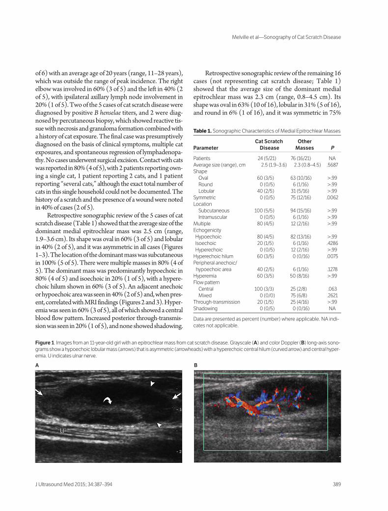
Retrospective sonographic review of the 5 cases of catscratch disease (Table 1) showed that the average size of thedominant medial epitrochlear mass was 2.5 cm (range,1.9–3.6 cm). Its shape was oval in 60% (3 of 5) and lobularin 40% (2 of 5), and it was asymmetric in all cases (Figures1–3). The location of the dominant mass was subcutaneousin 100% (5 of 5). There were multiple masses in 80% (4 of5). The dominant mass was predominantly hypoechoic in80% (4 of 5) and isoechoic in 20% (1 of 5), with a hypere-choic hilum shown in 60% (3 of 5). An adjacent anechoicor hypoechoic area was seen in 40% (2 of 5) and, when pres-ent, correlated with MRI findings (Figures 2 and 3). Hyper-emia was seen in 60% (3 of 5), all of which showed a centralblood flow pattern. Increased posterior through-transmis-sion was seen in 20% (1 of 5), and none showed shadowing.
Retrospective sonographic review of the remaining 16cases (not representing cat scratch disease; Table 1)showed that the average size of the dominant medialepitrochlear mass was 2.3 cm (range, 0.8–4.5 cm). Itsshape was oval in 63% (10 of 16), lobular in 31% (5 of 16),and round in 6% (1 of 16), and it was symmetric in 75%
J Ultrasound Med 2015; 34:387–394 389
Melville et al—Sonography of Cat Scratch Disease
Table 1. Sonographic Characteristics of Medial Epitrochlear Masses
Cat Scratch Other
Parameter Disease Masses P
Patients 24 (5/21) 76 (16/21) NA
Average size (range), cm 2.5 (1.9–3.6) 2.3 (0.8–4.5) .5687
Shape
Oval 60 (3/5) 63 (10/16) >.99
Round 0 (0/5) 6 (1/16) >.99
Lobular 40 (2/5) 31 (5/16) >.99
Symmetric 0 (0/5) 75 (12/16) .0062
Location
Subcutaneous 100 (5/5) 94 (15/16) >.99
Intramuscular 0 (0/5) 6 (1/16) >.99
Multiple 80 (4/5) 12 (2/16) >.99
Echogenicity
Hypoechoic 80 (4/5) 82 (13/16) >.99
Isoechoic 20 (1/5) 6 (1/16) .4286
Hyperechoic 0 (0/5) 12 (2/16) >.99
Hyperechoic hilum 60 (3/5) 0 (0/16) .0075
Peripheral anechoic/
hypoechoic area 40 (2/5) 6 (1/16) .1278
Hyperemia 60 (3/5) 50 (8/16) >.99
Flow pattern
Central 100 (3/3) 25 (2/8) .063
Mixed 0 (0/0) 75 (6/8) .2621
Through-transmission 20 (1/5) 25 (4/16) >.99
Shadowing 0 (0/5) 0 (0/16) NA
Data are presented as percent (number) where applicable. NA indi-
cates not applicable.
Figure 1. Images from an 11-year-old girl with an epitrochlear mass from cat scratch disease. Grayscale (A) and color Doppler (B) long-axis sono-
grams show a hypoechoic lobular mass (arrows) that is asymmetric (arrowheads) with a hyperechoic central hilum (curved arrow) and central hyper-
emia. U indicates ulnar nerve.
A B
3403jum363-518 copy_Layout 1 2/20/15 3:14 PM Page 389
(12 of 16; Figures 4 and 5). The location of the dominantmass, which was multiple in 12% (2 of 16), was subcuta-neous in 94% (15 of 16) and intramuscular in 6% (1 of 16).The dominant mass was predominantly hypoechoic in82% (13 of 16), hyperechoic in 12% (2 of 16), and iso -echoic in 6% (1 of 16). None of the masses showed ahyperechoic hilum. An adjacent anechoic or hypoechoicarea was seen in 6% (1 of 16). Hyperemia was seen in 50%(8 of 16), which showed a central blood flow pattern in25% (2 of 8) and a peripheral pattern in 75% (6 of 8).Increased posterior through-transmission was seen in 25%(4 of 16), and none showed shadowing.
When comparing the sonographic findings betweenthe cat scratch disease and non–cat scratch disease cases,there were significant differences (P < .05) with regard tosymmetry of the medial epitrochlear mass and the presence
of a hyperechoic hilum; cases of cat scratch disease showedmass asymmetry and the presence of a hyperechoic hilum.Furthermore, the data were analyzed to determine whetherthere were significant differences in the various parametersmeasured as a function of a cat scratch disease versus non–cat scratch disease diagnosis (Table 2). Once again, sym-metry and a hyperechoic hilum were significant parameters(P < .05); in addition, the presence of a peripheral anechoicor hypoechoic area was found to be statistically significant.Logistic regression for these 3 parameters with a cat scratchdisease versus non–cat scratch disease diagnosis as thedependent variable indicated that asymmetry followed by ahyperechoic hilum and then a peripheral anechoic orhypoechoic region were the best predictors of a cat scratchdisease versus non–cat scratch disease diagnosis (r2 = 0.783;log likelihood = 2.502).
Melville et al—Sonography of Cat Scratch Disease
J Ultrasound Med 2015; 34:387–394390
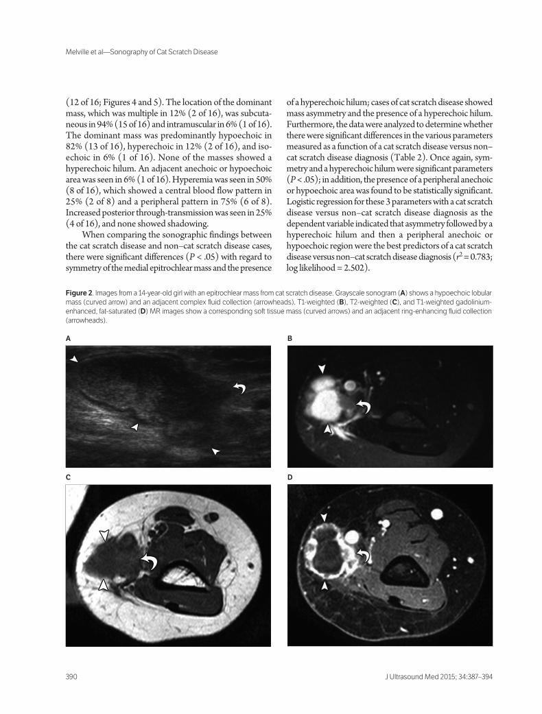
Figure 2. Images from a 14-year-old girl with an epitrochlear mass from cat scratch disease. Grayscale sonogram (A) shows a hypoechoic lobular
mass (curved arrow) and an adjacent complex fluid collection (arrowheads). T1-weighted (B), T2-weighted (C), and T1-weighted gadolinium-
enhanced, fat-saturated (D) MR images show a corresponding soft tissue mass (curved arrows) and an adjacent ring-enhancing fluid collection
(arrowheads).
A B
C D
3403jum363-518 copy_Layout 1 2/20/15 3:14 PM Page 390

Discussion
Abnormal medial epitrochlear lymph node enlargement isa characteristic finding of cat scratch disease; however,imaging findings may appear confusing and potentially bemisinterpreted for other disease types such as malignancy,leading to unnecessary biopsy. The results of our retro-spective study showed that identification of a mass in theexpected location of a medial epitrochlear lymph node thatshowed asymmetry and a hyperechoic hilum was charac-
teristic of cat scratch disease, unlike other causes of lymphnode enlargement.
Cat scratch disease usually presents with regional lym-phadenopathy, painful swelling, and nonspecific clinicalsigns and symptoms as result of B henselae infection. A defin-itive diagnosis is based on polymerase chain reaction forDNA analysis or serologic testing, as isolating Bartonellaspecies in culture is difficult and usually unsuccessful; histo-logic examination reveals typical but nonspecific features,including granulomas with central necrosis at later stages.14,15
J Ultrasound Med 2015; 34:387–394 391
Melville et al—Sonography of Cat Scratch Disease
Figure 3. Images from a 28-year-old man with an epitrochlear mass from
cat scratch disease. Grayscale long-axis sonogram (A) shows an oval
asymmetric hypoechoic mass (arrowheads) with a hyperechoic hilum
(arrow) and an adjacent hypoechoic complex fluid collection (curved
arrow). Proton density-weighted (B), T2-weighted (C), and T1-weighted
gadolinium-enhanced, fat-saturated (D) MR images show a correspon-
ding soft tissue mass (arrowheads) and an adjacent ring-enhancing fluid
collection (curved arrows).
A
B
C D
3403jum363-518 copy_Layout 1 2/20/15 3:14 PM Page 391

The presumptive diagnosis of cat scratch disease is fre-quently made on the basis of suggestive clinical features,including lymph node enlargement, cat exposure, and anupper extremity inoculation site.1 Nearly 75% of involvedlymph nodes are located in the neck or upper extremity,most commonly at the medial epitrochlear region.2,16
The peak age range for cat scratch disease is reportedas between 2 and 14 years, and 40% of our study group (2of 5) belonged to this age group.2,17 Between 95% and 99%of cat scratch disease cases follow contact with a cat, andapproximately 75% report a cat scratch.5 In our studygroup, 80% (4 of 5) reported cat contact, with 40% (2 of 5)reporting a scratch. Our study group suggests that patientsoutside the peak age range and without a recalled history ofa cat scratch may be more likely to undergo imaging. Atyp-ical presentations may occur in 5% to 11% of patients, due tosystemic dissemination, hematogenous osteomyelitis, or
hepatosplenic involvement, and may occur more com-monly in immune-compromised patients; however, noneof our patients had clinical features to suggest this compli-cation.2,3 Hepatosplenic involvement may be identified onsonography as multiple small hypoechoic nodules, whichmay eventually calcify.18,19 The presence of these lesionsmay further support the diagnosis of cat scratch disease,particularly when clinical suspicion for malignancy remainslow. Most cases of cat scratch disease confined to theepitrochlear or axillary lymph nodes resolve within 3months.
Epitrochlear nodes, which are also known as thecubital or supraepitrochlear lymph nodes, constitute partof the upper extremity superficial lymphatic system and arelocated in the subcutaneous tissue on the medial aspect ofthe elbow approximately 4 to 5 cm above the trochlea.20
Although the association of cat scratch disease with medial
Melville et al—Sonography of Cat Scratch Disease
J Ultrasound Med 2015; 34:387–394392
Figure 4. Images from a 34-year-old man with a biopsy-proven hyper-
plastic lymph node. Grayscale long-axis sonogram (A) shows a hypo -
echoic mass (arrowheads) with a mixed pattern of hyperemia (not shown).
T1-weighted (B), T2-weighted (C), and T1-weighted gadolinium-
enhanced, fat- saturated (D) MR images show a corresponding soft tissue
mass (arrows).
A
C
D
B
3403jum363-518 copy_Layout 1 2/20/15 3:14 PM Page 392

epitrochlear lymph node enlargement is established in theradiology and infectious disease literature, complex nodalmasses with suppuration may mimic aggressive soft tissuemasses,8,11 especially if specific lymph node features are notidentified on MRI, which can occur when inflammation isextensive. Although the appearance of such lymph nodelesions on MRI has been described, the sonographic fea-tures of cat scratch disease have not been fully evalu-ated.10,12
Reported sonographic features of involved regionallymph nodes in cat scratch disease without suppurationinclude a hypoechoic mass with or without increased pos-terior through-transmission.12 Although the lymph nodesin our patients with cat scratch disease were also com-monly hypoechoic (80%), those in patients with othercauses of epitrochlear masses were also commonly hypoe-choic (82%). Similarly, the previously described increasedposterior through-transmission was not seen in these casesas a specific feature, as this finding was present in 20% ofmasses from cat scratch disease and 25% from other causes.
An additional finding, the presence of a markedly increasedDoppler signal within involved nodes, has been previouslyidentified as a suggestive feature of cat scratch disease, pos-sibly related to neovascularization resulting from Bartonellainfection, which may also produce vasoproliferative lesionsinvolving the skin, brain, bone marrow, and mucosal sur-faces.21–23 The conspicuity of this feature on powerDoppler imaging has been described as the “fire” pattern.12
In our study, central hyperemia was noted in 60% of catscratch disease cases (3 of 5); however, hyperemia waspresent in 50% of non–cat scratch disease cases (8 of 16),although it was central in only 25%.
Our retrospective review comparing the sonographicfeatures of epitrochlear masses from cat scratch disease toother causes revealed 2 significant findings: asymmetry andthe presence of an echogenic hilum. The cause of asym-metry is not clear but may explain the finding of an adjacentfluid collection, which was found to be substantial whencomparing parameters as a function of a cat scratch diseaseversus non–cat scratch disease diagnosis and was presentin 40% of cat scratch disease cases but only 6% of caseswith other causes. The presence of an adjacent or intran-odal fluid collection is believed to indicate nodal suppu-ration or necrosis, which is seen in up to 35% of cases.4,24
This appearance corresponds to the reported appearanceof cat scratch disease on MRI, with mixed enhancing solidand fluid components (Figures 2 and 3).10 The other sig-nificant finding of a hyperchoic hilum seen in cat scratchdisease was not unexpected; however, it was surprising thatno other causes of an epitrochlear mass showed this find-ing, even the cases of lymph node hyperplasia.
This retrospective study had several limitations.Despite the frequency of cat scratch disease, there wereonly 5 patients with cat scratch disease undergoing sono-
J Ultrasound Med 2015; 34:387–394 393
Melville et al—Sonography of Cat Scratch Disease
Figure 5. Images from a 54-year-old man with a biopsy-proven
epitrochlear mass from sarcoidosis. Grayscale (A) and color Doppler
(B) long-axis sonograms show an oval symmetric hypoechoic mass
(arrows) with a mixed pattern of hyperemia.
A
B
Table 2. Analysis of Parameters as a Function of a Cat Scratch
Disease Versus Non–Cat Scratch Disease Diagnosis
Parameter Statistical Results
Size t = 0.217; P = .8301
Shape χ2 = 0.404; P = .8172
Symmetry χ2 = 8.750; P = .0031
Location χ2 = 0.5668; P = .5668
Multiplicity χ2 = 0.836; P = .3606
Echogenicity χ2 = 1.382; P = .5011
Hyperechoic hilum χ2 = 11.20; P = .0008
Peripheral anechoic/hypoechoic area χ2 = 3.544; P = .0500
Hyperemia χ2 = 0.153; P = .6959
Flow pattern χ2 = 5.565; P = .0619
Through-transmission χ2 = 0.053; P = .8188
3403jum363-518 copy_Layout 1 2/20/15 3:14 PM Page 393

Melville et al—Sonography of Cat Scratch Disease
J Ultrasound Med 2015; 34:387–394394
graphic evaluations over a 13-year period, which reflectsthe fact that most of these cases are diagnosed on the basisof clinical and laboratory findings and are often not imaged.Due to the self-limiting nature of this condition, no cases ofcat scratch disease underwent surgical excision, limitingpathologic correlation; however, positive diagnoses wereobtained by serologic tests, clinical histories, and patientfollow-up. In addition, as sonographic examinations wereperformed as part of routine patient care, there is intrinsicvariability in the technique between sonographers; how-ever, there is less operator dependence when imaging a softtissue mass compared to imaging complex anatomic struc-tures such as the rotator cuff. Last, interobserver andintraobserver variability of sonographic findings were notassessed.
In summary, the results of our study show that thesonographic finding of epitrochlear lymph node enlarge-ment due to cat scratch disease most commonly is that ofa hypoechoic lobular or oval mass with central hyperemiaand a possible adjacent fluid collection; however, the find-ings of an asymmetric shape and a hyperechoic hilum dif-ferentiate cat scratch disease from other etiologies. Byrecognizing the common sonographic appearance of catscratch disease, an additional clinical history for cat expo-sure and serologic testing for B henselae may be consideredto avoid unnecessary biopsy or resection.
References
1. Florin TA, Zaoutis TE, Zaoutis LB. Beyond cat scratch disease: wideningspectrum of Bartonella henselae infection. Pediatrics 2008; 121:e1413–e1425.
2. Carithers HA. Cat-scratch disease: an overview based on a study of 1200patients. Am J Dis Child 1985; 139:1124–1133.
3. Rohr A, Saettele MR, Patel SA, Lawrence CA, Lowe LH. Spectrum ofradiological manifestations of paediatric cat-scratch disease. Pediatr Radiol2012; 42:1380–1384.
4. Wang CW, Chang WC, Chao TK, Liu CC, Huang GS. Computedtomography and magnetic resonance imaging of cat-scratch disease: areport of two cases. Clin Imaging 2009; 33:318–321.
5. Margileth AM. Cat scratch disease. Adv Pediatr Infect Dis 1993; 8:1–21.6. Luddy RE, Sutherland JC, Levy BE, Schwartz AD. Cat-scratch disease
simulating malignant lymphoma. Cancer 1982; 50:584–586.7. Nimityongskul P, Anderson LD, Sri P. Cat-scratch disease: orthopaedic
presentation. Orthop Rev 1992; 21:55–59.8. Fox BC, Gurtler RA. Cat-scratch disease mimicking rhabdomyosarcoma.
Orthop Rev 1993; 22:1148–1149.9. Lefkowitz M, Wear DJ. Cat-scratch disease masquerading as a solitary
tumor of the breast. Arch Pathol Lab Med 1989; 113:473–475.
10. Gielen J, Wang XL, Vanhoenacker F, et al. Lymphadenopathy at themedial epitrochlear region in cat-scratch disease. Eur Radiol 2003;13:1363–1369.
11. Dong PR, Seeger LL, Yao L, Panosian CB, Johnson BL Jr, Eckardt JJ.Uncomplicated cat-scratch disease: findings at CT, MR imaging, and radi-ography. Radiology 1995; 195:837–839.
12. Garcia CJ, Varela C, Abarca K, Ferres M, Prado P, Vial PA. Regionallymphadenopathy in cat-scratch disease: ultrasonographic findings.Pediatr Radiol 2000; 30:640–643.
13. Barr LL, Kirks DR. Ultrasonography of acute epitrochlear lymphadenitis.Pediatr Radiol 1993; 23:72–73.
14. Regnery RL, Olson JG, Perkins BA, Bibb W. Serological response to“Rochalimaea henselae” antigen in suspected cat-scratch disease. Lancet1992; 339:1443–1445.
15. Bass JW, Vincent JM, Person DA. The expanding spectrum of Bartonellainfections, II: cat-scratch disease. Pediatr Infect Dis J 1997; 16:163–179.
16. Margileth AM. Dermatologic manifestations and update of cat scratchdisease. Pediatr Dermatol 1988; 5:1–9.
17. Zangwill KM, Hamilton DH, Perkins BA, et al. Cat scratch disease inConnecticut: epidemiology, risk factors, and evaluation of a newdiagnostic test. N Engl J Med 1993; 329:8–13.
18. Talenti E, Cesaro S, Scapinello A, Perale R, Zanesco L. Disseminatedhepatic and splenic calcifications following cat-scratch disease. PediatrRadiol 1994; 24:342–343.
19. Larsen CE, Patrick LE. Abdominal (liver, spleen) and bone manifesta-tions of cat scratch disease. Pediatr Radiol 1992; 22:353–355.
20. Catalano O, Nunziata A, Saturnino PP, Siani A. Epitrochlear lymph nodes:anatomy, clinical aspects, and sonography features. Pictorial essay. J Ultrasound2010; 13:168–174.
21. Spinella A, Lumetti F, Sandri G, Cestelli V, Mascia MT. Beyond catscratch disease: a case report of Bartonella infection mimicking vasculiticdisorder. Case Rep Infect Dis 2012; 2012:354625.
22. Conley T, Slater L, Hamilton K. Rochalimaea species stimulate humanendothelial cell proliferation and migration in vitro. J Lab Clin Med 1994;124:521–528.
23. Stockmeyer B, Schoerner C, Frangou P, Moriabadi T, Heuss D, Harrer T.Chronic vasculitis and polyneuropathy due to infection with Bartonellahenselae. Infection 2007; 35:107–109.
24. Margileth AM, Hayden GF. Cat scratch disease: from feline affection tohuman infection. N Engl J Med 1993; 329:53–54.
3403jum363-518 copy_Layout 1 2/20/15 3:14 PM Page 394


![Cat Scratch (Bartonella henselae) - Biocare Medical · Cat Scratch (Bartonella henselae) [H2A10] is a mouse monoclonal antibody that is intended for laboratory use in the qualitative](https://img.pdfslide.net/doc/110x75/606375af7209417ea40878e9/cat-scratch-bartonella-henselae-biocare-medical-cat-scratch-bartonella-henselae.jpg)