Embed Size (px)
Citation preview
1
Andreotti
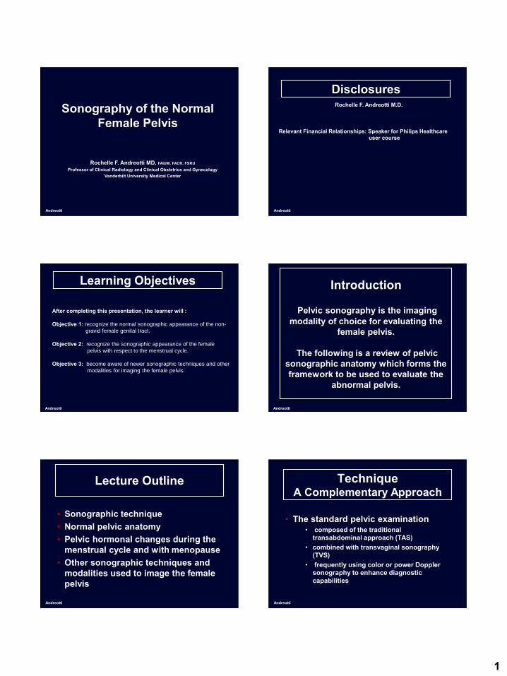
Sonography of the Normal Female Pelvis
Rochelle F. Andreotti MD, FAIUM, FACR, FSRUProfessor of Clinical Radiology and Clinical Obstetrics and Gynecology
Vanderbilt University Medical Center
Andreotti
DisclosuresRochelle F. Andreotti M.D.
Relevant Financial Relationships: Speaker for Philips Healthcare user course
Learning Objectives
After completing this presentation, the learner will :
Objective 1: recognize the normal sonographic appearance of the non-
gravid female genital tract.
Objective 2: recognize the sonographic appearance of the female
pelvis with respect to the menstrual cycle.
Objective 3: become aware of newer sonographic techniques and other
modalities for imaging the female pelvis.
Andreotti
Introduction
Pelvic sonography is the imaging modality of choice for evaluating the
female pelvis.
The following is a review of pelvic sonographic anatomy which forms the framework to be used to evaluate the
abnormal pelvis.
Andreotti
Andreotti
Lecture Outline
• Sonographic technique• Normal pelvic anatomy• Pelvic hormonal changes during the
menstrual cycle and with menopause• Other sonographic techniques and
modalities used to image the female pelvis
Andreotti
TechniqueA Complementary Approach
• The standard pelvic examination • composed of the traditional
transabdominal approach (TAS) • combined with transvaginal sonography
(TVS) • frequently using color or power Doppler
sonography to enhance diagnostic capabilities

2
Andreotti
Transabdominal Sonography
Andreotti
Classic TechniqueTransabdominal Sonography
• Uses a distended bladder as window to pelvic structures for a global view
• Visualization limited by attenuation from the body wall and the distance from the area of interest of the transducer
• Unable to use higher frequency transducers and benefit from their inherent higher axial and lateral resolution
Andreotti
Transabdominal Sonography
• Sagittal and transverse views of the pelvis
Bladder
UterusCX
Vagina
Ovaries
Andreotti
Limited Technique Transabdominal
• Used to complement TVS
• Initial evaluation without dedicated bladder filling
• For global view of pelvis
Andreotti
Transvaginal Sonography (TVS)
Andreotti
TechniqueTransvaginal sonography
• Gives a more detailed evaluation of pelvic architecture using higher frequency transducers (>5 MHZ) at closer proximity to pelvic structures
• Field of view is limited

3
Andreotti
TechniqueContraindications
• Premenarchal patients• The majority of virginal patients • Any patient who does not willingly
consent to vaginal examination
Andreotti
Transvaginal Sonography
right left
anterior
posterior
anterior
posterior
cephalad
Andreotti
Sagittal view
• Uterine axis rotated 90 degrees counterclockwise from the TAS image on the sagittal view
90º
Andreotti
Transverse Uterine View(coronal view of pelvis)
• Uterine orientation on transverse view (coronal view of the pelvis) is usually the same as TAS image
Andreotti
The Normal Sonographic Appearance of the Non-Gravid
Genital Tract
Andreotti
Pelvis
Bladder
Ovaries
Sigmoid & rectum
Vagina
Broad ligament

4
Andreotti
Pelvic FloorSagittal midline
urethravagina
rectum
Andreotti
Pelvic FloorTransperineal coronal view
urethra
vagina
rectum
Andreotti
Pelvic Vasculature
Uterine
Artery & vein
0varian artery
& vein
Pampiniform plexus
Infundibulo-pelvic
ligament
Utero-ovarian ligament
Arcuate and Radial
Branches
Andreotti
Pelvic Vasculature
• Uterine and ovarian artery
Pampiniform plexus
UT
Uterine
Artery
0varian artery
OV
OV
Andreotti
Pelvic Vasculature
• Uterine artery branches
Arcuate and Radial
Arteries
Andreotti
Uterus
• Consists of:– Cervix- the lower cylindrical portion
which projects into the vagina– Body or corpus– Isthmus- narrow area connecting the
body and cervix

5
Andreotti
Cervix
CX
Vag90º
Andreotti
Cervix
Cervical mucus
Andreotti
Cervix
Nabothian cysts• Result of
occlusion of endocervical glands
• Have no clinical significance
Andreotti
Uterus
• Uterine size and consistency• Position• Endometrium
Andreotti
Uterine size
• Measurement may be performed by TAS or TVS in sagittal, transverse and anterior-posterior dimensions
• Uterine length is often measured more accurately by TAS since the cervix may not be completely included on the transvaginal image
Andreotti
Uterine Size
• Uterine length measured more accurately by TAS when the cervix is not completely included on the transvaginal image

6
Andreotti
Uterine consistency
Myometrium best evaluated by TVS forimproved resolution of architecture
Andreotti
Uterine Consistency
• The myometrium is divided into an echogenic outer layer (o) and intermediate layer (m), and hypoechoic inner layer (i)
omi
Arcuate artery
Andreotti
Uterine ConsistencyCoronal view
• The inner layer appears as a thin hypoechoic halo surrounding the endometrium
• Best seen on coronal view
Andreotti
Uterine MeasurementsPremenstrual patients
• Ave volume approximately 1 cc up to age 9• Larger cervix compared to corpus
Andreotti
Uterine Measurements
• Normal measurements in menstrating females –vary with parity– 6-10.5 cm Length– 3-6 cm Transverse diameter– 2-5 cm A-P diameter
Andreotti
Uterine Measurements
• Normal measurements in postmenopausal females– 3.5-7.5 cm Length– 2-4 cm Transverse diameter– 1.7-3.3 AP diameter

7
Andreotti
Uterine MeasurementsPremenopausal Postmenopausal
4.8 cm L x 2.4 cm AP x 2.5 cm TRV7.1 cm L x 4.3 cm AP x 3.5 cm TRV
Andreotti
Uterine Position
Variable changing with degree of bladder and rectal distention
• Flexion- axis of uterine body relative to cervix
• Version- axis of cervix relative to the vagina
Andreotti
Uterine Position
• Anteversion/anteflexion-most common position
TAS
TVS
Andreotti
Uterine Position
• Retroversion/retroflexion
TAS
TVS
Andreotti
Uterine Position
• Retroverted/retroflexed uterus image orientation
posterior
Andreotti
Uterine Position
• Neutral position• Demonstrates coronal rather than
transverse view of uterus
8
Andreotti
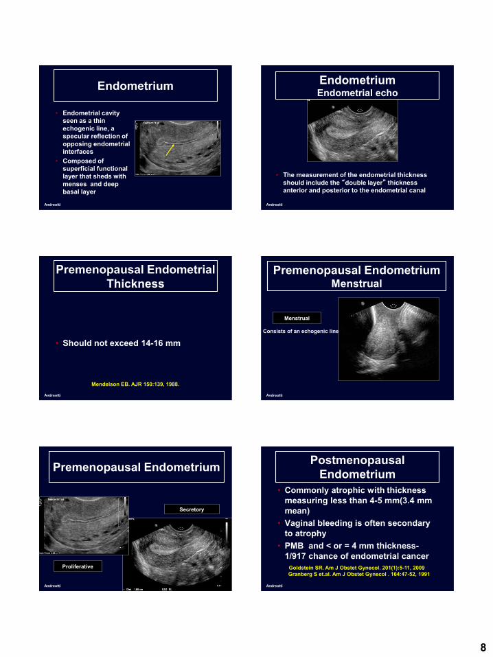
Endometrium
• Endometrial cavity seen as a thin echogenic line, a specular reflection of opposing endometrial interfaces
• Composed of superficial functional layer that sheds with menses and deep basal layer
Andreotti
EndometriumEndometrial echo
• The measurement of the endometrial thickness should include the “double layer” thickness anterior and posterior to the endometrial canal
Andreotti
Premenopausal Endometrial Thickness
• Should not exceed 14-16 mm
Mendelson EB. AJR 150:139, 1988.
Andreotti
Premenopausal EndometriumMenstrual
Consists of an echogenic line
Menstrual
Andreotti
Premenopausal Endometrium
Proliferative
Secretory
Andreotti
Postmenopausal Endometrium
• Commonly atrophic with thickness measuring less than 4-5 mm(3.4 mm mean)
• Vaginal bleeding is often secondary to atrophy
• PMB and < or = 4 mm thickness-1/917 chance of endometrial cancer
Goldstein SR. Am J Obstet Gynecol. 201(1):5-11, 2009Granberg S et.al. Am J Obstet Gynecol . 164:47-52, 1991

9
Andreotti
The Asymptomatic Thickened Postmenopausal Endometrium
• Significance of thickening debatable• No prospective studies performed to
determine significant thickness• Texture most important-polyp?
fibroid?• Routine biopsy not recommended
Andreotti
Postmenopausal Endometrium
Andreotti
Ovary
• The ovaries are ellipsoid and can be identified in menstruating females by the presence of follicles
Andreotti
OvaryLocation
• The location of ovaries is variable• Often seen in the ovarian fossa
(Waldeyer’s Fossa), especially in nulliparous females
• Waldeyer’s Fossa bounded by the obliterated umbilical artery anteriorly, the ureter and internal iliac artery posteriorly and the external iliac vein superiorly
Andreotti
OvaryLocation
• Waldeyer’s Fossa by transvaginal color Doppler sonography
Int iliac artery
Ext iliac veinovary
Andreotti
Ovary
Ovarian Volumes
“Bigger than we think”
Cohen H. Radiology 177: 189, 1990.
10
Andreotti
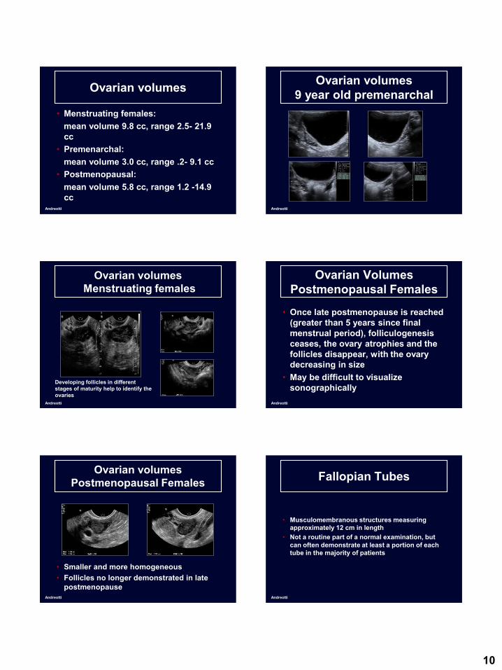
Ovarian volumes
• Menstruating females:mean volume 9.8 cc, range 2.5- 21.9 cc
• Premenarchal:mean volume 3.0 cc, range .2- 9.1 cc
• Postmenopausal:mean volume 5.8 cc, range 1.2 -14.9 cc
Andreotti
Ovarian volumes9 year old premenarchal
Andreotti
Ovarian volumesMenstruating females
Developing follicles in different stages of maturity help to identify the ovaries
Andreotti
Ovarian VolumesPostmenopausal Females
• Once late postmenopause is reached (greater than 5 years since final menstrual period), folliculogenesis ceases, the ovary atrophies and the follicles disappear, with the ovary decreasing in size
• May be difficult to visualize sonographically
Andreotti
Ovarian volumesPostmenopausal Females
• Smaller and more homogeneous• Follicles no longer demonstrated in late
postmenopauseAndreotti
Fallopian Tubes
• Musculomembranous structures measuring approximately 12 cm in length
• Not a routine part of a normal examination, but can often demonstrate at least a portion of each tube in the majority of patients

11
Andreotti
Fallopian Tubes
• intramural, (1) isthmic (2) and ampullary (3) portions
12
3
Andreotti
Fallopian Tubes
• Identified by its tubular structure (arrow) which can be followed to the uterine cornua (C)
C
Andreotti
Cul-de-sac
• Physiologic fluid in cul-de-sac seen in menstruating females (<15 ml)
Andreotti
Sonographic Changes in the Appearance of the Female Pelvis with Respect to the Menstrual Cycle and with
Age
Andreotti
Cyclic Hormonal changes
Andreotti
Cyclic Changes of the Ovaries
• Follicular phase- Enlargement of ovarian follicles with usually one dominant, preovulatory follicle ( 20 mm average diameter) prior to ovulation

12
Andreotti
Cyclic Changes of the Ovaries
• Developing follicles in the early follicular phase
Andreotti
Cyclic Changes of the Ovaries
• One or two dominant follicles usually seen after day 10
Andreotti
Cyclic Changes of the Ovaries
• Ovulatory follicle usually 18-25 mm average diameter (ave 20 mm) Andreotti
Cyclic Changes of the Ovaries
• Ovulatory follicle containing a cumulus oophorus• Separation of the granulosal layer of the follicular
wall from the thecal layer (containing ovum) 24 hours prior to ovulation
Andreotti
Cyclic Changes of the Ovaries
Luteal phase- LH surge triggering ovulation with ruptured follicle becoming postovulatory corpus luteum• Crenulated thick walled cystic
structure• Peripheral vascularity by color
Doppler
Andreotti
Cyclic Changes of the Ovaries
• Post-ovulatory corpus luteum

13
Andreotti
Cyclic Changes of the Ovaries
• Pre-menstrual corpus luteum-fills in with echoes no longer appearing cystic
Andreotti
FOLLICLE EVALUATIONS
• Monitoring size and number of follicles for evaluation of ovulation in:
Normal cyclesOvulation induction cyclesIVF cycles
• Mature follicle (18-25mm)
Andreotti
Cyclic Changes of the Endometrium
• Menstrual phase• Proliferative phase• Secretory phase
Variations in thickness and architecture*
*Lyons EA, Radiol Clin North Am 30:663, 1992.
Andreotti
Cyclic Changes of the Endometrium
• Menstrual phase: Thin slightly irregular echogenic surface due to sloughing of the functional layer of the endometrium
Andreotti
Cyclic Changes of the Endometrium
• Proliferative phase: Thickens with an echogenic border but hypoechoic inner layer reflecting increase in length of glands (4-10 mm)
Peri-ovulatoryEarly
Andreotti
Cyclic Changes of the Endometrium
• Secretory phase: Increase in echogenicity reflecting tortuosity and distention of glands with mucin (7-14 mm)

14
Andreotti
Perimenopause
• Unopposed estrogen stimulation: Increase in echogenicity with thickening that can be greater than 14 mm
• Cannot differentiate from hyperplasia
Andreotti
Early Postmenopause
• Within 5 years of last menstrual period
• Occasional follicular development with subsequent ovulation
• Resulting in cyclic changes of the endometrium and menstruation
Andreotti
Postmenopausal Simple Cysts
• Simple cysts (thin walled, anechoic)often seen in postmenopausal ovaries
• Follicles, paraovarian or paratubal, ovarian surface epithelial inclusion cysts, cystadenomas
• Simple cysts <1 cm considered almost certainly benign and clinically unimportant
Andreotti
Postmenopausal Simple Cysts
Andreotti
Other Techniques for Imaging the Female Pelvis
Andreotti
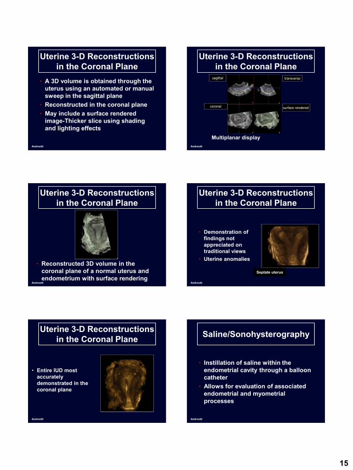
Uterine 3-D Reconstructions in the Coronal Plane
• 2D imaging limited by constraints of the vaginal probe
• Volume imaging allows routine visualization of the coronal plane
15
Andreotti
Uterine 3-D Reconstructions in the Coronal Plane
• A 3D volume is obtained through the uterus using an automated or manual sweep in the sagittal plane
• Reconstructed in the coronal plane• May include a surface rendered
image-Thicker slice using shading and lighting effects
Andreotti
Uterine 3-D Reconstructions in the Coronal Plane
sagittal transverse
coronal
Multiplanar display
surface rendered
Andreotti
Uterine 3-D Reconstructions in the Coronal Plane
• Reconstructed 3D volume in the coronal plane of a normal uterus and endometrium with surface rendering
Andreotti
Uterine 3-D Reconstructions in the Coronal Plane
• Demonstration of findings not appreciated on traditional views
• Uterine anomalies
Septate uterus
Andreotti
Uterine 3-D Reconstructions in the Coronal Plane
• Entire IUD most accurately demonstrated in the coronal plane
Andreotti
Saline/Sonohysterography
• Instillation of saline within the endometrial cavity through a balloon catheter
• Allows for evaluation of associated endometrial and myometrial processes

16
Andreotti
Saline/Sonohysterography
• Fluid distending the endometrial cavity
Andreotti
Saline/Sonohysterography
• 3D reconstructions of the distended cavity in the coronal plane
Andreotti
Contrast Agents
• Microbubble contrast material can be used to enhance the microvascular circulation
• Although little support, a few studies suggest usefulness in differentiation of benign versus malignant ovarian masses*
*Fleischer AC, et al., J Ultrasound Med 2008; 27:1011. Andreotti
Contrast Agents
• Pre and post-contrast images of a normal ovary• Enhancement of veins surrounding the ovary without
significant enhancement within the ovary Images courtesy of Andrej Lyschik, Dept. of Radiology, Vanderbilt University Medical Center
ov
PostPre
ov
Andreotti
Contrast Agents
• Pre and post-contrast images of ovarian carcinoma
• Marked enhancement of ovarian neoplasia
Images courtesy of Andrej Lyschik, Dept. of Radiology, Vanderbilt University Medical Center
PostPre
ov ov
Andreotti
Contrast Agents
• Comparison of contrast enhancement of normal ovary and carcinoma
Images courtesy of Andrej Lyschik, Dept. of Radiology, Vanderbilt University Medical Center
Normal Carcinoma
ovov

17
Andreotti
Contrast Agents
• Contrast enhancement of the normal myometriumImages courtesy of Andrej Lyschik, Dept. of Radiology, Vanderbilt University Medical Center
Pre Post
Andreotti
Other Imaging ModalitiesMRI and CT
Andreotti
Computed Tomography (CT)
• Sonography is initial exam of choice for evaluation of the pelvis
• Computed tomography is used frequently in patients suspected of GI or GU abnormalities
Andreotti
Computed Tomography (CT)
• Transverse image of pelvis with contrast
Ut
Ov
Andreotti
Computed Tomography (CT)
• Reconstruction in the sagittal plane
Bladder
Uterus
Andreotti
Pelvic MRI
• MRI can be a problem solving technique in the pelvis when US is not definitive
• Pelvic anatomy can be better defined with MRI than CT

18
Andreotti
Pelvic MRI
• Sag T2 weighted image
• High intensity endometrium and myometrium
Endometrium
Andreotti
Pelvic MRI
• Transverse T2 weighted image• Low intensity junctional zone(JZ)
separating endometrium and myometrium
Endometrium JZ
Ovarian cyst
Andreotti
Pelvic MRI
• Sag and transverse T2 weighted images of the right ovary
JZMFollicles Follicles
Andreotti
Conclusions• Using transabdominal, transvaginal and color
Doppler sonography, the architecture of
female pelvic organs is well demonstrated.
• One should be familiar with the normal pelvic
findings including the cyclic changes of the
uterus and ovaries in order to differentiate
these from true abnormalities.
• Newer sonographic techniques as well as
other radiologic modalities also play a role in
pelvic evaluation
THANK YOU!
Key References
Andreotti
1. Benacerraf BR, Abuhamad AZ, Bromley B, Goldstein SR, Groszman Y, Shipp
TD, Timor-Trisch IE. Consider ultrasound first for imaging the female pelvis.
Am J Obstet Gynecol 2015; 212: 450-5
2. Cohen HL, Tice HM, Mandel FS. Ovarian volumes measured by US: bigger
than we think. Radiology. 1990;177(1):189-192.
3. Levine D, Brown DL, Andreotti RF, et al. Management of asymptomatic
ovarian and other adnexal cysts imaged at US: Society of Radiologists in
Ultrasound Consensus Conference Statement. Radiology. 2010;256(3):943-
954.
4. Lyons EA, Gratton D, harrington C, Transvaginal sonography of normal pelvic
anatomy. Radiol Clin North Am 30:663, 1992.
5. Merz E, Miric-Tesanic D, Bahlmann F, Weber G, Wellek S. Sonographic size
of uterus and ovaries in pre- and postmenopausal women. Ultrasound in
obstetrics & gynecology : the official journal of the International Society of
Ultrasound in Obstetrics and Gynecology. 1996;7(1):38-42.