Embed Size (px)
Citation preview
1
Manuscript M1:00609 revised (June 1st, 2001)
SopE and SopE2 from S. typhimurium activate different sets of RhoGTPases of the host
cell.
Andrea Friebel, Heiko Ilchmann, Martin Aepfelbacher, Kristin Ehrbar, Werner Machleidt‡
and Wolf-Dietrich Hardt¶
Max von Pettenkofer-Institut, Ludwig Maximilians Universität, Pettenkoferstr. 9a, 80336
München, Germany. ‡Adolf-Butenandt-Institut für Physiologische Chemie, Physikalische
Biochemie und Zellbiologie, Ludwig-Maximilians-Universität München, Schillerstr. 42, D-
80336 München, Germany.
running title: Differential RhoGTPase specificity of SopE and SopE2
¶Corresponding author: Wolf-Dietrich Hardt, Max von Pettenkofer-Institut, Ludwig
Maximilians Universität, Pettenkoferstr. 9a, 80336 München, Germany. fax: 89-5160-5223,
phone: 89-5160-5263, e-mail: [email protected]
*This work was supported by grants from the Deutsche Forschungsgemeinschaft
(Nachwuchsgruppe of WDH; BIAcore 2000-work: Sonderforschungsbereich 469 of the LMU
München, grant A-6/Machleidt)
Abbreviations: aa: amino acid; GDP: guanosine di-phosphate; mant: -O-(N-
methylanthraniloyl); mGDP: mant-GDP; GEF: guanine nucleotide exchange factor; GST:
glutathione S-transferase; S. typhimurium: Salmonella enterica subspecies I serovar
Typhimurium; HUVEC: human umbilical vein endothelial cell.
Copyright 2001 by The American Society for Biochemistry and Molecular Biology, Inc.
JBC Papers in Press. Published on July 5, 2001 as Manuscript M100609200 by guest on June 18, 2018
http://ww
w.jbc.org/
Dow
nloaded from
2
ABSTRACT
The bacterial enteropathogen Salmonella typhimurium employs a specialized type III
secretion system to inject toxins into host cells, which trigger signalling cascades leading to
cell death in macrophages, secretion of pro-inflammatory cytokines or rearrangements of the
host cell cytoskeleton and thus to bacterial invasion. Two of the injected toxins, SopE and the
69% identical protein SopE2, are highly efficient guanine nucleotide exchange factors (GEF)
for the RhoGTPase Cdc42 of the host cell. However, it has been a puzzle why S. typhimurium
might employ two toxins with redundant function. We hypothesized that SopE and SopE2
might have different specificities for certain host cellular RhoGTPases. In vitro guanine
nucleotide exchange assays and surface plasmon resonance measurements revealed that SopE
is an efficient GEF for Cdc42 and Rac1, while SopE2 was interacting efficiently only with
Cdc42, but not with Rac1. Affinity precipitation of Cdc42·GTP and Rac1·GTP from lysates
and characteristic cytoskeletal rearrangements of infected tissue culture cells confirmed that
SopE is highly efficient at activating Cdc42 and Rac1 in vivo, while SopE2 was efficiently
activating Cdc42, but not Rac1. We conclude that the translocated effector proteins SopE and
SopE2 allow S. typhimurium to specifically activate different sets of RhoGTPase signalling
cascades.
by guest on June 18, 2018http://w
ww
.jbc.org/D
ownloaded from
3
INTRODUCTION
The RhoGTPase subfamily of the Ras superfamily of small GTP-binding proteins comprises
more than 10 different proteins (1). They act as molecular switches and cycle between GDP-
bound (inactive) and GTP-bound (active) conformations (2). Activation and inactivation of
RhoGTPases is controlled by guanine nucleotide exchange factors (GEFs), GTPase-activating
enzymes and guanine dissociation inhibitors (3, 4). The eukaryotic GEFs for RhoGTPases
share two common sequence motives, the DH (dbl-homology) and the PH (pleckstrin
homology) domains, which are responsible for targeting, binding the RhoGTPases and
catalysis of RhoGTPase·GDP�RhoGTPase·GTP exchange (5). Only the active GTP-bound
RhoGTPases can interact with downstream elements of signal transduction cascades
mediating the cellular responses. GTPases of the Rho subfamily are central switches in the
signalling cascades regulating motility, cellular adhesion, cell shape, cytokinesis, cell
contraction and gene expression (6, 7). Each of the RhoGTPases can regulate a specific set of
downstream signalling cascades leading to activation of specific cellular functions (4). For
example, in Swiss 3T3 cells, activation of Cdc42 leads to the formation of filopodia,
activation of Rac1 induces formation of lamellipodia and activation of RhoA leads to the
formation of stress fibers and focal adhesions (8, 9, 10, 11). Thus, selective activation of
specific RhoGTPases leads to specific cellular responses.
Invasion of the gram negative bacterial enteropathogen S. typhimurium (Salmonella enterica
subspecies I serovar Typhimurium) into non-phagocytic mammalian cells is studied as a
model system for the ”trigger mechanism” of bacterial invasion (12). For triggering invasion,
S. typhimurium employs the specialized type III secretion system encoded in "Salmonella
pathogenicity island I" (SPI1) to inject/translocate a set of at least nine different bacterial
toxins (called "effector proteins") into host cells (13, 14, 15, 16, 17, 18, 19, 20, 21, 22). Inside
the host cell, the effector proteins activate signalling cascades leading to a variety of
responses including cytoskeletal rearrangements and bacterial internalization/invasion (12).
It has been shown that Cdc42 is a key element in the Salmonella induced signalling cascades
leading to transcriptional activation and bacterial invasion: disruption of Cdc42 signalling by
by guest on June 18, 2018http://w
ww
.jbc.org/D
ownloaded from
4
transfection with dominant negative Cdc42N17 alleles interferes with bacterial invasion and
activation of c-jun-kinase and PAK signalling (23, 24, 25). Rac1 plays a less prominent role
and disruption of Rac1 signalling merely leads to reduced S. typhimurium invasion rates,
while inhibition of RhoA signalling does not affect invasion at all (23). This suggests that the
translocated effector proteins of S. typhimurium may preferentially address certain
RhoGTPase signalling pathways.
Recent work has demonstrated that S. typhimurium (strain SL1344) relies mainly on a set of
three translocated effector proteins to trigger invasion: a triple mutant S. typhimurium strain
lacking SopB, SopE and SopE2 is non-invasive, even though the SPI1 type III secretion
system is still fully functional (22, 24, 26, 27, 28, 29, 30). SopB has PI-phosphatase activity
and a sopB--mutant is less invasive than wildtype S. typhimurium (28, 31). Until now,
however, the molecular mechanism explaining the role of SopB in triggering invasion is
unclear.
SopE and SopE2 of S. typhimurium are 69% identical (21, 22, 27). Besides the lack of any
recognizable sequence similarity to proteins with DH- or PH-domains, both proteins are
highly efficient GEFs for Cdc42 in vitro (22, 24, 32). In fact, the catalytic parameters of SopE
mediated guanine nucleotide exchange of Cdc42 are similar to those reported for the active
domains of eukaryotic GEFs for members of the Ras-superfamily (32). The specificity of
SopE and SopE2 for other GTPases of the Rho subfamily has not been studied in detail. If
both bacterial virulence factors had different preferences for different RhoGTPases, SopE and
SopE2 might provide S. typhimurium with a means to differentially activate specific
signalling pathways inside the host cell.
In the present study we have analyzed the specificity of SopE and SopE2 for the RhoGTPases
Cdc42 and Rac1. Biochemical analyses of purified recombinant proteins and analysis of
RhoGTPase activity in infected tissue culture cells revealed that SopE is an efficient activator
for both Cdc42 and Rac1 in vitro and in vivo. In contrast, SopE2 efficiently activates Cdc42,
but not Rac1. This demonstrates for the first time that expression of two homologous
translocated effectors with GEF activity for RhoGTPases allows S. typhimurium to
differentially activate specific signalling pathways within host cells.
by guest on June 18, 2018http://w
ww
.jbc.org/D
ownloaded from
5
MATERIALS AND METHODS
Bacterial Strains
All S. typhimurium strains used in this study have been described (26) and are isogenic
derivatives of the virulent wild-type strain SL1344 (33). M516 (SL1344, sopB, sopE::aphT,
sopE2::tetr) lacks the three major effector proteins necessary for tissue culture cell invasion
(26). Transformation of M516 with pM136 (pBAD24, expresses SopE1-240-M45 under the
control of the native sopE-promotor; (22)) or with pM226 (pBAD24, expresses SopE21-240-M45
under the control of the native sopE2-promotor; (22)) complements the invasion defect. For
tissue culture cell infection experiments the bacteria were grown in high salt media as
described (22).
Preparation of recombinant proteins
Preparation of recombinant proteins was performed essentially as described (32, 34). Briefly,
all proteins used in this study were overexpressed as GST fusion proteins, recovered from
bacterial extracts by binding to Glutathione-Sepharose 4B (Amersham Pharmacia Biotech)
and either eluted with 20mM glutathione (GST, GST-SopE78-240, GST-SopE269-240, GST-
Cdc42Hs1-192 and GST-Rac11-191) or cleaved off the column by digestion with thrombin
protease (SopE78-240 and SopE269-240, Cdc42Hs1-192 and H-Ras) or with factor Xa (Rac11-191).
Proteins were concentrated by ultrafiltration (MWCO 8000), snap frozen in liquid nitrogen
and stored at -80°C.
Due to design of the expression vectors, the proteins carry the following additional amino
acids: GST-SopE78-240 carries PGISGGGGGILEFEM between the thrombin cleavage site and
L78 of SopE; GST-SopE269-240 carries PGISGGGGGIL between the thrombin cleavage site
and G69 of SopE2; SopE78-240 carries the additional N-terminal amino acids:
GSPGISGGGGGILEFEM; SopE269-240 carries the additional N-terminal amino acids:
by guest on June 18, 2018http://w
ww
.jbc.org/D
ownloaded from
6
GSPGISGGGGGIL; GST-Rac11-191 carries GIDPGAT between the factor Xa recognition site
and M1 of Rac1; Rac11-191 carries the N-terminal amino acids GIDPGAT; H-Ras carries the
additional N-terminal amino acids GS; GST-Cdc42Hs1-192 carries RRASVGSKIISA between
the thrombin recognition site and M1 of Cdc42. Cdc42Hs1-192 carries the N-terminal amino
acids GSRRASVGSKIISA.
The expression vector for the GST-fusion protein with the Rac1 and Cdc42 binding region (aa
56-272) of human PAK1B was generously provided by E. Sander and J.G. Collard (35) and
purification of the protein bound to Glutathione-Sepharose beads was performed as described
(35).
Preparation of mGDP Cdc42Hs1-192 complex
To remove the associated GDP, GST-Cdc42Hs1-192 bound to a Glutathione-Sepharose 4B
column was washed with 10ml buffer D (50mM Tris-HCl pH 7.6, 100mM NaCl, 2mM
EDTA, 2mM DTT). Afterwards, beads were incubated as a batch in the presence of a 2.5-fold
molar excess of fluorescent mGDP (Molecular Probes, Netherlands) for 10 min at 22°C in
buffer D. After addition of excess MgCl2, mGDP⋅Cdc42Hs1-192 was cleaved off the column
material using thrombin (Amersham Pharmacia Biotech) in buffer A (4°C; over night) and
unbound mGDP was removed by gel filtration chromatography. Fractions containing the
mGDP⋅Cdc42Hs1-192 complex were identified by fluorescence spectroscopy, pooled,
concentrated by ultrafiltration (Millipore Ultrafree-15, MWCO 8000), snap frozen and stored
at -80°C.
Preparation of mGDP Rac11-191 complex
For preparation of mGDP⋅Rac11-191 we devised a new method based on the high stability of
the SopE⋅Rac1 complex (32). 2 mg GST-SopE78-240 was bound to a Glutathione-Sepharose 4B
column. Rac11-191 (in buffer A: 50mM Tris-HCl pH 7.6, 100mM NaCl, 5mM MgCl2, 2mM
by guest on June 18, 2018http://w
ww
.jbc.org/D
ownloaded from
7
DTT) was applied to the column and unbound Rac11-191 was removed by washing with 10ml
of buffer A. The bound Rac11-191 was eluted as mGDP·Rac11-191 complex using buffer A
(20°C) supplemented with 200µM mGDP and purified and concentrated as described above
for mGDP⋅Cdc42Hs1-192.
Filter binding assays
Filter binding assays were performed in buffer B (50mM Tris-HCl, pH 7.6, 50mM NaCl,
5mM MgCl2, 5mM DTT) as described (22, 32).
Surface Plasmon Resonance
Association and dissociation reactions involving GST-Cdc42Hs1-192, GST-Rac11-191,
SopE78-240 and SopE269-240 were analyzed in buffer E (10mM HEPES/NaOH, pH 7.3, 150mM
NaCl, 5mM MgCl2, 0.005% Igepal CA-630 (Sigma)) using surface plasmon resonance
(BIAcore 2000 system) as described recently (32).
Fluorescence spectrometry
Fluorescence measurements were performed at 20°C in buffer F (40mM HEPES/NaOH pH
7.3, 100mM NaCl, 5mM MgCl2) on an Aminco Bowman Series 2 Fluorescence Spectrometer
(excitation: 366nm; emission: 440nm). Increasing concentrations of either mGDP⋅Rac11-191 or
mGDP⋅Cdc42Hs1-192 were premixed with SopE78-240 or SopE269-240 (final concentrations:
25nM). Reactions were started by addition of unlabelled GDP (1mM final concentration) and
dissociation of mGDP was recorded as decreased fluorescence at 440nm.
GST-PAK-CRIB affinity purification assay
To measure the amounts of Cdc42·GTP and Rac1·GTP in infected tissue culture cells we
performed affinity purification assays as described (35). Briefly, confluent COS7 cells grown
by guest on June 18, 2018http://w
ww
.jbc.org/D
ownloaded from
8
in DMEM (5% FBS) were infected with S. typhimurium at a multiplicity of infection of 50
bacteria per cell. Cells were washed with cold PBS and lysed in 1ml GST-fish buffer (10%
glycerol, 50mM Tris-HCl pH7.6, 100mM NaCl, 1% Igepal CA-630, 2mM MgCl2
supplemented with „complete“ protease inhibitor cocktail (Roche)), lysates were cleared by
centrifugation (4000x g; 4°C) and the activated Rac1/Cdc42 was recovered by binding to
immobilized GST-PAK-CRIB-fusion protein (see above; 30', 4°C). The beads were washed
and the amount of activated Cdc42·GTP and Rac1·GTP was determined by Western blot
analysis using mouse-α-Rac1 (1:2500 in PBS, 5% non-fat milk; Upstate Biotechnology) or
mouse-α-Cdc42 (1:500, Transduction Laboratories) antibodies . SopE1-240-M45 and SopE21-240-
M45 proteins were detected using mouse-αM45 antibody (1:100 in PBS, 5% non-fat milk;
(36)), a secondary horseradish peroxidase conjugated α-mouse antibody (1:12000; Dianova)
and the ECL Plus detection kit, as recommended by the manufacturer (Amersham Pharmacia
Biotech).
Actin cytoskeletal rearrangements in HUVEC tissue culture cells
Human umbilical vein endothelial cells (HUVEC) were grown to confluency in endothelial
cell growth medium (EGM, Promo Cell, supplements: ECGS/H2, 10% FCS) on gelatin
coated plastic coverslips (Thermanox, Nalge Nunc International) as described previously (37).
The culture medium was replaced with serum free EGM and cells were infected with S.
typhimurium (MOI = 50) for 40 minutes, washed, fixed with PBS, 4% paraformaldehyde,
permeabilized with PBS, 0.1% Triton X-100 and stained with rhodamine-phalloidin
(Molecular Probes, 1:20 in PBS, 3% BSA). Bacteria were stained with α-Salmonella O-
1,4,5,12(8) antiserum (Difco, 1:400 in PBS, 3% BSA) and a secondary α-rabbit-FITC
conjugate (Sigma, 1:250 in PBS, 3% BSA). Coverslips were mounted and analyzed by
fluorescence microscopy. Cells with obvious rearrangments in the actin cytoskeleton (ca. 35%
of all cells ) were evaluated and classified based on their cytoskeletal structure.
by guest on June 18, 2018http://w
ww
.jbc.org/D
ownloaded from
9
RESULTS
SopE and SopE2 display differential specificities for Cdc42Hs and Rac1.
Previous work had shown that SopE and SopE2 are efficient guanine nucleotide exchange
factors for Cdc42 (22, 32). Guanine nucleotide exchange factor activity for other RhoGTPases
has not been studied in detail. Here, we have compared the GEF-activity of SopE78-240 and
SopE269-240 on Rac11-191 and Cdc42Hs1-192. Cdc42Hs1-192, Rac11-191 or H-Ras were loaded with
[3H]GDP and we determined the rates of SopE78-240 or SopE269-240 mediated [3H]GDP release
using filter binding assays (Fig. 1; MATERIALS AND METHODS). In line with earlier
results (22), 1µM SopE78-240 and SopE269-240 catalyzed fast [3H]GDP release from Cdc42Hs1-
192 (Fig. 1a (�); 1d (x)). In contrast, [3H]GDP release from Rac11-191 was much faster in the
presence of SopE78-240 (Fig. 1b (�)) than in the presence of 1µM SopE269-240 (Fig. 1e (x)).
Therefore, SopE78-240 is a highly efficient GEF for Cdc42Hs1-192 and Rac11-191, whereas
SopE269-240 acts equally efficient on Cdc42Hs1-192, but is much less active on Rac11-191 in
vitro.
Association/dissociation kinetics of the complexes between SopE, SopE2, Rac1 and
Cdc42Hs.
To analyze the binding specificity of SopE and SopE2 we have measured the kinetics of
formation and dissociation of the complexes between Cdc42 (or Rac1) and SopE2 (or SopE)
using surface plasmon resonance. This technique allows one to study binding/dissociation
kinetics by measuring the change in mass on the surface of a sensor chip.
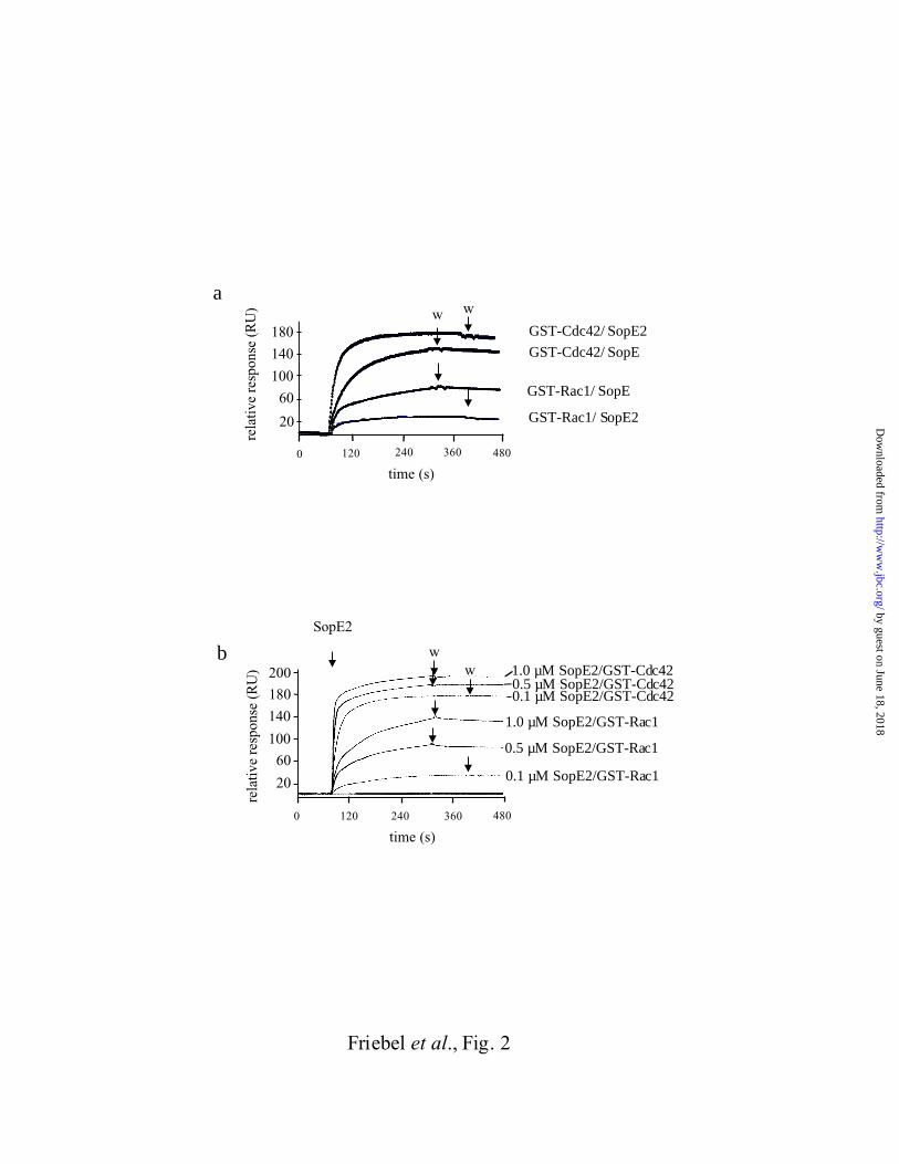
GST-Cdc42Hs1-192 or GST-Rac11-191 fusion protein (or GST as a control) was bound to a
sensor chip and we measured the kinetics of binding of SopE78-240 (or SopE269-240; 100nM;
Fig. 2a). The observed rates of complex formation between GST-Cdc42Hs1-192, GST-Rac11-
191, SopE78-240 and SopE269-240 were dependent on the concentration of the RhoGTPase
applied (Fig. 2b; data not shown). From the binding curves we calculated the kinetic constants
for complex formation (kass) assuming simple one step bimolecular association reactions:
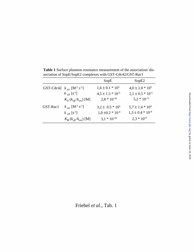
SopE78-240 binds with similar kinetics to GST-Cdc42Hs1-192 and to GST-Rac11-191 (Tab. 1). In
by guest on June 18, 2018http://w
ww
.jbc.org/D
ownloaded from
10
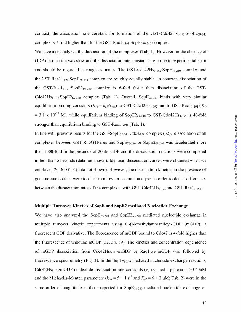
contrast, the association rate constant for formation of the GST-Cdc42Hs1-192·SopE269-240
complex is 7-fold higher than for the GST-Rac11-191·SopE269-240 complex.
We have also analyzed the dissociation of the complexes (Tab. 1). However, in the absence of
GDP dissociation was slow and the dissociation rate constants are prone to experimental error
and should be regarded as rough estimates. The GST-Cdc42Hs1-192·SopE78-240 complex and
the GST-Rac11-191·SopE78-240 complex are roughly equally stable. In contrast, dissociation of
the GST-Rac11-191·SopE269-240 complex is 6-fold faster than dissociation of the GST-
Cdc42Hs1-192·SopE269-240 complex (Tab. 1). Overall, SopE78-240 binds with very similar
equilibrium binding constants (KD = koff/kass) to GST-Cdc42Hs1-192 and to GST-Rac11-191 (KD
= 3.1 x 10⁻¹⁰ M), while equilibrium binding of SopE269-240 to GST-Cdc42Hs1-192 is 40-fold
stronger than equilibrium binding to GST-Rac11-191 (Tab. 1).
In line with previous results for the GST-SopE78-240·Cdc42∆C complex (32), dissociation of all
complexes between GST-RhoGTPases and SopE78-240 or SopE269-240 was accelerated more
than 1000-fold in the presence of 20µM GDP and the dissociation reactions were completed
in less than 5 seconds (data not shown). Identical dissociation curves were obtained when we
employed 20µM GTP (data not shown). However, the dissociation kinetics in the presence of
guanine nucleotides were too fast to allow an accurate analysis in order to detect differences
between the dissociation rates of the complexes with GST-Cdc42Hs1-192 and GST-Rac11-191.
Multiple Turnover Kinetics of SopE and SopE2 mediated Nucleotide Exchange.
We have also analyzed the SopE78-240 and SopE269-240 mediated nucleotide exchange in
multiple turnover kinetic experiments using O-(N-methylanthraniloyl-GDP (mGDP), a
fluorescent GDP derivative. The fluorescence of mGDP bound to Cdc42 is 4-fold higher than
the fluorescence of unbound mGDP (32, 38, 39). The kinetics and concentration dependence
of mGDP dissociation from Cdc42Hs1-192·mGDP or Rac11-191·mGDP was followed by
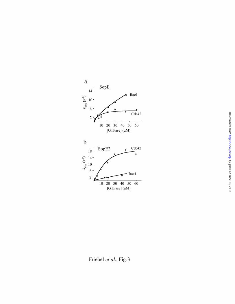
fluorescence spectrometry (Fig. 3). In the SopE78-240 mediated nucleotide exchange reactions,
Cdc42Hs1-192·mGDP nucleotide dissociation rate constants (v) reached a plateau at 20-40µM
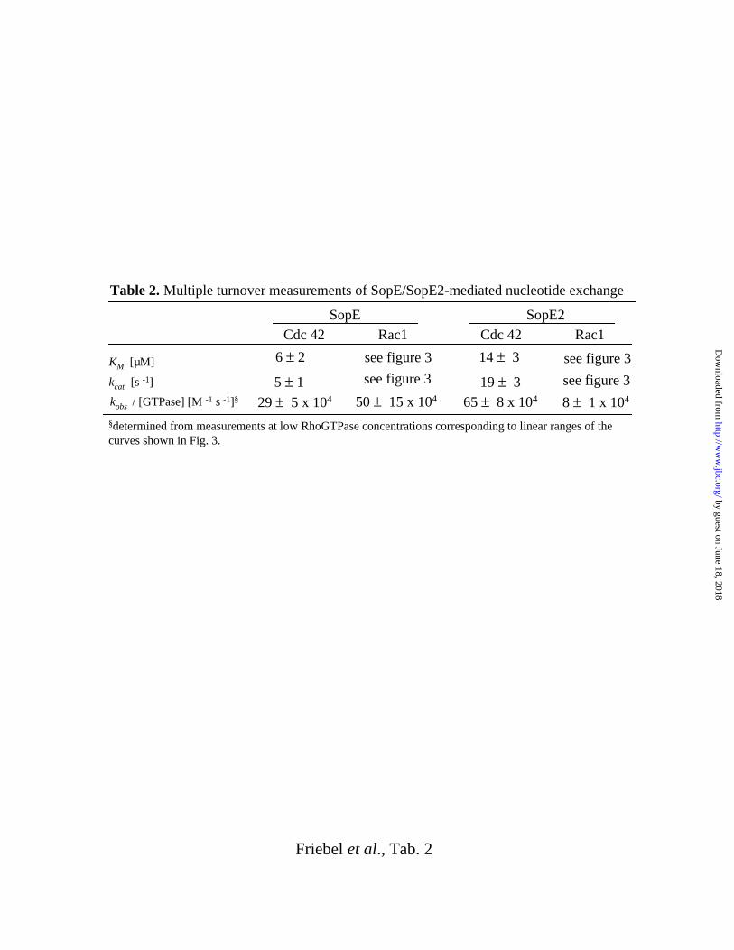
and the Michaelis-Menten parameters (kcat = 5 ± 1 s-1 and KM = 6 ± 2 µM; Tab. 2) were in the
same order of magnitude as those reported for SopE78-240 mediated nucleotide exchange on
by guest on June 18, 2018http://w
ww
.jbc.org/D
ownloaded from
11
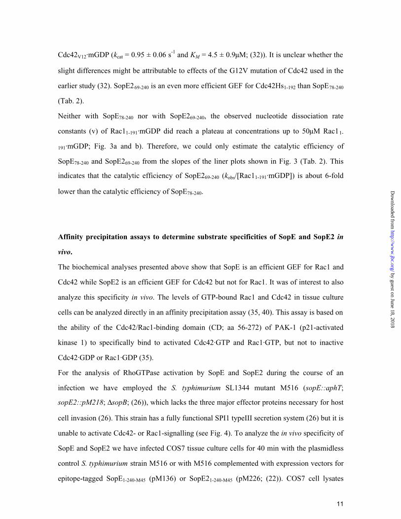
Cdc42V12·mGDP (kcat = 0.95 ± 0.06 s-1 and KM = 4.5 ± 0.9µM; (32)). It is unclear whether the
slight differences might be attributable to effects of the G12V mutation of Cdc42 used in the
earlier study (32). SopE269-240 is an even more efficient GEF for Cdc42Hs1-192 than SopE78-240
(Tab. 2).
Neither with SopE78-240 nor with SopE269-240, the observed nucleotide dissociation rate
constants (v) of Rac11-191·mGDP did reach a plateau at concentrations up to 50µM Rac11-
191·mGDP; Fig. 3a and b). Therefore, we could only estimate the catalytic efficiency of
SopE78-240 and SopE269-240 from the slopes of the liner plots shown in Fig. 3 (Tab. 2). This
indicates that the catalytic efficiency of SopE269-240 (kobs/[Rac11-191·mGDP]) is about 6-fold
lower than the catalytic efficiency of SopE78-240.
Affinity precipitation assays to determine substrate specificities of SopE and SopE2 in
vivo.
The biochemical analyses presented above show that SopE is an efficient GEF for Rac1 and
Cdc42 while SopE2 is an efficient GEF for Cdc42 but not for Rac1. It was of interest to also
analyze this specificity in vivo. The levels of GTP-bound Rac1 and Cdc42 in tissue culture
cells can be analyzed directly in an affinity precipitation assay (35, 40). This assay is based on
the ability of the Cdc42/Rac1-binding domain (CD; aa 56-272) of PAK-1 (p21-activated
kinase 1) to specifically bind to activated Cdc42·GTP and Rac1·GTP, but not to inactive
Cdc42·GDP or Rac1·GDP (35).
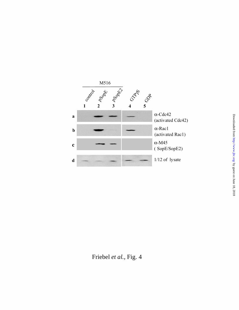
For the analysis of RhoGTPase activation by SopE and SopE2 during the course of an
infection we have employed the S. typhimurium SL1344 mutant M516 (sopE::aphT;
sopE2::pM218; ∆sopB; (26)), which lacks the three major effector proteins necessary for host
cell invasion (26). This strain has a fully functional SPI1 typeIII secretion system (26) but it is
unable to activate Cdc42- or Rac1-signalling (see Fig. 4). To analyze the in vivo specificity of
SopE and SopE2 we have infected COS7 tissue culture cells for 40 min with the plasmidless
control S. typhimurium strain M516 or with M516 complemented with expression vectors for
epitope-tagged SopE1-240-M45 (pM136) or SopE21-240-M45 (pM226; (22)). COS7 cell lysates
by guest on June 18, 2018http://w
ww
.jbc.org/D
ownloaded from
12
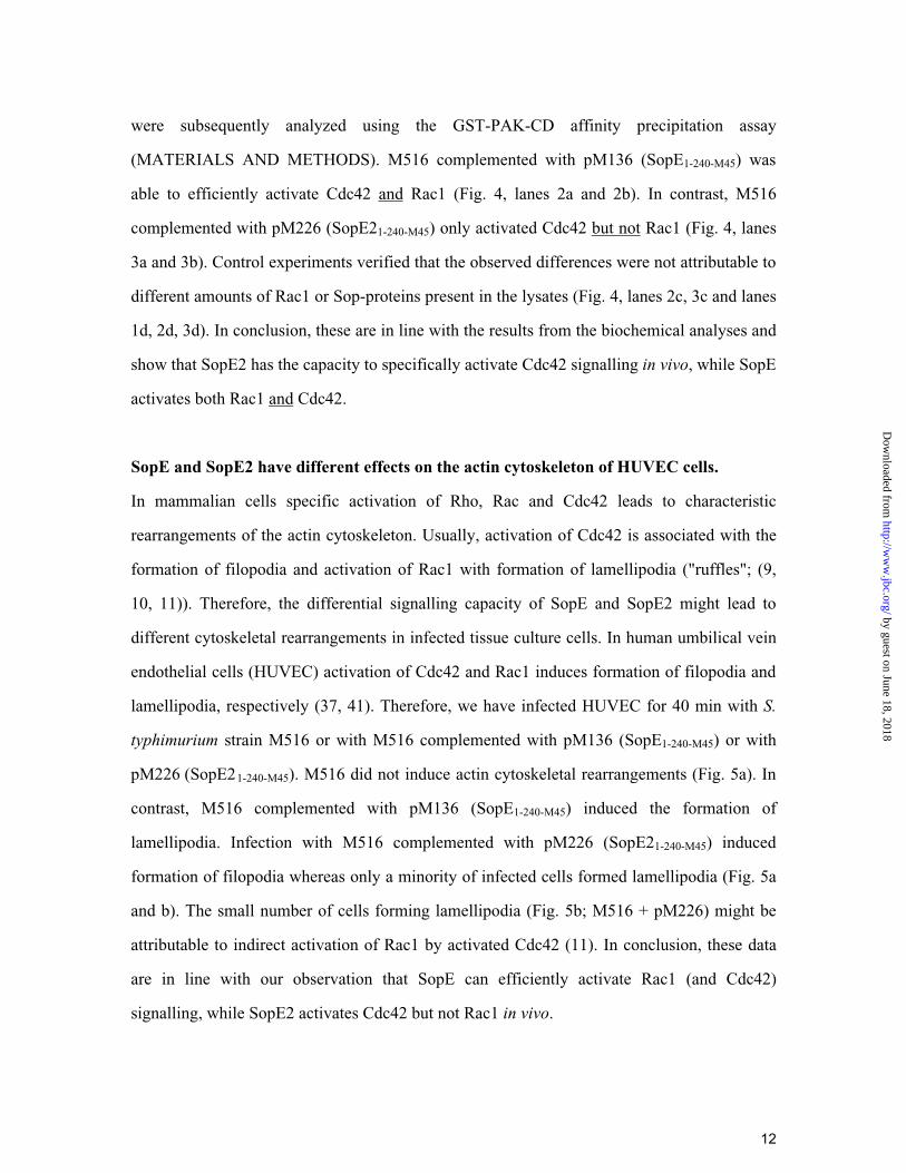
were subsequently analyzed using the GST-PAK-CD affinity precipitation assay
(MATERIALS AND METHODS). M516 complemented with pM136 (SopE1-240-M45) was
able to efficiently activate Cdc42 and Rac1 (Fig. 4, lanes 2a and 2b). In contrast, M516
complemented with pM226 (SopE21-240-M45) only activated Cdc42 but not Rac1 (Fig. 4, lanes
3a and 3b). Control experiments verified that the observed differences were not attributable to
different amounts of Rac1 or Sop-proteins present in the lysates (Fig. 4, lanes 2c, 3c and lanes
1d, 2d, 3d). In conclusion, these are in line with the results from the biochemical analyses and
show that SopE2 has the capacity to specifically activate Cdc42 signalling in vivo, while SopE
activates both Rac1 and Cdc42.
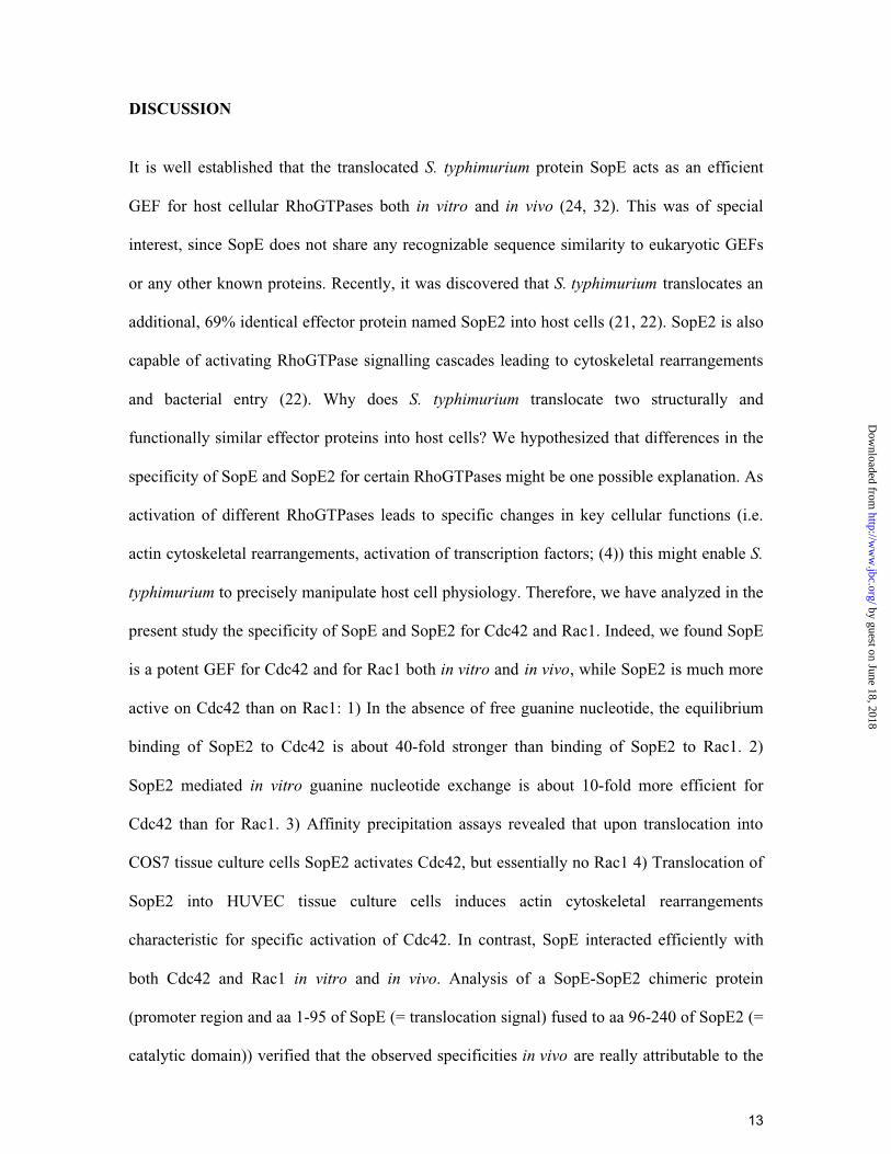
SopE and SopE2 have different effects on the actin cytoskeleton of HUVEC cells.
In mammalian cells specific activation of Rho, Rac and Cdc42 leads to characteristic
rearrangements of the actin cytoskeleton. Usually, activation of Cdc42 is associated with the
formation of filopodia and activation of Rac1 with formation of lamellipodia ("ruffles"; (9,
10, 11)). Therefore, the differential signalling capacity of SopE and SopE2 might lead to
different cytoskeletal rearrangements in infected tissue culture cells. In human umbilical vein
endothelial cells (HUVEC) activation of Cdc42 and Rac1 induces formation of filopodia and
lamellipodia, respectively (37, 41). Therefore, we have infected HUVEC for 40 min with S.
typhimurium strain M516 or with M516 complemented with pM136 (SopE1-240-M45) or with
pM226 (SopE21-240-M45). M516 did not induce actin cytoskeletal rearrangements (Fig. 5a). In
contrast, M516 complemented with pM136 (SopE1-240-M45) induced the formation of
lamellipodia. Infection with M516 complemented with pM226 (SopE21-240-M45) induced
formation of filopodia whereas only a minority of infected cells formed lamellipodia (Fig. 5a
and b). The small number of cells forming lamellipodia (Fig. 5b; M516 + pM226) might be
attributable to indirect activation of Rac1 by activated Cdc42 (11). In conclusion, these data
are in line with our observation that SopE can efficiently activate Rac1 (and Cdc42)
signalling, while SopE2 activates Cdc42 but not Rac1 in vivo.
by guest on June 18, 2018http://w
ww
.jbc.org/D
ownloaded from
13
DISCUSSION
It is well established that the translocated S. typhimurium protein SopE acts as an efficient
GEF for host cellular RhoGTPases both in vitro and in vivo (24, 32). This was of special
interest, since SopE does not share any recognizable sequence similarity to eukaryotic GEFs
or any other known proteins. Recently, it was discovered that S. typhimurium translocates an
additional, 69% identical effector protein named SopE2 into host cells (21, 22). SopE2 is also
capable of activating RhoGTPase signalling cascades leading to cytoskeletal rearrangements
and bacterial entry (22). Why does S. typhimurium translocate two structurally and
functionally similar effector proteins into host cells? We hypothesized that differences in the
specificity of SopE and SopE2 for certain RhoGTPases might be one possible explanation. As
activation of different RhoGTPases leads to specific changes in key cellular functions (i.e.
actin cytoskeletal rearrangements, activation of transcription factors; (4)) this might enable S.
typhimurium to precisely manipulate host cell physiology. Therefore, we have analyzed in the
present study the specificity of SopE and SopE2 for Cdc42 and Rac1. Indeed, we found SopE
is a potent GEF for Cdc42 and for Rac1 both in vitro and in vivo, while SopE2 is much more
active on Cdc42 than on Rac1: 1) In the absence of free guanine nucleotide, the equilibrium
binding of SopE2 to Cdc42 is about 40-fold stronger than binding of SopE2 to Rac1. 2)
SopE2 mediated in vitro guanine nucleotide exchange is about 10-fold more efficient for
Cdc42 than for Rac1. 3) Affinity precipitation assays revealed that upon translocation into
COS7 tissue culture cells SopE2 activates Cdc42, but essentially no Rac1 4) Translocation of
SopE2 into HUVEC tissue culture cells induces actin cytoskeletal rearrangements
characteristic for specific activation of Cdc42. In contrast, SopE interacted efficiently with
both Cdc42 and Rac1 in vitro and in vivo. Analysis of a SopE-SopE2 chimeric protein
(promoter region and aa 1-95 of SopE (= translocation signal) fused to aa 96-240 of SopE2 (=
catalytic domain)) verified that the observed specificities in vivo are really attributable to the
by guest on June 18, 2018http://w
ww
.jbc.org/D
ownloaded from
14
catalytic C-terminal domains (data not shown). Altogether, SopE and SopE2 provide S.
typhimurium with a means to specifically activate either Cdc42 and Rac1 or Cdc42 but not
Rac1.
Eukaryotic GEFs share a common functional unit (DH- plus PH-domain) which facilitates
GTPase-binding and catalysis. Yet, the eukaryotic GEFs for RhoGTPases display different
specificities for different subsets of RhoGTPases (5, 42). The 3D-structure of Rac1
complexed with the DH- and PH-domains of the eukaryotic GEF Tiam1 has identified the
amino acid residues determining the binding specificity (43). Unfortunately, as SopE and
SopE2 do not share any recognizable sequence similarity with eukaryotic GEFs, it is
impossible to predict the amino acid residues responsible for the different substrate
preferences of SopE and SopE2 from the data of Worthylake et al. (43).
Do all Salmonella strains address Cdc42 and Rac1? Phylogenetic analyses have shown that
sopE2 is present in all contemporary Salmonella lineages (21, 22, 26). Therefore, the capacity
to directly activate Cdc42 but not Rac1 (via SopE2) inside cells of the animal host is common
to all Salmonellae. In contrast, sopE is encoded in the genome of a bacteriophage which is
only present in very few Salmonella strains, including the S. typhimurium strain SL1344 used
in this study (26, 27, 44, 45). A second S. typhimurium strain (ATCC 14028) that is
commonly used to study virulence mechanisms does not carry SopEΦ and does not express
SopE (Mirold and Hardt, unpublished). Therefore, the capacity to directly activate Rac1-
signalling (via SopE) is not strictly required for Salmonella virulence per se. However, it is
well possible that SopE2 can activate Rac1 via an indirect mechanism as it is known that
specific activation of Cdc42 will finally result in activation Rac1in Swiss 3T3 fibroblasts (11).
Nonetheless, expression of SopE improves virulence. Interestingly, SopE-expressing S.
typhimurium strains are associated with severe epidemics (44). It has been speculated that the
improved epidemic virulence of these strains might simply be attributable to a higher "sopE"-
gene dosage and higher total amounts of SopE-like proteins delivered into host cells (44).
by guest on June 18, 2018http://w
ww
.jbc.org/D
ownloaded from
15
However, the data presented here suggest that the improved virulence of sopE-positive S.
typhimurium strains is much rather linked to the capacity of these strains to directly activate
Rac1 and Cdc42 (via SopE) inside host cells.
Indeed, there are several lines of evidence from tissue culture experiments suggesting that
direct activation of Cdc42 and Rac1 are needed to optimize host cell invasion: 1. Disruption
of the sopE gene in S. typhimurium strain SL1344 leads to a 2-fold decreased invasiveness
into COS7 tissue culture cells (27), while a sopE2 mutant is equally invasive as the wildtype
SL1344 strain (22). This argues that the presence of SopE can fully compensate for the loss of
SopE2, while SopE2 (possibly due to its inability to directly activate Rac1) cannot completely
compensate for the loss of SopE. 2. Complementation of a non-invasive S. typhimurium strain
(M516 = SL1344, sopE-, sopE2-, sopB-) which lacks all three translocated effector proteins
triggering bacterial entry with a SopE expression vector is 2-fold more efficient than
complementation with a SopE2 expression vector (26). 3. Disruption of Cdc42-signalling
inside COS7 cells by transfection with Cdc42N17 expression vectors is 2-fold more efficient at
blocking S. typhimurium SL1344 invasion than disruption of Rac1 signalling via Rac1N17
(23). In conclusion these observations suggest that the capacity to directly activate Rac1 (in
addition to Cdc42) via SopE improves S. typhimurium virulence.
Taken together, our results show for the first time that S. typhimurium can specifically
activate different RhoGTPases of the host cell via the translocated effector proteins SopE and
SopE2. This allows the bacteria to fine tune host cellular responses very precisely. Future
work will have to address how SopE/SopE2 triggered signalling may be further modulated by
the other translocated effector proteins like the actin binding protein SipA (19), the PI-
phosphatase SopB (Norris et al., 1998) or the GTPase activating protein SptP (46). This will
further advance our current knowledge of the intricate network of responses triggered by the
translocated effector proteins of S. typhimurium in order to alter host cell signalling in a very
precise manner.
by guest on June 18, 2018http://w
ww
.jbc.org/D
ownloaded from
16
REFERENCES
1. Ridley, A. J. (2000) in GTPases (Hall, A., ed.), pp. 89-136, Oxford University Press,
Oxford.
2. Bourne, H. R., Sanders, D. A. and McCormick, F. (1991) Nature 349, 117-127
3. Boguski, M. S., and McCormick, F. (1993) Nature 366, 643-654
4. Bishop, A. L., and Hall, A. (2000) Biochem. J. 348, 241-255
5. Cerione, R. A., and Zheng, Y. (1996) Curr. Opin. Cell Biol. 8, 216-222
6. Hall, A. (1998) Science 279, 509-514
7. Van Aelst, L. and D'Souza-Schorey, C. (1997) Genes Dev. 11, 2295-2322
8. Ridley, A. J., and Hall, A. (1992) Cell 70, 389-399
9. Ridley, A. J., Paterson, H. F., Johnston, C. L., Diekmann, D., and Hall, A. (1992) Cell 70,
401-410
10. Kozma, R., Ahmed, S., Best, A., and Lim, L. (1995) Mol. Cell. Biol. 15, 1942-1952
11. Nobes, C. D., and Hall, A. (1995) Cell 81, 53-62
12. Galán, J. E. (1999) Curr. Opin. Microbiol. 2, 46-50
13. Wood, M. W., Rosqvist, R., Mullan, P. B., Edwards, M. H., and Galyov, E. E. (1996)
Mol. Microbiol. 22, 327-338
14. Galyov, E. E., Wood, M. W., Rosqvist, R., Mullan, P. B., Watson, P. R., Hedges, S., and
Wallis, T.S. (1997) Mol. Microbiol. 25, 903-912
15. Collazo, C. M., and Galán, J. E. (1997) Mol. Microbiol. 24, 747-756
16. Hardt, W.-D., and Galán, J. E. (1997) Proc. Natl. Acad. Sci. U.S.A. 94, 9887-9892
17. Jones, M. A., Wood, M. W., Mullan, P. B., Watson, R., Wallis, T. J., and Galyov, E. E.
(1998) Infect. Immun. 66, 5799-5804
18. Fu, Y., and Galán, J. E. (1998) Mol. Microbiol. 27, 359-368
19. Zhou, D., Mooseker, M. S., and Galán, J. E. (1999) Science 283, 2092-2095
by guest on June 18, 2018http://w
ww
.jbc.org/D
ownloaded from
17
20. Miao, E. A., Scherer, C. A., Tsolis, R. M., Kingsley, R. A., Adams, L. G., Bäumler, A. J.,
and Miller, S. I. (1999) Mol. Microbiol. 34, 850-864
21. Bakshi, C. S., Singh, V. P., Wood, M. W., Jones, P. W., Wallis, T.S., and Galyov, E. E.
(2000) J. Bacteriol. 182, 2341-2344
22. Stender, S., Friebel, A., Linder, S., Rohde, M., Mirold, S., and Hardt, W.-D. (2000) Mol.
Microbiol. 36, 1206-1221
23. Chen, L. M., Hobie, S., and Galán, J. E. (1996) Science 274, 2115-2118
24. Hardt, W.-D., Chen, L. M., Schuebel, K.E. Bustelo, X. R., and Galán, J. E. (1998) Cell 93,
815-826
25. Chen, L. M., Bagrodia, S., Cerione, R.A., and Galán, J.E. (1999) J. Exp. Med. 189, 1479-
1488
26. Mirold, S., Ehrbar, K., Weissmüller, A., Prager, R., Tschäpe, H., Rüssmann, H., and
Hardt, W.-D. (2001) J. Bacteriol.183: 2348-2358
27. Hardt, W.-D., Urlaub, H., and Galán, J E. (1998) Proc. Natl. Acad. Sci. USA 95, 2574-
2579
28. Hong, K. H., and Miller, V. L. (1998) J. Bacteriol. 180, 1793-1802
29. Galán, J. E., and Zhou, D. (2000) Proc. Natl. Acad. Sci. USA 97, 8754-8761
30. Zhou, D., Chen, L. M. , Hernandez, L., Shears, S. B., and Galán, J. E. (2001) Mol.
Microbiol. 39, 248-259
31. Norris, F. A., Wilson, M.P., Wallis, T. S., Galyov, E. E., and Majerus, P. W. (1998) Proc.
Natl. Acad. Sci. USA 95, 14057-14059
32. Rudolph, M. G., Weise, C., Mirold, S., Hillenbrand, B., Bader, B., Wittinghofer, A., et al.
(1999) J. Biol. Chem. 274, 30501-30509
33. Hoiseth, S.K. and Stocker, B.A. (1981) Nature 291, 238-239
34. Friebel, A. and Hardt, W.D. (2000) Meth. Enzymol. 325, 82-91
by guest on June 18, 2018http://w
ww
.jbc.org/D
ownloaded from
18
35. Sander, E. E., van Delft, S., ten Klooster, J. P., Reid, T., van der Kammen, R. A.,
Michiels, F., and Collard, J. G. (1998) J. Cell. Biol. 143, 1385-1398
36. Obert, S., O'Connor, R. J., Schmid, S., and Hearing, P. (1994) Mol. Cell. Biol. 2, 1333-
1346
37. Aepfelbacher, M., Essler, M., Huber, E., Sugai, M., and Weber, P. C. (1997) Arterioscler.
Thromb. Vasc. Biol. 17: 1623-1629
38. Leonard, D. A., Evans, T., Hart, M., Cerione, R. A., and Manor, D. (1994) Biochemistry
33, 12323-12328
39. Rudolph, M. G., Bayer, P., Abo, A., Kuhlmann, J., Vetter, I. R., and Wittinghofer, A.
(1998) J. Biol. Chem. 273, 18067-18076
40. Azim, A.C., Barkalow, K.L. and Hartwig, J.H. (2000) Meth. Enzymol. 325, 257-263
41. Andor, A., Trülzsch, K., Essler, M., Roggenkamp, A., Wiedemann, A., Heesemann, J.,
and Aepfelbacher, M. (2001) Cell. Microbiol. 3: 301-310
42. Stam, J. C., and Collard, J. G. (1999) Prog. Mol. Subcell. Biol. 22, 51-83
43. Worthylake, D. K., Rossman, K. L. and Sondek, J. (2000) Nature 408, 682-688
44. Mirold, S., Rabsch, W., Rohde, M., Stender, S., Tschäpe, H., Rüssmann, H. et al. (1999)
Proc. Natl. Acad. Sci. USA 96, 9845-9850
45. Prager, R., Mirold, S., Tietze, E., Strutz, U., Knüppel, B., Rabsch, W., Hardt, W-D., and
Tschäpe, H. (2000) Int. J. Med. Micro. 290: 605-617
46. Fu, Y., and Galán, J. E. (1999) Nature 401: 293-297
by guest on June 18, 2018http://w
ww
.jbc.org/D
ownloaded from
19
FIGURES AND LEGENDS
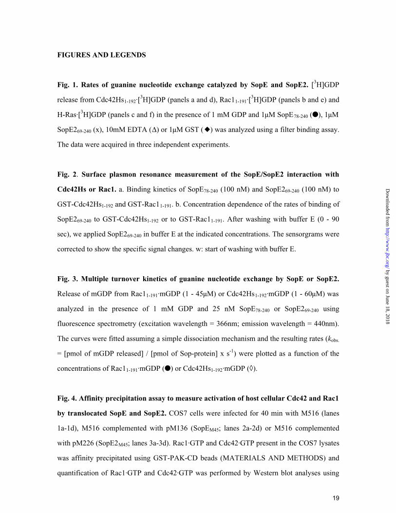
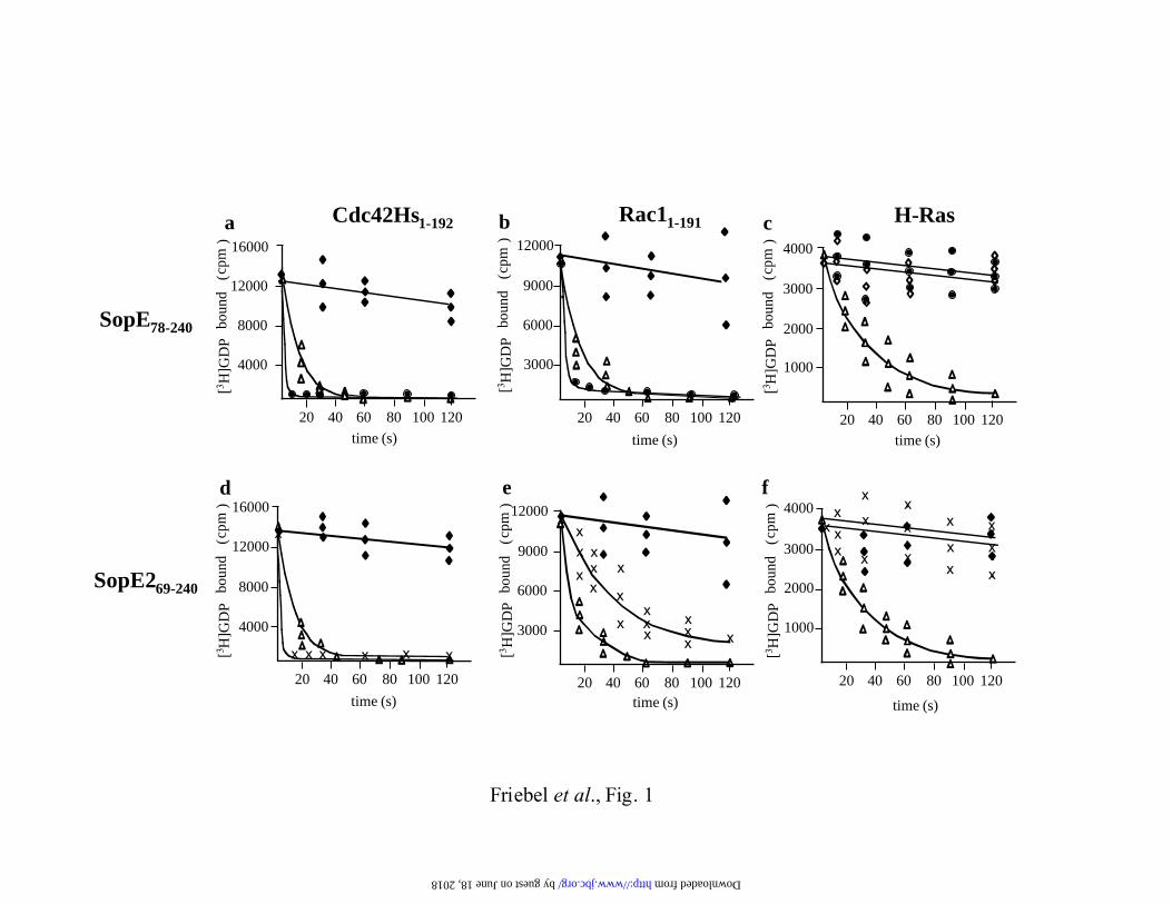
Fig. 1. Rates of guanine nucleotide exchange catalyzed by SopE and SopE2. [3H]GDP
release from Cdc42Hs1-192·[3H]GDP (panels a and d), Rac11-191·[
3H]GDP (panels b and e) and
H-Ras·[3H]GDP (panels c and f) in the presence of 1 mM GDP and 1µM SopE78-240 (�), 1µM
SopE269-240 (x), 10mM EDTA (∆) or 1µM GST (�) was analyzed using a filter binding assay.
The data were acquired in three independent experiments.
Fig. 2. Surface plasmon resonance measurement of the SopE/SopE2 interaction with
Cdc42Hs or Rac1. a. Binding kinetics of SopE78-240 (100 nM) and SopE269-240 (100 nM) to
GST-Cdc42Hs1-192 and GST-Rac11-191. b. Concentration dependence of the rates of binding of
SopE269-240 to GST-Cdc42Hs1-192 or to GST-Rac11-191. After washing with buffer E (0 - 90
sec), we applied SopE269-240 in buffer E at the indicated concentrations. The sensorgrams were
corrected to show the specific signal changes. w: start of washing with buffer E.
Fig. 3. Multiple turnover kinetics of guanine nucleotide exchange by SopE or SopE2.
Release of mGDP from Rac11-191·mGDP (1 - 45µM) or Cdc42Hs1-192·mGDP (1 - 60µM) was
analyzed in the presence of 1 mM GDP and 25 nM SopE78-240 or SopE269-240 using
fluorescence spectrometry (excitation wavelength = 366nm; emission wavelength = 440nm).
The curves were fitted assuming a simple dissociation mechanism and the resulting rates (kobs.
= [pmol of mGDP released] / [pmol of Sop-protein] x s-1) were plotted as a function of the
concentrations of Rac11-191·mGDP (�) or Cdc42Hs1-192·mGDP (◊).
Fig. 4. Affinity precipitation assay to measure activation of host cellular Cdc42 and Rac1
by translocated SopE and SopE2. COS7 cells were infected for 40 min with M516 (lanes
1a-1d), M516 complemented with pM136 (SopEM45; lanes 2a-2d) or M516 complemented
with pM226 (SopE2M45; lanes 3a-3d). Rac1·GTP and Cdc42·GTP present in the COS7 lysates
was affinity precipitated using GST-PAK-CD beads (MATERIALS AND METHODS) and
quantification of Rac1·GTP and Cdc42·GTP was performed by Western blot analyses using
by guest on June 18, 2018http://w
ww
.jbc.org/D
ownloaded from
20
specific mouse-α-Cdc42 (lane a) or mouse-α-Rac1 (lane b) antibodies. Lane c: relative
amounts of Sop-proteins (or Rac1; lane d) present in the lysates detected by Western blot
using a specific mouse-α-M45 (lane c) or a mouse-α-Rac1 (lane d) antibody. Lane 4a-4d:
Positive control. Rac1 and Cdc42 in lysate was activated by loading with GTPγS. Lane 5a-5d:
Negative control. Rac1 and Cdc42 in lysate was inactivated by loading with GDP. The assay
shown is representative for the five independent experiments performed.
Fig. 5. SopE/SopE2 induced rearrangements in the HUVEC actin cytoskeleton. a.
HUVECs were infected for 40 min with the indicated S. typhimurium strains. Cells were
fixed, f-actin was stained with rhodamine-phalloidin (red) and bacteria were stained with a
polyclonal α-Salmonella antiserum and a secondary α-rabbit FITC antibody (green;
MATERIALS AND METHODS). b. Quantitative analysis of the SopE/SopE2 induced
cytoskeletal rearrangements. Cells with altered actin cytoskeletal morphology (ca. 35% of all
cells) were classified based on their morphological features: profound membrane ruffling (i.e.
Fig. 5a, panel 2), weak filopodia formation (<20 filopodia per cell), pronounced filopodia
formation (>20 filopodia per cell; i.e. Fig. 5a, panel 3). For each S. typhimurium strain at least
100 cells with altered actin cytoskeletal morphology were evaluated in three independent
experiments (experimental error: ±10%). *no cytoskeletal rearrangements observed.
ACKNOWLEDGEMENTS
We would like to thank Dr. Irmgard Assfalg-Machleidt for her helpful instructions to perform
and evaluate surface plasmon resonance measurements on the BIAcore 2000 instrument.
by guest on June 18, 2018http://w
ww
.jbc.org/D
ownloaded from
SopE78-240
SopE269-240
Friebel et al., Fig. 1
Cdc42Hs1-192 H-Ras
1000
2000
3000
4000
4000
8000
12000
16000
[3 H]G
DP
boun
d cp
m(
)
time (s)
20 40 60 100 12080
Rac11-191
3000
6000
9000
12000
time (s)
20 40 60 100 12080 20 40 60 80 100 120time (s)
4000
8000
12000
16000
time (s)
3000
6000
9000
12000
time (s)
x
x
x x
x
xx x x xxx
x
x
x
x
x
x
x
x
x
x
x
x
1000
2000
3000
4000
time (s)
xxx
xx
x
x
x
xx
xxx
x
20 40 60 100 1208020 40 60 100 1208020 40 60 100 12080
a b c
d e f
[3 H]G
DP
boun
d cp
m(
)
[3 H]G
DP
boun
d cp
m(
)
[3 H]G
DP
boun
d cp
m(
)
[3 H]G
DP
boun
d cp
m(
)
[3 H]G
DP
boun
d cp
m(
)
xx
by guest on June 18, 2018 http://www.jbc.org/ Downloaded from
aw
GST-Rac1/ SopE
GST-Cdc42/ SopE
GST-Cdc42/ SopE2
GST-Rac1/ SopE2
w
rela
tive
resp
onse
(RU
)
20
180
140
100
60
0
time (s)
240120 360 480
time (s)
rela
tive
resp
onse
(RU
)
20
180
140
100
60
0 240120 360 480
200
0.1 µM SopE2/GST-Rac1
0.5 µM SopE2/GST-Rac1
1.0 µM SopE2/GST-Rac1
0.1 µM SopE2/GST-Cdc420.5 µM SopE2/GST-Cdc421.0 µM SopE2/GST-Cdc42
ww
bSopE2
Friebel et al., Fig. 2
by guest on June 18, 2018http://w
ww
.jbc.org/D
ownloaded from
SopE SopE2
GST-Cdc42 1,6 ± 0.1 * 105
4,5 ± 1.5 * 10-5 2,1 ± 0.5 * 10-5
4,0 ± 1.0 * 105k ass [M-1 s-1]
k off [s-1]
2,8 * 10-10 5,2 * 10-11KD (koff /kass) [M]
GST-Rac1
1,3 ± 0.4 * 10-4
5,7 ± 1.4 * 1043,2 ± 0.5 * 105
1,0 ±0.2 * 10-4
k ass [M-1 s-1]
k off [s-1]
2,3 * 10-93,1 * 10-10KD (koff /kass) [M]
Friebel et al., Tab. 1
Table 1 Surface plasmon resonance measurement of the association/ dis-sociation of SopE/SopE2 complexes with GST-Cdc42/GST-Rac1
by guest on June 18, 2018http://w
ww
.jbc.org/D
ownloaded from
Friebel et al., Fig.3
2
6
10
14
18
10 20 30 40 50 60
SopE2
[GTPase] (µM)
2
6
10
14
10 20 30 40 50 60
SopE
[GTPase] (µM)
k obs.
(s-1
)
Cdc42
Rac1
k obs.
(s-1
)a
b
Rac1
Cdc42
by guest on June 18, 2018http://w
ww
.jbc.org/D
ownloaded from
kcat [s -1]
KM [µM]
kobs / [GTPase] [M -1 s -1]§
5 ± 1
6 ± 2
29 ± 5 x 10⁴
see figure 3
see figure 3
50 ± 15 x 104
SopE2
see figure 3
Rac1
8 ± 1 x 10⁴
see figure 3
Cdc 42
19 ± 3
14 ± 3
65 ± 8 x 10⁴
Friebel et al., Tab. 2
SopERac1Cdc 42
Table 2. Multiple turnover measurements of SopE/SopE2-mediated nucleotide exchange
§determined from measurements at low RhoGTPase concentrations corresponding to linear ranges of thecurves shown in Fig. 3.
by guest on June 18, 2018http://w
ww
.jbc.org/D
ownloaded from
α-M45 ( SopE/SopE2)
1 2 3
c
Friebel et al., Fig. 4
1/12 of lysated
α-Cdc42 (activated Cdc42)
a
α-Rac1 (activated Rac1)
b
4 5
pSop
E
pSop
E2
GTP
γS G
DP
cont
rol
M516
by guest on June 18, 2018http://w
ww
.jbc.org/D
ownloaded from
and Wolf-Dietrich HardtAndrea Friebel, Heiko Ilchmann, Martin Aepfelbacher, Kristin Ehrbar, Werner Machleidt
host cellSopE and SopE2 from S. typhimurium activate different sets of RhoGTPases of the
published online July 5, 2001J. Biol. Chem.
10.1074/jbc.M100609200Access the most updated version of this article at doi:
Alerts:
When a correction for this article is posted•
When this article is cited•
to choose from all of JBC's e-mail alertsClick here
by guest on June 18, 2018http://w
ww
.jbc.org/D
ownloaded from