Embed Size (px)
Citation preview

Sparing and Salvaging Metals in ChloroplastsCrysten E. Blaby-Haas and Sabeeha S. Merchant
University of California, Los Angeles, CA, USA
1 Introduction 512 Metal Sparing and Salvaging within the
Chloroplast 523 Back-Ups 564 Reference Organisms for Sub-Cellular Metal Sparing
and Salvaging 575 Copper 576 Iron 607 Zinc 628 Acknowledgments 629 Abbreviations and Acronyms 62
10 References 62
1 INTRODUCTION
Of the essential inorganic minerals, the micronutri-ents (manganese, iron, copper, and zinc) have held specialinterest in plant physiology because of their chemistry, dualityas both nutrient and toxin, availability, and biogeochemicalhistory. The green tissues of plants readily exhibit visualsymptoms of metal deficiency, and even before the role ofmetals in photosynthesis was understood, the importance ofmetals in plant nutrition was already evident. Chlorosis is acommon symptom of poor metal nutrition, which manifests asa yellowing of green tissues owing to decreased abundance ofchlorophyll. Because of this readily scored phenotype, the ear-liest work on trace metals focused on descriptive observationsof metal nutrition.
It was not until the early to mid-1800s with thepopularization of the mineral theory of plant nutrition, that aserious look at the requirement of metal salts for plant growthand health began (Figure 1). In the 1840s, Eusebe Gris reportedthat the application of iron salts to either the roots or directlyto the leaves of some chlorotic plants resulted in reversing thesymptom. As a result, iron deficiency became synonymouswith chlorosis. Julius von Sachs is accredited with establishingthe essentiality of iron in plant growth, and 40-years laterBenjamin Moore reported that iron is indeed in the chloroplastand proposed that it plays a direct role in photosynthesis. In thenext 30 years, the essentiality of manganese, copper, and zincin plant growth and more specifically in photosynthesis was
Metals in Cells. Edited by Valeria Culotta and Robert A. Scott. © 2013 John Wiley & Sons, Ltd. ISBN 978-1-119-95323-4
reported. While the necessity of metal salts in plant growthand in photosynthesis was apparent, the actual biologicalrole of these ions remained elusive until the second half ofthe twentieth century, with the discovery of metal ions ascofactors for chloroplast-localized proteins.
We now know that linear electron flow requiresthe direct involvement of manganese, iron, and copper(Figure 2). A Mn4CaO5 cluster on the donor side of eachphotosystem II (PSII) monomer unit in the dimer catalyzesthe extraction of electrons from water, initiating the sequenceof electron flow. Within PSII, these electrons move to QA (aone-electron acceptor plastoquinone) and then to QB (a two-electron acceptor plastoquinone). A bicarbonate ion ligatedto a non-heme iron is thought to facilitate this transfer. Thecytochrome b6f dimer contains both heme and iron–sulfurclusters. Electron transfer between cytochrome b6f and theacceptor side of photosystem I (PSI) can involve either asmall copper protein, plastocyanin (land plants, algae andcyanobacteria), or an equivalent heme protein (algae andcyanobacteria). At the terminal end of linear electron flow, PSIcontains three 4Fe–4S clusters involved in electron transferwithin the complex, and these ultimately reduce the 2Fe–2Sprotein ferredoxin, which provides the reducing power forseveral metabolic reactions including NADP+ reduction.
In addition to the thylakoid membrane, many metal-dependent proteins localize to other chloroplast compartments(Table 1). They do not necessarily participate in photosyntheticelectron transfer directly, but they serve as support staff

52 METALS IN CELLS
Aristotle proposes that plants assimilate matterfrom the soil
1648 – Jean Baptista van Helmont – the willowthree experiment; plant mass is not derived fromsoil
1772 – Joseph Priestley – the mouse in bell jarexperiment; credited for discovering that plantsproduce oxygen
1809 – Albrecht Thaer – the Humus Theory –the majority of plant dry matter is derived from"soil nutritive juices"
1826 and 1828 – Carl Sprengel refutes theHumus Theory – soluble salts in the humuswere the real nutrients – proposes theMineral Theory of Nutrition
1837 – Hugo von Mohl – first definitive descrip-tion of chloroplasts
1840 – Justus von Liebig popularizes theMineral Theory – carbon comes from carbondioxide, hydrogen from water and othernutrients from solubilized salts in soil andwater
1843 – Eusébe Gris – chlorosis caused byinadequate supply of Fe1861 – Julius von Sachs – chloroplasts are siteof photosynthesis
1869 – Julius von Sachs – Fe is essentialelement for plant growth
1881 – Theodor Engelmann – oxygen evolutionby isolated chloroplasts in light
1914 – Benjamin Moore – Fe is in chloroplasts,and Fe is essential for photosythesis
1921 – René Wurmser – photosynthesis as aredox reaction
1922 – James McHargue – Mn required forplant growth
1931 – C.B. Lipman and G. MacKinney – Cuis required for plant growth
1937 – Hill reaction published
1937 – A. Pirson – Mn essential for oxygenicphotosynthesis
1939 – D.I. Arnon and P.R. Stout – define theterm “essential mineral element”
1939 – Arthur Neish – Fe and Cu are concen-trated in the chloroplast compared to the rest ofthe leaf
1949 – D.I. Arnon – discovers frist Cu enzymein isolated choloroplasts
1951 – R. Hill and R. Scarisbrick – Cyto-chrome f discovered
1960 – S. Katoh – discovers plastocyanin
1962 – K. Tagawa and D.I. Arnon – ferre-doxin recognized as an iron-dependentelectron carrier in the chloroplast
1968 – G.M. Cheniae and I.F. Martin – Mnrequired for water splitting and O2 evolution
1971 – A.J. Bearden and R. Malkin – discov-ery of PSI Fe/S clusters
350 BC
1800
1820
1840
1860
1880
1900
1920
1940
1960
1980
to ensure that the chloroplast is as productive as possiblein situations of environmental light fluctuation or whendetrimental by-products of the light reactions, such as reactiveoxygen species, accumulate. The chloroplast is also thesite of many metabolic pathways that are dependent on atransition metal at one or more steps. These include fattyacid biosynthesis, amino acid biosynthesis, nitrate and sulfateassimilation, and secondary metabolite synthesis. Metal ionsalso play central roles as cofactors in protein structure (suchas zinc in ribosomes) and regulation.
2 METAL SPARING AND SALVAGING WITHINTHE CHLOROPLAST
Plants regulate metal assimilation and distributionto ensure a balance between supply and demand, but whendemand exhausts the external provisions, mechanisms forconserving, redistributing, and prioritizing the metal cofactorare activated. These mechanisms include metal sparing, whichis a regulated reduction in the abundance of metal-dependentproteins, and metal salvaging, which involves degradation ofmetal-bound proteins and recycling of the limiting cofactor.Metal-sparing mechanisms result in a decreased numberof metal-dependent proteins through repression of geneexpression or degradation of transcripts and apoprotein,while metal salvaging results in liberation and redistributionof the precious metal. Both metal sparing and salvagingultimately lead to allocation of the limiting nutrient awayfrom unnecessary proteins and toward indispensable proteins.An attempt is made to preserve core metal-dependent functionsduring the shortage, and when metal is resupplied to deficientcells, the cellular landscape is poised to prioritize metalcofactor delivery to those key proteins.
Well-characterized examples of metal-sparing andmetal-salvaging mechanisms at the sub-cellular level existbecause of the availability of single-cell reference organismssuch as cyanobacteria and algae. Here, we will focus on sub-cellular acclimation to metal deficiency specifically in contextof the chloroplast, although these strategies may operate toeconomize and re-distribute metal at every level in the plant.
←−−−−−−−−−−−−−−−−−−−−−−−−−−−−−−−−−−−−−−−−−−−Figure 1 Historically significant events in understanding the roleof trace metals in plant nutrition. The origin of scholarly discourseon plant nutrition is commonly traced to Aristotle and his studentswho formalized the role of soil in plant survival. Aristotle’s notionthat plants absorb their matter from soil was refuted 2000 years laterby Jean Baptista van Helmont’s five-year willow tree experiments,although he erroneously concluded that plant matter was absorbedfrom water. In the next 300 years, our current understanding of plantphysiology was shaped with the discovery of photosynthesis and therole of inorganic minerals as nutrients and then, more specifically, asprotein cofactors. Seminal discoveries in plant metal nutrition are inbold

SPARING AND SALVAGING METALS IN CHLOROPLASTS 53
Ferritin
Iron buffer C. reinhardtii:iron recycling to the mitochondria
depending on trophicstatus (?)
C. reinhardtii:iron salvagedfor prioritized
proteins
Cyanobacteria/diatoms:
Fe CuZn Mn Ni SOD
Stroma
Fd
Fld
Fd replaced with Fld
Fe
FeFe
PSII
4 Mn 4 Mn
FeFe
Fe Fe4Fe
4Fe4Fe
Cu PC PSI
Fe Fe
Fe
Fe Fe2Fe 2Feb6f
Fe
Fe Fe
Fe
Fe2Fe
Fe
Fe
F
Iron deficiency
Copper deficiency PC spared andcopper recycled
Copper prioritized to PC
PSI and cytochrome b6f with 12 iron atomseach are major targets for iron salvaging
Fe
Cu
Cu
CuPPO
Thylakoid lumen
PC
Land plantsPC Cyt c6
Cyanobacteria / algae
(a)
(b)
Figure 2 Iron and copper sparing and salvaging within the chloroplast. Assuming 1:1:1:1:1 stoichiometry of functional complexes (dimersfor PSII and Cytb6f and either monomer or trimer for PSI), linear electron flow from PSII to ferredoxin is estimated to require 56 iron ions(using plastocyanin; 57 with cytochrome c6) in cyanobacteria and 30 iron ions (using plastocyanin; 31 using cytochrome c6) in land plantsand algae. Iron found in Cytc550, which is only present in the PSII complex of cyanobacteria and some red and brown algae, is surroundedby a dashed circle. (a) The iron-salvaging pathways as described for C. reinhardtii, cyanobacteria, and diatoms. In C. reinhardtii, iron issalvaged from the degradation of the ETC complexes and buffered by ferritin. A hierarchy of iron reallocation is established and involvesmaintenance of FeSOD in the chloroplast and respiration in the mitochondria during photoheterotrophic growth. Replacement of SOD isoforms(either transiently or permanently) and a switch between ferredoxin and flavodoxin are also popular metal-sparing strategies in photosyntheticmicrobes, particularly phytoplankton. (b) The two major copper-sparing and -salvaging pathways are shown. During copper deficiency inland plants, plastocyanin maturation is prioritized through downregulation of dispensable copper-bound proteins (dotted red line). In somecyanobacteria and algae, plastocyanin is dispensable and copper may be recycled to other compartments (dotted red line)
2.1 The Membranes of the Chloroplast—Barriers toMetal Mobilization
As pointed out, the importance of metals as essentialplant nutrients was reported in the literature over a centuryand a half ago. However, we are just now beginning tounderstand how the plant ensures proper acquisition anddistribution of these nutrients to the correct proteins in thecorrect compartments.
To maintain metal homeostasis, photosynthetic eu-karyotes have a unique challenge to overcome. The chloroplastis composed of three membranes: the outer- and inner-envelope membranes and the thylakoid membrane, and thesemembranes delineate three compartments: the intermembrane(interenvelope) space, the stroma, and the thylakoid lumen.The chloroplast machinery translocates most (if not all) metal-dependent proteins unfolded and cofactor free. Within thechloroplast, the imported polypeptide is processed to its ma-ture form and sorted to its final destination, which may be
the inter-membrane space, envelope membrane, stroma, thy-lakoid membrane, or lumen. For metalloproteins, insertion ofthe cofactor is usually one of the very last steps in protein mat-uration [except for lumen proteins that are translocated by thetwin-arginine translocase (TAT) pathway described below]and occurs once the protein has reached its final destination.This has been experimentally demonstrated for cytochromec6 heme attachment, Mn4CaO5 cluster assembly in PSII, andcopper insertion in plastocyanin.1
Therefore, the cofactor must be present in thefinal compartment. The outer envelope of archaeplastidicchloroplasts is analogous to the outer membrane of bacteria,and metal-chelates may freely diffuse across this porousmembrane into the intermembrane space. The inner membraneis analogous to the plasma membrane of bacteria, but howmetal crosses this membrane is largely unknown. A P1B-typeATPase PAA1 is proposed to pump copper into the stroma,as is a second ATPase HMA1, which is also implicated in

54 METALS IN CELLS
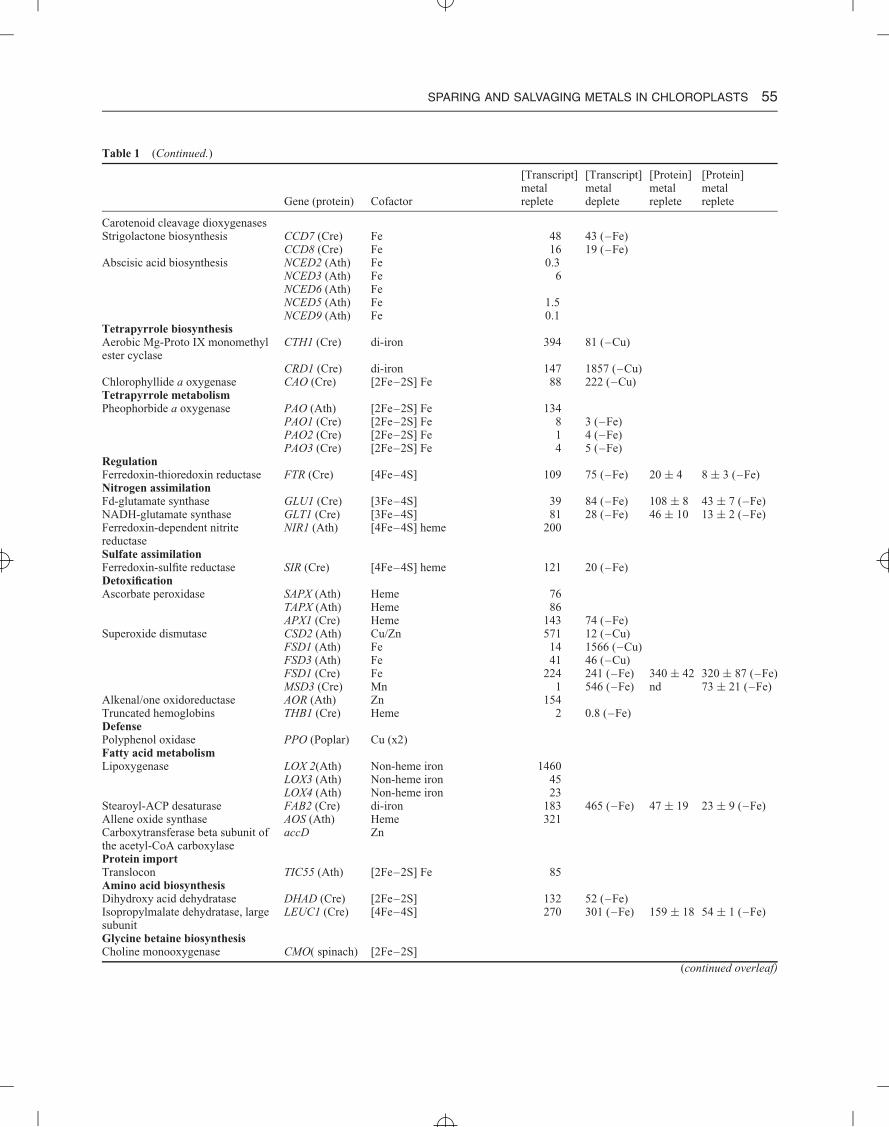
Table 1 Metal-dependent pathways in the chloroplast
[Transcript] [Transcript] [Protein] [Protein]metal metal metal metal
Gene (protein) Cofactor replete deplete replete deplete
The light reactionsPSII psbE (Cytb559) hemeLp, Ga dimer: 4Fe, 8Mn psbFCb, Ra, Dt dimer: 6Fe, 8Mn psbA (D1) Fe–S ↓ (–Fe)
psbD (D2)psbA (D1) Mn4CaO5psbC (CP43)
Cyanobacterial/diatom-specific psbV (Cytc550) HemeCytb6f petB (Cytb6) Heme (×3)Dimer: 12Fe petA (Cytf) Heme ↓ (–Fe)
PETC (Rieskeiron–sulfurprotein)
[2Fe–2S]
Plastocyanin (1Cu) Lp, Ga, Dt, Cb PETE1 (Ath) Cu 182 129 (–Cu)PETE2 (Ath) Cu 1681 1529 (–Cu)PCY1 (Cre) Cu 7100 6200 (–Cu) 1350 nd (–Cu)
Cyt c6 (1Fe) Ga, Ra, Dt, Cb CYC6 (Cre) Heme 0.8 2677 (–Cu) nd 285 (–Cu)PSI psaC [4Fe–4S] (×2)Lp, Ga monomer: 12Fe psaA [4Fe–4S] ↓ (–Fe)Cbtrimer: 36Fe psaBFd (2Fe) Characterized and chloroplast-localized in A. thaliana
FD1 (Ath) [2Fe–2S] 81FD2 (Ath) [2Fe–2S] 1230FDC1 (Ath) [2Fe–2S] 59FDC2 (Ath) [2Fe–2S] 35Characterized and chloroplast-localized in C. reinhardtiiPETF (Cre) [2Fe–2S] 8278 5065 (–Fe) 87 ± 35 nd (–Fe)FDX2 (Cre) [2Fe–2S] 8 3.1 (–Fe)FDX3 (Cre) [2Fe–2S] 21 144 (–Fe) nd 45 ± 7 (–Fe)FDX5 (Cre) [2Fe–2S] 17 28 (–Fe)FDX6 (Cre) [2Fe–2S] 41 362 (–Fe) ↓ (–Fe)
Carbon metabolismAssociates with glyceraldehyde-3-phosphate dehydrogenase andphosphoribulokinase
CP12-1 (Ath) Cu 523 483 (–Cu)CP12-2 (Ath) Cu 180 181 (–Cu)CP12-3 (Ath) Cu 6 6 (–Cu)
Carbonic anhydrase CAH1 (Cre) Zn 12CAH8 (Cre) Zn 101
Alcohol dehydrogenase ADH1 (Cre) Fe 205Alternative electron transferType I NAD(P)H dehydrogenase NDF4 (Ath) [2Fe–2S] 34Missing from Cre ndhI [4Fe–4S] (x2)
ndhK [4Fe–4S]Plastid terminal oxidase (PTOX) IM1 (Ath) di-iron 48
PTO1 (Cre) di-iron 87 42 (–Fe)PTO2 (Cre) di-iron 27 11 (–Fe)
Succinate dehydrogenase SDH2 (Cre) [2Fe–2S] [4Fe–4S] [3Fe–4S] 324 190 (–Fe)SDH3 (Cre) Heme 324 147 (–Fe)SDH4 (Cre) 314 150 (–Fe)
Carotenoid biosynthesisCarotenoid hydroxylase CYP97A3 (Cre) Heme 14 43 (–Fe)
CYP97B3 (Cre) Heme 24 21 (–Fe)CYP97C1 (Cre) Heme 19 63 (–Fe)
4-Hydroxy-3-methylbut-2-en-1-yldiphosphate synthase
HDS (Cre) [4Fe–4S] 49 110 (–Fe) 125 ± 37 23 ± 7 (–Fe)
4-Hydroxy-3-methylbut-2-enyldiphosphate reductase
HDR (Cre) [4Fe–4S] 95 102 (–Fe) 61 ± 7 nd (–Fe)
Carotenoid metabolismβ-Carotene isomerase D27 (Cre) Fe 27 18 (–Fe)
SPARING AND SALVAGING METALS IN CHLOROPLASTS 55
Table 1 (Continued.)
[Transcript] [Transcript] [Protein] [Protein]metal metal metal metal
Gene (protein) Cofactor replete deplete replete replete
Carotenoid cleavage dioxygenasesStrigolactone biosynthesis CCD7 (Cre) Fe 48 43 (–Fe)
CCD8 (Cre) Fe 16 19 (–Fe)Abscisic acid biosynthesis NCED2 (Ath) Fe 0.3
NCED3 (Ath) Fe 6NCED6 (Ath) FeNCED5 (Ath) Fe 1.5NCED9 (Ath) Fe 0.1
Tetrapyrrole biosynthesisAerobic Mg-Proto IX monomethylester cyclase
CTH1 (Cre) di-iron 394 81 (–Cu)
CRD1 (Cre) di-iron 147 1857 (–Cu)Chlorophyllide a oxygenase CAO (Cre) [2Fe–2S] Fe 88 222 (–Cu)Tetrapyrrole metabolismPheophorbide a oxygenase PAO (Ath) [2Fe–2S] Fe 134
PAO1 (Cre) [2Fe–2S] Fe 8 3 (–Fe)PAO2 (Cre) [2Fe–2S] Fe 1 4 (–Fe)PAO3 (Cre) [2Fe–2S] Fe 4 5 (–Fe)
RegulationFerredoxin-thioredoxin reductase FTR (Cre) [4Fe–4S] 109 75 (–Fe) 20 ± 4 8 ± 3 (–Fe)Nitrogen assimilationFd-glutamate synthase GLU1 (Cre) [3Fe–4S] 39 84 (–Fe) 108 ± 8 43 ± 7 (–Fe)NADH-glutamate synthase GLT1 (Cre) [3Fe–4S] 81 28 (–Fe) 46 ± 10 13 ± 2 (–Fe)Ferredoxin-dependent nitritereductase
NIR1 (Ath) [4Fe–4S] heme 200
Sulfate assimilationFerredoxin-sulfite reductase SIR (Cre) [4Fe–4S] heme 121 20 (–Fe)DetoxificationAscorbate peroxidase SAPX (Ath) Heme 76
TAPX (Ath) Heme 86APX1 (Cre) Heme 143 74 (–Fe)
Superoxide dismutase CSD2 (Ath) Cu/Zn 571 12 (–Cu)FSD1 (Ath) Fe 14 1566 (–Cu)FSD3 (Ath) Fe 41 46 (–Cu)FSD1 (Cre) Fe 224 241 (–Fe) 340 ± 42 320 ± 87 (–Fe)MSD3 (Cre) Mn 1 546 (–Fe) nd 73 ± 21 (–Fe)
Alkenal/one oxidoreductase AOR (Ath) Zn 154Truncated hemoglobins THB1 (Cre) Heme 2 0.8 (–Fe)DefensePolyphenol oxidase PPO (Poplar) Cu (x2)Fatty acid metabolismLipoxygenase LOX 2(Ath) Non-heme iron 1460
LOX3 (Ath) Non-heme iron 45LOX4 (Ath) Non-heme iron 23
Stearoyl-ACP desaturase FAB2 (Cre) di-iron 183 465 (–Fe) 47 ± 19 23 ± 9 (–Fe)Allene oxide synthase AOS (Ath) Heme 321Carboxytransferase beta subunit ofthe acetyl-CoA carboxylase
accD Zn
Protein importTranslocon TIC55 (Ath) [2Fe–2S] Fe 85Amino acid biosynthesisDihydroxy acid dehydratase DHAD (Cre) [2Fe–2S] 132 52 (–Fe)Isopropylmalate dehydratase, largesubunit
LEUC1 (Cre) [4Fe–4S] 270 301 (–Fe) 159 ± 18 54 ± 1 (–Fe)
Glycine betaine biosynthesisCholine monooxygenase CMO( spinach) [2Fe–2S]
(continued overleaf)
56 METALS IN CELLS
Table 1 (Continued.)
[Transcript] [Transcript] [Protein] [Protein]metal metal metal metal
Gene (protein) Cofactor replete deplete replete replete
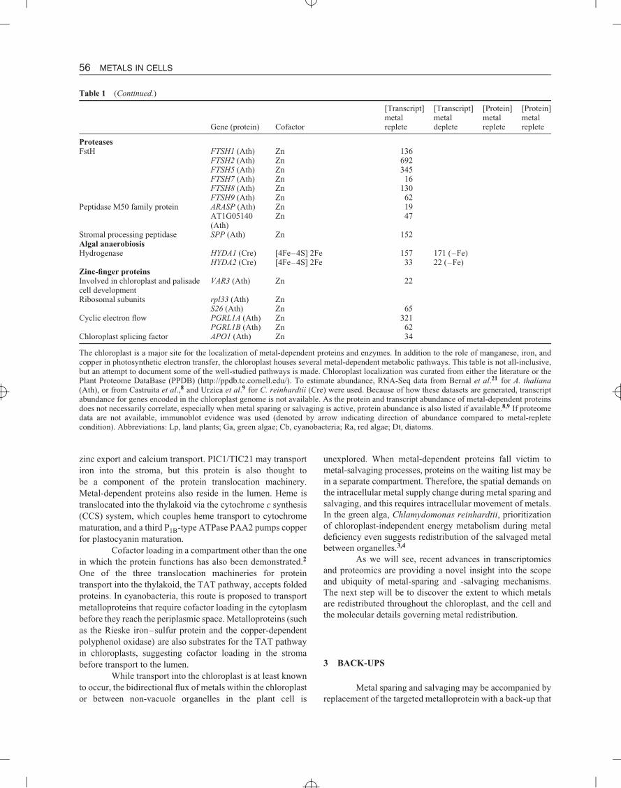
ProteasesFstH FTSH1 (Ath) Zn 136
FTSH2 (Ath) Zn 692FTSH5 (Ath) Zn 345FTSH7 (Ath) Zn 16FTSH8 (Ath) Zn 130FTSH9 (Ath) Zn 62
Peptidase M50 family protein ARASP (Ath) Zn 19AT1G05140(Ath)
Zn 47
Stromal processing peptidase SPP (Ath) Zn 152Algal anaerobiosisHydrogenase HYDA1 (Cre) [4Fe–4S] 2Fe 157 171 (–Fe)
HYDA2 (Cre) [4Fe–4S] 2Fe 33 22 (–Fe)Zinc-finger proteinsInvolved in chloroplast and palisadecell development
VAR3 (Ath) Zn 22
Ribosomal subunits rpl33 (Ath) ZnS26 (Ath) Zn 65
Cyclic electron flow PGRL1A (Ath) Zn 321PGRL1B (Ath) Zn 62
Chloroplast splicing factor APO1 (Ath) Zn 34
The chloroplast is a major site for the localization of metal-dependent proteins and enzymes. In addition to the role of manganese, iron, andcopper in photosynthetic electron transfer, the chloroplast houses several metal-dependent metabolic pathways. This table is not all-inclusive,but an attempt to document some of the well-studied pathways is made. Chloroplast localization was curated from either the literature or thePlant Proteome DataBase (PPDB) (http://ppdb.tc.cornell.edu/). To estimate abundance, RNA-Seq data from Bernal et al.21 for A. thaliana(Ath), or from Castruita et al.,8 and Urzica et al.9 for C. reinhardtii (Cre) were used. Because of how these datasets are generated, transcriptabundance for genes encoded in the chloroplast genome is not available. As the protein and transcript abundance of metal-dependent proteinsdoes not necessarily correlate, especially when metal sparing or salvaging is active, protein abundance is also listed if available.8,9 If proteomedata are not available, immunoblot evidence was used (denoted by arrow indicating direction of abundance compared to metal-repletecondition). Abbreviations: Lp, land plants; Ga, green algae; Cb, cyanobacteria; Ra, red algae; Dt, diatoms.
zinc export and calcium transport. PIC1/TIC21 may transportiron into the stroma, but this protein is also thought tobe a component of the protein translocation machinery.Metal-dependent proteins also reside in the lumen. Heme istranslocated into the thylakoid via the cytochrome c synthesis(CCS) system, which couples heme transport to cytochromematuration, and a third P1B-type ATPase PAA2 pumps copperfor plastocyanin maturation.
Cofactor loading in a compartment other than the onein which the protein functions has also been demonstrated.2
One of the three translocation machineries for proteintransport into the thylakoid, the TAT pathway, accepts foldedproteins. In cyanobacteria, this route is proposed to transportmetalloproteins that require cofactor loading in the cytoplasmbefore they reach the periplasmic space. Metalloproteins (suchas the Rieske iron–sulfur protein and the copper-dependentpolyphenol oxidase) are also substrates for the TAT pathwayin chloroplasts, suggesting cofactor loading in the stromabefore transport to the lumen.
While transport into the chloroplast is at least knownto occur, the bidirectional flux of metals within the chloroplastor between non-vacuole organelles in the plant cell is
unexplored. When metal-dependent proteins fall victim tometal-salvaging processes, proteins on the waiting list may bein a separate compartment. Therefore, the spatial demands onthe intracellular metal supply change during metal sparing andsalvaging, and this requires intracellular movement of metals.In the green alga, Chlamydomonas reinhardtii, prioritizationof chloroplast-independent energy metabolism during metaldeficiency even suggests redistribution of the salvaged metalbetween organelles.3,4
As we will see, recent advances in transcriptomicsand proteomics are providing a novel insight into the scopeand ubiquity of metal-sparing and -salvaging mechanisms.The next step will be to discover the extent to which metalsare redistributed throughout the chloroplast, and the cell andthe molecular details governing metal redistribution.
3 BACK-UPS
Metal sparing and salvaging may be accompanied byreplacement of the targeted metalloprotein with a back-up that
SPARING AND SALVAGING METALS IN CHLOROPLASTS 57
uses a different metal cofactor (one that is available) or nocofactor at all. Specific examples will be given in the followingsections. By expressing a back-up, the cell can reduce metalexpenditure without sacrificing its function.
The existence of metalloprotein back-ups underscoresthe redundancy of metal-catalyzed chemistry—two metals cancatalyze the same reaction. However, except for documentedcases of cambialism, metalloproteins are generally specific forone metal. This means that a separate protein must be producedto catalyze the reaction with the alternative non-limitingcofactor. Even when a protein is genuinely cambialistic, thevarious metal ions may not provide the same level of activity.There are very few examples—perhaps only two—in theliterature of cambialism as a proposed mechanism to overcomemetal-limitation (vide infra).
Carbonic anhydrases are responsible for the re-versible hydration of CO2 to bicarbonate and a proton.Classically, these enzymes use a zinc-bound hydroxide tocatalyze this reaction, but during growth under low zinc withcadmium supplementation, the carbonic anhydrase CDCA1from the marine diatom Thalassiosira weissflogii containscadmium instead of zinc and is still active. This unique classof carbonic anhydrase is a structural mimic of the unrelatedβ-carbonic anhydrase dimer and can readily exchange cad-mium for zinc to yield an even more active enzyme.5 Thecambialistic nature of this enzyme, which is important incarbon sequestering, is thought to represent a zinc-sparingmechanism and provide a selective advantage for diatomsgrowing in zinc-poor ocean waters.
Superoxide dismutase (SOD) from several bacterialspecies is a second example of cambialistic metal sparing.Divergent evolution has led to the iron- and manganese-dependent SODs, while convergent evolution has led to theunrelated classes of CuZnSODs and NiSODs. As we will see,these classes are commonly used as back-ups for each otherin metal-sparing strategies, but only a unique subclass of theiron- and manganese-dependent SODs is cambialistic. Thecambialistic SODs are found mainly in prokaryotes and mayresemble an ancestor that existed before the evolution of thetwo distinct iron and manganese types.6 While the distinctSODs are selective for one metal or the other, the cambialisticSODs are active with either cofactor. The concentration ofmanganese and iron in the culture medium determines whichcofactor the cambialistic SOD will use.
Back-up proteins generally arise by convergentevolution and do not share sequence similarity. A notableexception is the manganese- and iron-dependent SOD familymentioned earlier. In most cases, the existence of unrelatedfunctionally equivalent protein families with different metalcofactors is known for decades before their role as a back-upenzyme during metal-deficiency acclimation is known. Thesestrategies are invariably uncovered through gene expressionanalysis, as the back-up is expressed only under metaldeficiency. Because they may not have recognizable domains,
several back-ups could remain unnoticed in the list of metal-deficiency-induced genes of unknown function.
An example is the folE2 gene commonly found inbacterial zinc regulons.7 GTP cyclohydrolase I (encoded byfolE in Escherichia coli) catalyzes the conversion of GTPto 7,8-dihydroneopterin triphosphate, a substrate for severalbiosynthetic pathways including de novo folate synthesis inbacteria and plants. Absence of the corresponding gene inseveral bacterial genomes led to the discovery of a non-homologous gene encoding a functionally equivalent enzymenamed folE2. Until the discovery of this function, folE2,referred to at that time as yciA, was a gene of unknown functionregulated by zinc nutrition. As these genomes contain the geneencoding the canonical enzyme, which is zinc-dependent, thedesignation of YciA/FolE2 as a zinc-independent back-up forFolE has been proposed.
4 REFERENCE ORGANISMS FOR SUB-CELLULARMETAL SPARING AND SALVAGING
Although cyanobacteria lack the complexity ofphotosynthetic eukaryotes, they are free-living relatives ofthe original chloroplast. Several cyanobacterial genomes aresequenced, and the fortuitous arrangement of co-regulatedgenes into operons can aid in functional genomics studies ofgenes conserved between the lineages.
With the seminal work on plastocyanin and cy-tochrome c6 substitution, C. reinhardtii has become a premierreference for probing analogous metal-responsive events. Inaddition to the genome, the transcriptomes of C. reinhardtiiunder copper- and iron-deficiency situations have beensequenced.8,9
The analyzes of individual phytoplankton and phy-toplankton communities have provided a novel insight intodistinctive strategies for acclimating to metal deficiency andnovel adaptations to metal-poor aquatic environments. Com-pared to life on land, aquatic environments have uniquemetal nutrient concentrations and speciations. Both spatialand temporal changes in metal concentration influence thebiochemistry of marine microorganisms, and in some cases,phytoplankton species have adapted to chronic metal defi-ciency by remodeling the genome to reflect the repertoirecorresponding to genes induced in terrestrial algae duringmetal deficiency. Many oceanic algal species have evolvedthe ability to grow at much lower external and intracellu-lar concentrations of iron, zinc, and manganese and, whenpossible, functionally equivalent proteins have permanentlyreplaced those that are dependent on these metals.
5 COPPER
Copper and iron are the only trace metals concentratedin the chloroplast compared to the rest of the leaf.10,11 This

58 METALS IN CELLS
Table 2 Transcripts involved in A. thaliana copper economy
Gene Description Cu−/Cu+ miRNA Location
AT2G28190 CSD2 (copper/zinc superoxide dismutase) 0.02 mir398 StromaAT2G29130 LAC2 (laccase; copper ion binding) 0.02 mir397 ApoplasticAT2G02850 ARPN (plantacyanin; copper ion binding) 0.06 mir408 ApoplasticAT1G08830 CSD1 (copper/zinc superoxide dismutase) 0.06 mir398 CytoplasmAT1G12520 CCS1 (copper chaperone for superoxide dismutase) 0.07 miR398 Stroma/cytoplasmAT2G30210 LAC3 (laccase; copper ion binding) 0.2 mir408 ApoplasticAT1G72230 Plastocyanin-like domain-containing protein 0.2 mir408 ApoplasticAT2G38080 IRX12/LAC4 (laccase; copper ion binding) 0.4 mir397 ApoplasticAT3G15640 Cytochrome c oxidase family protein (Cox5b-1) 0.4 mir398 MitochondrionAT5G60020 LAC17 (laccase; copper ion binding) 0.6 mir397 ApoplasticAT2G44790 UCC2 (uclacyanin; copper ion binding) 0.5 mir408 Apoplastic
The fold difference in transcript abundance from shoots of plants grown in the absence (Cu−) and presence (Cu+) of copper supplementationis given according to Bernal et al.21
observation can be attributed to the prevalence of these metalsin the electron transport chain (ETC), but while iron is centralto multiple steps in linear electron flow, only one step andonly one protein, plastocyanin, is dependent on copper. Inland plants and most algae, plastocyanin is responsible forthe oxidation of the b6f complex and reduction of PSI duringlinear electron flow (Figure 2). Exceptions include some redand brown algae that are solely reliant on cytochrome c6 (iron-dependent) for this step. In most chloroplasts, a bottleneck inphotosynthesis can form during copper deficiency, whereelectron flow through plastocyanin becomes the limitingstep. Therefore, copper-sparing mechanisms in chloroplastsdescribed so far center on this small soluble protein inthe thylakoid lumen. The best-characterized responses area plastocyanin/cytochrome c6 switch in algae and microRNAs(miRNAs)-imposed copper economy in land plants (Figure 2and Table 2).
5.1 Cytochrome c6 Replaces Plastocyanin DuringCopper Deficiency
In the late 1970s, it became apparent that a solubleheme protein substitutes for plastocyanin in response to coppernutrition in some algae and cyanobacteria. This heme protein,originally referred to as cytochrome c-552 or c-553 based onα-absorption maximum and eventually renamed cytochromec6 based on function, serves as a back-up protein and facilitatesan effective copper-sparing mechanism: electron transfer canoccur without interruption, absence of plastocyanin minimizesthe chloroplast demand for copper, and available copper maybe redirected to other proteins. Plastocyanin is estimated tobe one of the most abundant proteins in the lumen, and thisswitch spares a significant amount of copper. The reciprocalaccumulation of cytochrome c6 and plastocyanin is widespreadamong algae and cyanobacteria, but was presumably lost fromland plants and lost or never gained by some red, green, andbrown algae, which contain either plastocyanin or cytochromec6.12
While copper regulates transcription of the geneencoding cytochrome c6 in both cyanobacteria and algae,the regulation of plastocyanin abundance occurs by variousmechanisms.13 However, each case represents a copper-sparing mechanism, because each results in a lower copperquota in the cell (for cyanobacteria) or the cell and thechloroplast (for algae). In the cyanobacteria Anabaena sp.PCC 7937, Anabaena sp. PCC 7120, Synechocystis sp.PCC 6803, and Prochlorothrix hollandica, plastocyaninmRNA abundance decreases in response to decreasing copperconcentrations. In green algae, three distinct mechanismshave been found. Plastocyanin abundance in Scenedesmusobliquus is regulated at the level of mRNA accumulation,whereas in Pediastrum boryanum, mRNA accumulates inboth copper-replete and -deficient cells, but the transcriptfrom copper-deficient cells is truncated, resulting in loss of theinitiation codon, and hence it is not translated. In C. reinhardtii,mRNA abundance is independent of the medium copperconcentration, and an unidentified protease, which is activespecifically during copper deficiency, targets plastocyaninfor degradation. This degradation is not an example ofmetal salvaging, because the protease (or proteases) targetsthe mature but cofactor-free form.14 Presumably, the costof maintaining plastocyanin expression, translation, andlocalization to the thylakoid in C. reinhardtii is offset bythe advantage of re-establishing plastocyanin function thatmuch faster after copper resupply.
This conclusion would suggest that there is an ad-vantage of using plastocyanin over cytochrome c6. We do notknow what the advantage is, but we have noted that there aremany other metabolic changes in the plastid coincident withthe use of cytochrome c6. One of these changes is in the levelof unsaturation of thylakoid membrane galactolipids, whichwould influence membrane fluidity—presumably of relevancefor a diffusible electron carrier.8 Another major change isthe upregulation of enzymes in tetrapyrrole biosynthesis.8
Specifically, expression of the gene CPX1 encoding copro-porhyrinogen (coprogen) III oxidase, which functions prior
SPARING AND SALVAGING METALS IN CHLOROPLASTS 59
to the branch point between heme and chlorophyll biosynthe-sis, increases in a copper-deficiency-dependent manner. Onethought is that this response may supply the extra hemerequired for cytochrome c6 maturation. Nevertheless, theextra heme is probably only marginal over the amount re-quired for other photosynthetic complexes, and CPX1 mRNA,protein, and activity increase about 5- to 10-fold. Further-more, the expression of three genes encoding enzymes inthe chlorophyll biosynthetic pathway is also increased dur-ing copper deficiency in C. reinhardtii: CRD1 encodes acomponent of the aerobic Mg-Proto IX monomethyl estercyclase, CGL78/YCF54 encodes an ortholog to a proteinthat forms a complex with and is required for functionof the Mg-Proto IX monomethyl ester cyclase,15 and CAOencodes chlorophyllide a oxygenase, which is involved inchlorophyll b biosynthesis. We note that all three enzymesare oxygen-dependent. CRD1, CGL78, and CPX1 are directtargets of the Cu-responsive transcription factor CRR1, sug-gesting a specific adaptation in C. reinhardtii of the tetrapyr-role pathway in response to copper deficiency—probablyto increase flux through chlorophyll biosynthesis. How-ever, the specific consequence of this adjustment remainsunknown.
5.1.1 Copper Recycling
Instead of copper salvaging, copper recycling mayexist in cyanobacteria and algae. Not only can photosynthesisstill function without plastocyanin because of the presence ofcytochrome c6, but plastocyanin ultimately serves as a copperstorage protein. The copper-bound plastocyanin presumablysuccumbs to normal protein turnover, and reestablishment ofthe cellular copper equilibrium would result in redirectionof copper away from the thylakoid. Whether altering thecopper distribution in the cell would occur in a passivemanner or involve regulation of transporters and/or metal-binding factors such as metal chaperones is presentlynot known. Targets for prioritized copper allocation inC. reinhardtii include cytochrome oxidase, which acquirescopper in the mitochondrion inter-membrane space, andferroxidase, which acquires copper in the trans-Golgi enroute to the plasma membrane. As ferroxidase is requiredfor high-affinity iron uptake and, therefore, dispensablein an iron-replete situation, the prioritized target may becytochrome oxidase. Indeed, ferroxidase protein abundancedecreases during growth in iron-replete, copper-limitedmedium.8 Under copper-replete conditions, plastocyaninis estimated to be at least 10-fold more abundant thancytochrome oxidase,14 but reduction of the copper quotamay be a larger advantage than copper recycling as copper-starved (successive culturing in copper-limited medium16) C.reinhardtii cultures do not display visible signs of copperdeficiency.
5.2 miRNAs and the Cu-Economy Model in Land Plants
The lifestyles of the fast-growing, motile, single-celled photosynthetic microbes and of slower-growing,stationary, multicellular land plants have resulted in quitedifferent evolutionary strategies to overcome fluctuations incopper availability. The most dramatic difference is that aplastocyanin back-up is not encoded in the genomes of landplants (as of August 2012), and cyanobacteria and algaesuccessfully cope with copper deficiency, whereas land plantsdo not. Copper-deficient plants show symptoms of stuntedgrowth, chlorosis, photosynthetic and morphological defects,and experience desiccation.17 Copper-sparing mechanisms doexist in land plants, but the inability to sacrifice plastocyaninwithout consequences on photosynthetic efficiency appears tohave a large impact on survival. The presumed goal of coppersparing and salvaging in land plants is to prioritize copperfor plastocyanin, but the plant can spare and salvage onlya finite amount of copper to maintain an adequate level ofplastocyanin. However, these mechanisms suffice in the faceof fluctuating copper availability and mild copper deficiency.
As characterized in Arabidopsis thaliana and Populustrichocarpa, post-transcriptional regulation of non-essentialcopper-dependent proteins and synthesis of back-up enzymesare the primary responses to copper-deficiency in land plants.18
In 2003, using DNA microarrays, Wintz et al.19 noted thatsub-optimal copper nutrition leads to a reduction in theabundance of the CuZnSOD-encoding transcripts (CSD1 andCSD2) and those for the associated copper chaperone (CCS).Downregulation of these genes was later found to resultfrom targeted transcript degradation due to a microRNA.miRNAs are short (ca. 21 nucleotides) non-coding RNAs thatusually negatively regulate protein abundance by targetingtranscripts for degradation or inhibiting translation.20 Whilethe transcription factor CRR1 regulates copper-dependentexpression of cytochrome c6 in C. reinhardtii, a CRR1ortholog in A. thaliana, SPL7, regulates the expression of thecopper miRNAs. Both regulators share a GTAC recognitionsite, which constitutes the core of a Cu-response element(CuRE). The element is located upstream of the transcriptionstart site.21
Copper limitation in A. thaliana induces the expres-sion of four Cu-miRNA families. These families are centralto the Cu-miRNA-mediated model for Cu-economy proposedby Burkhead et al.18 (Table 2). The transcripts for copperproteins that are dispensable such as several phytocyanins(targets of miR408) and laccases (targets of miR397, miR408,and miR857), and copper proteins that can be replaced withcopper-independent back-ups such as CuZnSOD (target ofmiR398) are degraded. The outcome is a reduced demand onthe copper pool, estimated to be by about 70%,21 and putativelya higher success of plastocyanin metallation. An alternativeor mutual result is that transcript degradation eliminates thecompetition, and during copper re-supply, plastocyanin ispositioned to preferentially acquire copper. Reflective of se-lectively reduced protein synthesis during copper deficiency,

60 METALS IN CELLS
photosynthetic efficiency recovers sooner than do the activitiesof CuZnSOD and polyphenol oxidase in P. trichocarpa.22
Of the known targets of Cu-miRNAs in A. thaliana,only CSD2 and its chaperone CCS1 are in the chloroplast.SOD is an important detoxification enzyme responsible forthe dismutation of superoxide to hydrogen peroxide. In greenleaves, nearly 90% of SOD activity is in the chloroplast,23 andbecause the reducing side of PSI produces superoxide, CSD2localizes to the stroma. Therefore, CSD2 may directly competefor copper heading toward plastocyanin in the thylakoidlumen. Land plants also encode iron-dependent superoxidedismutases (FeSODs), and copper deficiency leads to anincrease in the abundance of the FeSOD-encoding transcript,FSD1, concurrently with the degradation of CSD2 transcript.21
The presumed role of FSD1 expression is compensation forthe lack of CuZnSOD activity.
The majority of transcripts targeted by Cu-miRNAsencode proteins that are or are predicted to be secreted(Table 2). Although these proteins are apoplastic, metallationwould occur in the secretory system before export,24
and the reduced synthesis of these proteins may lead torerouting of copper to the chloroplast. The presence ofmiR397, miR398, and miR408 gene families is conservedin P. trichocarpa, which contains an additional miRNAfamily, miR1444. miR1444 is upregulated during copperdeficiency and regulates the abundance of transcripts encodingpolyphenol oxidase.22 Polyphenol oxidases are binuclearcopper proteins in the thylakoid lumen, which are absentin both A. thaliana and C. reinhardtii. The presence ofthis miRNA family reveals tailoring of Cu-economy to generepertoire.
6 IRON
Studies in the 1940s established that chloroplastscontain roughly 80% of the cellular iron found in greenleaves.25,26 The majority of this iron is present in the proteinsof the photosynthetic ETC (Figure 2). The simplest calculationof iron content per ETC (23–24 iron) assumes a molar ratio of1:1 for each iron-containing complex, and that each complexis a monomer. Therefore, this value is an underestimatebecause both PSII and cytochrome b6f are dimers, PSI is atrimer in cyanobacteria but a monomer in eukaryotes, and thestoichiometries of these complexes are generally not 1:1. Forinstance, the PSI:PSII ratio in cyanobacteria can vary from4:1 to 1:1 depending on the nutritional supply of iron, and aconstitutive ratio of 1:10 was found for a diatom adapted tochronically low-iron abundance. Regardless of the absoluteiron requirement of the ETC, the chloroplastic demand isrelatively high, and the bioavailability of iron in aerobicsoils is low. Therefore, acquired iron deficiency compared todeficiency in other transition metals is common in agriculture.Iron deficiency also contributes to reduced primary production
in the oceans. Then, it is of no surprise that iron was one ofthe first inorganic nutrients studied in plant fitness.
Considering the historical significance of iron inplant nutrition, sparing and salvaging in the context of thechloroplast is underexplored in land plants. This gap inknowledge may be because of the difficulty in controllingiron deficiency, which is necessary to avoid noise fromthe individual context of different cell types in a tissue.Therefore, iron sparing and salvaging in the context ofphotosynthesis is best understood in cyanobacteria andalgae, where homogeneous populations grown under strictlydefined conditions of iron nutrition are easily attained.Well-characterized examples include remodeling of thephotosynthetic machinery and back-ups.
6.1 Iron Sparing and Salvaging in C. reinhardtii:Remodeling of the Thylakoid Membrane ProteinContent
Physiology and expression of key genes involved inacclimating to iron status define four stages of iron nutritionin C. reinhardtii.27 Cells will accumulate two- to five-foldmore iron in the iron-excess stage (200 μM medium ironcontent) compared to the iron-replete stage (20 μM mediumiron content), and, as a consequence, are sensitive to excessexcitation energy. In the iron-replete situation, genes encodingiron uptake pathways are expressed at basal levels, and luxuryiron consumption corresponds to one-sixth of the availablemedium iron content (ca. 3 μM iron).
As the cells become iron deficient (3–1 μM mediumiron content), classic iron-deficiency chlorosis is not evident,but a programmed response that includes induction of ironuptake and remodeling of the photosynthetic architecture isinitiated.28 Various iron-containing proteins in the chloroplastare degraded, including PSI, the cytochrome b6f complex, andferredoxin, and the light-harvesting complex associated withthe remaining PSI is remodeled. ETC remodeling is not uniqueto C. reinhardtii. Similar iron-deficiency responses such asdecreasing the ratio of PSI to PSII are observed in other algaeand cyanobacteria.29
In C. reinhardtii, not all iron-dependent proteins inthe chloroplast are reduced in abundance. Maintenance ofFeSOD suggests salvaging of iron within the chloroplast,while maintenance of respiration during growth on acetatesuggests salvaging of iron between the chloroplast andmitochondrion.3,27 Iron released from the degradation ofETC complexes is buffered by ferritin, an iron-storageprotein whose expression is increased during iron deficiencyin C. reinhardtii. In contrast, ferritin expression in othereukaryotes, including land plants, is increased during ironexcess.30 Ferritin, by binding the released iron, serves as abuffer during the salvage process.
The fourth stage, iron limitation (≤0.5 μM mediumiron content), is marked by chlorosis and diminished growthrate. Loss of PSI and cytochrome complexes is evident

SPARING AND SALVAGING METALS IN CHLOROPLASTS 61
with a reduction in abundance to <1% in acetate-grownC. reinhardtii cells compared to iron-replete cultures. In theabsence of acetate, however, these complexes are maintained,suggesting that the complexes are sacrificed in the previoussituation for iron salvaging.
A recent analysis of transcript abundance using theRNA-Seq method during the three iron nutrition stages hasfurther illuminated iron-sparing and -salvaging strategies ofC. reinhardtii. Generally, transcripts encoding proteins withFe–S clusters were reduced in abundance, while transcriptsencoding some heme-bound proteins were increased inabundance (Table 1). This trend may represent routing ofiron away from a subset of proteins and ensure maintenanceof another subset. The reduction of Fe–S proteins in thechloroplast may also be an attempt to control photooxidativestress. Fe–S clusters are particularly labile in the presenceof superoxide, which is produced by the photoreduction ofdioxygen by PSI, especially when the function of the Fe–Sclusters in PSI is compromised by iron deficiency. Superoxidedestroys solvent-exposed Fe–S clusters (including potentiallythose from PSI), releasing Fe3+, which can then react withhydrogen peroxide, creating the highly cytotoxic hydroxylradical, which cannot be enzymatically destroyed.
6.2 Back-Ups
6.2.1 Ferredoxin/Flavodoxin—Iron Salvaging
Ferredoxin and flavodoxin are iron-dependent andiron-independent electron transfer proteins, respectively.Positioned at the terminal end of linear electron flow, theyserve as switchboards providing reducing power for severalpathways involved in metabolism and regulation. Flavodoxin,known at the time of its discovery as phytoflavin, was firstisolated from the cyanobacterium Synechococcus elongatusand found to the substitute for ferredoxin in the photoreductionof NADP+ by isolated chloroplasts.31 The connection to ironsparing was made by Knight et al.32, who isolated flavodoxinfrom the bacterium Clostridium pasteurianum grown in low-iron medium. They proposed that flavodoxin has a similarrole during iron deficiency as ferredoxin has during ironsufficiency, and thus the name ‘‘flavor-’’, as it contains a flavinand ‘‘-doxin’’ because of its equivalence with ferredoxin.Schonheit et al.33 found that ferredoxin was actively degradedwhen the C. pasteurianum culture consumed the bulk of theiron from the medium. The cells continued to grow and asecond Fe–S protein, pyruvate synthase, was maintained.They proposed an iron-salvaging mechanism during whichiron is recycled from ferredoxin.
The genomes of some algae, particularly marinediatoms, contain genes for both ferredoxin and flavodoxin.34
The presence of both genes does not necessarily equate tothe ability of that organism to functionally substitute one forthe other in response to iron. While reciprocal expressionis found in the oceanic diatom T. weissflogii, flavodoxin is
not induced during iron deficiency in the coastal relativeThalassiosira pseudonana. Flavodoxin induction by irondeficit is widespread in bacteria and proposed as a markerof iron deficiency in the oceans. Less well explored, mainlybecause of the dearth of robust genetic tools, is the abilityof flavodoxin to functionally substitute for ferredoxin inalgae.
Flavodoxin is missing from the genomes of se-quenced land plants and most algae. Nevertheless, transgenictobacco lines expressing a cyanobacterial flavodoxin in chloro-plasts gained a fitness advantage when grown on iron-deficientmedium.35 This and other studies of recombinant flavodoxinhave incited curiosity as to why flavodoxin was lost during thetransition to land. The simplest answer is that during the evo-lution of modern plants, flavodoxin did not provide a selectivebenefit, and as in the case of so many genes inherited fromthe original cyanobacterial symbiont, the flavodoxin gene wassimply lost.
Even in the absence of flavodoxin, iron sparinginvolving ferredoxin does occur in C. reinhardtii. Of theferredoxins in the chloroplast, ferredoxin-2 is predicted toreduce nitrate reductase and, therefore, may be unnecessaryduring growth in the presence of ammonium and can be sparedif iron nutrition is suboptimal.
6.2.2 Superoxide Dismutase
With four distinct isoforms having four differentmetal cofactor requirements, when multiple SODs make theirway into the same genome, the stage is set for a metal-sparingstrategy. As we saw with copper economy, the CuZnSOD inthe chloroplast of land plants is downregulated reciprocallywith the induction of FeSOD. In bacteria, where CuZnSODis missing, the reciprocal presence of FeSOD in iron-repletemedium and MnSOD in iron-deficient medium is widespread.
Some algae also lack CuZnSOD and rely solely onFeSOD (or MnSOD in the case of T. pseudonana) activity inthe chloroplast. Therefore, the induction of an MnSOD duringiron deficiency in C. reinhardtii pointed to an iron-sparingmechanism in the chloroplast.36 However, recent work hasshown that while the MnSOD is produced and localizes tothe chloroplast, FeSOD is actually maintained while otheriron-dependent proteins are lost.27
6.2.3 Fructose-Bisphosphate Aldolase
The expression of a metal-free isozyme of fructose-bisphosphate aldolase has also been proposed to serve as aback-up for the metal-dependent isozyme in the chloroplastduring iron limitation.37 While the expression of the metal-free form is responsive to iron nutrition in numerous systems,dependence of the metal-dependent form on iron has notbeen shown. Therefore, whether expression of fructose-bisphosphate aldolase is an example of iron sparing or involvedin metabolic remodeling is yet to be determined.

62 METALS IN CELLS
6.3 Chronic Iron Limitation
Iron fertilization experiments in the 1990s establishedthat photosynthesis in the open ocean is chronically limited bythe abundance and bioavailability of iron. The phytoplanktonspecies in these communities (cyanobacteria, diatoms, andalgae) have adapted to the iron deficit with permanent iron-sparing: reduction in the abundance of iron-requiring proteinsand reduction in the total number of genes encoding iron-requiring proteins. Putative sodN genes are present in thepico-prymnesiophyte metagenome, the four pico-prasinophytegenomes, and Phaeodactylum tricornutum, and while theymay also contain genes for CuZnSOD or MnSOD, thesegenomes do not contain genes for FeSOD.38 Like landplants, some diatoms that inhabit the open ocean useplastocyanin instead of cytochrome c6, while their coastalcousins constituently use cytochrome c6. Other differencesinclude the ratio of iron-rich ETC complexes. The oceanicdiatom Thalassiosira oceanica has up to fivefold lower PSI andup to sevenfold lower cytochrome b6f complex concentrationsthan the coastal diatom T. weissflogii.39
In nitrogen fixation, the nitrogenase enzyme sys-tem catalyzes the reduction of dinitrogen to ammonia. Thissystem consists of two proteins: a 4Fe:4S enzyme and amolybdenum–iron enzyme with an 8Fe:7S cluster and aMo:7Fe:9S cofactor. In the cyanobacterium, Crocosphaerawatsonii, nitrogen fixation occurs at night and transcripts in-volved in photosynthesis decline, while transcripts for thenitrogenase system increase in the evening as photosynthesiswinds down. The most obvious explanation for temporal sep-aration of these two processes is the incompatibility betweenmolecular oxygen and the nitrogenase complex. Neverthe-less, recent quantification of protein abundance during themetabolic switch has led to the hypothesis that cyanobac-teria recycle iron between the nitrogenase proteins andphotosynthesis.40 This process, referred to as ‘‘hot-bunking,’’is estimated to reduce the cellular iron quota by around 40%and appears to be an adaptation to the low bioavailability ofiron in the open ocean, as regulation by iron status is notindicated.
7 ZINC
Although zinc is an abundant metal cofactor, fewexamples of zinc sparing and salvaging in plants are available,and none is presently known to operate in the chloroplast. Onehurdle to the study of sub-cellular zinc homeostasis is the lackof techniques applicable to the study of this spectroscopicallysilent ion. In addition, the ease with which the zinc cofactorloads into the apoprotein in vitro generally precludes theneed to add exogenous zinc to enzyme assays. Therefore,several enzymes may be erroneously labeled as not requiringa cofactor. In bacteria, zinc sparing and salvaging are well-known strategies for acclimation to zinc limitation, and this
is mainly because of the identification of zinc-responsiveregulons using bioinformatic approaches.13 Examples ofthese include reciprocal expression of zinc-dependent vszinc-independent isoforms of ribosomal subunits, metabolicenzymes, and regulatory proteins. Perhaps, the role of zinc inthe chloroplast has been overlooked by the plant world becauseof its inability to perform redox chemistry (in fact, zinc is nota transition metal), but interest in zinc and acclimation to zincdeficiency is gaining prominence.41 Diatoms have providedus with several examples of strategies to overcome zinclimitation: the use of a cadmium-activated carbonic anhydrasediscussed earlier and a cobalt-substituted carbonic anhydrase.Whether these scenarios occur in the chloroplast is yet tobe determined. The abundance of the carbonic anhydrases inalgae for operation of the carbon concentrating mechanismssuggests that they may be sources for zinc salvage. Yet, theirfunction is essential in low CO2 environments, and back-upversions of these enzymes may well occur in the algae of theplant lineage as well.
8 ACKNOWLEDGMENTS
The research on copper and zinc homeostasis issupported by the National Institutes of Health GM042143 toS.S.M., while the work on iron and manganese homeostasis issupported by the Division of Chemical Sciences, Geosciences,and Biosciences, Office of Basic Energy Sciences of the USDepartment of Energy through Grant DE-FD02-04ER15529to S.S.M. C.E.B.-H. is supported by a Kirschstein NationalResearch Service Award GM100753.
9 ABBREVIATIONS AND ACRONYMS
CCS = cytochrome synthesis; CuRE = Cu-responseelement; ETC = electron transport chain; FeSODs = iron-dependent superoxide dismutases; miRNAs = microRNAs;miRNAs = microRNAs; PPDB = Plant Proteome DataBase;PSII = photosystem II; PSI = photosystem I; SOD =Superoxide dismutase; TAT = twin-arginine translocase.
10 REFERENCES
1. S. Merchant and B. W. Dreyfuss, Annu. Rev. Plant Physiol.Plant Mol. Biol., 1998, 49, 25.
2. T. Palmer and B. C. Berks, Nat. Rev. Microbiol., 2012, 10(7),483.
3. A. M. Terauchi, G. Peers, M. C. Kobayashi, K. K. Niyogi, andS. S. Merchant, Photosynth. Res., 2010, 105(1), 39.
4. B. Naumann, A. Busch, J. Allmer, E. Ostendorf, M. Zeller, H.Kirchhoff, and M. Hippler, Proteomics, 2007, 7(21), 3964.
5. Y. Xu, L. Feng, P. D. Jeffrey, Y. Shi, and F. M. Morel, Nature,2008, 452(7183), 56.
SPARING AND SALVAGING METALS IN CHLOROPLASTS 63
6. M. E. Martin, B. R. Byers, M. O. Olson, M. L. Salin, J. E.Arceneaux, and C. Tolbert, J. Biol. Chem., 1986, 261(20),9361.
7. B. Sankaran, S. A. Bonnett, K. Shah, S. Gabriel, R. Reddy,P. Schimmel, D. A. Rodionov, V.de Crecy-Lagard, J. D.Helmann, D. Iwata-Reuyl, and M. A. Swairjo, J. Bacteriol.,2009, 191(22), 6936.
8. M. Castruita, D. Casero, S. J. Karpowicz, J. Kropat, A. Vieler,S. I. Hsieh, W. Yan, S. Cokus, J. A. Loo, C. Benning, M.Pellegrini, and S. S. Merchant, Plant Cell, 2011, 23(4), 1273.
9. E. I. Urzica, D. Casero, H. Yamasaki, S. I. Hsieh, L. N. Adler,S. J. Karpowicz, C. E. Blaby-Haas, S. G. Clarke, J. A. Loo, M.Pellegrini, and S. S. Merchant, Plant Cell, 2012, 24(10), 3921.
10. F. R. Whatley, L. Ordin, and D. I. Arnon, Plant Physiol., 1951,26(2), 414.
11. A. C. Neish, Biochem. J., 1939, 33(3), 300.
12. C. J. Howe, B. G. Schlarb-Ridley, J. Wastl, S. Purton, andD. S. Bendall, J. Exp. Bot., 2006, 57(1), 13.
13. S. S. Merchant and J. D. Helmann, Adv. Microb. Physiol.,2012, 60, 91.
14. H. H. Li and S. Merchant, J. Biol. Chem., 1995, 270(40),23504.
15. S. Hollingshead, J. Kopecna, P. J. Jackson, D. P. Canniffe, P.A. Davison, M. J. Dickman, R. Sobotka, and C. N. Hunter, J.Biol. Chem., 2012, 287(33), 27823.
16. J. M. Quinn and S. Merchant, Methods Enzymol., 1998, 297,263.
17. C. Piper, J. Agric. Sci., 1942, 32, 143.
18. J. L. Burkhead, K. A. Reynolds, S. E. Abdel-Ghany, C. M.Cohu, and M. Pilon, New Phytol., 2009, 182(4), 799.
19. H. Wintz, T. Fox, Y. Y. Wu, V. Feng, W. Chen, H. S. Chang,T. Zhu, and C. Vulpe, J. Biol. Chem., 2003, 278(48), 47644.
20. O. Voinnet, Cell, 2009, 136(4), 669.
21. M. Bernal, D. Casero, V. Singh, G. T. Wilson, A. Grande, H.Yang, S. C. Dodani, M. Pellegrini, P. Huijser, E. L. Connolly,S. S. Merchant, and U. Kramer, Plant Cell, 2012, 24(2), 738.
22. K. Ravet, F. L. Danford, A. Dihle, M. Pittarello, and M. Pilon,Plant Physiol., 2011, 157(3), 1300.
23. C. Jackson, J. Dench, A. L. Moore, B. Halliwell, C. H. Foyer,and D. O. Hall, Eur. J. Biochem., 1978, 91(2), 339.
24. N. J. Robinson and D. R. Winge, Annu. Rev. Biochem., 2010,79, 537.
25. L. Jacobson, Plant Physiol., 1945, 20(2), 233.
26. K. Noack and H. Jiebich, Naturwissenschaften, 1941, 29, 302.
27. M. D. Page, M. D. Allen, J. Kropat, E. I. Urzica, S. J.Karpowicz, S. I. Hsieh, J. A. Loo, and S. S. Merchant, PlantCell, 2012, 24, 2649.
28. S. Merchant, M. Allen, J. Kropat, J. Moseley, J. Long, S.Tottey, and A. Terauchi, Biochim. Biophys. Acta Mol. Cell.Res., 2006, 1763(7), 578.
29. M. J. Behrenfeld and A. J. Milligan, Annu. Rev. Mar. Sci.,2012.
30. J. F. Briat, C. Duc, K. Ravet, and F. Gaymard, Biochim.Biophys. Acta, 2010, 1800(8), 806.
31. R. M. Smillie, Plant Physiol., 1965, 40(6), 1124.
32. E. Knight, A. J. D’Eustachio, and R. W. Hardy, Biochim.Biophys. Acta, 1966, 113(3), 626.
33. P. Schonheit, A. Brandis, and R. K. Thauer, Arch. Microbiol.,1979, 120(1), 73.
34. A. F. Lodeyro, R. D. Ceccoli, J. J. Pierella Karlusich, and N.Carrillo, FEBS Lett., 2012, 586(18), 2917.
35. V. B. Tognetti, M. D. Zurbriggen, E. N. Morandi, M. F. Fillat,E. M. Valle, M. R. Hajirezaei, and N. Carrillo, Proc. Natl.Acad. Sci. U. S. A., 2007, 104(27), 11495.
36. M. D. Allen, J. Kropat, S. Tottey, J. A. Del Campo, and S. S.Merchant, Plant Physiol., 2007, 143(1), 263.
37. M. Lommer, M. Specht, A. S. Roy, L. Kraemer, R. Andreson,M. A. Gutowska, J. Wolf, S. V. Bergner, M. B. Schilhabel, U.C. Klostermeier, R. G. Beiko, P. Rosenstiel, M. Hippler, andJ. Laroche, Genome Biol., 2012, 13(7), R66.
38. M. L. Cuvelier, A. E. Allen, A. Monier, J. P. McCrow, M.Messie, S. G. Tringe, T. Woyke, R. M. Welsh, T. Ishoey, J.H. Lee, B. J. Binder, C. L. DuPont, M. Latasa, C. Guigand,K. R. Buck, J. Hilton, M. Thiagarajan, E. Caler, B. Read, R.S. Lasken, F. P. Chavez, and A. Z. Worden, Proc. Natl. Acad.Sci. U. S. A., 2010, 107(33), 14679.
39. R. F. Strzepek and P. J. Harrison, Nature, 2004, 431(7009),689.
40. M. A. Saito, E. M. Bertrand, S. Dutkiewicz, V. V. Bulygin, D.M. Moran, F. M. Monteiro, M. J. Follows, F. W. Valois, andJ. B. Waterbury, Proc. Natl. Acad. Sci. U. S. A., 2011, 108(6),2184.
41. S. A. Sinclair and U. Kramer, Biochim. Biophys. Acta, 2012,1823(9), 1553.