Embed Size (px)
Citation preview

Contents lists available at ScienceDirect
Neuropsychologia
journal homepage: www.elsevier.com/locate/neuropsychologia
Spatial distortion related to time compression during spatiotemporalproduction in Parkinson's disease
Motoyasu Honmaa,⁎, Yuki Muraib,c, Shuhei Shimab, Yuko Yotsumotob, Takeshi Kurodaa,Akinori Futamuraa, Azusa Shiromarua, Ikuya Murakamid, Mitsuru Kawamuraa,⁎
a Department of Neurology, Showa University School of Medicine, Tokyo, Japanb Department of Life Sciences, The University of Tokyo, Tokyo, Japanc Japan Society for the Promotion of Science, Tokyo, Japand Department of Psychology, The University of Tokyo, Tokyo, Japan
A R T I C L E I N F O
Keywords:Spatiotemporal productionParkinson's diseaseAgingStriatal dopamine transporter
A B S T R A C T
To produce coordinated manual actions within specific space and time, their relationship must be properly dealtwith in a sensorimotor system. This study examined how such a coordination system might be impaired innormal aging and in Parkinson's disease (PD). Using a tablet device, young participants, elderly participants, andpatients with PD were tested for concurrent production of distance and duration as well as single production ofdistance or duration alone. Results were analyzed in relation to deficiency of presynaptic dopamine transporter(DaT) in the striatum. We observed different patterns of impairment between normal aging and PD. Elderlyparticipants exhibited duration overproduction when they had to produce distance and duration concurrently,but were normal in single production of either distance or duration. In contrast, PD patients exhibited normaldistance production and marked underproduction of duration when either distance or duration was producedalone, but both duration and distance were underproduced when they were concurrently produced. Thesefindings suggest that aging yields impaired performances in both elderly people and PD patients, but thattemporal underproduction in PD patients entrains spatial production as if the distance to be produced were madeconsistent with their duration underproduction. We also observed that striatal DaT deficit was correlated withthe extent of duration underproduction in PD patients. The deficit may be associated with the severe timecompression and the entrainment during spatiotemporal production in PD patients.
1. Introduction
Comprehension and production of distance and duration are es-sential for coordinated action control; without them, it would be vir-tually impossible to move an object to a certain location at certaintiming without any sensory cue to location or time. Manually produceddistance and duration are also important tools for information sharingand nonverbal communications with others in social activities. Whatmechanisms underlie such spatiotemporal production? Space and timeare known as closely coupled psychological dimensions, as demon-strated by psychophysical (Morrone et al., 2005; Frassinetti et al., 2009;Cai and Connell, 2015), neuropsychological (Cappelletti et al., 2009),and neuroimaging studies (Bonato et al., 2012). Classical studies havealso documented that spatial and temporal dimensions are interrelatedin various contexts, such as time influencing space perception (the taueffect; Helson, 1930) and space influencing time perception (the kappaeffect; Cohen et al., 1953). Studies on the neural basis of space
perception have put emphasis on the connections from the visual toparietal cortex (PC) and then to prefrontal cortex (PFC) (Quintana andFuster, 1993; Chafee and Goldman-Rakic, 1998), whereas time per-ception may involve striatal networks interconnected with the hippo-campus, PC, and PFC (Buhusi and Meck, 2005); the PC, PFC, and theircombination may play an important role in spatiotemporal integration(Oliveri et al., 2009). Some researchers have also proposed “a theory ofmagnitude” which states that space, time, and number are representedin equivalent formats and processed in a common analog magnitudesystem implemented in the PC (Walsh, 2003; Bueti and Walsh, 2009).What remains to be elucidated is the way these internally representedspatial and temporal values are expressed by human motor control suchas hand movement.
To examine this, we measured performance of concurrent produc-tion of space and time in a single action by asking participants to makehorizontal movement of a hand for a certain travelling size and for acertain time interval, hereafter called “distance” and “duration”
http://dx.doi.org/10.1016/j.neuropsychologia.2017.06.004Received 19 December 2016; Received in revised form 3 June 2017; Accepted 6 June 2017
⁎ Correspondence to: Department of Neurology, Showa University School of Medicine, 1-5-8 Hatanodai, Shinagawa-ku, Tokyo 142-8666, Japan.E-mail addresses: [email protected] (M. Honma), [email protected] (M. Kawamura).
Neuropsychologia 102 (2017) 61–69
Available online 07 June 20170028-3932/ © 2017 Elsevier Ltd. All rights reserved.
MARK

respectively, and compared the performance of this task with that ofsingle production of distance or duration. If each dimension is pro-cessed separately, the motor outputs for all tasks should be similar.Conversely, they should differ if the computations for the two dimen-sions compete for common cognitive resources during the concurrentproductions, as seen in behavioral performances in dual tasks in general(Hartley, 2001; Pashler, 1994). In such cases, spatial and temporalproductions can interact in various ways; for example, the productionof one dimension may exhibit a greater dispersion whereas the pro-duction of the other dimension is relatively unaffected. However, themotor outputs would be effectively similar between the concurrent andsingle tasks for healthy young people, given that the tasks are simpleenough for them to execute concurrently with a negligible effect ofresource competition. In contrast, age-related decline may make theeffect of resource competition more explicit and thereby impair theconcurrent productions of distance and duration. Therefore, compar-ison of results between young and elderly may elicit identifiable fea-tures of spatiotemporal production.
To examine whether conditions affecting time production also af-fects space production in such a cognitively challenging situation ofconcurrent productions, we attempted to test concurrent production inpatients with conditions that are known to affect time production. Someneurological diseases accompany disordered temporal processing, andthis is particularly true for Parkinson's disease (PD), which is marked bydifficulties in both comprehension and production of time (Allman andMeck, 2012; Piras et al., 2014). Patients with PD have decreased levelsof dopamine (DA) (Haber, 2014) and may further develop disordersrelated to striatal proteins such as presynaptic dopamine transporter(DaT), which is responsible for the incorporation and transmission ofDA components (Vaughan and Foster, 2013). Patients with PD tend tounderestimate time intervals (Lange et al., 1995; Smith et al., 2007),and administration of a DA agonist leads to a shift toward normal inproduced duration (Pastor et al., 1992), indicating that DA levels areassociated with time perception. Furthermore, these characteristicssuggest that the basal ganglia are involved in temporal processing(Koch et al., 2008; Torta et al., 2010). On the other hand, distanceproduction has not been tested in patients with PD. We predicted thatPD patients would accurately produce distance but would under-produce duration in the single production task, as has been reportedpreviously (Allman and Meck, 2012). What prediction could be madefor the concurrent production task? If the computations for productionsof space and time are independent of each other with a negligible effectof resource competition, patients would correctly produce distance butunderproduce duration. Conversely, if spatial and temporal processesdo interact with each other under the condition of resource competi-tion, both distance and duration would differ between single and con-current production tasks. The concurrent production in patients ex-hibiting disordered mental time may thus shed light on the underlyingmechanism of spatiotemporal processing.
Productions of distance and duration in concurrent production taskcould be generally less accurate than those in single tasks for both theelderly participants and PD patients, since aging per se would poten-tially impair performances requiring sensorimotor coordination(Salthouse, 1996; Hartley, 2001). Studies of spatiotemporal compre-hension have revealed that temporal representation more heavily de-pend on spatial representation, than vice versa (Boroditsky, 2000;Casasanto et al., 2010). If this were also true for spatiotemporal pro-duction in normal aging, the spatial aspects of internal informationwould play a more dominant role than the temporal aspects. For thesame reason, in patients with PD, the concurrent production of distanceand duration may also be impaired. However, it is possible that dis-ordered temporal processing associated with PD still yields severe timecompression even with accurate spatial production.
We conducted behavioral experiments to identify the effects of PDand aging on spatial and temporal productions by contrasting theirperformances with those for normal controls, and used brain imaging to
identify the effects of striatal DaT deficit on manual productions inpatients with PD. We also confirmed that, when a spatial and/or tem-poral cue was visually available during task, all the participants had anability to understand the task, to follow object movement, and to attendto the cue. Furthermore, since elderly people and PD patients maypresent cognitive deficits, such as inefficient learning in visual dis-crimination (Price and Shin, 2009) and motor skills (Vandenbosscheet al., 2013; Gobel et al., 2013), we examined whether the participantsimproved distance and/or duration production after feedback.
2. Material and methods
2.1. Participants
This study was approved by the ethics committees of ShowaUniversity Hospital and of the University of Tokyo and was conductedaccording to the principles of the Declaration of Helsinki. All partici-pants provided written informed consent. Clinical neurologists re-cruited 39 patients with PD who met the diagnostic criteria of theParkinson's Disease Society Brain Bank (Daniel and Lees, 1993), and 19(mean age = 72.63) of them were selected as the participants of thisstudy as having no signs of dementia as determined by two cognitiveassessment batteries, the Mini-Mental Status Examination (MMSE;score> 25) testing individual memory, attention, and language abil-ities (Folstein et al., 1975), and the Montreal Cognitive Assessment(MoCA; score> 25) testing short-term memory, visuospatial abilities,executive functions, attention, concentration, working memory, andlanguage abilities (Nasreddine et al., 2005). We also recruited 18 el-derly controls (EC: mean age = 67.72) and 20 young controls (YC:mean age = 18.45) with no neurological disease history and no signs ofdementia (Table 1). The difference in age between EC and PD groupswas not significant (unpaired t-test: t35 = 1.973, P> 0.05). Handednesswas assessed by verbal report from the participants and all of them werevery confident that they exclusively used the right hand for writing indaily life. The PDs and ECs showed no brain abnormalities on magneticresonance imaging with fluid attenuated inversion recovery and diffu-sion-weighted imaging. PD severity was measured using the UnifiedParkinson's Disease Rating Scale (UPDRS) (Martinez-Martin et al.,1994), the Hoehn–Yahr scale, and disease duration. All patients weretaking a DA agonist (carbidopa/levodopa equivalent daily dose), whichhad no influence on DaT imaging (Kägi et al., 2010), and participated inbehavioral experiments under the On condition under which medicinewas being administered.
Table 1Participant details.
YC (n = 20) EC (n = 18) PD (n = 19)
Age (years) 18.45 (0.60) 67.72 (6.59) 72.63 (6.91)
SexFemale 10 9 11Male 10 9 8
Hand dominanceRight 20 18 19Left 0 0 0
MMSE 29.75 (0.44) 27.67 (0.84) 27.68 (1.29)MoCA 28.65 (0.99) 27.33 (1.41) 27.67 (1.23)UPDRS – – 39.7 (27.59)Hoehn-Yahr stage – – 2.7 (0.91)PD duration (years) – – 7.2 (4.65)
YC: Young controls. EC: Elderly controls. PD: Patients with Parkinson's disease. MMSE:Mini-Mental State Examination. MoCA: Montreal Cognitive Assessment. UPDRS: UnifiedParkinson's Disease Rating Scale. The standard deviations are shown in parentheses.
M. Honma et al. Neuropsychologia 102 (2017) 61–69
62
2.2. Behavioral measurements
Participants were asked to produce a specified distance and/orduration with their right hand holding a stylus pen above an electronictablet (Intuos4 Extra Large, WACOM Corporation, Saitama, Japan;spatial precision±0.25 mm, sampling rate 200 points/s, screen size488 mm × 305 mm, frame size 623 mm × 462 mm). At the beginningof each trial, the distance and/or duration to produce were instructedverbally by the experimenter (Supplementary Video 1). In all condi-tions, they started each trial when they were ready, tapped the tabletwith the pen at the beginning and end of the pen's trajectory, and re-ceived no feedback unless specified otherwise. The direction of the penin the distance production task was from left to right in all trials and thepen trajectory was recorded. In the “single production” task, eitherdistance or duration production was tested. To produce the specifieddistance, participants were asked to move the pen at a speed as constantas possible over a distance of 10 cm (“S10”) or 20 cm (“S20”) (Fig. 1A).To produce the specified duration, participants were asked to wait for10 s (“T10”) or 20 s (“T20”), without actually moving the pen (Fig. 1B).In the “concurrent production” task (Fig. 1C), participants were askedto move the pen at a speed as constant as possible for the specifieddistance, just spending the specified duration. Trials for this task hadfour combinations: movement for 10 cm in 10 s (“S10T10”), movementfor 10 cm in 20 s (“S10T20”), movement for 20 cm in 10 s (“S20T10”),and movement for 20 cm in 20 s (“S20T20”). In the conditions re-quiring the movement of the pen, participants were asked to move it asconstantly as possible and to keep it 5–10 mm above the surface of thetablet (to avoid undesirable occasional contact to the surface due totremor symptoms that must be especially cared for in patients with PD).The (x, y) coordinates (trajectory) of the tip of the pen and the tappingactions (contact) were recorded independently. In the “feedback” ses-sion, a “ruler” cue (Fig. 1D), a “clock” cue (Fig. 1E), or both cues(Fig. 1F) were provided during each trial. The “clock” cue was a
drawing of an analog clock with its dial scales in seconds, with itsmaximum value set at 60 s, and its second hand in smooth rotation aswith a real clock. The “ruler” cue was a drawing of a ruler with its tickscales in millimeters and with its maximum value set at 30 cm.
Supplementary material related to this article can be found online athttp://dx.doi.org/10.1016/j.neuropsychologia.2017.06.004.
The experiment consisted of four consecutive sessions: (1) a testsession, (2) the first “feedback” session, (3) the second “feedback”session, and (4) a retest session (Fig. 1G). Each of the four sessionsconsisted of the eight conditions described above (“S10”, “S20”, “T10”,“T20”, “S10T10”, “S10T20”, “S20T10”, and “S20T20”). The trial orderin each session was randomized. The feedback effect was defined as thedifference in performance between the test and retest session. Forsimplicity, the data for the first and second “feedback” sessions weremerged in subsequent analyses as if just one session were carried out.
2.3. DaT imaging
DaT scanning used ioflupane (123I-FP-CIT), a radio-iodinated co-caine analog (Kagi et al., 2010; Tatsch and Poepper, 2013). It has a highaffinity for the DaT protein expressed on presynaptic nerve endings inthe striatum originating in projections of dopaminergic neurons fromthe substantia nigra. The radiation bound to DaT thus reflects thenumber of dopaminergic neurons in the striatum. Three hours afterinjection of ioflupane (167 MBq), single photon emission computedtomography imaging was performed using a triple-headed gammacamera (GCA-9300R, Toshiba Medical Systems Corporation, Tokyo,Japan), using fan beam collimators (N2). Ninety projection images wereobtained over 360 degrees by rotating each head through 120 degrees,following a circular contour, with the radius of rotation minimized foreach patient. The matrix size was 128 × 128, and a magnificationfactor of 1.00 rendered a pixel size of 1.72 mm. Radiation counts wereacquired within a 10% symmetrical energy window centered around
Fig. 1. Schematic illustration of trials and experimental flow. (A)The single production task for distance. Distance was produced bymoving the pen at any speed for a specified distance. (B) Thesingle production task for duration. Duration was produced bytapping the tablet at the same position when a specified durationwas felt to have elapsed. (C) The concurrent production task.Distance and duration were produced by moving the pen for aspecified distance, just spending a specified duration. (D) Thefeedback task for the single distance production. Distance wasproduced by moving the pen for a specified distance in referenceto the position information on the “ruler” cue. (E) The feedbacktask for the single duration production. Duration was produced bywaiting for a specified duration to elapse in reference to the timeinformation on the “clock” cue. (F) The feedback task for con-current production. Distance and duration were produced bymoving the pen with reference to spatial as well as temporal in-formation provided by “ruler” and “clock” cues. (G) Experimentalflow.
M. Honma et al. Neuropsychologia 102 (2017) 61–69
63

159 keV. Image post-processing was performed using DaTView soft-ware (Nihon Medi-Physics, Tokyo, Japan). Raw projections were fil-tered prior to reconstruction using a Butterworth filter, with a cut-offfrequency of 0.76 cycles/cm and order of 4. Trans-axial slices coveringthe whole brain were reconstructed using OS-EM (four iterations andeight subsets), and the range covered the whole brain with 1 pixel-thickslices. Each striatal volume was set at 11.2 ml in the right and lefthemispheres. Radiation bound to DaT was expressed by a specificbinding ratio (SBR): the ratio of the radiations in the striatum to thosein the whole brain, as calculated using the Bolt method (Tossici-Boltet al., 2006). The DaT imaging was conducted within 3 months before/after behavioral tests; the chosen criterion of 3 months was well justi-fied by the known dynamics of DaT over months (Ahlskog, 2003).
2.4. Statistics
Analysis of variance (ANOVA) and post-hoc t-tests were performedfor behavioral data and screening scores. Data for the eight conditions(“S10,” “S20,” “T10,” “T20,” “S10T10,” “S10T20,” “S20T10,” and“S20T20”) were analyzed independently. In addition, the distance andduration in the concurrent production task (“S10T10,” “S10T20,”“S20T10,” and “S20T20”) were analyzed separately. To determine theeffect of condition, distance production for 10 cm (analysis comparingacross the “S10,” “S10T10,” and “S10T20” conditions hereafter calledthe “S10-related” combination) and that for 20 cm (analysis comparingacross the “S20,” “S20T10,” and “S20T20” conditions hereafter calledthe “S20-related” combination) were analyzed separately. Likewise,duration production for 10 s (analysis comparing across the “T10,”“S10T10,” and “S20T10” conditions hereafter called the “T10-related”combination) and that for 20 s (analysis comparing across the “T20,”“S10T20,” and “S20T20” conditions hereafter called the “T20-related”combination) were analyzed separately.
3. Results
3.1. Cognitive assessment
The scores of the cognitive assessments were significantly lower inthe EC and PD groups, although all the participants performed abovecut-off (score> 25). In the MMSE (Fig. 2A), one-way ANOVA for par-ticipant group (YC, EC, and PD groups) confirmed a main effect (F2, 54= 32.893, p<0.0001, η2 = 0.549). Post-hoc t-tests showed that the ECand PD groups exhibited lower scores than the YC group (YC-EC: t36 =9.695, adjusted p<0.0001; YC-PD: t37 = 6.741, adjusted p<0.0001;EC-PD: t35 = 0.049, adjusted p = 0.961). Similarly, in the MoCA(Fig. 2B), the ANOVA confirmed a main effect (F2, 54 = 8.419,p<0.001, η2 = 0.238). Post-hoc t-tests showed that the EC and PDgroups exhibited lower scores than the YC group (YC-EC: t36 = 3.354,adjusted p<0.005; YC-PD: t37 = 4.043, adjusted p<0.0001; EC-PD:t35 = 0.282, adjusted p = 0.779). These results indicate that both the
EC and PD groups had similarly compromised cognitive functioning ascompared with the YC.
3.2. Behavior measurement
Ten trials (0.55%) out of a total of 1824 (4 sessions × 8 conditions× 57 participants) were excluded from the analysis because of missingdata due to technical problems, but it was unlikely that the exclusionsystematically biased the results because these trials occurred randomlyacross conditions and participant groups (1 EC and 1 PD in the “S10”condition of the test session; 1 PD in the “T10” of the test session; 1 ECin the “S10T20” of the test session; 1 EC in the “S20T20” of the testsession; 1 EC in the “S10” of the feedback session; 1 PD in the “S20” ofthe retest session; 1 EC and 1 PD in the “T20” of the test session; 1 EC inthe “S10T20” of the retest session). Two-way (3 groups and 3 sessions)ANOVAs were performed for each of the eight conditions (seeSupplementary Table 1). In addition, two-way (3 groups and 3 condi-tions) ANOVAs were performed for each of the four combinations of thetest session to determine the difference in performance between thesingle and concurrent production tasks (see Supplementary Table 2).
3.2.1. Production with feedbackThe produced distance and duration did not differ among groups in
the feedback session (Fig. 3: middle plots along the horizontal axis ineach panel), in which the spatial and/or temporal cue was visuallyavailable. Post-hoc t-tests confirmed the absence of significant differ-ences in produced distance/duration in all conditions (all adjustedp>0.05). Thus, all participants were able to produce the distance andduration with comparable accuracy in the presence of spatial and/ortemporal cues.
3.2.2. Single productionIn the test session, the produced distance in the “S10” and “S20”
conditions did not differ among groups (Fig. 3A and B). For the pro-duced duration in the “T10" and “T20” conditions (Fig. 3C and D),however, the PD group yielded a shorter duration compared with theYC and EC groups. An ANOVA confirmed a main effect of group (seeSupplementary Table 1 for statistics), and post-hoc t-tests showed thatthe duration was shorter in the PD group relative to the YC and ECgroups for both the “T10” (PD-YC: t36 = 6.330, p<0.0001; PD-EC: t34= 4.478, p<0.0001) and “T20” (PD-YC: t37 = 6.387, p<0.0001; PD-EC: t35 = 4.262, p<0.0001) conditions. In brief, in the single pro-duction task, the PD patients produced a normal distance but shorterduration.
3.2.3. Concurrent productionTo evaluate performances of the concurrent production task in the
test session in relation to those for the single production task(Supplementary Table 2), separate analyses were made depending onwhat kind of production was requested (see Methods).
For the PD group, the produced distance in the concurrent produc-tion task was significantly shorter than that in the single productiontask (Fig. 4A and B). In the “S10-related” combination analysis(Fig. 4A), the distances in the “S10T10” and “S10T20” were shorter thanthat in the “S10” condition only in the PD group. A two-way ANOVA forgroup as an across-participant factor and condition (“S10,” “S10T10,”and “S10T20” conditions) as a within-participant factor revealed maineffects of group and condition, as well as their interaction. Multiplecomparison tests with Bonferroni correction for group showed that thedistance for the PD group was shorter than those for the YC (adjustedp<0.05) and EC (adjusted p<0.05) groups. Post-hoc paired t-testswithin the PD group showed that, compared with the “S10” condition,the distances under the “S10T10” (t17 = 2.710, adjusted p<0.05) and“S10T20” (t17 = 4.293, adjusted p<0.05) conditions were shorter. Theresults of distance in the “S20-related” combination analysis were es-sentially the same as above (Fig. 4B): compared with the “S20”
Fig. 2. Group comparison in cognitive assessments. (A) MMSE and (B) MoCA scoresplotted for the three groups. Asterisks indicate significant differences (p<0.05). Errorbars indicate SEM. YC = young control. EC = elderly control. PD = patients withParkinson’s disease.
M. Honma et al. Neuropsychologia 102 (2017) 61–69
64

condition, the distances under the “S20T10” (t18 = 2.670, adjustedp<0.05) and “S20T20” (t18 = 8.30, adjusted p<0.001) conditionswere shorter. These results show that, in the test session, the patientsunderproduced the distance in the concurrent production task, relativeto the single production task.
When similar analyses were also made in produced duration, it wasfound that, for the EC group, the produced duration in the concurrentproduction task was longer than in the single production task (Fig. 4Cand D). In the “T10-related” combination analysis (Fig. 4C), the dura-tions in the “S10T10” and “S20T10” conditions were longer than that inthe “T10” condition only in the EC group. A two-way ANOVA revealedmain effects of group and condition, as well as their interaction. Mul-tiple comparisons showed that the duration for the PD group wasshorter than those for the YC (adjusted p<0.05) and EC (adjustedp<0.05) groups. Post-hoc tests within the EC group showed that,compared with the “T10” condition, the durations under the “S10T10”(t17 = 3.701, adjusted p<0.05) and “S20T10” (t17 = 4.334, adjustedp<0.05) conditions were longer. The results of duration in the “T20-related” combination analysis were essentially the same as above(Fig. 4D): compared with the “T20” condition, the durations under the“S10T20” (t16 = 3.032, adjusted p<0.05) and “S20T20” (t17 = 8.238,adjusted p<0.001) conditions were longer. In contrast to the perfor-mances for the EC group, the produced duration for the PD group wasstable across conditions in both the “T10-related” and “T20-related”combination analysis (Fig. 4C and D); that is, duration was stably un-derproduced.
Why did the PD group underproduce distance in the concurrentproduction task? Increases in random errors that are usually seen in thedual-task procedure (Yordanova et al., 2015; Corp et al., 2016) as
opposed to single tasks cannot explain the results because the durationproduction in the patients reduced the accuracy of distance productionduring the concurrent task, not the precision; SEM did not significantlydiffer across conditions. Furthermore, the underproduction of durationcannot be explained by the dual-task procedure simply exacerbatingalready impaired performances in single tasks because there was nosignificant across-group difference in produced duration under thesingle-task conditions (“S10” and “S20”). If anything, the PD groupyielded a statistically unsupported but slightly longer distance than theYC group in these single-production conditions; nevertheless, produceddistance became markedly shorter for the PD group in the concurrentproduction.
3.2.4. Effects of feedbackEffects of feedback on later performance were examined by com-
paring performances between the test and retest sessions. In the singleproduction task, the produced distance during retest was longer thanthat during test only for the YC group (Fig. 3A and B), whereas theproduced duration did not differ (Fig. 3C and D). Post-hoc tests showedthat, only for the YC group, the distance during retest was longer thanthat during test in the “S10” (post-hoc paired t-test, t19 = 3.157, ad-justed p<0.05) and “S20” (t19 = 2.425, adjusted p<0.05) conditions.The same analyses were also performed for the “T10” and “T20” con-ditions, but none of the effects were significant.
The same analysis was also performed for the performances in theconcurrent production task. Effects of feedback on the distance pro-duction were observed for the YC and PD groups (Fig. 3E, G, I, and K).Only for the YC and PD groups, post-hoc tests showed that the distanceduring retest was longer than that during test in the “S10T10” (PD: t18
Fig. 3. Distance and duration production in test, feedback, and retest sessions. (A, B) Produced distance in the single production task. (C, D) Produced duration in the single productiontask. (E, G, I, and K) Produced distance in the concurrent production task. (F, H, J, and L) Produced duration in the concurrent production task. Connecting brackets indicate significantdifferences (p<0.05). Error bars indicate SEM. YC = young control. EC = elderly control. PD = patients with Parkinson’s disease.
M. Honma et al. Neuropsychologia 102 (2017) 61–69
65

= 2.659, adjusted p<0.05), “S10T20” (YC: t19 = 3.480, adjustedp<0.05; PD: t18 = 5.353, adjusted p<0.05), “S20T10” (YC: t19 =4.384, adjusted p<0.05; PD: t18 = 3.602, adjusted p<0.05), and“S20T20” (YC: t19 = 1.975, adjusted p<0.05; PD: t18 = 6.587, ad-justed p<0.01) conditions. In contrast, effects of feedback on theduration production were observed only for the EC group (Fig. 3F, H, J,and L). Only for the EC group, post-hoc tests showed that the durationduring retest was shorter than that during test in the “S10T10” (t17 =2.861, adjusted p<0.05), “S10T20” (t17 = 2.861, adjusted p<0.05),“S20T10” (t17 = 2.633, adjusted p<0.05), and “S20T20” (t16 = 2.959,adjusted p<0.05) conditions.
In the concurrent task, duration production in the EC group anddistance production in the PD group were improved after the experienceof the feedback session. For both groups, the dimension in which theperformance was improved after the feedback session was consistentwith the dimension in which the performance was reduced during theconcurrent task in the initial test session.
3.3. Correlation between striatal DaT and production
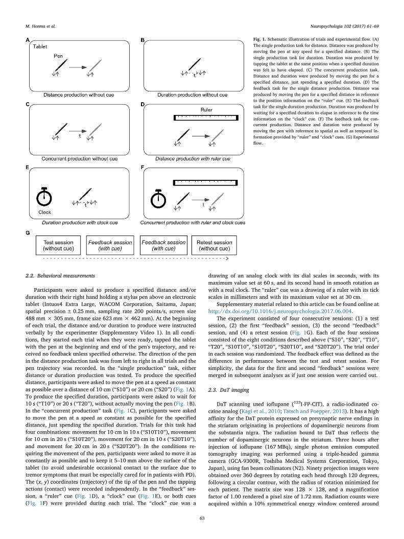
The DaT imaging indicated that there was little accumulation ofradiation in the striatum in the patients (Fig. 5A). The SBRs in the 19patients negatively correlated with the UPDRS (Fig. 5B). For the singleproduction task (Fig. 6A and B), the produced durations under the “T10”and “T20” conditions in the test session were strongly correlated withSBR. For the concurrent production task (Fig. 6C and D), the durationsunder the “S10T10” and “S20T20” conditions were correlated withSBR, but those under the “S10T20” and “S20T10” conditions were not(Table 2). Produced distances under all the conditions, on the otherhand, lacked correlations with SBR. Note that participants were re-quested to move the pen at a constant speed of 1 cm/s in the “S10T10”and “S20T20” conditions, whereas the requested speeds in the“S10T20” and “S20T10” conditions were 0.5 cm/s and 2 cm/s, re-spectively.
These results indicate that striatal DaT deficit is related to duration
production in both single and concurrent tasks, although this re-lationship may be speed dependent.
4. Discussion
In a manual action to move a pen for a specified distance and in aspecified duration, we observed differences in performance betweenelderly and young participants as well as between PD patients and theyoung participants. Elderly participants exhibited impaired durationproduction when they had to produce distance and duration con-currently. On the other hand, no impairment was observed for singleproduction of either distance or duration. In contrast, PD patients ex-hibited normal distance production and marked duration under-production in single production task, but both distance and durationwere underproduced in concurrent production task. Therefore, whenparticipants were asked for concurrent production of space and time,they should have suffered from competition of limited cognitive
Fig. 4. Comparison between the single and concurrent productions. (A) Distance in the conditions belonging to the “S10-related" analysis. (B) Distance in the conditions belonging to the“S20-related” analysis. (C) Duration in the conditions belonging to the “T10-related” analysis. (D) Duration in the conditions belonging to the “T20-related” analysis. Asterisks indicatesignificant differences (*p<0.05). Error bars indicate SEM. YC = young control. EC = elderly control. PD = patient with Parkinson’s disease.
Fig. 5. Striatal DaT deficit in PD. (A) Binding radiation accumulation in a PD patient withHoehn-Yahr stage 3 on striatal DaT imaging with coronal view. The numbers indicatebinding radiation counts per pixel. The imaging show little accumulation of radiation inthe striatum. (B) The specific binding ratio (SBR) in the striatum in PD was correlated toUPDRS. r and p indicate Pearson’s correlation coefficent and the uncorrected p value,respectively.
M. Honma et al. Neuropsychologia 102 (2017) 61–69
66
resources as seen in performances in dual tasks in general, but yieldedperformance changes as a systematic increase in constant error in onedirection rather than random error. These findings suggest that normalaging yields impaired performances in both elderly participants and PDpatients. Temporal underproduction in the PD patients appeared to“entrain” spatial production during spatiotemporal processing, as if thedistance to be produced were made consistent with the abnormallyshorter duration in perception and performance in these patients. Wealso observed that reduced striatal DaT was correlated with the extentof duration underproduction in PD patients.
Performance did not differ between PD patients and controls whenspatial or temporal visual cue was presented, suggesting that all parti-cipants understood the task, were able to move the pen, and were ableto pay attention to the cues, appropriately calibrating their motor ac-tions in reference to the cue. According to cognitive assessment scores,both elderly participants and patients exhibited similarly compromisedfunctioning as compared to young controls. This is consistent withprevious studies showing normal aging to be associated with generaldecline of cognitive functioning (Axelrod et al., 1992; Freitas et al.,
2011). However, none of our participants had any signs of dementia;their cognitive assessment scores were well within normal range.
Duration underproduction for PD patients in the single productiontask was similar to those reported in previous examinations of timeproduction in PD patients (Pastor et al., 1992; Lange et al., 1995; Smithet al., 2007), altogether suggesting that they exhibit dysfunctionaltemporal processing (i.e., time compression). Furthermore, distanceproduction in the single production task was normal for all participants,suggesting that purely spatial processing is unaffected by normal agingand PD.
Aging and PD affected spatiotemporal production performance dif-ferently. It may be because of aging per se if their performances simi-larly differ from those for young controls, but aging cannot explain whythe patients and the age-equated elderly controls exhibited differentpatterns of results. Compared to the single production task, producedduration in the concurrent production task without cues was longer inthe elderly group, although produced distance in this group was nearlyidentical between the concurrent and single production tasks, in-dicating that normal aging is associated with worse temporal processingin concurrent spatiotemporal production. In spatiotemporal compre-hension, it is known that spatial representations have precedence overtemporal ones in accuracy and precision (Boroditsky, 2000; Casasantoet al., 2010). In spatiotemporal production also, it is possible that el-derly people spend a greater effort to accomplish accurate spatialproduction at the expense of some extra physical time exceeding a re-quested duration for production. In healthy elderly adults, spatial in-formation may be used as a stable base on which further spatiotemporalprocessing can be based. In contrast, the patients produced shorterdistances in the concurrent task than in the single distance task. How-ever, duration remained stably underproduced in both the singleduration task and the concurrent task, indicating disruption of spatialproduction only during the concurrent task, during which temporalunderproduction in PD patients appeared to “entrain” spatial under-production.
For the spatial underproduction during the concurrent task in PDpatients, one might argue that group differences in single productioncould have been simply exacerbated with increasing task complexity,leading to a more pronounced deficit in the concurrent task. Our datashowed that the deficit of duration production remained stable betweenthe single and concurrent tasks, therefore the observed spatial under-production in the concurrent task might have reflected an impact ofincreased task demands on impaired sensorimotor systems, such that amajor fraction of cognitive resources is spent to prevent duration pro-duction from overt exacerbation at the expense of distance production.However, this scenario has a difficulty in explaining why distanceproduction became consistently shortened, rather than extended ormore prone to random error (Yordanova et al., 2015; Corp et al., 2016),in the concurrent task. If the spatial production error was simply causedby lesser allocation of cognitive resources to the spatial task in thecondition of increased task demands, the error could be observed asspatial overproduction in some patients and underproduction in others,but the results unequivocally indicated distance underproduction in allpatients. Therefore, we consider it more likely that the spatial under-production occurred as an entrainment by temporal underproduction inPD patients. Nevertheless, in future studies we should recruit a largernumber of patients to clarify to what extent the "entrained" distanceunderproduction in PD patients may correlate with their impairedduration production. Currently some conditions seemed to yield a slighttendency that those patients who exhibited more pronounced durationunderproduction also produced a shorter distance in the dual task ascompared with the single task (e.g., the "S10T10" condition, r = 0.40, p= 0.094), and our claim will be strengthened if such interobservercorrelations are found to be the case.
With regard to cue feedback, in the single production task, an effectof feedback on distance production was observed in young participants.This suggests that young people have cognitive flexibility for distance
Fig. 6. Correlation between striatal DaT and produced duration. Each panel plots anacross-patient scattergram between DaT as indexed by SBR in the striatum and producedduration in the behavioral conditions designated in each (A) “T10”, (B) “T20”, (C)“S10T10”, and (D) “S20T20”. The line indicates the best-fit linear regression. r and pindicate Pearson’s correlation coefficent and the uncorrected p value, respectively.
Table 2Correlation of DaT and distance/duration.
Task Condition (Test session) Dimension r p
Single S10 Distance 0.167 0.509S20 Distance 0.425 0.069T10 Duration 0.597 0.009T20 Duration 0.549 0.015
Concurrent S10T10 Distance −0.114 0.643Duration 0.518 0.023
S10T20 Distance 0.218 0.371Duration 0.339 0.156
S20T10 Distance −0.040 0.870Duration 0.341 0.153
S20T20 Distance 0.217 0.372Duration 0.625 0.004
M. Honma et al. Neuropsychologia 102 (2017) 61–69
67

production. On the other hand, the absence of feedback effect in elderlyand PD participants may indicate inadequacy of the feedback sessionfor them, because they experienced only two trials per condition duringthe two blocks of the feedback session. Future research is required toexamine how many trials are actually needed for these participants toexhibit a feedback effect, if any. In contrast, no participants yielded anyfeedback effect on duration production. This suggests that internal re-presentation of duration may be stable within individuals. In the con-current production task, effect of feedback on distance production wasobserved in young and PD participants, whereas effect of feedback onduration production was observed in elderly participants. The feedbackeffect on distance production in young participants may be similar tothat for the single task. However, for PD patients, the improvement ondistance production may be viewed as some alleviation of spatio-temporal entrainment after experience of the feedback session. On theother hand, the feedback effect on duration production in elderly par-ticipants may be viewed as some calibration to counteract erroneousduration production.
Mental time is thought to be processed by a complex neural networkinvolving the frontal cortex, hippocampus, basal ganglia, and cere-bellum (Hinton &Meck, 2004; Buhusi and Meck, 2005). Furthermore,the corpus striatum plays a central role in processing of time around afew tens of seconds (Honma et al., 2016). Correlation between DaT andproduced duration in the single production task in the PD group sug-gests that disordered temporal processing is associated with striatalexpression of DaT, which transports DA. On the other hand, the pre-sence of correlation of DaT and produced duration at average speed(1 cm/s) of pen movement but the absence of correlation at faster(2 cm/s) and slower (0.5 cm/s) speeds in the concurrent productiontask suggests that this DaT-duration relationship is velocity sensitive.Moving the pen at a faster or slower speed may require greater atten-tional resources. For example, in the case of faster speed, transient at-tention may be required to achieve that speed as quickly as possible,whereas sustained attention may be required to maintain a steady statemovement in the case of slower speed. In addition, no correlation be-tween DaT and produced distance suggests that striatal dopaminergicneurotransmission does not play a marked role in spatial production.Although spatial processing occurs via connections from the visualcortex to the PC and then to the PFC (Quintana and Fuster, 1993;Chafee and Goldman-Rakic, 1998), the spatial production examinedhere may be unrelated to the PFC and striatum, for the patients with PDexhibited normal distance production in the single production task.
Our findings support the hypothesis that spatial and temporal pro-cesses interact during spatiotemporal processing, because one produc-tion was distorted so as to be more consistent with the other productionduring the concurrent task. PD patients, in particular, underproducedduration appeared to disrupt normal production of distance. Patientswith PD may strictly adhere to temporal aspects, to which spatial as-pects are entrained during spatiotemporal processing, possibly due tosevere time compression associated with DA and/or DaT deficits. It isknown that poverty of DA linked to deficient DaT protein in thestriatum leads to hyper-activity or fast signal cycles in the globus pal-lidus and subthalamic nuclei (Delong, 1990; Bergman et al., 1994; Razet al., 2000). Time compression may be associated with hyper-activityor fast signal cycles via a loop system such as striatum–pallidus–tha-lamus–cortex (Galvan et al., 2015). Furthermore, time compressionmay demand a high load for temporal processing, and the severe timecompression may consolidate in patients with PD due to their prolongedsymptom. Consequently, duration production may be given a higherpriority than distance production during concurrent processing.
The current research revealed new aspects of spatiotemporal pro-cessing that are hardly noticed with healthy young adult participantsalone. However, it remains to be seen whether young adults processspace and time dimensions separately or interactively, since they ac-curately produce both distance and duration. It is possible that inter-actions between spatial and temporal processes occur at some early
stage but are not made explicit as interactions in performance levels inyoung adults. It is also possible that each dimension is processed se-parately with sufficiently rich sensorimotor systems separately allo-cated to each processing (Pashler, 1994). Future research is required toreveal a possible mechanism for spatiotemporal production by in-creasing workloads, perhaps with extreme or variable speeds duringtask.
Our study has several limitations. First, the duration task was re-stricted to 10 s or 20 s. In the timing literature, it is known that PDpatients overproduce a shorter duration (e.g., 8 s) and underproduce alonger duration (e.g., 21 s) (migration effect) (Malapani et al., 1998),and this effect occurs only when durations exceed 2 s (Koch et al.,2008). If we had reduced duration to a few seconds or within a sub-second range, different kinds of entrainment might have shown up.Second, PD patients were studied while medicated, because some pa-tients suffered from too large tremor to manage the pen tablet under theOff condition, in which medicine was not being administered. As do-pamine replacement promotes striatal functioning but can adverselyaffect frontal functions (Damier, 2015), comparative performance inthe Off state is ideally required to determine the effects of medication.Finally, the absence of DaT scan data for normal controls was an in-evitable limitation due to our own hospital's ethical guidelines.Nevertheless, our findings may help bridge the gap between striatalDaT/DA and spatiotemporal production.
Our approach may shed new light on the way temporal processingaffects spatial processing, and vice versa, when they are concurrentlyactive, and may be applicable to a wide variety of symptoms withdistorted production, such as epilepsy (Drane et al., 1999), depression(Thönes and Oberfeld, 2015), schizophrenia (Su et al., 2015), and at-tention-deficit hyperactivity disorder (Suarez et al., 2013), amongothers. Furthermore, this easy-to-use approach using a tablet devicemay be useful in identifying previously undiagnosed disorders of spa-tiotemporal production and may reveal a mechanism underlying in-tegration of space and time.
Acknowledgements
This study was supported by Grant-in-aids for Scientific Research(KAKENHI) for Innovative Areas ‘‘The Science of Mental Time’’[25119003 and 25119006], Scientific Research (C) [23591283],Exploratory Research [24650142], and Young Scientists [26860677].This study was also supported in part by the Showa University MedicalFoundation.
Appendix A. Supplementary material
Supplementary data associated with this article can be found in theonline version at http://dx.doi.org/10.1016/j.neuropsychologia.2017.06.004.
References
Ahlskog, J.E., 2003. Slowing Parkinson's disease progression: recent dopamine agonisttrials. Neurology 60, 381–389.
Allman, M.J., Meck, W.H., 2012. Pathophysiological distortions in time perception andtimed performance. Brain 135, 656–677.
Axelrod, B.N., Goldman, R.S., Henry, R.R., 1992. Sensitivity of the mini-mental stateexamination to frontal lobe dysfunction in normal aging. J. Clin. Psychol. 48, 68–71.
Bergman, H., Wichmann, T., Karmon, B., Delong, M.R., 1994. The primate subthalamicnucleus. II. Neuronal activity in the MPTP model of parkinsonism. J. Neurophysiol.72, 507–520.
Bonato, M., Zorzi, M., Umilta, C., 2012. When time is space: evidence for a mental timeline. Neurosci. Biobehav. Rev. 36, 2257–2273.
Boroditsky, L., 2000. Metaphoric structuring: understanding time through spatial meta-phors. Cognition 75, 1–28.
Bueti, D., Walsh, V., 2009. The parietal cortex and the representation of time, space,number and other magnitudes. Philos. Trans. R. Soc. Lond. Ser. B Biol. Sci. 364,1831–1840.
Buhusi, C.V., Meck, W.H., 2005. What makes us tick? Functional and neural mechanismsof interval timing. Nat. Rev. Neurosci. 6, 755–765.
M. Honma et al. Neuropsychologia 102 (2017) 61–69
68

Cai, Z.G., Connell, L., 2015. Space-time interdependence: evidence against asymmetricmapping between time and space. Cognition 136, 268–281.
Cappelletti, M., Freeman, E.D., Cipolotti, L., 2009. Dissociations and interactions betweentime, numerosity and space processing. Neuropsychologia 47, 2732–2748.
Casasanto, D., Fotakopoulou, O., Boroditsky, L., 2010. Space and time in the child's mind:evidence for a cross-dimensional asymmetry. Cogn. Sci. 34, 387–405.
Chafee, M.V., Goldman-Rakic, P.S., 1998. Matching patterns of activity in primate pre-frontal area 8a and parietal area 7ip neurons during a spatial working memory task.J. Neurophysiol. 79, 2919–2940.
Cohen, J., Hansel, C.E., Sylvester, J.D., 1953. A new phenomenon in time judgment.Nature 172, 901.
Corp, D.T., Rogers, M.A., Youssef, G.J., Pearce, A.J., 2016. The effect of dual-task diffi-culty on the inhibition of the motor cortex. Exp. Brain Res. 234, 443–452.
Damier, P., 2015. Why do Parkinson's disease patients sometimes make wrong decisions?J. Parkinson's Dis. 5, 637–642.
Daniel, S.E., Lees, A.J., 1993. Parkinson's Disease Society Brain Bank, London: overviewand research. J. Neural Transm. Suppl. 39, 165–172.
Delong, M.R., 1990. Primate models of movement disorders of basal ganglia origin.Trends Neurosci. 13, 281–285.
Drane, D.L., Lee, G.P., Loring, D.W., Meador, K.J., 1999. Time perception followingunilateral amobarbital injection in patients with temporal lobe epilepsy. J. Clin. Exp.Neuropsychol. 21, 385–396.
Folstein, M.F., Folstein, S.E., Mchugh, P.R., 1975. "Mini-mental state". A practical methodfor grading the cognitive state of patients for the clinician. J. Psychiatr. Res. 12,189–198.
Frassinetti, F., Magnani, B., Oliveri, M., 2009. Prismatic lenses shift time perception.Psychol. Sci. 20, 949–954.
Freitas, S., Simoes, M.R., Alves, L., Santana, I., 2011. Montreal Cognitive Assessment(MoCA): normative study for the Portuguese population. J. Clin. Exp. Neuropsychol.33, 989–996.
Galvan, A., Devergnas, A., Wichmann, T., 2015. Alterations in neuronal activity in basalganglia-thalamocortical circuits in the parkinsonian state. Front. Neuroanat. 9, 5.
Gobel, E.W., et al., 2013. Implicit perceptual-motor skill learning in mild cognitive im-pairment and Parkinson's disease. Neuropsychology 27, 314–321.
Hartley, A.A., 2001. Age differences in dual-task interference are localized to response-generation processes. Psychol. Aging 16, 47–54.
Haber, S.N., 2014. The place of dopamine in the cortico-basal ganglia circuit.Neuroscience 282, 248–257.
Helson, H., 1930. The tau effect–an example of psychological relativity. Science 71,536–537.
Honma, M., Kuroda, T., Futamura, A., Shiromaru, A., Kawamura, M., 2016. Dysfunctionalcounting of mental time in Parkinson's disease. Sci. Rep. 6, 25421.
Kagi, G., Bhatia, K.P., Tolosa, E., 2010. The role of DAT-SPECT in movement disorders. J.Neurol. Neurosurg. Psychiatry 81, 5–12.
Koch, G., et al., 2008. Impaired reproduction of second but not millisecond time intervalsin Parkinson's disease. Neuropsychologia 46, 1305–1313.
Lange, K.W., Tucha, O., Steup, A., Gsell, W., Naumann, M., 1995. Subjective time esti-mation in Parkinson's disease. J. Neural Transm. Suppl. 46, 433–438.
Malapani, C., et al., 1998. Coupled temporal memories in Parkinson's disease: a dopa-mine-related dysfunction. J. Cogn. Neurosci. 10, 316–331.
Martinez-Martin, P., et al., 1994. Unified Parkinson's disease rating scale characteristicsand structure. The cooperative multicentric group. Mov. Disord. 9, 76–83.
Morrone, M.C., Ross, J., Burr, D., 2005. Saccadic eye movements cause compression oftime as well as space. Nat. Neurosci. 8, 950–954.
Nasreddine, Z.S., et al., 2005. The montreal cognitive assessment, MoCA: a brief screeningtool for mild cognitive impairment. J. Am. Geriatr. Soc. 53, 695–699.
Oliveri, M., Koch, G., Caltagirone, C., 2009. Spatial-temporal interactions in the humanbrain. Exp. Brain Res. 195, 489–497.
Pashler, H., 1994. Dual-task interference in simple tasks: data and theory. Psychol. Bull.116, 220–244.
Pastor, M.A., Artieda, J., Jahanshahi, M., Obeso, J.A., 1992. Time estimation and re-production is abnormal in Parkinson's disease. Brain 115 (Pt 1), 211–225.
Piras, F., et al., 2014. Time dysperception perspective for acquired brain injury. Front.Neurol. 4, 217.
Price, A., Shin, J.C., 2009. The impact of Parkinson's disease on sequence learning: per-ceptual pattern learning and executive function. Brain Cogn. 69, 252–261.
Quintana, J., Fuster, J.M., 1993. Spatial and temporal factors in the role of prefrontal andparietal cortex in visuomotor integration. Cereb. Cortex 3, 122–132.
Raz, A., Vaadia, E., Bergman, H., 2000. Firing patterns and correlations of spontaneousdischarge of pallidal neurons in the normal and the tremulous 1-methyl-4-phenyl-1,2,3,6-tetrahydropyridine vervet model of parkinsonism. J. Neurosci. 20,8559–8571.
Salthouse, T.A., 1996. The processing-speed theory of adult age differences in cognition.Psychol. Rev. 103, 403–428.
Smith, J.G., Harper, D.N., Gittings, D., Abernethy, D., 2007. The effect of Parkinson'sdisease on time estimation as a function of stimulus duration range and modality.Brain Cogn. 64, 130–143.
Su, L., et al., 2015. Temporal perception deficits in schizophrenia: integration is theproblem, not deployment of attentions. Sci. Rep. 5, 9745.
Suarez, I., Lopera, F., Pineda, D., Casini, L., 2013. The cognitive structure of time esti-mation impairments in adults with attention deficit hyperactivity disorder. Cogn.Neuropsychol. 30, 195–207.
Tatsch, K., Poepperl, G., 2013. Nigrostriatal dopamine terminal imaging with dopaminetransporter SPECT: an update. J. Nucl. Med. 54, 1331–1338.
Thones, S., Oberfeld, D., 2015. Time perception in depression: a meta-analysis. J. Affect.Disord. 175, 359–372.
Torta, D.M., et al., 2010. Dissociation between time reproduction of actions and of in-tervals in patients with Parkinson's disease. J. Neurol. 257, 1356–1361.
Tossici-Bolt, L., Hoffmann, S.M., Kemp, P.M., Mehta, R.L., Fleming, J.S., 2006.Quantification of [123I]FP-CIT SPECT brain images: an accurate technique formeasurement of the specific binding ratio. Eur. J. Nucl. Med. Mol. Imaging 33,1491–1499.
Vandenbossche, J., et al., 2013. Impaired implicit sequence learning in Parkinson's dis-ease patients with freezing of gait. Neuropsychology 27, 28–36.
Vaughan, R.A., Foster, J.D., 2013. Mechanisms of dopamine transporter regulation innormal and disease states. Trends Pharmacol. Sci. 34, 489–496.
Walsh, V., 2003. A theory of magnitude: common cortical metrics of time, space andquantity. Trends Cogn. Sci. 7, 483–488.
Yordanova, J., Kirov, R., Kolev, V., 2015. Increased performance variability as a markerof implicit/explicit interactions in knowledge awareness. Front. Psychol. 6, 1957.
M. Honma et al. Neuropsychologia 102 (2017) 61–69
69