Embed Size (px)
Citation preview

1521-009X/44/7/1014–1019$25.00 http://dx.doi.org/10.1124/dmd.115.068809DRUG METABOLISM AND DISPOSITION Drug Metab Dispos 44:1014–1019, July 2016Copyright ª 2016 by The American Society for Pharmacology and Experimental Therapeutics
Special Section on Pediatric Drug Disposition and Pharmacokinetics
Human Intestinal PEPT1 Transporter Expression and Localization inPreterm and Term Infants
Miriam G. Mooij, Barbara E. A. de Koning, Dicky J. Lindenbergh-Kortleve, Ytje Simons-Oosterhuis,Bianca D. van Groen, Dick Tibboel, Janneke N. Samsom, and Saskia N. de Wildt
Intensive Care and Department of Pediatric Surgery (M.G.M., B.E.A.K., B.D.G., D.T., S.N.W.), and Department of Pediatrics (D.J.L.-K.,Y.S.-O., J.N.S.), Erasmus MC-Sophia Children’s Hospital, Rotterdam, The Netherlands; and Department of Pharmacology and
Toxicology, Radboudumc, Nijmegen, The Netherlands (S.N.W.)
Received December 4, 2015; accepted April 13, 2016
ABSTRACT
The intestinal influx oligopeptide transporter peptide transporter 1(PEPT1) (SLC15A1) is best known for nutrient-derived di- andtripeptide transport. Its role in drug absorption is increasinglyrecognized. To better understand the disposition of PEPT1 sub-strate drugs in young infants, we studied intestinal PEPT1 mRNAexpression and tissue localization across the pediatric age range.PEPT1 mRNA expression was determined using real-time reverse-transcription polymerase chain reaction in small intestinal tissuescollected from surgical procedures (neonates and infants) or bi-opsies (older children and adolescents). PEPT1 mRNA relative tovillin mRNA expression was compared between neonates/infantsand older children/adolescents. PEPT1 was visualized in infanttissue using immunohistochemical staining. Other transporters[multidrug resistance protein 1 (MDR1), multidrug resistance–likeprotein 2 (MRP2), and organic anion transporter polypeptide 2B1
(OATP2B1)] were also stained to describe the localization in relationto PEPT1. Twenty-six intestinal samples (n = 20 neonates/infants,n = 2 pediatric, n = 4 adolescents) were analyzed. The young infantsamples were collected at a median (range) gestational age atbirth of 29.2 weeks (24.7–40) and postnatal age of 2.4 weeks (0–16.6). The PEPT1 mRNA expression of the neonates/infants wasonly marginally lower (0.8-fold) than the older children (P < 0.05).Similar and clear apical PEPT1 and MRP2 staining, apical andlateral MDR1 staining, and intraepithelial OATP2B1 staining atthe basolateral membrane of the enterocyte were detected in 12infant and 2 adolescent samples. Although small intestinal PEPT1expression tended to be lower in neonates than in older children,this difference is small and tissue distribution is similar. This findingsuggests similar oral absorption of PEPT1 substrates across thepediatric age range.
Introduction
The influx oligopeptide transporter peptide transporter 1 (PEPT1)(SLC15A1) is a member of the solute carrier superfamily and is situatedon the apical membrane of the enterocyte. Its expression in the humanadult jejunum is at least more than 2–10 times higher than that of othertransporters such as those from the ATP-binding cassette transporterfamily [multidrug resistance protein 1 (MDR1), multidrug resistance–like protein 2 (MRP2), or breast cancer resistance protein] or from thesolute carrier family [organic anion transporter polypeptides (OATPs)](Hilgendorf et al., 2007). In histologically normal intestinal biopsiesfrom 10 adults, PEPT1 was expressed most abundantly in duodenumand ileum with a mean relative mRNA expression of 4 compared with,0.5 in colon (Meier et al., 2007). In six adult intestine organ donors thePEPT1 protein expression accounted for approximately 50% of the totalexpression of all transporter proteins in the small intestine. In colon,PEPT1 represents 5% of all transporter proteins (Drozdzik et al., 2014).PEPT1 is best known for its function as a nutrient-derived di- and
tripeptide transporter, but may also have a role in (pro)drug transport
because it is the most abundant peptide transporter in the gut and drugproperties mimicking di- and tripeptides may allow uptake by thepeptide transporter (Brandsch, 2013). Its role as intestinal transporter hasbeen demonstrated for several (pro)drugs. In PepT1 (ortholog of humanPEPT1) knockout mice, the small intestinal uptake of the prodrugvalacyclovir was attributed to 90% of the PepT1 transporter (Yang andSmith, 2013). Consequently, these mice had a delayed Tmax anddecreased Cmax of acyclovir (active metabolite of valacyclovir) relativeto wild-type mice (Yang et al., 2013). However, in human adults PEPT1mRNA expression was not correlated with valacyclovir pharmacoki-netic parameters, even though in vitro valacyclovir was a PEPT1substrate (Landowski et al., 2003). This suggests that valacyclovir maynot be a PEPT1 substrate in human adults, or that PEPT1 mRNAexpression may not correlate to PEPT1 activity. The role of PEPT1in valacyclovir pharmacokinetics in humans may differ from that inanimals and remains to be elucidated. Several b-lactam antibiotics haveappeared to be PEPT1 substrates in Caco-2 cells with varying affinities,e.g., ceftibuten, cyclacillin, cefadroxil, cefaclor, benzylpenicillin, andcephalexin. Their structure resembles the tripeptide structure withadditional groups (Brandsch et al., 2008). The angiotensin convertingenzyme-inhibitor fosinopril is a PEPT1 substrate; other angiotensindx.doi.org/10.1124/dmd.115.068809.
ABBREVIATIONS: GAPDH, glyceraldehyde-3-phosphate dehydrogenase; IHC, immunohistochemistry; MDR, multidrug resistance protein; MRP,multidrug resistance–like protein; OATP, organic anion transporter polypeptide; PEPT, peptide transporter.
1014
at ASPE
T Journals on M
ay 1, 2018dm
d.aspetjournals.orgD
ownloaded from
converting enzyme inhibitors might be substrates, although this needs tobe confirmed (Brandsch et al., 2008).Most drugs prescribed to children are administered orally. Some
PEPT1 substrates are dosed to children even very early in life.Considering the wide age-related variation in the processes affectingoral drug absorption—including gastric pH, gastric motility, and drugmetabolizing enzyme activity—age-related changes in membrane trans-porters are also very likely (Mooij et al., 2012). A previous study by ourgroup Mooij et al. (2014) showed transporter-dependent maturation ingene expression in young infants for MDR1, MRP2, and OATP2B1, butoverall data on the intestinal expression of membrane transporters duringgrowth and development are very scarce (Brouwer et al., 2015).To the best of our knowledge, studies on the development of intestinal
PEPT1 in humans are lacking. From a pharmacological point a view, it isimportant to elucidate the development of PEPT1 expression for knownsubstrates. However, this may also be important for the development ofnew drugs in which PEPT1 could enhance oral absorption.PEPT1 developmental changes have been studied in several animal
species. A developmental pattern of PepT1 mRNA and protein ex-pression has been shown in the duodenum, jejunum, ileum, and colon ofrats (Shen et al., 2001). In the small intestine of newborn rats theexpression peaked 3–5 days after birth, after which it rapidly decreased
and increased again by the time animals were weaning. Shen et al.(2001) ascribed the increase postpartum to suckling. In another study inrats, PepT1 small intestinal mRNA expression was stable from postnatalday 4 until day 21, and then decreased from postnatal day 50 onward(Rome et al., 2002). In neonatal miniature pigs, PepT1-mediateddipeptide (3H-glycylsarcosine) disappearance in the ileal segment washighest in the youngest age group (1 week), but PepT1 expression inpostweaned pigs was higher than in sucklings (Nosworthy et al., 2013).These data suggest that in the first weeks of life, intestinal PEPT1 isimportant for nutritional intake and later for diet transition followingweaningComparative mRNA expression of various peptide transporters in
mice, rats, and human adults shows a PEPT1 expression in all species,but expression levels varied in relation to that of other peptidetransporters (cadherin transporter and PEPT2) (Kim et al., 2007). Thissuggests that animal data cannot be directly extrapolated to humans andthat human studies are needed. To our knowledge expression or activityof PEPT1 in human fetal or pediatric population has not been describedthus far, let alone a developmental expression pattern. To betterunderstand the disposition of PEPT1 substrate drugs in neonates andyoung infants, we aimed to compare intestinal PEPT1 mRNA expres-sion and tissue localization in these age groups with those in older
TABLE 1
Patients’ characteristics
Subject IHC mRNA Gender Ethnicity DiagnosisGestational Ageat Birth (weeks)a
PostnatalAge (weeks)a
Nutritionb Pathology ReportResectionArea
1 * Male Caucasian NEC And stoma closure 25.3 0.2 and 6.9 EF and PN Necrotizing enterocolitisand stoma closure
Jejunum
2 * Male Caucasian Stoma closure (history of NEC) 30.3 5.7 EF — Ileum3 * Male Caucasian Volvulus jejunum, malrotation 39.3 3.9 PN — Jejunum4 * Male Caucasian Stoma closure (history of NEC) 25.6 16.6 EF — Ileum5 * * Male Unknown NEC 30.9 8.9 EF and PN Resection stenosis with
necrotizing enterocolitisCecum
6 * * Male Unknown Jejunum atresia 35.7 0 PN Stenosis Jejunum7 * Male Unknown NEC in patient with complex
cor vitium death, enterobactersepsis
33.0 0.9 EF NEC Ileum
8 * * Female Caucasian NEC 26.9 2.7 EF and PN NEC Ileum9 * * Male Unknown CHD complicated by volvulus
and intestinal necrosis afterhernia repair
36.9 2.0 EF and PN Ischemia Jejunum
10 * * Female Caucasian NEC 26.4 2.0 PN NEC Ileum11 * Male Unknown NEC 25.3 3.1 ? Ischemic enteritis Jejunum12 * Female Caucasian Stoma closure (history of NEC) 24.7 9.57 PN — Ileum13 * Male Caucasian Midgut volvulus 28.1 3.86 PN Infarction and peritonitis Ileum14 * * Male Unknown Meckel’s diverticulum 40.0 3.1 EF Meckel’s diverticulum
with ulcerations andnormal small intestine
Ileum
15 * * Male Caucasian Jejunum atresia 38.3 0.1 PN stenosis Jejunum16 * * Male Caucasian Jejunum atresia 38.0 0.1 PN Small reactive changes Jejunum17 * Male Caucasian Jejunum atresia 38.3 0 PN Atresia with extensive
reactive changesJejunum
18 * * Male Unknown Ileum atresia 38.9 0.1 EF Ileumatresia, no ischemia Ileum19 * Female Caucasian NEC 24.9 1.1 PN NEC Ileum20 * Female Unknown Stoma closure (history of NEC) 25.6 13.9 EF Ileum21 * Male Caucasian Crohn’s disease: active Adolescent 15 years ? Ileocaecal resection Ileum22 * Female Caucasian Ulcerative colitis: active Adolescent 17 years ? Ileoanal pouch surgery Ileum23 * Caucasian Biopsy in case of abdominal
complaints; non-IBDAdolescent 9 years ? Normal Ileum
24 * Caucasian Biopsy in case of abdominalcomplaints; non-IBD
Adolescent 10 years ? Normal Ileum
25 * Caucasian Biopsy in case of abdominalcomplaints; non-IBD
Adolescent 16 years ? Normal Ileum
26 * Caucasian Biopsy in case of abdominalcomplaints; non-IBD
Adolescent 17 years ? Normal Ileum
CHD, congenital hernia diaphragmatic; EF, enteral feeding; IBD, inflammatory bowel disease; NEC, necrotizing enterocolitis; PN, parenteral nutrition.aUnless noted otherwise.bUp to 3 days before date of sampling EF or PN.*available results for subject; ?, unknown nutritional information; —, no pathology report available.
Human Intestinal PEPT1 in Infants 1015
at ASPE
T Journals on M
ay 1, 2018dm
d.aspetjournals.orgD
ownloaded from
children and adolescents. To describe PEPT1 protein staining in relationto other transporters with known mRNA expression data, we also aimedto detect MDR1, MRP2, and OATP2B1 protein in intestine.
Material and Methods
Tissue Samples. Intestinal tissue samples were obtained surgically at time ofresection (neonates/infants/adolescents) or as biopsies during ileocolonoscopies(older children/adolescents: subjects 23–26) (Table 1). For mRNA isolation,postresection, tissue was snap frozen in liquid nitrogen and stored at280�C. Forimmunohistochemical analysis, tissue was immediately put in 4% formaldehydein phosphate-buffered saline and processed to paraffin cubes.
The collection of neonatal/infant intestinal tissue and the use of leftovermaterial were approved by the Central Committee of Research involving HumanSubjects (The Hague, Netherlands) (Puiman et al., 2011). The Erasmus MCresearch ethics board in two other protocols approved collection of intestinalresidual tissue from adolescent patients and endoscopy biopsies of older childrenand adolescents. Informed consent was obtained from all parents/caregivers andchildren older than 12 years of age for use of leftover tissue and clinical data.
Real-Time Reverse-Transcription Polymerase Chain Reaction. Isolationand cDNA synthesis have been previously described (Mooij et al., 2014). In brief,frozen tissue samplesweremechanically homogenized on ice. RNAwas extractedusing the RNeasy Mini Kit (Qiagen, Hilden, Germany). To digest genomic DNAremnants, RNA was treated with DNase. The RNA integrity numbers of thesamples were analyzed using the 2100 BioAnalyzer (Agilent, Santa Clara, CA),and a value of,5 was considered poor quality and reason to discard the sample.The mRNA expression was measured by SYBR Green (Applied Biosystems,Thermo Fisher Scientific Inc., Waltham, MA) quantitative real-time reversetranscription polymerase chain reaction with a 7900 Sequence Detector (ABIPrism, Applied Biosystems, Thermo Fisher Scientific Inc., Waltham, MA).
Primers were used for PEPT1, villin, and glyceraldehyde-3-phosphatedehydrogenase (GAPDH), and sequences were designed using the Oligo 6.22software (Molecular Biology Insights Inc., Colorado Springs, CO). The primersequenceswere the following: GAPDH forward 59-GTCGGAGTCAACGGATT-39and reverse 59-AAGCTTCCCGTTCTCAG-39; villin forward 59-TGCCAACAC-CAAGAGACT-39 and reverse 59-TCCCAATCCAGAAGAAGAC-39; and PEPT1forward 59-TTGGCCCAATGTCTCA-39 and reverse 59-GGCCCTGCTTGAAGTC-39. The melting curve was analyzed after every polymerase chain reaction toconfirm product specificity. GAPDH and villin mRNA expressions were used asthe endogenous control. PEPT1 transcript levels were normalized to villintranscript levels (ratio PEPT1/villin), and relative expression was comparedacross the age groups (neonates/young infants versus children/adolescents).
Immunohistochemistry (IHC). Intestinal sections were dewaxed for IHC,and endogenous peroxidases were quenched with 3% H2O2 in methanol for 20minutes. Antigens were retrieved using Pepsin (0.1% in 0.01 N HCL) pretreat-ment for 7 minutes in a stove at 37�C. The sections were blocked for 1 hour in10% normal human serum and 10% normal rabbit serum diluted in 10 mM Tris(pH 8), 5 mM EDTA (pH 8), 0.15 M NaCl, 0.25% gelatin, and 0.05% Tween-20.Primary antibodies (goat anti-PEPT1 C-20; Santa Cruz Biotechnology, Inc.,Heidelberg, Germany) were incubated over night at 4�C in 2% human serum.Immunoreactive sites were detected with biotinylated secondary rabbit anti-goatserum using the Vectastain ABC Elite Kit (Vector Laboratories, Burlingame,CA), and 3,39-diaminobenzidine tetrahydrochloride solution (Sigma-Aldrich,Zwijndrecht, Netherlands). The nuclei were counterstained with hematoxylin(Vector Laboratories). A negative control staining lacking the primary antibodywas performed for every slide. A matched goat-antibody negative control wasperformed to assess background staining. Images were acquired and analyzedwith a Leica microscope and LAS-AF image acquisition software (LeicaMicrosystems, Wetzlar, Germany). IHC was also performed on the same tissuesamples for three other membrane transporters with known intestinal mRNAexpression data (MDR1, MRP2, and OATP2B1) to compare tissue distribution ofPEPT1 with these transporters. In the case of MDR1 and OATP2B1 staining,microwave pretreatment in citrate buffer (10 mM, pH 6.0) was used to retrieveantigens. In the case of MRP2, pepsin (which is similar to PEPT1) pretreatmentwas used. Primary antibodies (mouse anti-MDR1 and mouse anti-MRP2) wereobtained from EMD Millipore (Billerica, MA), and rabbit anti-OATP2B1 wasobtained from Abnova (Taipei City, Taiwan). PEPT1 staining intensity was micro-scopically scored by two independent observers (low 1, moderate 2 or high 3).
Statistical Analysis. Data are presented as median and range, unless indicatedotherwise. Group comparison (neonates/young infants and older children/adolescents) was made using the nonparametric Mann-Whitney U test. Withinthe neonates/young infants group, the association between postmenstrual age(gestational plus postnatal age) and PEPT1 mRNA expression was assessed usingSpearman’s rho correlation. All statistical analyses were performed usingGraphPad Prism software (version 5.00.2, La Jolla, CA) and IBM SPSS Statisticssoftware (SPSS Statistics for Windows, version 21.0; IBM, Armonk, NY). Thelevel of significance was set at P , 0.05.
Results
Descriptive Results. Twenty-six samples (n = 20 neonates/infants,n = 2 children, and n = 4 adolescents) were collected (Table 1). The agesof the young infants ranged from gestational age at birth [median (range)29.2 weeks (24.7–40)] to postnatal age [2.4 weeks (0–16.6)]. The mainreasons for resection were stoma closure [in patients with history ofnecrotizing enterocolitis (n = 5), current necrotizing enterocolitis (n = 6),and intestinal atresia (n = 5)]. Other reasons for resection were intestinalvolvulus (n = 3) and Meckel’s diverticulum (n = 1). Samples of twochildren (9 and 10 years old) and two adolescents (16 and 17 years old),from whom frozen biopsy tissue was available for mRNA, underwentendoscopy for suspicion of inflammatory bowel disease, and werepreviously classified as histologically normal. The two samples for IHCwere collected from two other children; one underwent ileocecalresection (age 15 years, history of Crohn’s disease, active disease attime of surgery) and the other underwent ileoanal pouch surgery (age 17years, history of ulcerative colitis, active disease at time of surgery).mRNA was analyzed in samples from which snap frozen tissue was
available, i.e., 17 young infant, 2 pediatric, and 2 adolescent samples.IHC was performed on samples from which paraffin-embedded tissuewas available, i.e., 12 young infant and two adolescent samples(Table 1). GAPDH mRNA strongly correlated with villin mRNA(n = 21, r = 0.6182, P , 0.01). One sample was excluded due to lowmRNA expression of villin and GAPDH, suggesting loss of enterocytes.Twenty samples remained for mRNA expression analysis.Nutritional intake up to 3 days before date of sampling (surgery or
biopsy) was fully enteral in six patients, parenteral nutrition was given innine patients, and four patients received both enteral and parenteralfeeding. Data on nutritional intake were lacking in seven subjects. Noinformation on concomitant medications was available.PEPT1 Gene Expression. PEPT1 mRNA was detected in all 20
samples. The relative intestinal PEPT1 mRNA expression (PEPT1/villin) in young infants slightly varied (0.15-fold) (Fig. 1). In theneonatal/infant group the PEPT1 expression was 0.8-fold lower than in
Fig. 1. Relationship between age and intestinal PEPT1 gene expression. RelativemRNA expression of PEPT1 in relation to age normalized to villin mRNAexpression.
1016 Mooij et al.
at ASPE
T Journals on M
ay 1, 2018dm
d.aspetjournals.orgD
ownloaded from
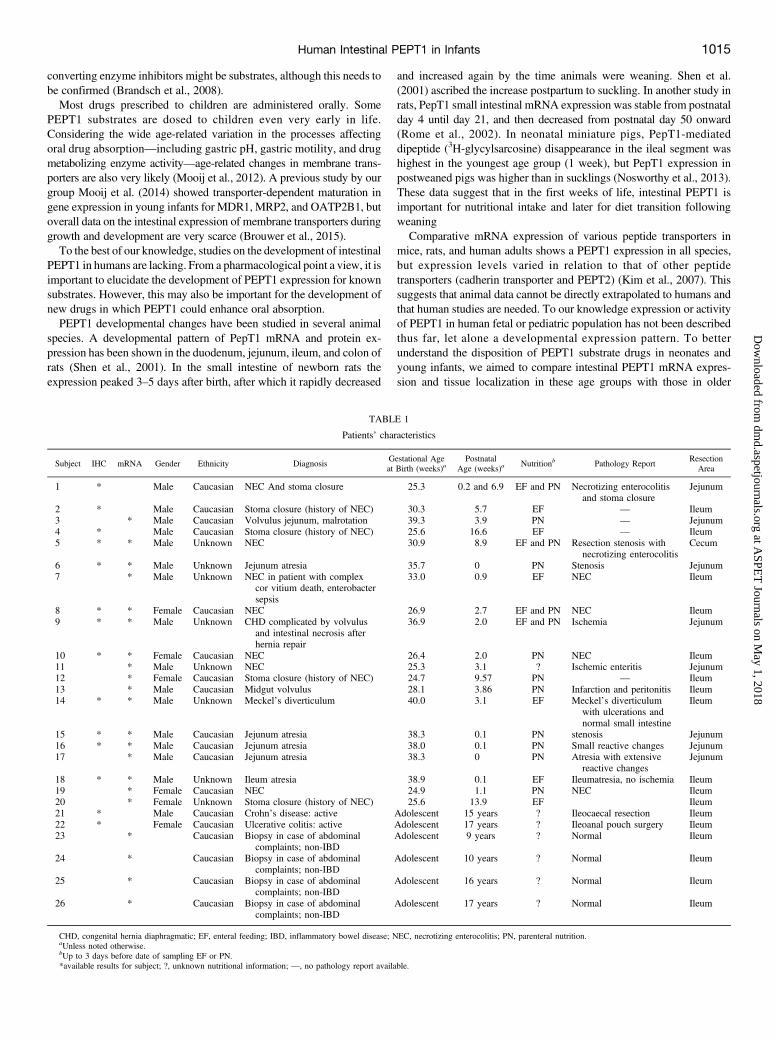
the older age group (P = 0.01), with median relative mRNA expressionsof 0.80 (range 0.77–0.92) and 1.02 (1.01–1.04), respectively. In theneonatal/infant group postmenstrual age was not correlated withPEPT1 mRNA expression (r = 0.453, P = 0.078).Presence of PEPT1 Protein in Enterocytes. PEPT1 staining was
present at the apical membrane in the brush border of the enterocyte in allbut one sample (Fig. 2). This sample did not show clear villin and wasalso excluded from PEPT1 mRNA analysis for low villin and GAPDHmRNA expression levels. PEPT1 apical localization was similar inneonatal and adolescent intestinal samples. No PEPT1 staining wasdetected in goblet cells, most likely due to the artificial effect ofenlargement of goblet cells during the process of paraffin embedding.No staining was observed at the basolateral membrane or at the tightjunctions.Microscopically, PEPT1 staining intensitywas variable amongsamples. Median PEPT1 staining was high in neonatal and infantilesamples (median 3, range 1–3), and low in the two adolescent samples(both 1).Presence of MDR1, MRP2, and OATP2B1 Protein in Entero-
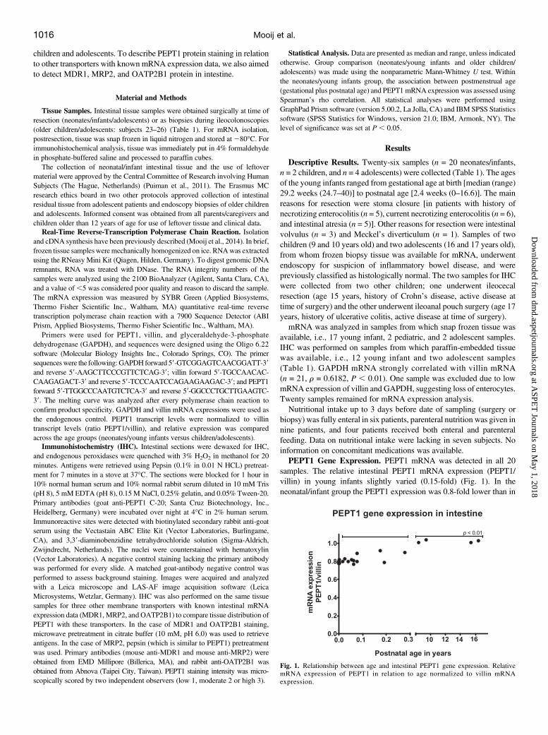
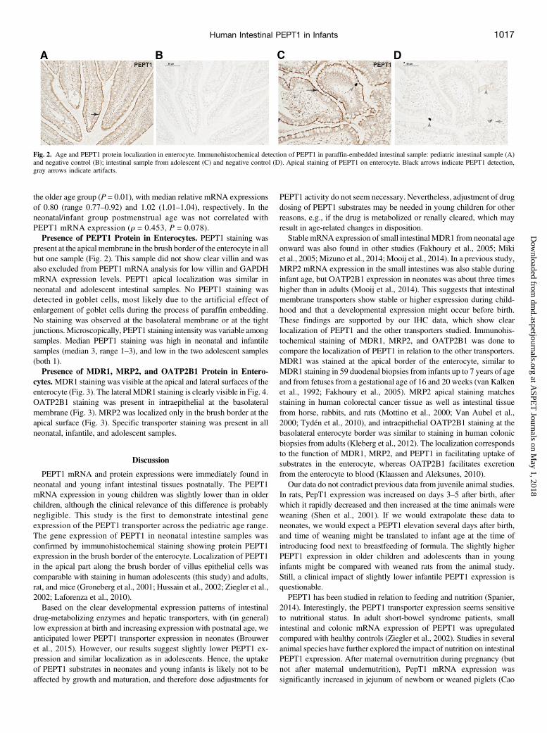
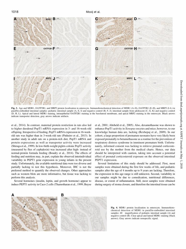
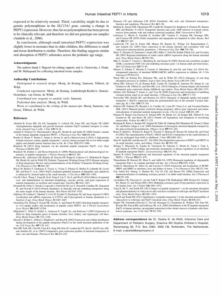
cytes.MDR1 staining was visible at the apical and lateral surfaces of theenterocyte (Fig. 3). The lateralMDR1 staining is clearly visible in Fig. 4.OATP2B1 staining was present in intraepithelial at the basolateralmembrane (Fig. 3). MRP2 was localized only in the brush border at theapical surface (Fig. 3). Specific transporter staining was present in allneonatal, infantile, and adolescent samples.
Discussion
PEPT1 mRNA and protein expressions were immediately found inneonatal and young infant intestinal tissues postnatally. The PEPT1mRNA expression in young children was slightly lower than in olderchildren, although the clinical relevance of this difference is probablynegligible. This study is the first to demonstrate intestinal geneexpression of the PEPT1 transporter across the pediatric age range.The gene expression of PEPT1 in neonatal intestine samples wasconfirmed by immunohistochemical staining showing protein PEPT1expression in the brush border of the enterocyte. Localization of PEPT1in the apical part along the brush border of villus epithelial cells wascomparable with staining in human adolescents (this study) and adults,rat, and mice (Groneberg et al., 2001; Hussain et al., 2002; Ziegler et al.,2002; Laforenza et al., 2010).Based on the clear developmental expression patterns of intestinal
drug-metabolizing enzymes and hepatic transporters, with (in general)low expression at birth and increasing expression with postnatal age, weanticipated lower PEPT1 transporter expression in neonates (Brouweret al., 2015). However, our results suggest slightly lower PEPT1 ex-pression and similar localization as in adolescents. Hence, the uptakeof PEPT1 substrates in neonates and young infants is likely not to beaffected by growth and maturation, and therefore dose adjustments for
PEPT1 activity do not seem necessary. Nevertheless, adjustment of drugdosing of PEPT1 substrates may be needed in young children for otherreasons, e.g., if the drug is metabolized or renally cleared, which mayresult in age-related changes in disposition.Stable mRNA expression of small intestinalMDR1 from neonatal age
onward was also found in other studies (Fakhoury et al., 2005; Mikiet al., 2005; Mizuno et al., 2014; Mooij et al., 2014). In a previous study,MRP2 mRNA expression in the small intestines was also stable duringinfant age, but OATP2B1 expression in neonates was about three timeshigher than in adults (Mooij et al., 2014). This suggests that intestinalmembrane transporters show stable or higher expression during child-hood and that a developmental expression might occur before birth.These findings are supported by our IHC data, which show clearlocalization of PEPT1 and the other transporters studied. Immunohis-tochemical staining of MDR1, MRP2, and OATP2B1 was done tocompare the localization of PEPT1 in relation to the other transporters.MDR1 was stained at the apical border of the enterocyte, similar toMDR1 staining in 59 duodenal biopsies from infants up to 7 years of ageand from fetuses from a gestational age of 16 and 20 weeks (van Kalkenet al., 1992; Fakhoury et al., 2005). MRP2 apical staining matchesstaining in human colorectal cancer tissue as well as intestinal tissuefrom horse, rabbits, and rats (Mottino et al., 2000; Van Aubel et al.,2000; Tydén et al., 2010), and intraepithelial OATP2B1 staining at thebasolateral enterocyte border was similar to staining in human colonicbiopsies from adults (Kleberg et al., 2012). The localization correspondsto the function of MDR1, MRP2, and PEPT1 in facilitating uptake ofsubstrates in the enterocyte, whereas OATP2B1 facilitates excretionfrom the enterocyte to blood (Klaassen and Aleksunes, 2010).Our data do not contradict previous data from juvenile animal studies.
In rats, PepT1 expression was increased on days 3–5 after birth, afterwhich it rapidly decreased and then increased at the time animals wereweaning (Shen et al., 2001). If we would extrapolate these data toneonates, we would expect a PEPT1 elevation several days after birth,and time of weaning might be translated to infant age at the time ofintroducing food next to breastfeeding of formula. The slightly higherPEPT1 expression in older children and adolescents than in younginfants might be compared with weaned rats from the animal study.Still, a clinical impact of slightly lower infantile PEPT1 expression isquestionable.PEPT1 has been studied in relation to feeding and nutrition (Spanier,
2014). Interestingly, the PEPT1 transporter expression seems sensitiveto nutritional status. In adult short-bowel syndrome patients, smallintestinal and colonic mRNA expression of PEPT1 was upregulatedcompared with healthy controls (Ziegler et al., 2002). Studies in severalanimal species have further explored the impact of nutrition on intestinalPEPT1 expression. After maternal overnutrition during pregnancy (butnot after maternal undernutrition), PepT1 mRNA expression wassignificantly increased in jejunum of newborn or weaned piglets (Cao
Fig. 2. Age and PEPT1 protein localization in enterocyte. Immunohistochemical detection of PEPT1 in paraffin-embedded intestinal sample: pediatric intestinal sample (A)and negative control (B); intestinal sample from adolescent (C) and negative control (D). Apical staining of PEPT1 on enterocyte. Black arrows indicate PEPT1 detection,gray arrows indicate artifacts.
Human Intestinal PEPT1 in Infants 1017
at ASPE
T Journals on M
ay 1, 2018dm
d.aspetjournals.orgD
ownloaded from
et al., 2014). In contrast, maternal protein restriction in rats also ledto higher duodenal PepT1 mRNA expression in 3- and 16-week-oldoffspring. Irrespective of feeding, PepT1mRNA expression in 16-week-old rats was higher than in 3-week-old rats (Pinheiro et al., 2013). Inanother study in adult rats on a protein-rich diet, PepT1 mRNA andprotein expressions as well as transporter activity were increased(Shiraga et al., 1999). In low-birth-weight piglets colonic PepT1 activity(measured by flux of cephalexin) was increased after high- instead ofnormal-protein formula feeding (Boudry et al., 2014). The effects offeeding and nutrition may, in part, explain the observed interindividualvariability in PEPT1 gene expression in young infants in the presentstudy. Unfortunately, the available nutritional data were too diverse andpartially lacking to test this hypothesis. Moreover, IHC is not thepreferred method to quantify the observed changes. Other approachessuch as western blots are more informative, but tissue was lacking toperform this analysis.Several hormones (insulin, leptin, and growth hormone) appear to
induce PEPT1 activity in Caco-2 cells (Thamotharan et al., 1999; Buyse
et al., 2001; Alteheld et al., 2005). Also, dexamethasone was shown toenhance PepT1 activity in Xenopus oocytes and mice; however, to ourknowledge human data are, lacking (Rexhepaj et al., 2009). In ourcohort, a large proportion of premature neonates have very likely beenexposed prenatally to betamethasone as a routine for the prevention ofrespiratory distress syndrome in imminent premature birth. Unfortu-nately, informed consent was lacking to retrieve prenatal corticoste-roid use by the mother from the medical charts. Hence, our datashould be interpreted with caution, taking into account a potentialeffect of prenatal corticosteroid exposure on the observed intestinalPEPT1 expression.Several limitations of this study should be addressed. First, most
samples were obtained during the first few weeks of life, and pediatricsamples after the age of 4 months up to 9 years are lacking. Therefore,the expression in this age range is still unknown. Second, variability inour samples might be due to comedication, nutritional differences,disease, or extent of inflammation. Still, most samples were obtainedduring surgery of stoma closure, and therefore the intestinal tissue can be
Fig. 3. Age and MDR1, OATP2B1, and MRP2 protein localization in enterocyte. Immunohistochemical detection of MDR1 (A–D), OATP2B1 (E–H), and MRP2 (I–L) inparaffin-embedded intestinal samples: pediatric intestinal sample (A, E, I) and negative control (B, F, J); intestinal sample from adolescent (C, G, K) and negative control(D, H, L). Apical and lateral MDR1 staining, intraepithelial OATP2B1 staining at the basolateral membrane, and apical MRP2 staining in the enterocyte. Black arrowsindicate transporter detection, gray arrows indicate artifacts.
Fig. 4. MDR1 protein localization in enterocyte. Immunohisto-chemical detection of MDR1 in paraffin-embedded intestinalsamples: 40� magnification of pediatric intestinal sample (A) andnegative control (B). Clear apical and lateral MDR1 staining (blackarrows). Gray arrows indicate nonspecific signal.
1018 Mooij et al.
at ASPE
T Journals on M
ay 1, 2018dm
d.aspetjournals.orgD
ownloaded from
expected to be relatively normal. Third, variability might be due togenetic polymorphisms in the SLC15A1 gene, causing a change inPEPT1 expression. However, thus far no polymorphism has been provento be clinically relevant, and therefore we did not genotype our samples(Anderle et al., 2006).In conclusion, although small intestinal PEPT1 expression was
slightly lower in neonates than in older children, this difference is smalland tissue distribution is similar. Therefore, this finding suggests similaroral absorption of PEPT1 substrates across the pediatric age range.
Acknowledgments
The authors thank J. Hagoort for editing support, and A. Gasiewska, J. Dunk,and M. Maliepaard for collecting intestinal tissue samples.
Authorship ContributionsParticipated in research design: Mooij, de Koning, Samsom, Tibboel, de
Wildt.Conducted experiments: Mooij, de Koning, Lindenbergh-Kortleve, Simons-
Oosterhuis, van Groen, de Wildt.Contributed new reagents or analytic tools: Samsom.Performed data analysis: Mooij, de Wildt.Wrote or contributed to the writing of the manuscript: Mooij, Samsom, van
Groen, Tibboel, de Wildt.
References
Alteheld B, Evans ME, Gu LH, Ganapathy V, Leibach FH, Jones DP, and Ziegler TR (2005)Alanylglutamine dipeptide and growth hormone maintain PepT1-mediated transport in oxida-tively stressed Caco-2 cells. J Nutr 135:19–26.
Anderle P, Nielsen CU, Pinsonneault J, Krog PL, Brodin B, and Sadée W (2006) Genetic variantsof the human dipeptide transporter PEPT1. J Pharmacol Exp Ther 316:636–646.
Boudry G, Rome V, Perrier C, Jamin A, Savary G, and Le Huerou-Luron I (2014) A high-proteinformula increases colonic peptide transporter 1 activity during neonatal life in low-birth-weightpiglets and disturbs barrier function later in life. Br J Nutr 112:1073–1080.
Brandsch M (2013) Drug transport via the intestinal peptide transporter PepT1. Curr OpinPharmacol 13:881–887.
Brandsch M, Knütter I, and Bosse-Doenecke E (2008) Pharmaceutical and pharmacological im-portance of peptide transporters. J Pharm Pharmacol 60:543–585.
Brouwer KL, Aleksunes LM, Brandys B, Giacoia GP, Knipp G, Lukacova V, Meibohm B, NigamSK, Rieder M, and de Wildt SN; Pediatric Transporter Working Group (2015) Human ontogenyof drug transporters: Review and recommendations of the Pediatric Transporter Working Group.Clin Pharmacol Ther 98:266–287.
Buyse M, Berlioz F, Guilmeau S, Tsocas A, Voisin T, Péranzi G, Merlin D, Laburthe M, LewinMJ, and Rozé C, et al. (2001) PepT1-mediated epithelial transport of dipeptides and cephalexinis enhanced by luminal leptin in the small intestine. J Clin Invest 108:1483–1494.
Cao M, Che L, Wang J, Yang M, Su G, Fang Z, Lin Y, Xu S, and Wu D (2014) Effects of maternalover- and undernutrition on intestinal morphology, enzyme activity, and gene expression ofnutrient transporters in newborn and weaned pigs. Nutrition 30:1442–1447.
Drozdzik M, Gröer C, Penski J, Lapczuk J, Ostrowski M, Lai Y, Prasad B, Unadkat JD, SiegmundW, and Oswald S (2014) Protein abundance of clinically relevant multidrug transporters alongthe entire length of the human intestine. Mol Pharm 11:3547–3555.
Fakhoury M, Litalien C, Medard Y, Cavé H, Ezzahir N, Peuchmaur M, and Jacqz-Aigrain E (2005)Localization and mRNA expression of CYP3A and P-glycoprotein in human duodenum as afunction of age. Drug Metab Dispos 33:1603–1607.
Groneberg DA, Döring F, Eynott PR, Fischer A, and Daniel H (2001) Intestinal peptide transport:ex vivo uptake studies and localization of peptide carrier PEPT1. Am J Physiol GastrointestLiver Physiol 281:G697–G704.
Hilgendorf C, Ahlin G, Seithel A, Artursson P, Ungell AL, and Karlsson J (2007) Expression ofthirty-six drug transporter genes in human intestine, liver, kidney, and organotypic cell lines.Drug Metab Dispos 35:1333–1340.
Hussain I, Kellett L, Affleck J, Shepherd J, and Boyd R (2002) Expression and cellular distributionduring development of the peptide transporter (PepT1) in the small intestinal epithelium of therat. Cell Tissue Res 307:139–142.
Kim HR, Park SW, Cho HJ, Chae KA, Sung JM, Kim JS, Landowski CP, Sun D, Abd El-Aty AM,and Amidon GL, et al. (2007) Comparative gene expression profiles of intestinal transporters inmice, rats and humans. Pharmacol Res 56:224–236.
Klaassen CD and Aleksunes LM (2010) Xenobiotic, bile acid, and cholesterol transporters:function and regulation. Pharmacol Rev 62:1–96.
Kleberg K, Jensen GM, Christensen DP, Lundh M, Grunnet LG, Knuhtsen S, Poulsen SS, HansenMB, and Bindslev N (2012) Transporter function and cyclic AMP turnover in normal colonicmucosa from patients with and without colorectal neoplasia. BMC Gastroenterol 12:78.
Laforenza U, Miceli E, Gastaldi G, Scaffino MF, Ventura U, Fontana JM, Orsenigo MN,and Corazza GR (2010) Solute transporters and aquaporins are impaired in celiac disease. BiolCell 102:457–467.
Landowski CP, Sun D, Foster DR, Menon SS, Barnett JL, Welage LS, Ramachandran C,and Amidon GL (2003) Gene expression in the human intestine and correlation with oralvalacyclovir pharmacokinetic parameters. J Pharmacol Exp Ther 306:778–786.
Meier Y, Eloranta JJ, Darimont J, Ismair MG, Hiller C, Fried M, Kullak-Ublick GA, and VavrickaSR (2007) Regional distribution of solute carrier mRNA expression along the human intestinaltract. Drug Metab Dispos 35:590–594.
Miki Y, Suzuki T, Tazawa C, Blumberg B, and Sasano H (2005) Steroid and xenobiotic receptor(SXR), cytochrome P450 3A4 and multidrug resistance gene 1 in human adult and fetal tissues.Mol Cell Endocrinol 231:75–85.
Mizuno T, Fukuda T, Masuda S, Uemoto S, Matsubara K, Inui KI, and Vinks AA (2014) De-velopmental trajectory of intestinal MDR1/ABCB1 mRNA expression in children. Br J ClinPharmacol 77:910–912.
Mooij MG, de Koning BA, Huijsman ML, and de Wildt SN (2012) Ontogeny of oral drugabsorption processes in children. Expert Opin Drug Metab Toxicol 8:1293–1303.
Mooij MG, Schwarz UI, de Koning BAE, Leeder JS, Gaedigk R, Samsom JN, Spaans E, vanGoudoever JB, Tibboel D, and Kim RB, et al. (2014) Ontogeny of human hepatic and intestinaltransporter gene expression during childhood: age matters. Drug Metab Dispos 42:1268–1274.
Mottino AD, Hoffman T, Jennes L, and Vore M (2000) Expression and localization of multidrugresistant protein mrp2 in rat small intestine. J Pharmacol Exp Ther 293:717–723.
Nosworthy MG, Bertolo RF, and Brunton JA (2013) Ontogeny of dipeptide uptake and peptidetransporter 1 (PepT1) expression along the gastrointestinal tract in the neonatal Yucatan mini-ature pig. Br J Nutr 110:275–281.
Pinheiro DF, Pinheiro PF, Buratini J, Jr, Castilho AC, Lima PF, Trinca LA, and Vicentini-PaulinoMdL (2013) Maternal protein restriction during pregnancy affects gene expression and immu-nolocalization of intestinal nutrient transporters in rats. Clin Sci (Lond) 125:281–289.
Puiman PJ, Burger-Van Paassen N, Schaart MW, De Bruijn AC, De Krijger RR, Tibboel D, VanGoudoever JB, and Renes IB (2011) Paneth cell hyperplasia and metaplasia in necrotizingenterocolitis. Pediatr Res 69:217–223.
Rexhepaj R, Rotte A, Kempe DS, Sopjani M, Föller M, Gehring EM, Bhandaru M, Gruner I, MackAF, and Rubio-Aliaga I, et al. (2009) Stimulation of electrogenic intestinal dipeptide transport bythe glucocorticoid dexamethasone. Pflugers Arch 459:191–202.
Rome S, Barbot L, Windsor E, Kapel N, Tricottet V, Huneau JF, Reynes M, Gobert JG, and ToméD (2002) The regionalization of PepT1, NBAT and EAAC1 transporters in the small intestine ofrats are unchanged from birth to adulthood. J Nutr 132:1009–1011.
Shen H, Smith DE, and Brosius FC, 3rd (2001) Developmental expression of PEPT1 and PEPT2 inrat small intestine, colon, and kidney. Pediatr Res 49:789–795.
Shiraga T, Miyamoto K, Tanaka H, Yamamoto H, Taketani Y, Morita K, Tamai I, Tsuji A,and Takeda E (1999) Cellular and molecular mechanisms of dietary regulation on rat intestinalH+/peptide transporter PepT1. Gastroenterology 116:354–362.
Spanier B (2014) Transcriptional and functional regulation of the intestinal peptide transporterPEPT1. J Physiol 592:871–879.
Thamotharan M, Bawani SZ, Zhou X, and Adibi SA (1999) Hormonal regulation of oligopeptidetransporter Pept-1 in a human intestinal cell line. Am J Physiol 276:C821–C826.
Tydén E, Bjornstrom H, Tjälve H, and Larsson P (2010) Expression and localization of BCRP,MRP1 and MRP2 in intestines, liver and kidney in horse. J Vet Pharmacol Ther 33:332–340.
Van Aubel RA, Hartog A, Bindels RJ, Van Os CH, and Russel FG (2000) Expression andimmunolocalization of multidrug resistance protein 2 in rabbit small intestine. Eur J Pharmacol400:195–198.
van Kalken CK, Giaccone G, van der Valk P, Kuiper CM, Hadisaputro MM, Bosma SA, ScheperRJ, Meijer CJ, and Pinedo HM (1992) Multidrug resistance gene (P-glycoprotein) expression inthe human fetus. Am J Pathol 141:1063–1072.
Yang B, Hu Y, and Smith DE (2013) Impact of peptide transporter 1 on the intestinal absorptionand pharmacokinetics of valacyclovir after oral dose escalation in wild-type and PepT1 knockoutmice. Drug Metab Dispos 41:1867–1874.
Yang B and Smith DE (2013) Significance of peptide transporter 1 in the intestinal permeability ofvalacyclovir in wild-type and PepT1 knockout mice. Drug Metab Dispos 41:608–614.
Ziegler TR, Fernández-Estívariz C, Gu LH, Bazargan N, Umeakunne K, Wallace TM, Diaz EE,Rosado KE, Pascal RR, and Galloway JR, et al. (2002) Distribution of the H+/peptide transporterPepT1 in human intestine: up-regulated expression in the colonic mucosa of patients with short-bowel syndrome. Am J Clin Nutr 75:922–930.
Address correspondence to: Dr. Saskia N. de Wildt, Intensive Care andDepartment of Pediatric Surgery, Erasmus MC-Sophia Children’s Hospital,Wytemaweg 80, P.O. Box 2060, 3000 CB, Rotterdam, The Netherlands.E-mail: [email protected]
Human Intestinal PEPT1 in Infants 1019
at ASPE
T Journals on M
ay 1, 2018dm
d.aspetjournals.orgD
ownloaded from