Embed Size (px)
Citation preview

Article
Specific Metabolites in a Phaeodactylum tricornutumStrain Isolated from Western Norwegian Fjord Water
Siv Kristin Prestegard 1,2,*, Svein Rune Erga 3, Pia Steinrücken 3, Svein Are Mjøs 4,Gjert Knutsen 3 and Jens Rohloff 5
Received: 16 September 2015; Accepted: 22 December 2015; Published: 30 December 2015Academic Editor: Véronique Martin-Jézéquel
1 Bergen Marine Biobank, Department of Biology, University of Bergen, Thormøhlensgate 53A/B,N-5020 Bergen, Norway
2 Applied Biotechnology, Uni Research Environment, Thormøhlensgate 49B, N-5006 Bergen, Norway3 Department of Biology, University of Bergen, Thormøhlensgate 53AB, N-5020 Bergen, Norway;
[email protected] (S.R.E.); [email protected] (P.S.); [email protected] (G.K.)4 Department of Chemistry, University of Bergen, Allégaten 42, N-5020 Bergen, Norway; [email protected] Department of Biology, Norwegian University of Science and Technology, N-7491 Trondheim, Norway;
[email protected]* Correspondence: [email protected]; Tel.: +47-55-58-26-64
Abstract: We have searched for special characteristics in growth, protein expression, fatty acidsand volatile organic compounds (VOCs) in a local Phaeodactylum tricornutum Bohlin strain (BergenMarine Biobank), by comparing it with a common accession strain (CCAP). Differences in growth andexpressed proteins were detected between the BMB strain and the CCAP strain, and the BMB strainreached the highest cell densities under the given growth conditions. Fatty acid (FA) analyses showedhighest relative eicosapentaenoic acid (EPA) levels in the exponential phase (25.73% and 28.31%), andhighest levels of palmitoleic acid (16:1 n-7) in the stationary phase (46.36% and 43.66%) in the BMBand CCAP strain, respectively. The most striking finding of the VOCs analyses was the relativelyhigh levels of ectocarpene, 6-((1E)-butenyl)-1,4-cycloheptadiene, hormosirene, and desmarestene andstructurally related compounds, which were exclusively detected in the BMB strain. Many of theVOCs detected in the CCAP and, in particular, in the BMB strain have been reported as antimicrobialagents. We suggest that the array of pheromones and antimicrobial substances could be part ofan allelopathic strategy of the BMB strain, dominated by oval cells, thus reflecting the benthic lifestage of this morphological form. These findings show the potential for bioactive metabolites in theBMB strain.
Keywords: Phaeodactylum tricornutum; two-dimensional gel electrophoresis (2DGE); volatile organiccompounds (VOCs); hormosirene; ectocarpene; fatty acids; eicosapentaenoic acid (EPA)
1. Introduction
The diatoms represent a large and extraordinary ecologically flexible group of unicellulareukaryotic microalgae. They have acquired genes from an eukaryotic heterotroph and a red algaethrough secondary endosymbiosis [1] and later through lateral gene transfers from bacteria [2].The complex genomes of diatoms, together with their ability for sexual reproduction [3], give thisgenetically highly diverse group many interesting metabolic properties.
Except for the synthesis of primary metabolites like pigments and certain proteins, it is currentlyimpossible to foresee how diatoms will respond metabolically to different sets of growth or stressconditions. The biogenesis of secondary metabolites in diatoms and microalgae in general isunpredictable and needs to be investigated thoroughly. The capability of cells to produce bioactive
Mar. Drugs 2016, 14, 9; doi:10.3390/md14010009 www.mdpi.com/journal/marinedrugs

Mar. Drugs 2016, 14, 9 2 of 17
compounds must therefore be examined empirically, and the increasing knowledge of diatommetabolites was excellently reviewed by Stonik, V. and I. Stonik [4]. The sequenced genome ofP. tricornutum in 2008 [2] has provided new insight into the molecular foundation of adaption andsurvival mechanisms to highly changing environments [5,6]. It has been shown that diatoms excretemany of their bioactive compounds, which can have a variety of influences on other species in theplankton community [7,8]. Recently, metabolic fingerprinting of metabolites that had been excreted intothe surrounding medium from the diatoms Skeletonema marinoi and Thalassiosira pseudonana revealedgrowth stage dependence [9]. Thus, volatile metabolites originating from exudates from living cells,can also give important information concerning physiological status, and several volatile compoundshave been reported to be derivatives from degradation of fatty acids [10,11].
The polymorphic diatom Phaeodactylum tricornutum Bohlin is recognized as a model organism forstudies on diatom physiology and genetics. It is also used on an industrial level for aquaculture feedbecause of its high content of polyunsaturated fatty acids (PUFAs), especially eicosapentaenoic acid(EPA), which is of high value as a food additive to human and animals [12,13].
Phaeodactylum tricornutum can be found in both brackish and marine waters worldwide [14].Environmental conditions have been reported to induce the transformation between the differentmorphological cell forms; oval, fusiform, triradiate and cruciform [15–18]. Oval cells of P. tricornutumare considered to represent a benthic life stage since they are most often observed when grown on agaror on the surface of culture containers [19,20].
From a local strain of P. tricornutum, isolated from fjord water in Western Norway [21], more than95% of the cells were reported to be oval when grown in liquid bubbled cultures [22]. Water extractsfrom this strain strongly inhibited blood platelet activation and induced cell death in leukaemia cells.Part of the activity was shown to originate from the high levels of the nucleoside adenosine, whilethe rest of the activity was shown to originate from other unidentified compounds [21]. The highlyinteresting metabolic and morphological properties of this isolate motivated us to search for additionalspecial characteristics in growth, protein expression, fatty acids and volatile organic compounds. Fattyacids are also interesting in terms of commercial applications, and they have been reported to beprecursors for many volatile metabolites [10,11]. Volatile organic compounds (VOC) are part of themetabolome in diatoms and show a high structural variety [10]. Therefore, we wanted to shed light onthe VOC fraction in order to reveal characteristic metabolites.
To improve the evaluation of our results, we compared it with a common CCAP strain (CCAP1052/1A from the Culture Collection of Algae and Protozoa in Oban, UK). Here, we present noveldata which make us believe that our local strain has potential for new bioactive metabolites, amongwhich the VOC should be specially emphasized. These compounds comprise both pheromones andantimicrobial substances, which are of great ecological and biomedical importance.
2. Results
2.1. Different Growth and Protein Expression of BMB and CCAP Strains
Growth curves for the BMB (BMB-E-0007) and CCAP (CCAP 1052/1A) strains are presented inFigure 1A,B. From cell counts, the growth rates corresponding to the sampling day for soluble proteinswere determined for both strains. The average growth rate was 0.58 ˘ 0.10 day´1 for the BMB strainand 1.07 ˘ 0.03 day´1 for CCAP from day one to day two. From day three to four, the average growthrate for the BMB strain was 1.01 ˘ 0.06 day´1 and 0.98 ˘ 0.03 day´1 for the CCAP strain. The CCAPcultures reached their maximal average cell densities of 3.1 ˆ 107 cells¨ mL´1 at day six, and then thecell numbers started to decrease slowly. The BMB strain cultures reached their maximal average celldensities of 4 ˆ 107 cells¨ mL´1 at day eleven, and at this time the cultures also obtained the maximaldry weights (Figure 1B). The CCAP strain reached it maximal dry weight at day nine.

Mar. Drugs 2016, 14, 9 3 of 17
Figure 1. Growth of the BMB and CCAP strains in batch cultures. Cells were grown in artificialseawater with the addition of Walne’s medium, 27 PSU at 20 ˝C and 249 µmol photons m´2¨ s´1. (A):Cells¨ mL´1. Data in Figure 1A are modified from [22]. Arrows shows the time-points for samplingof total soluble proteins. (B): Dry weight mL´1 algae culture. The data in Figure 1A,B are from thesame experiments.
The BMB strain was dominated by oval cells (more than 95%), and the CCAP strain was dominatedby cells with fusiform morphology (Figure 2A,B). Triradiate cells could occur in the CCAP strain, butit was never observed in the BMB strain.
Total soluble proteins separated by 2DGE from the different growth phases from the BMB andCCAP strains are presented in Figure 3A–F. The protein profiles differed according to growth phasesand strain. Total soluble protein and number of resolved spots are presented in Table 1. At day two,more protein spots were resolved from the CCAP strain (372 ˘ 41) than from the BMB strain (197 ˘ 11),while at day four the number increased for the BMB strain (289 ˘ 18) and were reduced for the CCAPstrain (242 ˘ 39).
Furthermore at day eight, in the stationary growth phase, especially the CCAP strain had stronglyreduced resolved protein profiles on the 2DGE gels (Figure 3F). The protein profiles from the BMB strainin the stationary growth phase also revealed reduced number of proteins, but to a lesser extent than inthe CCAP profiles (Figure 3C). Differences in the total soluble protein from samples preparations inthe CCAP strain (3.12 ˘ 0.30) at day 2 compared to the BMB strain (2.56 ˘ 0.45) were not significant.Furthermore, the levels decreased by 24 times until day eight in the CCAP strain. The total solubleprotein from the BMB strain also decreased towards day eight, but only by 10 times (Table 1). InFigure 3A–C), an example of a protein that appears to be specific for the BMB strain with an apparentmolecular mass (Mw) of 33 kDa and pI of about 6.2 is marked. The 33 kDa protein was absent from the2DGE gels separating proteins from the CCAP strain at any growth stage.

Mar. Drugs 2016, 14, 9 4 of 17
Figure 2. Microscope photos of BMB strain (A) and CCAP (B). A Zeiss Axio Imager Z1 microscope(Carl Zeiss) was used to photograph representative cells from the two strains (100ˆ magnification withimmersion oil, Bright Field (A) and Differential interference contrast (B)).
Figure 3. Representatives of protein profiles on 2DGE from BMB strain; (A): day 2; (B): day 4; (C): day 8and of the CCAP strain; (D): day 2; (E): day 4; (F): day 8. Arrows with cross sign show that protein waspresent only in BMB strain.

Mar. Drugs 2016, 14, 9 5 of 17
Table 1. Total soluble proteins cell´1 for samples from BMB and CCAP strain of Phaeodactylumtricornutum for different time points during growth in batch cultures (n = 3). Numbers of resolvedspots using 2DGE are listed for the different samples (n = 3).
Strain Day Total Soluble Protein, pg/Cell Resolved Protein Spots by 2DGE
BMB 2 2.56 ˘ 0.45 197 ˘ 114 0.57 ˘ 0.06 289 ˘ 188 0.25 ˘ 0.04 198 ˘ 19
CCAP 2 3.12 ˘ 0.30 372 ˘ 414 1.10 ˘ 0.10 242 ˘ 398 0.13 ˘ 0.02 90 ˘ 6
2.2. Fatty Acid Content and Composition of BMB and CCAP Strains
Samples taken from exponential and stationary phases of the BMB and CCAP strain were analysedfor total fatty acid (FA) content and the relative composition of different FAs. In Figure 4A,B, 19 ofthe detected FA with relatively levels higher than 1% are shown. The two strains possess a similar FAcomposition in both growth phases. In the exponential phase (Figure 4A), eicosapentaenoic acid (EPA)(20:5 n-3) was the most dominant FA, with 25.73% ˘ 1.35% for the BMB strain and 28.31% ˘ 0.68% forthe CCAP strain. The average cell specific content of EPA is 0.85 and 1.08 pg¨ cell´1 for exponentiallygrowing BMB and CCAP strains, respectively, and 0.59 and 0.66 pg¨ cell´1, respectively, for stationarycells (see Figure 4A–C). In the stationary phase (Figure 4B), palmitoleic acid (16:1 n-7) became thepredominant FA with 46.36% ˘ 0.16% of the total FA in the BMB strain and 43.66% ˘ 1.05% in theCCAP strain. Also, the levels of the saturated palmitic acid (16:0) had increased to 29.3% and 32.1% inthe BMS and CCAP strain, respectively. EPA levels were reduced to 6.17% ˘ 0.06% for the BMB strainand 8.26% ˘ 0.21% for the CCAP strain in the stationary phase.
Figure 4. Fatty acid (FA) profiles given as % of total FA of BMB and CCAP strain in exponential phase(A) and in stationary phase (B). Total FA per cell in BMB and CCAP strain in exponential and stationaryphase (C) and % of total FA per dry weight (D).
Mar. Drugs 2016, 14, 9 6 of 17
In the exponential growth phase, a higher relative fraction of the fatty acids consisted of longchain fatty acids and polyunsaturated FAs, whereas in the stationary phase, a higher fraction of theshorter fatty acids (C16 and 16:1 n-7) was present. The total amount of fatty acids per cell more thandoubled from exponential phase to stationary phase for both strains (Figure 4C). In the stationaryphase, the BMB strain had a slightly higher total FA per cell (9.51 pg ˘ 0.82) than the CCAP strain(8.02 pg ˘ 0.26). In the exponential phase, 10.98% ˘ 1.1% of the dry biomass in the BMB strain was FA,and in the stationary phase it had increased to 31.01% ˘ 3.31%. In the CCAP strain, 6.4% ˘ 2.27% ofthe dry algae biomass was fatty acids in the exponential phase and 34.44% ˘ 2.26% in the stationaryphase (Figure 4D).
2.3. Differences in Volatile Organic Compounds (VOCs) from BMB and CCAP Strains
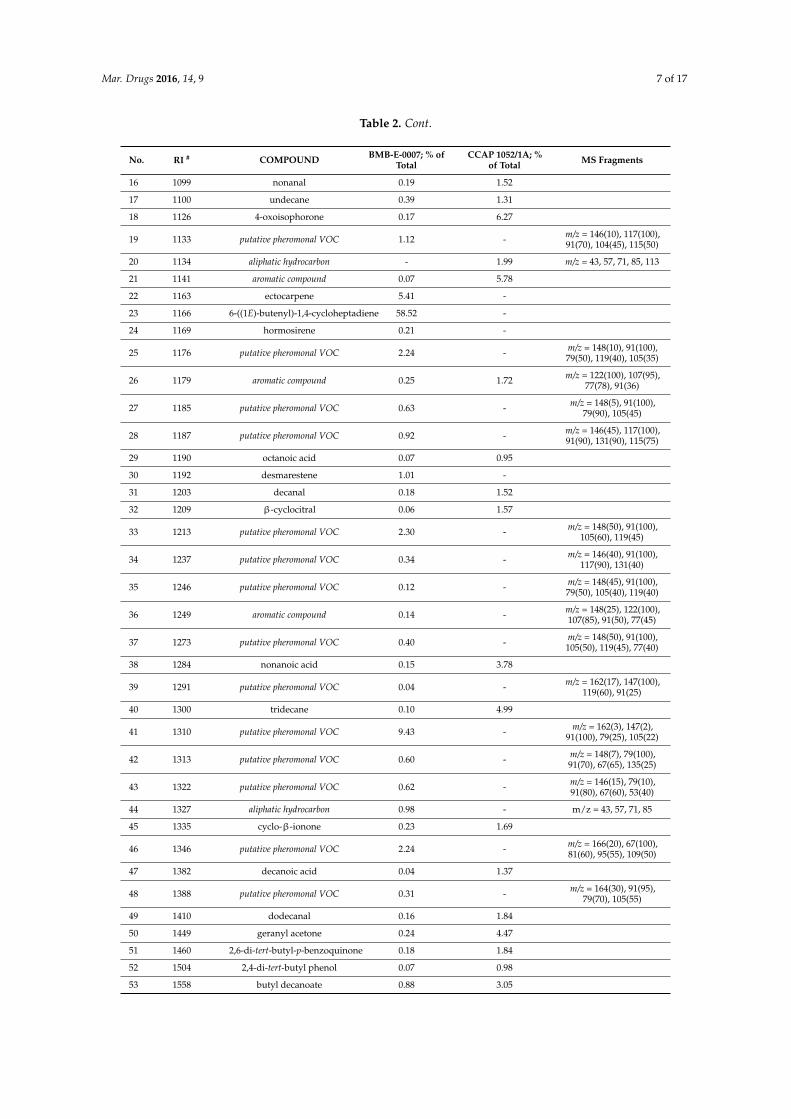
VOCs from early stationary cultures of BMB and CCAP strain of P. tricornutum were collectedand analysed by headspace SPME–GC-MS. A broad range of 61 structurally different VOCs weretentatively identified, comprising aliphatics (6 alkanes, 4 aldehydes, 1 ketone, 4 acids, 2 esters),sulphides (3), terpenes (3 monoterpenes, 1 sesquiterpene, 1 triterpene), aromatics (3), furans (1),quinones (1), carotenoid-derived volatiles (6), and alicyclic olefins (18). Furthermore, 7 non-identifiedpeaks were structurally assigned as aliphatic hydrocarbons (4) and aromatics (3) (Table 2). Ingeneral, a distinct higher number of VOCs could be isolated from the BMB strain (55) comparedto the CCAP strain of P. tricornutum (36). The most striking observation in the BMB strain was theabundance of the algal pheromones ectocarpene, 6-((1E)-butenyl)-1,4-cycloheptadiene, hormosireneand desmarestene. Further 14 non-identified pheromonal volatiles were structurally annotated basedon their characteristic and similar MS fragmentation patterns and the occurrence of the molecularions of identified pheromones with (M+) m/z = 148 (compounds No. 22–24) and (M+) m/z = 146(compound No. 30) (Table 2).
The content of 6-((1E)-butenyl)-1,4-cycloheptadiene comprised 58.5% of the detected VOCs inthe BMB strain. The gas chromatographic separation of pheromones of the BMB strain is depicted inFigure 5. These alicyclic olefins were totally absent in the volatile profiles of the CCAP strain.
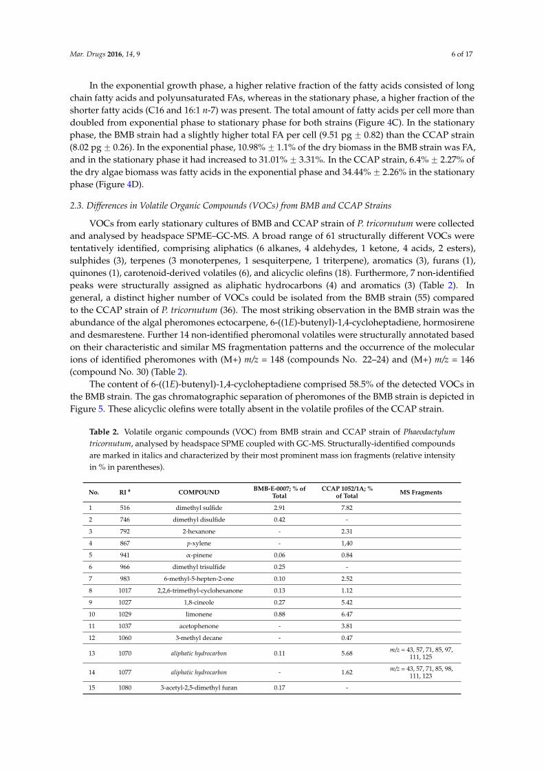
Table 2. Volatile organic compounds (VOC) from BMB strain and CCAP strain of Phaeodactylumtricornutum, analysed by headspace SPME coupled with GC-MS. Structurally-identified compoundsare marked in italics and characterized by their most prominent mass ion fragments (relative intensityin % in parentheses).
No. RI # COMPOUND BMB-E-0007; % ofTotal
CCAP 1052/1A; %of Total MS Fragments
1 516 dimethyl sulfide 2.91 7.82
2 746 dimethyl disulfide 0.42 -
3 792 2-hexanone - 2.31
4 867 p-xylene - 1,40
5 941 α-pinene 0.06 0.84
6 966 dimethyl trisulfide 0.25 -
7 983 6-methyl-5-hepten-2-one 0.10 2.52
8 1017 2,2,6-trimethyl-cyclohexanone 0.13 1.12
9 1027 1,8-cineole 0.27 5.42
10 1029 limonene 0.88 6.47
11 1037 acetophenone - 3.81
12 1060 3-methyl decane - 0.47
13 1070 aliphatic hydrocarbon 0.11 5.68 m/z = 43, 57, 71, 85, 97,111, 125
14 1077 aliphatic hydrocarbon - 1.62 m/z = 43, 57, 71, 85, 98,111, 123
15 1080 3-acetyl-2,5-dimethyl furan 0.17 -
Mar. Drugs 2016, 14, 9 7 of 17
Table 2. Cont.
No. RI # COMPOUND BMB-E-0007; % ofTotal
CCAP 1052/1A; %of Total MS Fragments
16 1099 nonanal 0.19 1.52
17 1100 undecane 0.39 1.31
18 1126 4-oxoisophorone 0.17 6.27
19 1133 putative pheromonal VOC 1.12 - m/z = 146(10), 117(100),91(70), 104(45), 115(50)
20 1134 aliphatic hydrocarbon - 1.99 m/z = 43, 57, 71, 85, 113
21 1141 aromatic compound 0.07 5.78
22 1163 ectocarpene 5.41 -
23 1166 6-((1E)-butenyl)-1,4-cycloheptadiene 58.52 -
24 1169 hormosirene 0.21 -
25 1176 putative pheromonal VOC 2.24 - m/z = 148(10), 91(100),79(50), 119(40), 105(35)
26 1179 aromatic compound 0.25 1.72 m/z = 122(100), 107(95),77(78), 91(36)
27 1185 putative pheromonal VOC 0.63 - m/z = 148(5), 91(100),79(90), 105(45)
28 1187 putative pheromonal VOC 0.92 - m/z = 146(45), 117(100),91(90), 131(90), 115(75)
29 1190 octanoic acid 0.07 0.95
30 1192 desmarestene 1.01 -
31 1203 decanal 0.18 1.52
32 1209 β-cyclocitral 0.06 1.57
33 1213 putative pheromonal VOC 2.30 - m/z = 148(50), 91(100),105(60), 119(45)
34 1237 putative pheromonal VOC 0.34 - m/z = 146(40), 91(100),117(90), 131(40)
35 1246 putative pheromonal VOC 0.12 - m/z = 148(45), 91(100),79(50), 105(40), 119(40)
36 1249 aromatic compound 0.14 - m/z = 148(25), 122(100),107(85), 91(50), 77(45)
37 1273 putative pheromonal VOC 0.40 - m/z = 148(50), 91(100),105(50), 119(45), 77(40)
38 1284 nonanoic acid 0.15 3.78
39 1291 putative pheromonal VOC 0.04 - m/z = 162(17), 147(100),119(60), 91(25)
40 1300 tridecane 0.10 4.99
41 1310 putative pheromonal VOC 9.43 - m/z = 162(3), 147(2),91(100), 79(25), 105(22)
42 1313 putative pheromonal VOC 0.60 - m/z = 148(7), 79(100),91(70), 67(65), 135(25)
43 1322 putative pheromonal VOC 0.62 - m/z = 146(15), 79(10),91(80), 67(60), 53(40)
44 1327 aliphatic hydrocarbon 0.98 - m/z = 43, 57, 71, 85
45 1335 cyclo-β-ionone 0.23 1.69
46 1346 putative pheromonal VOC 2.24 - m/z = 166(20), 67(100),81(60), 95(55), 109(50)
47 1382 decanoic acid 0.04 1.37
48 1388 putative pheromonal VOC 0.31 - m/z = 164(30), 91(95),79(70), 105(55)
49 1410 dodecanal 0.16 1.84
50 1449 geranyl acetone 0.24 4.47
51 1460 2,6-di-tert-butyl-p-benzoquinone 0.18 1.84
52 1504 2,4-di-tert-butyl phenol 0.07 0.98
53 1558 butyl decanoate 0.88 3.05
Mar. Drugs 2016, 14, 9 8 of 17
Table 2. Cont.
No. RI # COMPOUND BMB-E-0007; % ofTotal
CCAP 1052/1A; %of Total MS Fragments
54 1592 tetradecanal 0.10 1.38
55 1595 dodecanoic acid 0.03 1.11
56 1815 (E,E)-farnesyl acetate 0.16 1.43
57 2000 eicosane 0.30 -
58 2178 ethyl hexadecanoate 0.44 2.67
59 2200 docosane 0.05 6.14
60 2832 squalene 2.24 -
61 3000 triacontane 0.34 1.13
sum % 100 100
Total MS detector response 1.56E + 07 1.28E + 06# Retention indices calculated based on a series of n-alkanes on an apolar DB-5 column.
Figure 5. Example of GC-MS chromatogram showing the separation pattern of pheromonal VOCs andmonoterpenes, detected in the BMB strain. Numbers indicate volatile compounds listed in Table 2.
3. Discussion
The interesting metabolic properties and the generally great phenotypic variation shownin P. tricornutum isolates motivated us to search for additional characteristics in growth, proteinexpression, FA profiles and VOCs in a BMB strain isolated from Western Norwegian fjord water. Allexperiments and analyses were run in parallel with a common CCAP strain for comparison. TheBMB strain was shown to be dominated by oval cells (Figure 2A), and the CCAP strain (Figure 2B) byfusiform cells. The oval cell form of P. tricornutum has in several studies been emphasized to be a benthiclife form, which is supported by the fact that fusiform cells grown on agar surfaces will transform intoslightly motile oval cells surrounded by extracellular polymeric substances. It was also reported thatoval cells transferred from agar into liquid medium would gradually revert to fusiform cells [19]. Itis therefore an interesting feature of the BMB strain that it can be maintained oval in liquid bubbledcultures. Epigenetic changes have recently been shown to have an important role in phenotypicvariation [23]. It is not unlikely that the polymorphism seen in P. tricornutum could be related toepigenetic phenomena. Epigenetic regulation has been shown for P. tricornutum, and extensive genemethylation correlated strongly with differential expression under specific conditions [24]. Proteomics
Mar. Drugs 2016, 14, 9 9 of 17
can be one approach to reveal epigenetic effects [25], and here we have shown differences in solubleproteins separated by 2DGE between the BMB and CCAP strain in different growth phases. Thesedifferences indicate that the two P. tricornutum strains are under different transcriptional or epigeneticregulation, but a more in depth study is required to be able to reveal more about regulation ofexpressed proteins.
Growth parameters like cell numbers and dry weight and also expressed proteins are importantparameters to describe physiological status of microalgae cultures. When the CCAP cultures had themaximal growth rates at day two, the highest number of expressed proteins on the 2DGE gels wasobserved (Figure 3D and Table 1). The successive increase in growth rate for the BMB strain fromday one to day two and from day three to four co-occurred with an increased number of proteins onthe 2DGE gels at day four (Figure 3B and Table 1). This is in accordance with the increased need forproteins in cell division when growth rates increase. A decreased number of separated proteins at dayfour is observed for the CCAP cells (Figure 3E), and this can indicate that a turnover of the proteins hadoccurred, a process that allows the cells to re-utilize amino acids and change protein content. Thesefindings are supported by a recent study of P. tricornutum and its responses to nitrogen deprivation,where the authors showed that following nitrogen deprivation, a reduction in biosynthesis and anincrease in recycling of N-containing compounds like amino acids, proteins and nucleic acid occurredat the transcript level [26]. It has also been shown that as the growth rate decreased, the overall rate ofprotein turnover decreased in P. tricornutum [27]. The highly reduced numbers of expressed proteinsfound in the early stationary phase (day 8) in our study have also been observed in a stationary cultureof the dinoflagellate Prorocentrum triestinum [28]. Nitrogen limitation has been shown to decrease thecell protein content and abundance of proteins like ribulose bisphosphate carboxylase/oxygenase [29].A characteristic protein observed for the BMB strain was one with molecular weight of about 33 kDaand pI of about 6.2 (Figure 3A–C). This finding did not correspond to the presence of a 21 kDa proteinfound in the fusiform cells detected from comparison of protein bands from clonal cultures of oval andfusiform cells in one-dimensional SDS-PAGE gels [30], hence the 33 kDa protein appears to be specificto the BMB strain.
Fatty acids are metabolites of high commercial interest and with many interesting cellularfunctions. In microalgae, they are building blocks in cellular lipids like the polar membrane lipids andthe neutral lipids [31]. Some fatty acids have also been shown to possess antibacterial properties [32].In our studies, the total amount of fatty acids per cell and per dry weight increased more than twotimes from exponential to stationary phase in both strains. At the same time, both strains showed asimilar shift in their relative FA composition. The higher relative content of long chain polyunsaturatedFA (especially EPA) in exponential phase changed to a higher fraction of monounsaturated andsaturated fatty acids, especially palmitoleic acid (16:1 n-7) in the stationary phase. Both the increasein total FA and the shift towards saturation and monounsaturation in the stationary phase havebeen documented for P. tricornutum by others [33,34] and can be related to the accumulation ofneutral storage lipids (triacylglycerols, TAG) as a response to nitrogen starvation in the stationaryphase. They serve as storage compounds for carbon and energy and predominantly consist more ofsaturated and monounsaturated fatty acids, while polyunsaturated fatty acids (PUFA) are typicallypresent in membrane lipids [31]. Interestingly, EPA has also been reported to have high antibacterialactivity [35,36]. In another study, it was found that extracts from fusiform cells had higher antibacterialactivity and higher content of the fatty acids EPA, hexadecatrienoic acid and palmitoleic acid thanextracts from oval cells [32]. It is interesting to note that here in our study the BMB strain (dominatedby oval cells) had higher relative amounts (Figure 4B) and cell specific amounts of palmitoleic acid(average 4.41 pg¨ cell´1 ) than the CCAP strain (average 3.50 pg¨ cell´1, dominated by fusiform cells)in the stationary phase.
Even if most of the VOCs described are highly volatile and hydrophobic, they are still slightlysoluble in water and can be bioactive, i.e., molecules can be sensed by other individuals of the samespecies or even other species in the context of defense.Volatile metabolites originating from exudates
Mar. Drugs 2016, 14, 9 10 of 17
from living cells can give important information concerning physiological status, and several volatilecompounds have been reported to be derivatives from degradation of fatty acids. More volatilemetabolites could be detected from the oval cells (55) than from the fusiform cells (36). Interestinglythe higher number of separated protein spots in the stationary phase of the BMB strain co-occurredwith an increased number of detected VOCs. The bouquet of cultures of the BMB P. tricornutum strainhas a distinct pleasant smell, probably from the high levels of 6-((1E)-butenyl)-1,4-cycloheptadieneor possibly from ectocarpene. The most striking findings of the VOCs analyses in our studywere the relative high levels of ectocarpene, 6-((1E)-butenyl)-1,4-cycloheptadiene, hormosirene,and desmarestene and related structurally-assigned compounds, which were exclusively detectedin the BMB cultures. The occurrence of ectocarpene in P. tricornutum and other diatom specieshave been reported earlier [37,38], and the production of hormosirene in the freshwater diatomGomphonema parvulum has also been reported [7]. Ectocarpene, hormosirene and the intermediate6-((1E)-butenyl)-1,4-cycloheptadiene have been identified from brown algae, where their biochemicalfunction are described to be coupled to male gametophyte release and sexual attraction [39–42].However, hormosirene has also been found in the brown algae of the genus Dictyopteris spp, showingno connection to sexual reproduction [43]. The occurrence of the pheromone dictyopterene C inP. tricornutum [37] could not be confirmed by our study. It is interesting to note that the pheromonedesmarestene has not been detected in diatoms so far. The presence of C11 unsaturated olefins(pheromones) in the volatile fraction from cultures of the BMB strain is likely to originate fromoxylipin chemistry. The biosynthesis of C11 unsaturated olefins in diatoms and brown algae is basedupon eicosanoid precursors, and the cleavage of these fatty acids results in C11 hydrocarbons andoxoacids [7,10]. We have shown that the BMB strain contained high levels of potential FA precursorsfor bioactive oxylipins.
It is tempting to suggest that the presence of the pheromone “cocktail” released from the ovalcells in the BMB strain reflects its benthic life stage where the oval cells are assumed to dominate, evenif they were grown in liquid cultures in our study. To our current knowledge, the fusiform and ovalcells, typically representing the CCAP and BMB strain, respectively, are not associated with sexualdifferentiation since comparison of the DNA content in three different cell types of P. tricornutumrevealed no significant differences [44]. These pheromones and aldehydes may act as allelochemicalsfor the organisms in order to protect themselves against competitors or grazers, or it may be part of aquorum sensing strategy. Quorum sensing involves the production and detection of certain signallingmolecules (also called auto-inducers and pheromones) by organisms [45]. The possible roles of thesepheromones should be examined further.
Diatoms belong to the large group of Heterokontophyta, a major line of eukaryoticalgae. Although there are phylogenetic differences between unicellular diatoms from the classBacillariophyceae and the brown algae from the class Phaeophyceae, they share common biochemicalfeatures such as chlorophylls, carotenoids, fatty acids and hydrocarbons [37], and can thus, probablyshare pathways leading to the biosynthesis of pheromonal-active VOCs. This is also true with regardto the identification of terpenoid structures in our study, whose biosynthesis and abundance have highsignificance in terrestrial plants. Three monoterpenes were detected in our P. tricornutum strains, whichare in accordance with the detection of terpenes such as α-pinene and limonene in diatoms and otherphytoplankton species, recently being reported [46]. In addition to this, the oxygenated monoterpene1,8-cineole and the triterpene squalene have been identified from a red algae species [47]. Anothercommon feature is the release of S-containing volatiles (sulphides), which are known to be producedby both phytoplankton [48] and other marine organisms [49]. In contrast, halogenated VOCs [50]could not be detected by HS-SPME in our study probably due to high volatility and low abundance ofthese structures.
Many of the detected VOCs in our study (Table 2) are chemicals reported to exert antibioticactivity, such as 1,8-cineole [51], α-pinene and limonene [52,53] and octanoic acid (caprylic acid) [54].Our findings indicate that P. tricornutum cells produce an arsenal of antimicrobial compounds

Mar. Drugs 2016, 14, 9 11 of 17
probably to protect themselves against bacteria. Several studies have reported the transformation ofpolyunsaturated fatty acid (PUFA) into unsaturated aldehydes upon mechanical damage of diatomcells. These unsaturated aldehydes have been shown to have a negative effect on the copepodreproductive process, and they induce apoptosis in human carcinoma cells [55]. They are thereforethought to be a part of the defence system in diatoms thus contributing to an increase in the overallsuccess of many diatom species [56]. Such unsaturated aldehydes were not detected in the VOCs fromthe strains in our study, but this is not unexpected since it depends upon mechanical breakage of cells,which typically occurs during grazing. However, water extracts from four isolates of P. tricornutum,including the BMB strain, strongly inhibited blood platelet activation and induced cell death inleukaemia cells. The origin of activity was not chemically identified, but it was shown not to originatefrom adenosine [21]. The apoptotic effects could come from oxylipins released when preparing theextracts, although further isolation and characterisation of the active compounds in water extractsmust be conducted to confirm this suggestion. It is interesting to note that all of the four P. tricornutumisolates from Western Norwegian fjords were dominated by oval cells, and it is therefore temptingto suggest that cells representing a benthic life stage of P. tricornutum contain a variety of bioactivemetabolites with the purpose of increasing the survival of cells living on surfaces in the intertidal zone.
We have presented data on growth and protein expression, which confirm that the BMB strainhad the potential to fully exploit the growth conditions in our experiments and thereby obtain highercell densities and dry-weights than the non-local strain, which is a prerequisite for biomass productionof microalgae for aquaculture or even for biofuel [57]. The BMB strain also showed high levels ofEPA, a known precursor of bioactive metabolites, often found in the volatile fraction of the culture.The results from the volatile profiling in our study suggest that further investigations of the diatomP. tricornutum should focus on asexual life stages both in pelagic and benthic environments, in orderto reveal novel VOCs patterns and potential allelopathic functions in inter-species communication.Furthermore as it is realized that the life span for antibiotics is limited, there is a continuous searchfor new candidates [58]. Therefore, further investigations on the benthic diatoms as a source for newantibiotics, anti-cancer and other bioactivities should be intensified, adding new focus to other partsof the metabolome like the volatile organic compounds. The volatile fraction of large scale culturesof microalgae can be an important by-product that can be exploited and add value to the cultivationof microalgae. Local isolates of P. tricornutum like the BMB strain should be investigated for theirbiomedical potential, such as products of oxylipin chemistry, like 2,4-decadienal, which have beenshown to induce apoptosis in cancer cells, and in copepods and sea urchin embryos [59]. Interestingly,many anti-cancer agents used by stalwart drugs share common properties with oxylipins derived fromdiatoms, including teratogenic and anti-mitotic activities [60].
In view of the wide distribution of diatoms, their role as the world’s most important primaryproducer and their metabolic capabilities, we strongly believe that increased knowledge of their biologyin combination with improved analytical chemical methods will reveal many new and importantmetabolites in the years to come.
4. Experimental Section
4.1. Cultivation of BMB and CCAP Strains for Growth and Proteins Analyses
The BMB strain of P. tricornutum was isolated and maintained in stock culture as strain ND58 asdescribed [21]. It is now an accession (BMB-E-0007) in Bergen Marine Biobank (BMB) at Department ofBiology, University of Bergen, Norway and Uni Research AS. The second strain of P. tricornutum wasstrain CCAP 1052/1A from the Culture Collection of Algae and Protozoa in Oban, UK. Both isolateshave been maintained under identical conditions since 1997 [22]. BMB and CCAP strain were grown asbatch cultures at 20 ˝C in sterile Walne’s medium [61] prepared with artificial seawater (ASW) [62] withsalinity of 27 PSU in 300 mL glass cylinders with 3.5 cm in inner diameter. Humidified air mixed withCO2 (1% CO2 final concentration) was filtered through bacterial filters (0.2 µm) and bubbled through
Mar. Drugs 2016, 14, 9 12 of 17
the cultures. Cultures were provided with continuous white light of 249 µmol photons m´2¨ s´1 atthe front of the cultures. Scalar irradiance was measured at the fronts of the cultures with a sphericalsensor (Biospherical Instruments Inc. QSL-100, San Diego, CA, USA).
Six parallel cultures, each consisting of 280 mL, were grown as batch cultures. For every samplingfor protein analyses, one of these cultures was harvested by centrifugation at 2000 rpm for 5 min in50 mL aliquots at day 2, 4 and 8. The cells were carefully washed twice in sterile ASW before pelleted,frozen in liquid nitrogen and stored at ´80 ˝C until analysed. The cultures were not axenic, butbacterial numbers were kept at a minimum using sterile technique, sterilized equipment and media.The presence of bacteria was observed daily in a microscope (100ˆ magnification with immersion oil).
A Zeiss Axio Imager Z1 microscope (Carl Zeiss) was used to photograph representative cells fromthe two strains (100ˆ magnification with immersion oil).
4.2. Growth Measurements
Cell counts, growth determinations and dry weight were performed as described in [22].
4.3. Preparation of Protein Samples
Each protein sample was prepared from three parallel extractions from three cell pellets, each froma culture volume of 50 mL. Each pellet was dissolved in 0.3 mL precipitation solution, consisting ofacetone with 10% TCA (Trichloroacetic acid) and 20 mM DTT (Dithiotreitol, GE Healthcare Bio-Sciences,Pittsburgh, PA, USA). The cells were disrupted in the precipitation solution using glass beads (0.45 mm,BioSpec Products, Bartesville, OK, USA), and they were mixed with a vortex blender seven times for1 min, allowing the samples to cool on ice between the pulses. The protein extracts were removed andthe beads were washed twice with 0.5 mL of the precipitation solution, and these washes were pooledwith the first extract and left to precipitate o/n at ´20 ˝C. The precipitated proteins were centrifugedat 4 ˝C at 17,500 g for 30 min and the protein pellets were re solubilised in 1.5 mL acetone (with 20 mMDTT) to remove TCA and left to precipitate for 2 h at ´20 ˝C. This was repeated at least twice oruntil the pellet appeared white with no remnants of pigments. The protein pellets were dissolved in0.4 mL rehydration buffer containing 2 M thiourea, 7 M urea, 4% CHAPS, 0.5% Triton X-100, 20 mMDTT and 0.5% Zoomr Carrier Ampholytes pH 3–10 or pH 4–7 from Invitrogen (Waltham, MA, USA)at room temperature overnight. Unsolved proteins were removed by centrifugation at 17,500 g at4 ˝C for 30 min. Quantification of protein in each parallel extraction was performed using the BioRad RC DC protein assay (Bio-Rad, Hercules, CA, USA). The calibration curves were prepared usingBSA dissolved in the same rehydration buffer as sample (0.2–1.5 mg/mL) and the absorption of thesolution was recorded at 750 nm using a Shimadzu spectrophotometer (UV-2401PC). After the proteinconcentrations had been measured, the three parallel protein extracts were pooled, representing thesample from that time-point and strain.
4.4. Two Dimensional Gel Electrophoresis (2DGE)
ZOOMr IPGRunner™ System from Invitrogen™ (USA) was used for the first dimensionisoelectric focusing (IEF) and second dimension SDS-PAGE. The 7 cm immobilised pH gradientgels (IPG) Zoomr Strips from Invitrogen™ pH 4–7 were used. Protein samples were applied to thestrips as described by the manufacturer, and the best resolution in silver stained gels was obtained byloading 10 µg protein to each gel (n = 3).
IEF running conditions were modified according to the manufacturer’s recommendations andrunning conditions were as follows: 0.5 h at 100 V, 1 h at 200 V and 5 h at 500 V using the PowerEaser
500 from Invitrogen. After IEF the strips were frozen as recommended [63] overnight at ´20 ˝C beforeequilibrated in DTT and IAA (Iodoacetamide, Sigma Aldrich, St. Louis, MO, USA) before running thesecond dimension as described in the manual. The second dimension was performed using NuPager
Novex 4%–12% Bis-Tris Zoomr Gels from Invitrogen and comigrated with a broad range molecularweight marker (2.5 to 200 kDa). SDS-PAGE was run for 50–60 min at 200 V.
Mar. Drugs 2016, 14, 9 13 of 17
4.5. Visualization, Imaging and Analysis of Gels
The gels were stained with a MS compatible SilverQuest™ Silver Staining Kit from Invitrogen(cat. no. LC6070). Gels were scanned using Bio imaging system from SynGene, and images weresaved in TIFF format before analysed with Delta 2D version 3.5 (DECODON). Images were filtered toremove the background noise, and gel pictures of three parallel gels from the same sample preparationwere fused to create a master gel representative for that particular sample. Another master gel wasprepared from three parallel gel pictures from the other sample preparation, and these two mastergels were again compared to monitor the reproducibility of the sample preparation procedure and the2DGE run. The master gels were compared, day 2 with day 4, day 4 with day 8 for BMB strain and thesame for the CCAP strain. Next, day 2 for oval cells were compared with day 2 for fusiform and so onand analysed for differences in the number of expressed protein spots (Figure 3).
4.6. Cultivation of Strains for Determination of Total Fatty Acids and Profiling
Two parallel batch cultures of the BMB (BMB-E-0007) and the CCAP strain were grown in sterileWalne’s medium as previous, but prepared with aged seawater and distilled water (80:20, v:v, 29 PSU)in 300 mL glass cylinders as described in Section 4.1, using a light intensity of 150 µmol photonsm´2¨ s´1. Cultures were sampled daily for cell counting, pH, and once during mid exponential (day 3)and late stationary phase (day 10) for fatty acid and dry weight analysis. Counts of algae cells wereperformed by flowcytometry (FCM, FACSCalibur, Becton and Dickinson, Franklin Lakes, NJ, USA)for 60 s at high flow velocity with cultures diluted to give 20–100 events/s. The exact flow rate R(µL¨ min´1) was calculated by applying culture medium to the FCM and using the following equation(Wi ´ Wf)/t¨ ρ, with Wi = initial weight (mg), Wf = final weight (mg), t = time (min), ρ = density(1 g¨ cm´3). Dry weight concentration was determined using 0.5 M ammonium formate as washingbuffer as described [64]. For fatty acids determination, 10 mL of microalgal culture were sampled in10 mL glass tubes (PYREX), centrifuged for 6 min at 4000 g and relived from the supernatant. Pelletswere stored in nitrogen atmosphere at ´20 ˝C until analysed.
4.7. Fatty Acid Extraction
Samples were derivatized to fatty acid methyl esters (FAME) by direct esterification accordingto [65]. Briefly, the sample pellet was dried in the 10 mL tubes by evaporating water under nitrogenatmosphere. Internal standard, 23:0 FAME dissolved in isooctane, was added (150 µL, 0.240 mg/mL or100 µL, 4.90 mg/mL for samples in exponential or stationary phase, respectively). After evaporatingthe solvent, 0.5 mL methylation reagent (2 M HCl in methanol) was immediately added to each tube.The tubes were flushed with nitrogen, sealed and incubated in oven at 90 ˝C for 2 h. After cooling toroom temperature, half of the methanol was evaporated and 0.5 mL water was added. The sampleswere thereafter extracted twice by 1 mL isooctane. Before analysis by gas chromatography (GC), thecombined extracts were further diluted by isooctane (1:2 or 1:20 for exponential or stationary phasesamples, respectively).
4.8. FAME Analysis by Gas Chromatography and Mass Spectrometry (GC-MS)
The FAMEs were analysed on a 7890 gas chromatograph (Agilent, Santa Clara, CA, USA) equippedwith autosampler, split-splitless injector, flame ionization detector (FID) and a 60 m BPX70 capillarycolumn (SGE, Ringwood, Australia) with internal diameter 0.25 mm and film thickness of 0.25 µm.Samples of 1 µL were injected splitless at 60 ˝C where the temperature was held for 3 min before itwas raised by 40 ˝C/min to 150 ˝C followed by 1.5 ˝C/min to 230 ˝C. Helium was used as carrier gasin constant flow mode with an estimated average velocity at injection of 30 cm/s. Injector and detectortemperatures were 250 ˝C and 300 ˝C, respectively.
Identification of FAMEs was performed by analyzing selected samples on an Agilent 7890/5977GC-MS system, using the same capillary column as in GC-FID. Conditions and methodology were
Mar. Drugs 2016, 14, 9 14 of 17
as described in [66]. Data handling of GC-FID and GC-MS data were performed in Chrombox C andChrombox Q, respectively (www.chrombox.org).
4.9. Sampling of Volatiles by Headspace Solid-Phase Microextraction (SPME)
Preliminary tests with non-polar and semi-polar SPME fibre types showed that the 75 µmCarboxen™/PDMS fibre (Supelco Inc., Bellefonte, PA, USA) showed highest sensitivity towardsthe broad range of structurally different volatile organic compounds (VOCs) from cultures of P.tricornutum. The fibre, mounted on a manual SPME holder (Supelco Inc.), was exposed to the GCinlet in a blank run for 2 min for thermal desorption at 220 ˝C prior to headspace sampling. 2.5 mLof each Phaeodactylum strain in early stationary phase (n = 3) was sealed in a 15 mL screw-cappedvial with a phenolic cap and a PTFE/silicone septum (Supelco Inc.). The SPME fibre was exposedto the sample for 7 h by manually penetrating the septum (0.25 cm depth). The SPME device wasimmediately inserted into the GC injector and the fibre thermally desorbed for 3 min at 220 ˝C.
4.10. Volatile Analysis by Gas Chromatography and Mass Spectrometry (GC-MS)
A Varian Star 3400 CX gas chromatograph (Varian Inc., Walnut Creek, CA, USA) coupled witha Varian Saturn 3 mass spectrometer were used for all analyses. An Agilent J & W DB-5 fused silicacapillary column was applied for the chromatographic separation of VOCs: 30 m ˆ 0.25 mm i.d.,0.25 µm film thickness. Helium was used as carrier gas (15 psi) at 50 mL/min through the injector and30 cm/s through the column. The injector temperature was set at 220 ˝C carried out in splitless modefor 2 min. The GC temperature was a held at 40 ˝C for 2 min, ramped from 40 ˝C to 220 ˝C at a rateof 4.5 ˝C/min, and finally held at 220 ˝C for 3 min. The MS detector was set at 220 ˝C and a massrange of m/z 25–300 was recorded. All mass spectra were acquired with electron impact ionization.VOCs were tentatively identified based on mass spectrum database search (NIST/EPA/NIH massspectral library v.2.0, 2005), in combination with retention indices and comparison of mass spectrafound in literature. Based on compound-characteristic MS fragmentation patterns, several VOCs werestructurally-annotated (Table 2) relating to the groups of aliphatic hydrocarbons, aromatic compoundsor pheromonal VOC.
Acknowledgments: This research was supported by the Norwegian Research Council (NFR), grantNo. 139710/140.
Author Contributions: Prestegard, S.K., Knutsen, G., Erga, S.R., Rohloff, J. conceived and designed theexperiments; Prestegard, S.K., Steinrücken, P., Mjøs, S.A. and Rohloff, J. performed the experiments; Prestegard,S.K., Erga, S.R., Steinrücken, P., Mjøs, S.A. and Rohloff, J. analyzed the data; Mjøs, S.A., Erga, S.R., Knutsen, G.and Rohloff, J. contributed reagents/materials/analysis tools; Writing of the paper; Prestegard, S.K., Rohloff, J.,Steinrücken, P., Erga, S.R., Knutsen, G., and Mjøs, S.A.
Conflicts of Interest: The authors declare no conflict of interest.
References
1. Medlin, L.K.; Kooistra, W.C.H.F.; Schmid, A.M.M. A review of the evolution of diatoms: A total approachusing molecules, morphology and geology. In The Origin and Early Evolution of the Diatoms; Witkowski, A.,Sieminska, J., Eds.; Polish Academy of Sciences: Cracow, Poland, 2000; pp. 13–35.
2. Bowler, C.; Allen, A.E.; Badger, J.H.; Grimwood, J.; Jabbari, K.; Kuo, A.; Maheswari, U.; Martens, C.;Maumus, F.; Otillar, R.P.; et al. The Phaeodactylum genome reveals the evolutionary history of diatomgenomes. Nature 2008, 456, 239–244. [CrossRef] [PubMed]
3. Mann, D.G. Patterns of sexual reproduction in diatoms. Hydrobiologia 1993, 269, 11–20. [CrossRef]4. Stonik, V.; Stonik, I. Low molecular weight metabolites from diatoms: Structures, biological roles and
biosynthesis. Mar. Drugs 2015, 13, 3672–3709. [CrossRef] [PubMed]5. Kroth, P.G.; Chiovatti, A.; Gruber, A.; Martin-Jezequel, V.; Moch, T.; Parker, M.S.; Stanley, M.S.; Kaplan, A.;
Caron, L.; Weber, T.; et al. A model for carbohydrate metabolism in the diatom Phaeodactylum tricornutumdeduced from comparative whole genome analysis. PLoS ONE 2008, 3, e1426. [CrossRef] [PubMed]

Mar. Drugs 2016, 14, 9 15 of 17
6. Allen, A.E.; LaRoche, J.; Maheswari, U.; Lommer, M.; Schauer, N.; Lopez, P.J.; Finazzi, G.; Fernie, A.R.;Bowler, C. Whole-cell response of the pennate diatom Phaeodactylum tricornutum to iron starvation.Proc. Natl. Acad. Sci. USA 2008, 105, 10438–10443. [CrossRef] [PubMed]
7. Pohnert, G.; Boland, W. Biosynthesis of the algal pheromone hormosirene by the freshwater diatomGomphonema parvulum (Bacillariophyceae). Tetrahedron 1996, 52, 10073–10082. [CrossRef]
8. Pohnert, G.; Lumineau, O.; Cueff, A.; Adolph, S.; Cordevant, C.; Lange, M.; Poulet, S. Are volatile unsaturatedaldehydes from diatoms the main line of chemical defence against copepods? Mar. Ecol. Prog. Ser. 2002, 245,33–45. [CrossRef]
9. Barofsky, A.; Vidoudez, C.; Pohnert, G. Metabolic profiling reveals growth stage variability in diatomexudates. Limnol. Oceanogr. Meth. 2009, 7, 382–390. [CrossRef]
10. Pohnert, G.; Boland, W. The oxylipin chemistry of attraction and defense in brown algae and diatoms.Nat. Prod. Rep. 2002, 19, 108–122. [PubMed]
11. Schobert, B.; Elstner, E.F. Production of hexanal and ethane by Phaeodactylum triconutum and its correlation tofatty-acid oxidation and bleaching of photosynthetic pigments. Plant Physiol. 1980, 66, 215–219. [CrossRef][PubMed]
12. Yongmanitchai, W.; Ward, O.P. Growth of and omega-3-fatty-acid production by Phaeodactylum tricornutumunder different culture conditions. Appl. Environ. Microbiol. 1991, 57, 419–425. [PubMed]
13. Atalah, E.; Cruz, C.M.H.; Izquierdo, M.S.; Rosenlund, G.; Caballero, M.J.; Valencia, A.; Robaina, L. Twomicroalgae Crypthecodinium cohnii and Phaeodactylum tricornutum as alternative source of essential fatty acidsin starter feeds for seabream (Sparus aurata). Aquaculture 2007, 270, 178–185. [CrossRef]
14. De Martino, A.; Meichenin, A.; Shi, J.; Pan, K.H.; Bowler, C. Genetic and phenotypic characterization ofPhaeodactylum tricornutum (Bacillariophyceae) accessions. J. Phycol. 2007, 43, 992–1009. [CrossRef]
15. Johansen, J.R. Morphological variability and cell-wall composition of Phaeodactylum tricornutum(Bacillariophyceae). Great Bas. Nat. 1991, 51, 310–315.
16. Lewin, J.C.; Lewin, R.A.; Philpott, D.E. Observations on Phaeodactylum tricornutum. J. Gen. Microbiol. 1958,18, 418–426. [CrossRef] [PubMed]
17. Borowitzka, M.A.; Volcani, B.E. Polymorphic diatom Phaeodactylum tricornutum ultrastructure of itsmorphotypes. J. Phycol. 1978, 14, 10–21. [CrossRef]
18. He, L.Y.; Han, X.T.; Yu, Z.M. A rare Phaeodactylum tricornutum cruciform morphotype: Culture conditions,transformation and unique fatty acid characteristics. PLoS ONE 2014, 9. [CrossRef]
19. Lewin, J.C. The taxonomic position of Phaeodactylum tricornutum. J. Gen. Microbiol. 1958, 18, 427–432.[CrossRef] [PubMed]
20. Round, F.E.; Crawford, R.M.; Mann, D.G. Diatoms: Biology and Morphology of the Genera; Cambridge UniversityPress: New York, USA, 1990; p. 560.
21. Prestegard, S.; Oftedal, L.; Coyne, R.T.; Nygaard, G.; Skjærven, K.H.; Knutsen, G.; Døskeland, S.O.;Herfindal, L. Marine benthic diatoms contain compounds able to induce leukemia cell death and modulateblood platelet activity. Mar. Drugs 2009, 7, 605–623. [CrossRef] [PubMed]
22. Prestegard, S.K.; Knutsen, G.; Herfindal, L. Adenosine content and growth in the diatom Phaeodactylumtricornutum (Bacillariophyceae): Effect of salinity, light, temperature and nitrate. Diatom Res. 2014, 29,361–369. [CrossRef]
23. Hildebrand, M.; Dahlin, K. Nitrate transporter genes from the diatom Cylindrotheca fusiformis(Bacillariophyceae): mRNA levels controlled by nitrogen source and by the cell cycle. J. Phycol. 2000,36, 702–713. [CrossRef]
24. Veluchamy, A.; Lin, X.; Maumus, F.; Rivarola, M.; Bhavsar, J.; Creasy, T.; O’Brien, K.; Sengamalay, N.A.;Tallon, L.J.; Smith, A.D.; et al. Insights into the role of DNA methylation in diatoms by genome-wide profilingin Phaeodactylum tricornutum. Nat. Commun. 2013, 4. [CrossRef] [PubMed]
25. Bull, A.T.; Ward, A.C.; Goodfellow, M. Search and discovery strategies for biotechnology: The paradigmshift. Microbiol. Mol. Biol. Rev. 2000, 64, 573–606. [CrossRef] [PubMed]
26. Alipanah, L.; Rohloff, J.; Winge, P.; Bones, A.M.; Brembu, T. Whole-cell repsonse to nitrogen deprivation inthe diatom Phaeodactylum tricornutum. J. Exp. Bot. 2015. [CrossRef] [PubMed]
27. Quigg, A.; Beardall, J. Protein turnover in relation to maintenance metabolism at low photon flux in twomarine microalgae. Plant Cell Environ. 2003, 26, 693–703. [CrossRef]

Mar. Drugs 2016, 14, 9 16 of 17
28. Chan, L.L.; Hodgkiss, I.J.; Wan, J.M.F.; Lum, J.H.K.; Mak, A.S.C.; Sit, W.H.; Lo, S.C.L. Proteomic study of amodel causative agent of harmful algal blooms, Prorocentrum triestinum II: The use of differentially expressedprotein profiles under different growth phases and growth conditions for bloom prediction. Proteomics 2004,4, 3214–3226. [CrossRef] [PubMed]
29. Geider, R.J.; Laroche, J.; Greene, R.M.; Olaizola, M. Response of the photosynthetic apparatus of Phaeodactylumtricornutum (Bacillariophyceae) to nitrate, phosphate, or iron starvation. J. Phycol. 1993, 29, 755–766.[CrossRef]
30. Gutenbrunner, S.A.; Thalhamer, J.; Schmid, A.M.M. Proteinaceous and immunochemical distinctions betweenthe oval and fusiform morphotypes of Phaeodactylum tricornutum (Bacillariophyceae). J. Phycol. 1994, 30,129–136. [CrossRef]
31. Olofsson, M.; Lamela, T.; Nilsson, E.; Berge, J.P.; del Pino, V.; Uronen, P.; Legrand, C. Seasonal variation oflipids and fatty acids of the microalgae Nannochloropsis oculata grown in outdoor large-scale photobioreactors.Energies 2012, 5, 1577–1592. [CrossRef]
32. Desbois, A.P.; Walton, M.; Smith, V.J. Differential antibacterial activities of fusiform and oval morphotypes ofPhaeodactylum tricornutum (Bacillariophyceae). J. Mar. Biol. Assoc. UK 2010, 90, 769–774. [CrossRef]
33. Breuer, G.; Lamers, P.P.; Martens, D.E.; Draaisma, R.B.; Wijffels, R.H. The impact of nitrogen starvation on thedynamics of triacylglycerol accumulation in nine microalgae strains. Bioresour. Technol. 2012, 124, 217–226.[CrossRef] [PubMed]
34. Liang, Y.; Beardall, J.; Heraud, P. Changes in growth, chlorophyll fluorescence and fatty acid compositionwith culture age in batch cultures of Phaeodactylum tricornutum and Chaetoceros muelleri (Bacillariophyceae).Bot. Mar. 2006, 49, 165–173. [CrossRef]
35. Desbois, A.P.; Lebl, T.; Yan, L.M.; Smith, V.J. Isolation and structural characterisation of two antibacterial freefatty acids from the marine diatom Phaeodactylum tricornutum. Appl. Microbiol. Biotechnol. 2008, 81, 755–764.[CrossRef] [PubMed]
36. Desbois, A.P.; Mearns-Spragg, A.; Smith, V.J. A fatty acid from the diatom Phaeodactylum tricornutum isantibacterial against diverse bacteria including multi-resistant Staphylococcus aureus (MRSA). Mar. Biotechnol.2009, 11, 45–52. [CrossRef] [PubMed]
37. Wendel, T.; Jüttner, F. Lipoxygenase-mediated formation of hydrocarbons and unsaturated aldehydes infreshwater diatoms. Phytochemistry 1996, 41, 1445–1449. [CrossRef]
38. Derenbach, J.B.; Pesando, D. Investigations into a small fraction of volatile hydrocarbons III. Two diatomcultures produce ectocarpene, a pheromone of brown algae. Mar. Chem. 1986, 19, 337–341. [CrossRef]
39. Stratmann, K.; Boland, W.; Müller, D.G. Biosynthesis of pheromones in female gametes of marine brownalgae (Phaeophyceae). Tetrahedron 1993, 49, 3755–3766. [CrossRef]
40. Kodama, K.; Matsui, K.; Hatanaka, A.; Kajiwara, T. Sex-attractants secreted from female gametes of Japanesebrown algae of the genus Scytosiphon. Phytochemistry 1993, 32, 817–819. [CrossRef]
41. Müller, D.G.; Kawai, H.; Stache, B.; Fölster, E.; Boland, W. Sexual pheromones and gamete chemotaxis inAnalipus japonicus (Phaeophyceae). Experientia 1990, 46, 534–536. [CrossRef]
42. Maier, I.; Müller, D.G. Sexual pheromones in algae. Biol. Bull. 1986, 170, 145–175. [CrossRef]43. Schnitzler, I.; Pohnert, G.; Hay, M.; Boland, W. Chemical defense of brown algae (Dictyopteris spp.) against
the herbivorous amphipod Ampithoe longimana. Oecologia 2001, 126, 515–521. [CrossRef]44. Marshall Darley, W. Deoxyribonucleic acid content of the three cell types of Phaeodactylum tricornutum Bohlin.
J. Phycol. 1968, 4, 219–220. [CrossRef]45. Bassler, B.L. How bacteria talk to each other: Regulation of gene expression by quorum sensing. Curr. Opin.
Microbiol. 1999, 2, 582–587. [CrossRef]46. Yassaa, N.; Peeken, I.; Zollner, E.; Bluhm, K.; Arnold, S.; Spracklen, D.; Williams, J. Evidence for marine
production of monoterpenes. Environ. Chem. 2008, 5, 391–401. [CrossRef]47. Kamenarska, Z.; Ivanova, A.; Stancheva, R.; Stoyneva, M.; Stefanov, K.; Dimitrova-Konaklieva, S.; Popov, S.
Volatile compounds from some black sea red algae and their chemotaxonomic application. Bot. Mar. 2006,49, 47–56. [CrossRef]
48. Vogt, M.; Turner, S.; Yassaa, N.; Steinke, M.; Williams, J.; Liss, P. Laboratory inter-comparison of dissolveddimethyl sulphide (DMS) measurements using purge-and-trap and solid-phase microextraction techniquesduring a mesocosm experiment. Mar. Chem. 2008, 108, 32–39. [CrossRef]

Mar. Drugs 2016, 14, 9 17 of 17
49. Liss, P.S.; Hatton, A.D.; Malin, G.; Nightingale, P.D.; Turner, S.M. Marine sulphur emissions. Philos. Trans. R.Soc. Lond. B Biol. Sci. 1997, 352, 159–168. [CrossRef]
50. Abrahamsson, K.; Choo, K.S.; Pedersen, M.; Johansson, G.; Snoeijs, P. Effects of temperature on the productionof hydrogen peroxide and volatile halocarbons by brackish-water algae. Phytochemistry 2003, 64, 725–734.[CrossRef]
51. Carson, C.F.; Mee, B.J.; Riley, T.V. Mechanism of action of Melaleuca alternifolia (tea tree) oil onStaphylococcus aureus determined by time-kill, lysis, leakage, and salt tolerance assays and electronmicroscopy. Antimicrob. Agents Chemother. 2002, 46, 1914–1920. [CrossRef] [PubMed]
52. Andrews, R.E.; Parks, L.W.; Spence, K.D. Some effects of Douglas fir terpenes on certain microorganisms.Appl. Envir. Microbiol. 1980, 40, 301–304.
53. Uribe, S.; Ramirez, J.; Pena, A. Effects of beta-pinene on yeast membrane functions. J. Bacteriol. 1985, 161,1195–1200. [PubMed]
54. Hismiogullari, S.E.; Elyurek, E.; Hismiogullari, A.A.; Sahin, F.; Basalan, M.; Yenice, S. Effects of caproic andcaprylic acids on microbial growth and cytotoxicity. J. Anim. Vet. Adv. 2008, 7, 731–735.
55. Miralto, A.; Barone, G.; Romano, G.; Poulet, S.A.; Ianora, A.; Russo, G.L.; Buttino, I.; Mazzarella, G.;Laabir, M.; Cabrini, M.; et al. The insidious effect of diatoms on copepod reproduction. Nature 1999, 402,173–176. [CrossRef]
56. Pohnert, G. Phospholipase A2 activity triggers the wound-activated chemical defense in the diatomThalassiosira rotula. Plant Physiol. 2002, 129, 103–111. [CrossRef] [PubMed]
57. Duong, V.T.; Li, Y.; Nowak, E.; Schenk, P.M. Microalgae isolation and selection for prospective biodieselproduction. Energies 2012, 5, 1835–1849. [CrossRef]
58. Cowan, M.M. Plant products as antimicrobial agents. Clin. Microbiol. Rev. 1999, 12, 564–582. [PubMed]59. Romano, G.; Russo, G.L.; Buttino, I.; Ianora, A.; Miralto, A. A marine diatom-derived aldehyde induces
apoptosis in copepod and sea urchin embryos. J. Exp. Biol. 2003, 206, 3487–3494. [CrossRef] [PubMed]60. Blagosklonny, M.V. Teratogens as anti-cancer drugs. Cell Cycle 2005, 4, 1518–1521. [CrossRef] [PubMed]61. Walne, P.R. Studies on the Food Value of Nineteen Genera of Algae to Juvenile Bivalves of the Genera Ostrea,
Crassostrea, Mercenaria and Mytilus; H.M. Stationery Office: London, UK, 1970; Volume 26, pp. 1–62.62. Kester, D.R.; Duedall, I.W.; Connors, D.N.; Pytkowicz, R.M. Preparation of artificial seawater. Limnol.
Oceanogr. 1967, 12, 176–179. [CrossRef]63. Wang, S.B.; Hu, Q.; Sommerfeld, M.; Chen, F. An optimized protocol for isolation of soluble proteins from
microalgae for two-dimensional gel electrophoresis analysis. J. Appl. Phycol. 2003, 15, 485–496. [CrossRef]64. Zhu, C.J.; Lee, Y.K. Determination of biomass dry weight of marine microalgae. J. Appl. Phycol. 1997, 9,
189–194. [CrossRef]65. Meier, S.; Mjos, S.A.; Joensen, H.; Grahl-Nielsen, O. Validation of a one-step extraction/methylation method
for determination of fatty acids and cholesterol in marine tissues. J. Chromatogr. A 2006, 1104, 291–298.[CrossRef] [PubMed]
66. Wasta, Z.; Mjos, S.A. A database of chromatographic properties and mass spectra of fatty acid methyl estersfrom omega-3 products. J. Chromatogr. A 2013, 1299, 94–102. [CrossRef] [PubMed]
© 2015 by the authors; licensee MDPI, Basel, Switzerland. This article is an open accessarticle distributed under the terms and conditions of the Creative Commons by Attribution(CC-BY) license (http://creativecommons.org/licenses/by/4.0/).