Embed Size (px)
Citation preview
Cell, Vol. 80, 729-738, March 10, 1995, Copyright © 1995 by Cell Press
Specific Recruitment of SH-PTP1 to the Erythropoietin Receptor Causes Inactivation of JAK2 and Termination of Proliferative Signals
Ursula KlingmOller,*t Ulrike Lorenz,~ Lewis C. Cantley,t Benjamin G. Neel,~ and Harvey F. Lodish*§ *Whitehead Institute for Biomedical Research Cambridge, Massachusetts 02142 tMolecular Medicine Unit Beth Israel Hospital Boston, Massachusetts 02215 tDepartment of Cell Biology Harvard Medical School Boston, Massachusetts 02215 §Department of Biology Massachusetts Institute of Technology Cambridge, Massachusetts 02138
Summary
The binding of erythropoietin (EPO) to its receptor (EPO-R) activates the protein tyrosine kinase JAK2. The mechanism of JAK2 inactivation has been unclear. We show that the hematopoietic protein tyrosine phos- phatase SH-PTP1 (also called HCP and PTP1C) associ- ates via its SH2 domains with the tyrosine-phosphor- ylated EPO-R. In vitro binding studies suggest that Y429 in the cytoplasmic domain of the EPO-R is the binding site for SH-PTP1. Mutant EPO-Rs lacking Y429 are unable to bind SH-PTP1; cells expressing such mu- tants are hypersensitive to EPO and display prolonged EPO-induced autophosphorylation of JAK2. Our re- sults suggest that activation of SH-PTP1 by binding to the EPO-R plays a major role in terminating prolifera- tive signals.
Introduction
Erythropoietin (EPO), a 34 kDa glycoprotein, is essential for the survival and proliferation of erythroid progenitor cells and their differentiation into erythrocytes. Like most receptors for hematopoietic growth factors, the EPO re- ceptor (EPO-R) (D'Andrea et al., 1989) is a type I trans- membrane protein and member of the cytokine receptor superfamily, which includes the receptors for granulocyte/ macrophage colony-stimulating factor, granulocyte col- ony-stimulatory factor, and the interleukins (ILs) IL-2, IL-3, IL-4, IL-5, IL-6, and IL-7. These receptors contain four con- served cysteines and a Trp-Ser-X-Trp-Ser motif in their extracellular domains (Cosman et al., 1990; Miyajima et al., 1992). The cytosolic domains of these receptors are dissimilar, and none contains intrinsic enzymatic activity. Nevertheless, binding of ligand induces rapid but transient tyrosine phosphorylation of a number of cellular proteins, including the receptors themselves; tyrosine phosphoryla- tion returns to basal levels after approximately 30 min (Mi- ura et al., 1991 ; Quelle and Wojchowski, 1991 ; Dusanter- Fourt et al., 1992; Linnekin et al., 1992). Thus, signaling through cytokine receptors is promoted by the activation
of one or more protein tyrosine kinases (PTKs) and, pre- sumably, is terminated by one or more protein tyrosine phosphatases (PTPs).
The PTK Janus kinase 2 (JAK2) has been strongly impli- cated in signal transduction by a number of cytokine recep- tors (Argetsinger et al., 1993; Witthuhn et al., 1993). Like JAK1, JAK3, and TYK2, the other members of the JAK family, JAK2 is composed of a C-terminal kinase domain, a kinase-like domain of unknown function, and an N-ter- minal -60 kDa segment (Firmbach-Kraft et al., 1990; Wilks et al., 1991; Takahashi and Shirasawa, 1994). The EPO-R associates with JAK2, and hormone binding to the EPO-R specifically, but transiently, increases JAK2 auto- phosphorylation and kinase activity (Witthuhn et al., 1993).
In many growth factor receptors, phosphotyrosine (pY) residues serve as docking sites for proteins involved in downstream signal propagation. These secondary signal- ing molecules, such as phospholipase C7, rasGTPase- activating protein, and the regulatory subunit of phos- phatidylinositol 3-kinase, contain Src homology 2 (SH2) domains. SH2 domains comprise - 100 amino acids that selectively bind with high affinity to pY residues localized within specific amino acid sequences (reviewed by Cant- ley et al., 1991; Pawson and Gish, 1992; Schlessinger, 1994).
Although term ination of signals generated by PTKs most likely involves tyrosine dephosphorylation, little is known about which specific PTPs regulate these pathways. The identification of a subclass of nontransmembrane PTPs, which contain SH2 domains, suggested a role for these phosphatases in signaling from activated receptors. SH- PTP2 (Syp, PTP1 D, PTP2C), the homolog of the Drosoph- ila corkscrew-encoded protein, is ubiquitously expressed and binds via its SH2 domains to several growth factor receptors. Recent studies suggest that SH-PTP2 is a posi- tive signal transducer in some receptor PTK pathways (re- viewed by Sun and Tonks, 1994).
Conversely, SH-PTP1 appears to be predominantly a negative regulator of growth factor signaling. Unlike SH- PTP2, SH-PTP1 is expressed predominantly in hemato- poietic cells, although it is also detected in some epithelial cells (Shen et al., 1991; Matthews et al., 1992; Plutzky et al., 1992; Yi et al., 1992). Several lines of evidence suggest that SH-PTP1 regulates multiple hematopoietic growth factor signaling pathways. SH-PTP1 is tyrosine phosphor- ylated upon stimulation of macrophages with colony- stimulating factor 1 (Yeung et al., 1992) and upon T cell receptor stimulation (Lorenz et al., 1994). SH-PTP1 has also been reported to associate with the IL-3 receptor 13 chain (Yi et al., 1993) and the receptor PTK c-Kit (Yi and Ihle, 1993), but the specific SH-PTPl-binding site in these receptors has not been identified. Most importantly, muta- tions in the SH-PTP1 gene are the cause of the motheaten phenotype in mice (Shultz et al., 1993; Tsui et al., 1993). Mice harboring a SH-PTP1 null mutation (motheaten) or a deletion in the phosphatase domain of SH-PTP1 (moth- eaten viable) display a panoply of hematopoietic abnor-
Cell 730
A ,
NH2"-I
NH2"-'~
NH2~
NH2'-~
NH2~
NH2"--I
EXTRACELLULAR
YY YY y ~ YYy, Y I I I ' - " COOH Wl EPO-R
y y F,~;,q',¥ .1~ COOH Y429,431F
YE YY Y Y Y Y F ] YI Y I~_.,_CO0 H I I I Y431F
YY FF l Y Y YY Y IYF- COOH Y 4 6 0 , 4 6 4 F
Iml }.-- COOH 1-441
Y ~ " - ~ t ~ - - c OOH 1-374
TM INTRACELLULAR
B
- +
pr~ - + - + - + - + - + E P O
-~-- pY-EPOR pY-EPOR-t
1 2 3 4 5 6 7 8 9 10 11 12
]P: ~EPOR
blot: o~PTyr
Figure 1. Tyros ine Phosphory lat ion of Wild-Type and Mutant EPO-Rs
Expressed in Ba/F3 Cells (A) Schematic diagram of mutant EPO-Rs. Open boxes symbolize the extracellular and intracellular domains of the EPO-R. Vertical lines in the extracellular domain represent conserved cysteine residues, and the closed box represents the WSXWS motif. The transmembrane domain (TM) is symbolized by a larger closed box. The mutant EPO-Rs are named either according to the amino acid position of the tyrosine (Y) exchanged to phenylalanine (F) or the last amino acid expressed in the deleted EPO-R. Abbreviation: wt, wild-type. (B) Wild-type (WT) and mutant EPORs are tyrosine phosphorylated upon ligand binding. Pools of approximately 1 x 10⁷ Ba/F3 cells ex- pressing the indicated EPO-Rs were stimulated for 5 rain at 37°C with 100 U of EPO/ml (plus sign) or were left unstimulated (minus sign). Detergent lysates were subjected to immunoprecipitation with anti- EPO-R (IP: (LEPOR) antiserum. The immunoprecipitates were sepa- rated by SDS-polyacrylamide gel electrophoresis and were analyzed by immunoblotting with the monoclonal anti-PTyr antibody 4G10 (blot: ePTyr). The positions of the tyrosine-phosphorylated wild-type (pY- EPOR) and 1-441 truncated EPO-R (pY-EPOR-t) are indicated with arrows. The positions of the protein molecular weight standards are indicated in kilodaltons (kDa).
malities, indicating a central role for SH-PTP1 in the regu- lation of hematopoiesis. In the erythroid lineage, absence or reduction of enzymatic acitvity of SH-PTP1 results in hypersensitivity to EPO of EPO-responsive colony-form- ing unit-erythroid (CFU-E) precursor cells (Shultz and Sid- man, 1987; van Zant and Shultz, 1989).
Here, we show that SH-PTP1 binds selectively to pY429 in the cytoplasmic domain of the EPO-R. This interaction mediates the dephosphorylation and inactivation of JAK2. In a separate study, we have shown that an 11-amino acid peptide corresponding to the segment surrounding pY429
directly activates the phosphatase activity of SH-PTP1 (D. Pei, U. L., U. K., B. G. N., and C. T. Walsh, submitted). Recruitment of SH-PTP1 to a segment of the EPO-R con- taining pY429, induced by EPO binding, causes dephos- phorylation of JAK2. Since stable expression of a mutant EPO-R lacking Y429 allows proliferation of cells in one-fifth to one-tenth the concentration of EPO required for cells expressing the wild-type EPO-R, we conclude that SH- PTPl-induced dephosphorylation of JAK2 is important for down-modulation of signals generated by the activated EPO-R.
Results
SH-PTP1 Associates with the EPO-R after EPO Addition To study proteins involved in EPO-R signaling, we ex- pressed the wild-type and mutant EPO-Rs in the pro-B cell line Ba/F3. In agreement with previous results (Miura et al., 1991; Quelle and Wojchowski, 1991), the addition of EPO to transfected cells expressing the wild-type EPO-R induced receptor phosphorylation on one or more tyrosine residues, as demonstrated by immunoblotting with the anti-pY monoclonal antibody 4G10 (anti-PTyr; Figure 1B, lanes 1 and 2). Tyrosine phosphorylation of the EPO-R is transient and returns to basal levels within 30 rain (Du- santer-Fourt et al., 1992; Linnekin et al., 1992), indicating that a pY phosphatase may be recruited to the receptor.
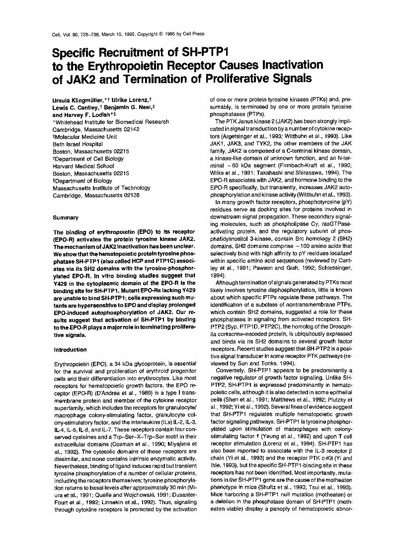
Since SH-PTP1 is expressed in hematopoietic cells, in- cluding Ba/F3, we asked whether SH-PTP1 associates with the EPO-R. The coimmunoprecipitation experiments in Figure 2 show a specific interaction between EPO-R and SH-PTP1. Ba/F3 cells expressing the wild-type EPO-R were either stimulated for 5 min with 100 U/ml EPO or were left unstimulated. Detergent lysates of these'cells were prepared and used for immunoprecipitation experi- ments. Analysis of anti-SH-PTP1 immunoprecipitates by immunoblotting with anti-PTyr and reprobing with a poly- clonal antiserum against SH-PTP1 revealed a low basal level of tyrosine phosphorylation of SH-PTP1, unchanged upon EPO addition (Figure 2, lanes 1,3, and 4). In Ba/F3 cells expressing the wild-type EPO-R and stimulated with EPO, the anti-SH-PTP1 antiserum also immunoprecipi- tated a tyrosine-phosphorylated protein, which migrated with an apparent molecular weight of 75 kDa (Figure 2, lane 4) and comigrated with the tyrosine-phosphorylated EPO-R (Figure 2, lane 6). To determine whether the 75 kDa SH-PTP 1 -associated protein was the EPO-R, anti-SH- PTP1 immunoprecipitates from lysates of cells expressing the wild-type EPO-R and stimulated with EPO were treated with 1% SDS and were heat denatured. Upon reimmuno- precipitation with anti-EPO-R antiserum, the 75 kDa tyro- sine-phosphorylated protein could be detected by anti-PTyr immunoblotting (Figure 2, lane 5). Tyrosine-phosphory- lated SH-PTP1 was not recovered in the second immuno- precipitation, indicating that the association of SH-PTP1 with the EPO-R was disrupted by the heat treatment. Sev- eral lines of evidence suggest that the 75 kDa tyrosine- phosphorylated protein complexed, after EPO addition, with SH-PTP1 is the tyrosine-phosphorylated EPO-R: it

Role of SH-PTP1 for EPO-R Signaling 731
W3" B
- - 4- 4-
p Y - E P O R
pY-SHPTP1 ~ ~
1 2 3 4
WT 1-441
rr
.EE_
.c e ~. kDa ~ ~ e ~ IP
+ + + :105 + " + + EPO
iili i~;~!~i;ii!i!i
- - 7 0 pY-EPOR-t
-~- pY-SHPTP1
5 6 7 8 9 10 11
blot: c~PTyr
pY-EPOR --~
EPOR
SHPTP1 4 , ~ - ~
blot: c~EPOR
blot: c~SH-PTP1
Figure 2. SH-PTP1 Coirnmunoprecipitates with Wild-Type and 1-441 Truncated EPO-R Control Ba/F3 cells (B) or cells expressing the wild-type (WT) or 1-441 mutant EPO-R were stimulated with 100 U of EPO/ml (plus sign) for 5 min at 37°C or were left unstimulated (minus sign). Lysates from 1 x 10⁷ cells were subjected to imrnunoprecipitation (IP) with antibodies against SH-PTP1 (~SHPTP1), EPO-R (~EPOR), or pre- immune serum (Preimrn.) from the same rabbit as the anti-EPO-R antibodies, as indicated above the panel. In lane 5, the sample was imrnunoprecipitated with an antibody against SH-PTP1, the immuno- precipitate was denatured in a solution containing SDS, and then it was reprecipitated with an antibody to the EPO-R. The im munoprecipitates were subjected to anti-PTyr immunoblot analysis (blot: aPTyr, top panel), and the filters were reprobed with an antibody to the EPO-R (blot: ~EPOR, middle panel) or to an anti-SH-PTP1 antiserum (blot: ~SH-PTP1, lower panel). The positions of tyrosine-phosphorylated forms of the wild-type EPO-R (pY-EPOR~ 1-441 EPO-R (pY-EPOR-t), and SH-PTP1 (pY-SHPTP1) are indica'e by arrows. The band with the lowest gel mobility detected by anti-EP, oR i: nmunoblotting (middle panel) is not identical with the tyrosine-phcsphorylated EPO-R as it migrates faster than the tyrosine-phospho" bated EPO-R detected by anti-PTyr immunoblotting. Furthermore, the intensity of this particular band is not enhanced upon EPO stimulation. The positions of the protein molecular weight standards are indicated in kilodaltons (kDa).
was reimmunoprecipitated with anti-EPO-R antiserum (Figure 2, lane 5); it was absent in the immunoprecipitation with preimmunserum (lane 7) and absent from the anti-SH- PTP1 immunoprecipitate from extracts of parental Ba/F3 cells (lane 3); and it was reduced in size upon deletion of the 40 C-terminal amino acids of the EPO-R (lanes 10 and 11). The tyrosine-phosphorylated EPO-R could not be detected directly by immunoblotting with our anti-EPO-R antiserum (Figure 2, compare blots of ~PTyr and aEPOR). One reason for this finding is that the sensitivity of the anti-PTyr monoclonal antibody is greater than that of our anti-EPO-R antibodies. Another is that only a small fraction of the EPO-R molecules, approximately 1000 receptors per cell, is present at the cell surface.
Thus, in response to EPO, the wild-type EPO-R be- comes tyrosine phosphorylated and forms a complex with SH-PTP1. SH-PTP1 was not detected in anti-EPO-R im- munoprecipitates (Figure 2, lane 6; data not shown), prob- ably because only a small fraction of the tyrosine- phosphorytated EPO-R associates with SH-PTP1 at any given time. These experiments did not determine whether
unphosphorylated EPO-R, before EPO addition, is com- plexed with SH-PTP1. However, the finding (below) that SH-PTP1 binding requires a specific pY in the EPO-R and is mediated by the SH2 domains of SH-PTP1 makes this unlikely, suggesting that SH-PTP1 associates with the EPO-R only after ligand-induced tyrosine phosphorylation of the receptor.
pY429 in the Cytoplasmic Domain of the EPO-R Mediates Binding of SH-PTP1 To determine which amino acid in the cytosolic domain of the EPO-R is responsible for binding SH-PTP1, we gen- erated a panel of EEPO-R deletion and tyrosine-to-phenyla- lanine point mutants, shown schematically in Figure 1A. To obtain stable cell lines, normally IL-3-dependent Ba/F3 cells were transfected with plasmids harboring the altered EPO-R cDNAs. Pools of cells were selected initially in G418 and IL-3. Each of these EPO-R mutants gave rise to comparable number of cell pools that were able to grow with EPO as the sole added g rowth factor (data not shown). Thus, none of the altered tyrosine residues nor the C-ter- minal - 100 amino acids of the EPO-R was essential for the generation of a proliferative signal in Ba/F3 cells.
Immunoprecipitation of cell lysates with anti-EPO-R anti- serum, followed by anti-PTyr immunoblotting showed that all of the mutant receptors tested in this experiment be- came tyrosine phosphorylated after EPO addition (see Fig- ure 1B, lanes 4, 6, 8, 10, and 12; compare with lane 2). EPO-induced tyrosine phosphorylation of the C-terminal deletion mutant 1-374 could not be detected (data not shown). All point mutants except Y429,431F generated a tyrosine-phosphorylated EPO-R with the same gel mobility as the wild-type EPO-R. Reproducibly, the tyrosine- phosphorylated mutant Y429,431F migrated as a diffuse species, somewhat slower than did the wild-type EPO-R (see Figure 1B, lanes 4 and 6). The altered gel mobility could be due to tyrosine phosphorylation on new sites, higher occupancy of the same sites phosphorylated in the wild-type EPO-R, or both, Of the 8 tyrosine residues in the EPO-R cytoplasmic domain, 4 are deleted in the C-ter- minal truncated mutant EPO-R 1-441. Nonetheless, this receptor also becomes tyrosine phosphorylated (see Fig- ure 1B, lanes 11 and 12). That all of the mutant receptors containing tyrosine-to-phenylalanine mutations or a dele- tion of the C-terminal 40 amino acids became tyrosine phosphorylated after EPO addition suggested either that we have not altered the target tyrosine for phosphorylation or, more likely, that the cytosolic domain of the EPO-R has multiple tyrosine-phosphorylation sites.
The finding that SH-PTP1 associates with tyrosine- phosphorylated wild-type EPO-R suggested that one or more tyrosine residues in the cytosolic domain of the EPO-R could be crucial for binding. The ability of the mu- tant EPO-Rs to form a complex with SH-PTP1 was ana- lyzed by coimmunoprecipitation. In Ba/F3 cells expressing EPO-R 1-441, a tyrosine-phosphorylated protein of ap- proximately 68 kDa (Figure 2, lane 10) coimmunoprecipi- tated with SH-PTP1. This protein represented the truncated 1-441 EPO-R because of the following: it comigrated with the tyrosine-phosphorylated EPO-R 1-441 (Figure 2, lane
Cell 732
~EPOR
kDa
c~SHPTP1 o~SHPTP1 1 st IP
0 E
o~ o 03
>- >-
p Y - E P O R - ~
EPOR- -~ -- 70 --
1 2 3 4 5 6
blot: c~PTyr
pY-EPOR--~-
EPOR -4~
blot: c~EPOR
.~- pY-EPOR
pY-SHPTP1
7 8 9 10
...... ~ - S H P T P 1
blot: c~SHPTP 1
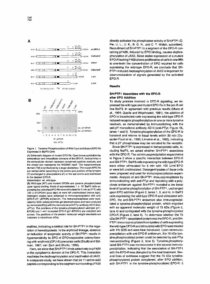
Figure 3. Mutation of Tyrosine Residues 429 and 431 to Phenylala- nine in the Cytoplasmic Domain of the EPO-R Abolishes Association with SH-PTP1 Parental Ba/F3 cells and Ba/F3 cells expressing wild-type (WT) or mutant EPO-Rs (Y429,431F and Y460,464F) were incubated with 100 U of EPO/ml for 5 rain at 37°C and were then lysed. Immunoprecip- itates (IPs) of the lysates with antibodies against EPO- R (aEPOR, lanes 1-4) or SH-PTP1 (~SHPTP1, lanes 5-8) were analyzed by anti-PTyr immunoblotting. In lanes 9 and 10, anti-SH-PTP1 immunoprecipitates of lysates of Ba/F3 cells expressing the wild-type (WT) EPO-R (as in lane 5) were boiled in 50 p_l of 1% SDS, diluted 10-fold with 1 x lysis buffer, and incubated with a mixture of EPO-R antibodies raised against N- and C-terminal EPO-R peptides (lane 9, ~EPOR) or preim- mune serum (Preimm., lane 10). These immunoprecipitates were ana- lyzed by anti-PTyr immunoblotting. The positions of the tyrosine- phosphorylated EPO-R (pY-EPOR) and tyrosine-phosphorylated SH-PTP1 (pY-SHPTP1) are marked with arrows. To show the equal efficiency of immunoprecipitation, the blots were either reprobed with anti-EPO-R antiserum (lanes 1-4) or anti-SH-PTP1 antiserum (lanes 5-10), as indicated beneath the panels. The positions of unphosphory- lated EPO-R (EPOR) and SH-PTP1 (SHPTP1) are indicated with arrows.
11); it was not precipitated from parental Ba/F3 cells or cells expressing the wild-type EPO-R (lanes 3 and 4); and it was absent in immunoprecipitates with preimmune serum (lane 8). The C-terminal 40 amino acids of the EPO-R thus are not required for interaction with SH-PTPI. This result eliminates the four tyrosine residues in the C-terminal seg- ment as essential docking sites for SH-PTPI.
Similar experiments showed that the tyrosine-phos- phorylated forms of all of the EPO-Rs with point mutations, except for Y429,431 F and Y429F, become complexed with SH-PTP1 after EPO addition (Figure 3; data not shown). Proteins from lysates of EPO-stimulated cells were immu- noprecipitated either with anti-SH-PTP1 (Figure 3, lanes 5-8) or anti-EPO-R antibodies (lanes 1-4) and were ana- lyzed by anti-PTyr immunoblotting. The mutant receptors Y429,431F and Y460, 464F were tyrosine phosphorylated
o~EPOR GST-N+C-SH2 GST-N-SH2 GST
105--
~-- pY-EPOR 70--
H~
2 3 4 5 6 7 8 9 10 11 12 13 14 15 16 17 18 19
blot: ~zPTyr
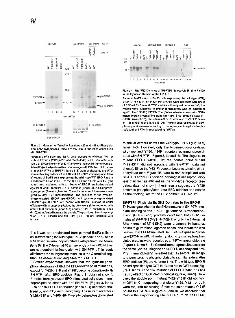
Figure 4. The SH2 Domains of SH-PTP1 Selectively Bind to PY429 in the Cytosolic Domain of the EPO-R Parental Ba/F3 cells or BedF3 cells expressing the wild-type (WT); Y429,431 F; Y431 F; or Y460,464F EPO-Rs were incubated with 100 U of EPO/ml for 3 rain at 37°C and were then lysed. In lanes 1-5, the lysates were subjected to immunoprecipitation with an antiserum against the EPO-R (~EPOR). The lysates were incubated with GST- fusion proteins containing both SH-PTP1 SH2 domains (GST-N- C-SH2, lanes 6-10), the N-terminal SH2 domain (GST-N-SH2, lanes 11-15), or GST alone (lanes 16-20). The immunoprecipitated or com- plexed proteins were analyzed by SDS-polyacrylamide gel electropho- resis and anti-PTyr immunoblotting ((~PTyr).
to similar extents as was the wild-type EPO-R (Figure 3, lanes 1-3). However, only the tyrosine-phosphorylated wild-type and Y460, 464F receptors coimmunoprecipi- tated with SH-PTP1 (Figure 3, lanes 5-8). The single point mutant EPO-R Y429F, like the double point mutant Y429,431F, did not associate with SH-PTP1 (data not shown). Since the Y431 F receptor became tyrosine phos- phorylated (see Figure 1B, lane 8) and complexed with SH-PTP1 after EPO addition, although i twas reproducibly less than half as efficient as the wild-type receptor (see below; data not shown), these results suggest that Y429 becomes phosphorylated after EPO addition and serves as the docking site for an SH2 domain in SH-PTP1.
SH-PTP1 Binds via Its SH2 Domains to the EPO-R To investigate whether the SH2 domains of SH-PTP1 me- diate binding to the EPO-R, glutathione S-transferase- fusion (GST-fusion) proteins containing both SH2 do- mains of SH-PTP1 (GST-N-C-SH2) or only the N-terminal SH2 domain (GST-N-SH2) were produced in bacteria, bound to glutathione-agarose beads, and incubated with lysates from EPO-stimulated Ba/F3 cells expressing wild- type EPO-R or EPO-R mutants. Bound tyrosine-phosphor- ylated proteins were revealed by anti-PTyr immunoblotting (Figure 4, lanes 6-15). Control immunoprecipitations from the same lysates using the anti-EPO-R antibody and anti- PTyr immunoblotting revealed that, as before, all recep- tors were tyrosine phosphorylated to a similar extent after EPO addition (Figure 4, lanes 1-4). The wild-type EPO-R bound specifically to GST-N-C, but not to GST alone (Fig- ure 4, lanes 6 and 16). Mutation of EPO-R Y461 or Y464 had no effect on GST-N-C binding (Figure 4, lane 9). How- ever, the double point mutant Y429,Y431F did not bind to GST-N-C, suggesting that either Y429, ¥431, or both were required for binding. Since the point mutant Y431F bound to GST-N-C (Figure 4, lane 8), we conclude that Y429 is the major binding site for SH-PTP1 on the EPO-R.
Role of SH-PTP1 for EPO-R Signaling 733
A
wt
Y429,431F
1 -374
B
~.PTyr ctJAK2 1400
- - ~ ~ ,~oo
1 oo0
. . . . .
E" 800
6 0 O
0' 5' 15 '30 '45 '60 ' 0' 5' 15 '30 '45 '60 ' 400
200
• Y429,431F
• , i . . . . i . . . . , . . . . J . . . . i . . . . i . . . . i . . . .
10 20 30 40 50 60 70 rain.
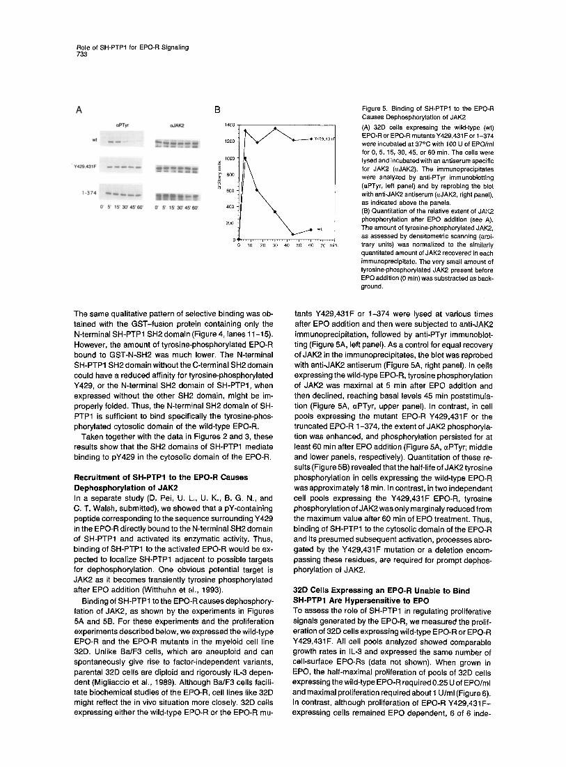
Figure 5. Binding of SH-PTP1 to the EPO-R Causes Dephosphorylation of JAK2 (A) 32D cells expressing the wild-type (wt) EPO-R or EPO-R mutants Y429,431F or 1-374 were incubated at 37°C with 100 U of EPO/ml for 0, 5, 15, 30, 45, or 60 min. The cells were lysed and incubated with an antiserum specific for JAK2 (~JAK2). The immunoprecipitates were analyzed by anti-PTyr immunoblotting 0zPTyr, left panel) and by reprobing the blot with anti-JAK2 antiserum (aJAK2, right panel), as indicated above the panels. (B) Quantitation of the relative extent of JAK2 phosphorylation after EPO addition (see A). The amount of tyrosine-phosphorylated JAK2, as assessed by densitometric scanning (arbi- trary units) was normalized to the similarly quantitated amount of JAK2 recovered in each immunoprecipitate. The very small amount of tyrosine-phosphorylated JAK2 present before EPO addition (0 rain) was substracted as back- ground.
The same qualitative pattern of selective binding was ob- tained with the GST-fusion protein containing only the N-terminal SH-PTP1 SH2 domain (Figure 4, lanes 11-15). However, the amount of tyrosine-phosphorylated EPO-R bound to GST-N-SH2 was much lower. The N-terminal SH-PTP1 SH2 domain without the C-terminal SH2 domain could have a reduced affinity for tyrosine-phosphorylated Y429, or the N-terminal SH2 domain of SH-PTP1, when expressed without the other SH2 domain, might be im- properly folded. Thus, the N-terminal SH2 domain of SH- PTP1 is sufficient to bind specifically the tyrosine-phos- phorylated cytosolic domain of the wild-type EPO-R.
Taken together with the data in Figures 2 and 3, these results show that the SH2 domains of SH-PTP1 mediate binding to pY429 in the cytosolic domain of the EPO-R.
Recruitment of SH-PTP1 to the EPO-R Causes Dephosphorylation of JAK2 In a separate study (D. Pei, U. L., U. K., B. G. N., and C. T. Walsh, submitted), we showed that a pY-containing peptide corresponding to the sequence surrounding Y429 in the EPO-R directly bound to the N-terminal SH2 domain of SH-PTP1 and activated its enzymatic activity. Thus, binding of SH-PTP1 to the activated EPO-R would be ex- pected to localize SH-PTP1 adjacent to possible targets for dephosphorylation. One obvious potential target is JAK2 as it becomes transiently tyrosine phosphorylated after EPO addition (Witthuhn et al., 1993).
Binding of SH-PTP1 to the EPO-R causes dephosphory- lation of JAK2, as shown by the experiments in Figures 5A and 5B. For these experiments and the proliferation experiments described below, we expressed the wild-type EPO-R and the EPO-R mutants in the myeloid cell line 32D. Unlike Ba/F3 cells, which are aneuploid and can spontaneously give rise to factor-independent variants, parental 32D cells are diploid and rigorously IL-3 depen- dent (Migliaccio et al., 1989). Although Ba/F3 cells facili- tate biochemical studies of the EPO-R, cell lines like 32D might reflect the in vivo situation more closely. 32D cells expressing either the wild-type EPO-R or the EPO-R mu-
tants Y429,431F or 1-374 were lysed at various times after EPO addition and then were subjected to anti-JAK2 immunoprecipitation, followed by anti-PTyr immunoblot- ting (Figure 5A, left panel). As a control for equal recovery of JAK2 in the immunoprecipitates, the blot was reprobed with anti-JAK2 antiserum (Figure 5A, right panel). In cells expressing the wild-type EPO-R, tyrosine phosphorylation of JAK2 was maximal at 5 min after EPO addition and then declined, reaching basal levels 45 min poststimula- tion (Figure 5A, ~zPTyr, upper panel). In contrast, in cell pools expressing the mutant EPO-R Y429,431F or the truncated EPO-R 1-374, the extent of JAK2 phosphoryla- tion was enhanced, and phosphorylation persisted for at least 60 min after EPO addition (Figure 5A, ~PTyr; middle and lower panels, respectively). Quantitation of these re- sults (Figure 5B) revealed that the half-life of JAK2 tyrosine phosphorylation in cells expressing the wild-type EPO-R was approximately 18 rain. In contrast, in two independent cell pools expressing the Y429,431F EPO-R, tyrosine phosphorylation of JAK2 was only marginaly reduced from the maximum value after 60 rain of EPO treatment. Thus, binding of SH-PTP1 to the cytosolic domain of the EPO-R and its presumed subsequent activation, processes abro- gated by the Y429,431F mutation or a deletion encom- passing these residues, are required for prompt dephos- phorylation of JAK2.
32D Cells Expressing an EPO-R Unable to Bind SH-PTP1 Are Hypersensitive to EPO To assess the role of SH-PTP1 in regulating proliferative signals generated by the EPO-R, we measured the prolif- eration of 32D cells expressing wild-type EPO-R or EPO-R Y429,431F. All cell pools analyzed showed comparable growth rates in IL-3 and expressed the same number of cell-surface EPO-Rs (data not shown). When grown in EPO, the half-maximal proliferation of pools of 32D cells expressing the wild-type EPO-R required 0.25 U of EPO/ml and maximal proliferation required about 1 U/ml (Figure 6). In contrast, although proliferation of EPO-R Y429,431F- expressing cells remained EPO dependent, 6 of 6 inde-

Cell 734
400000 - --0"-' EPO-R
E + EPO.R Y429,431F ~ ~ . o ; . . . . . . . . .
" / / . [ -
¢~ 200000- /
, / / ~ 1ooooo- /
o C - . . . . . . . . .
o units EPO/ml
Figure 6. EPO-Dependent Proliferation of 32D Cells Expressing the Wild-Type EPO-R and the EPO-R Mutant Y429,431F The graph shows the proliferation of I representative out of 6 indepen- dent cell pools expressing the wild-type EPO-R or the EPO-R mutant Y429, 431 F, in the presence of 0-5 U of EPO/ml measured by [3H]thym- idine incorporation. Each data point represents the mean ± SD of triplicate determinations• Abbreviation: cpm, counts per minute.
pendent cell pools expressing the mutant receptors dis- played a leftward shift in their EPO dose-response curve. Pools of 32D cells expressing the mutant EPO-R Y429,431F achieved half-maximal proliferation in as little as 0.025 U of EPO/ml, reaching a maximum rate at 0.1 U of EPO/ml (Figure 6). At the highest EPO concentrations employed, the rate of proliferation of cell pools expressing the wild-type EPO-R or the EPO-R mutant Y429,431F was the same. Thus, expression of an EPO-R unable to bind SH-PTP1 allows cells to proliferate in one-fifth to one-tenth the normal amount of EPO, indicating that the recruitment of SH-PTP1 to the EPO-R is important for down-modula- tion of intracellular signaling through the EPO-R.
Discussion
Binding of ligand to members of the cytokine receptor su- perfamily induces the tyrosine phosphorylation of several proteins, including the receptors themselves• Since phos- phorylation is transient, presumably PTPs cause protein dephosphorylation and signal termination. We have iden- tifed the first high affinity binding site for a specific PTP within the cytoplasmic domain of a cytokine receptor: SH- PTP1 specifically binds, via its SH2 domains, to a phos- phorylated peptide containing Y429 in the cytoplasmic do- main of the EPO-R. This association requires binding of EPO to the receptor, indicating that SH-PTP1 is selectively recruited to the activated signaling complex. Binding of SH-PTP1 to pY429 activates SH-PTP1 enzymatic activity (D. Pei, U. L., U. K., B. G. N., and C. T. Walsh, submitted) and causes dephosphorylation and inactivation of JAK2. Studies in which JAK2, SH-PTP1, and SH-PTP2 were ex- pressed in insect cells revealed that coexpression of SH- PTP1, but not its close relative SH-PTP2, leads to specific JAK2 dephosphorylation and inactivation of its kinase ac-
tivity (data not shown). Most importantly, activation of SH- PTP1 by binding to the EPO-R is essential for appropriate down-modulation of intracellular proliferative signals since cells expressing mutant EPO-Rs that are unable to bind SH-PTP1 (Y429,431F and 1-374) display prolonged JAK2 activation. This allows such cells to proliferate in one-fifth to one-tenth the concentration of EPO required for cells expressing the wild-type EPO-R. Experiments are in prog- ress to address whether cells expressing the single point mutant EPO-R Y429F display the same kinetics of JAK2 dephosphorylation and EPO hypersensitivity as do cells harboring the double point mutant EPO-R Y429,431F.
The binding specificity of the SH2 domains of SH-PTP1 has been an open question. In vitro binding studies using a degenerate phosphotyrosyl peptide library showed that the SH2 domains of SH-PTP1 bind to phosphopeptides with the general sequence motif pY-hydrophobic-XXX- hydrophobic, in which XXX can be any amino acid. The preferred amino acid at positions +1 and +3 in the target sequence for the SH-PTP1 SH2 domains was reported to be phenylalanine (Songyang et al., 1994). According to this prediction, upon phosphorylation, 5 of the 8 tyrosine residues in the cytosolic domain of the EPO-R (residues Y343, Y401, Y429, Y431, and Y479) might bind SH-PTPI.
Here, we measured the association of SH-PTP1 with wild-type and mutant EPO-Rs expressed in transfected Ba/F3 cells as well as the association in vitro of purified SH2 domains of SH-PTP1 with the tyrosine-phosphor- ylated cytosolic domains of wild-type and mutant EPO-Rs. Both studies showed that Y429 in the EPO-R is essential for binding to SH-PTP1. Using synthetic peptides, we showed that only pY429, in the sequence pY-Leu-Tyr- Leu-VaI-Val, is capable of binding SH-PTP1. A phospho- peptide containing the sequence corresponding to this segment of the EPO-R specifically binds the N-terminal SH2 domain of SH-PTP1 and activates its phosphatase activity (D. Pei, U. L., U. K., B. G. N., and C. T. Walsh, submitted). The binding affinity of phosphopeptides de- rived from the IL-3 receptor 13 chain and the IL-2 receptor 13 chain to the N-terminal SH2 domain of SH-PTP1 was at least 5-fold lower (D. Pei, U. L., U. K., B. G. N., and C. T. Walsh, submitted). Since the Y431F mutant EPO-R bound to the SH2 domains of SH-PTP1 about half as effi- ciently as did the wild type EPO-R, it is possible that, phos- phorylated or unphosphoryiated, Y431 participates in binding to the SH2 domains• By analogy with results of phosphotyrosyl peptide competition assays performed on SH-PTP2 (Case et al., 1994) and the recent solution of the crystal structure of the SH-PTP2 N-terminal SH2 do- main (Lee et al., 1994), it is likely that the affinity of the SH-PTP1 SH2 domains for phosphotyrosyl peptides is also influenced by the amino acid residues at the +4 and +5 positions from the target pY. We have not yet studied the role of these residues (433 and 434) in the EPO-R. Interestingly, other cytokine receptors reported to form complexes with SH-PTP1 (e.g., c-Kit and the IL-3 receptor 13 chain) contain sequnces resembeling the SH-PTP1- binding site in the EPO-R.
Identification of the SH-PTPl-binding site on the EPO-R allowed us to examine the physiological role of association
Role of SH-PTP1 for EPO-R Signaling 735
of SH-PTP1 with the EPO-R. At least one function of recep- tor-associated SH-PTP1 appears to be the dephosphoryla- tion and inactivation of JAK2. Most likely, JAK2 is a direct substrate of SH-PTP1, a contention supported by the re- sults of our insect cell reconstitution studies (data not shown). Our results thus identify the first PTK substrate of a nontransmembrane PTP. Previous studies have es- tablished that the transmembrane PTPs CD45 (Ledbetter et al., 1993) and HPTPa (den Hertog et al., 1993) dephos- phorylate the negative regulatory tyrosines of members of the Src subfamily of nontransmembrane PTKs, resulting in PTK activation. Conversely, SH-PTPl-mediated de- phosphorylation of JAK2 results in JAK2 inactivation. Moreover, Janus-type kinases are not the only non- transmembrane PTKs that are negatively regulated by SH- PTP1. Recently, we found that activation of the Src family member Lck is prolonged upon thymocyte activation in motheaten mice (U. L., K. Ravichandran, S. Burakoff, and B. G. N., unpublished data).
Binding of SH-PTP1 to other cytokine receptors proba- bly would have similar consequences. As the IL-3 receptor 13 chain has been reported to activate JAK2 (Silvennoinen et al., 1993) and to associate with SH-PTP1 (Yi et al., 1993), it is likely that signaling through this receptor is regulated in a similar fashion as is the EPO-R. Yi et al. (1993) reported that DA-3 cells with decreased SH-PTP1 protein levels, due to the expression of antisense SH- PTP1 RNA, show hyperphosphorylation of the the IL-3 receptor 13 chain and a slight increase in proliferation in response to IL-3. They suggested that the IL-3 receptor
chain is a direct SH-PTP1 target. However, in the context of our results, it is unclear whether the IL-3 receptor 13 chain hyperphosphorylation observed by these researchers was due to decreased dephosphorylation by SH-PTP1 or in- stead was a secondary consequence of prolonged JAK2 activation. It will be important to determine whether SH- PTP1 dephosphorylates one or more of these sites. Fur- thermore, it should be noted that, whereas Yi et al. (1993) studied the effect of decreasing the total level of SH-PTP1 expression within a cell, our studies focus on the specific role of receptor-bound SH-PTPI. This may account for the profound enhancement of cytokine sensitivity we have observed.
Cells expressing EPO-Rs incapable of SH-PTP1 binding manifest prolonged JAK2 activation. As JAK2 is believed to be crucial for the generation of a proliferative signal by the EPO-R (Witthuhn et al., 1993), we speculated that the extended activity of JAK2 would alter the growth properties of these cells. Indeed, such cells proliferate in EPO con- centrations 5-fold to 10-fold lower than the 0.5-1.0 U/ml of EPO normally required for proliferation of cells express- ing the wild-type EPO-R. The notion that abnormal JAK2 regulation and hyperproliferation are causally related is supported by recent studies in Drosophila. A mutation in the hopscotch locus, which encodes a JAK homolog, that generates a constitutively active JAK results in hematopoi~ etic tumors (Hanratty and Dearolf, 1993).
Our results provide a molecular explanation for the hy- persensitivity of erythroid precursor cells to EPO observed in two genetic syndromes. In mice, a central role for SH-
PTP1 in EPO-R signaling was suggested by studies of mice that either lack SH-PTP1 (motheaten) or are impaired in SH-PTP1 phosphatase activity (motheaten viable). The CFU-E erythroid progenitors from these mice are stimu- lated by lower than normal concentrations of EPO, and there are increased numbers of splenic CFU-Es in vivo (van Zant and Shultz, 1989). A possible explanation for these observations is that occupancy by EPO of fewer than normal cell surface EPO-Rs is sufficient to generate a proliferative signal and that SH-PTP1 is an important down-modulator of EPO-R signaling. However, it was also conceivable that the enhanced sensitivity of motheaten CFU-Es to EPO results from dysregulation of another sig- nal transduction pathway that served to lower the thresh- old for EPO-R stimulation. Our results clearly suggest that SH-PTP1 is a direct regulator of EPO-R signaling.
Members of a large Finnish family with autosomal domi- nant benign erythrocytosis have a mutation in one allele of the EPO-R, which introduces a premature stop codon and generates an EPO-R lacking the C-terminal 70 amino acids (de la Chapelle et al., 1993). This deleted segment includes the binding site for SH-PTP1 that we have defined in the murine EPO-R. The only pathophysiologic manifes- tation of this trait is a high hematocrit and a corresponding increase in the concentration of hemoglobin in the blood. Similar to the erythroid progenitors from motheaten mice, cultured erythroid progenitors from these patients are stimulated by lower than normal concentrations of EPO. The inability of SH-PTP1 to bind to the truncated EPO-R or to be activated could account for the enhanced respon- siveness of these cells to EPO. Therefore, it will be of great interest to address whether polycythemia syndromes are also caused by an alteration in the EPO-R or by an impair- ment of SH-PTP1 or its regulation.
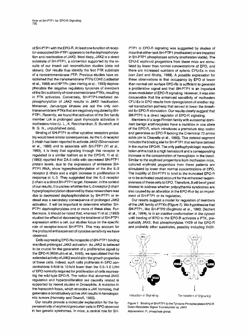
Our results suggest a model for regulation of members of the JAK family of PTKs (Figure 7). We hypothesize that SH-PTP1, like SH-PTP2 (Sugimoto et al., 1993; Dechert et al., 1994), is in an inactive conformation in the cytosol until binding of EPO to the EPO-R activates a PTK, pre- sumably JAK2, that phosphorylates Y429 of the EPO-R and probably other substrates, possibly including Y431.
Induction of Signaling Termination of Signaling
Figure 7. BindingofSH-PTP1 totheTyrosine-Phosphorylated EPO-R Down-Modulates Signal Transduction by JAK2 Abbreviation: P, phosphate group.
Cell 736
E n g a g e m e n t o f the SH-PTP1 S H 2 doma in (s ) by the EPO-R
both t r ans loca tes the p h o s p h a t a s e to the recep to r -
s igna l ing c o m p l e x and ac t i va tes the p h o s p h a t a s e act iv i ty
in c lose p rox im i t y to poss ib le subs t ra tes ; o n e such sub-
s t ra te is JAK2. T h e t ime lag b e t w e e n assoc ia t i on of SH-
PTP1 wi th the EPO-R and re turn o f JAK2 phospho ry l a t i on
to basa~l leve ls cou ld be c a u s e d by act iva t ion or inhib i t ion of
SH-PTP1 act iv i ty by o the r d o w n s t r e a m s igna l - t r ansduc ing
mo lecu les . D e p h o s p h o r y l a t i o n o f JAK2 l eads to te rm ina -
t ion o f its s igna l ing capab i l i t y and he lps set the no rma l
ce l lu la r level o f EPO sensi t iv i ty . S ince a pY-con ta in ing
pep t ide de r i ved f rom res idue Y 4 2 9 o f the EPO-R spec i f i -
ca l ly ac t i va tes SH-PTP1 and s ince o v e r e x p r e s s i o n of SH-
PTP1 r e p r e s s e s g rowth of a mye lo id cel l l ine, t he rapeu t i c
agen ts m im ick ing the act iv i ty med ia ted by th is pY pep t ide
cou ld be of t he rapeu t i c impor tance .
Experimental Procedures
Plasmids and Mutagenesis To convert tyrosine residues to phenylalanines in the cytosolic domain of the EPO-R, point mutations were generated by overlap extension using the PCR with synthetic oligonucleotides encoding the desired amino acid substitutions (Higuchi et al., 1988). The C-terminal trun- cated EPO-R 1-374 was created by introducing an oligonucleotide that contained a stop codon into the Hindlll site at nucleotide position 1221 in the EPO-R cDNA. Mutant EPO-R cDNAs were identified by sequencing using the dideoxynucleotide chain termination method and were subcloned into the eukaryotic expression vector pXM- EPO-R (D'Andrea et al., 1989; Longmore and Lodish, 1991) by ligating the ApaI-EcoRI fragment of the mutant EPO-R cDNAs into Apal- and EcoRI-cut pXM-EPO-R DNA. The construction of the C-terminal trun- cated EPO-R mutant (1-441) was described previously (Yoshimura et al., 1990b).
Cell Culture and Transfections Wild-type or mutant EPO-R cDNAs in pXM were cotransfected with the selectable marker pRclCMV(neo) (Invitrogen) into the IL-3-depen- dent pro-B cell line Ba/F3 or into the myeloid cell line 32D by electro- potation, and stable cell lines were selected in G418 as described (Sk0da et al., 1993). To identify pools of transfected cells expressing an EPO-R, and thus competent to grow in the presence of EPO, neomy- cin-resistant colonies were transferred to medium containing 0.3 U of EPO/ml (Arris Pharmaceuticals) as the sole growth factor. EPO-respon- sive cell pools were further characterized by immunoprecipitation and immunoblotting with anti-EPO-R antiserum. Cell pools that showed comparable surface expression of the wild-type or mutant EPO-Rs were identified by saturation binding of l~l-labeled EPO at 4°C (Hilton et al., 1988). IL-3-dependent and EPO-dependent cells were maintained as described previously (Yoshimura et al., 1990a).
Immunoprecipitations and Immunoblotting Antibodies directed against the EPO-R were either raised against two peptides, corresponding to the N- and the C-termini of the EPO-R (an!i-N-EPO-R and anti-C-EPO-R; Yoshimura et al., 1990a), or against a GST-fusion protein containing the EPO-R extracellular domain (anti- EPO-R). The anti-EPO-R antiserum was affinity purified using the ex- tracellular domain of the EPO-R as a fusion protein, with the maltose- binding protein coupled to CN Br-activated Sepharose 4B (Pharmacia).
Populations of Ba/F3 cells expressing wild-type or mutant EPO-Rs were washed with RPMI medium and were incubated at a density of 5 x 10⁷ cells/ml with 100 U of EPO/ml for the indicated times. Extracts were directly prepared by the addition of an equal volume of 2 x lysis buffer (2°/0 NP-40, 20% glycerol, 300 mM NaCI, 100 mM Tris-HCI [pH 7.4], 100 mM NaF, 2 mM ZnCI2, 2 mM Na3VO4, 10 p.g/ml leupeptin, and 1 mM AEBSF [ICN Biomedicals]). For immunoprecipitations, the cell extracts (250 p.I) were incubated with 2 pg of affinity-purified poly- clonal rabbit antibodies against human SH-PTP1 (Lorenz et al., 1994), 1 pl of JAK2 antiserum (Upstate Biotechnology), or 3 p.g of affinity- purified antibodies against the EPO-R, as indicated. Immune corn-
plexes were recovered by binding to protein A-Sepharose beads (Boehringer Mannheim). Protein A-bound immunoprecipitates were washed three times with 1 x lysis buffer and once with TNE (50 mM Tris-HCI [pH 8.0], 150 mM NaCI, 1 mM Na3VO4, 1 mM ZnCI2, 1 mM EDTA), and they were eluted by boiling in SDS sample buffer. Samples were separated on 10% Iow-bis SDS-polyacrylamide gels, transferred to nitrocellulose membranes (Schleicher & SchQII), and incubated with the indicated antiserum in 150 mM NaCI, 10 mM Tris-HCI (pH 8.0), 1% BSA, and 0.2% Tween-20 for 2 hr at room temperature. Antibodies used for immunoblotting were a polyclonal rabbit antiserum against SH-PTP1 (1 to 1000) (Lorenz et al., 1994), affinity-purified anti-EPO-R antiserum (1 to 5000), anti-C-EPO-R antiserum (1 to 1000), anti-JAK2 antiserum (1 to 1000), and the monoclonal anti-PTyr antibody 4G10 (anti-pTyr; 1 pg/ml). Bound antibodies were detected by incubation with horseradish peroxidase-coupled secondary antibodies (anti- rabbit or anti-mouse, as appropriate) (Amersham) and the enhanced chemiluminescence system (DuPont-NEN), as detailed by the manu- facturer.
To reprobe immunoblots, filters were incubated in 62.5 mM Tris (pH 6.8) plus 0.1 M I~-mercaptoethanol plus 20/0 SDS for 30 rain at 65°C and then were washed extensively in 10 mM Tris (pH 8) plus 150 mM NaCI.
GST-Fusion Proteins The N- and C-terminal SH2 domains of murine SH-PTP1 were cloned into the EcoRI site of the bacterial expression vector pGEX-2T (Phar- macia). For the N-terminal SH2 domain as well as for both of the SH2 domains together (N-C), the cDNA fragments corresponding to amino acids 1-121 and 1-220, respectively, were amplified by PCR using Vent DNA polymerase (New England Biolabs). The amplified DNA fragments were sequenced by fluorescent dye technology on a 373A DNA sequencer (Applied Biosystems). E. coil transformants express- ing the GST-SH-PTP1 fusion proteins were induced with 0.5 mM IPTG (Sigma) at 30°C for 12 hr, harvested by centrifugation, and lysed as described previously (Songyang et al., 1993). Cells were stimulated with 100 U of EPO/ml for 5 rain at 37°C prior to detergent lysis. GST- fusion proteins bound to glutathione-agarose beads (approximately 1 p.g of fusion protein per binding reaction) were incubated for 3 hr at 4°C with 250 pl of lysate, equivalent to 1 x 10⁷ cells, in 1 x lysis buffer. After washing the beads several times with lysis buffer, ad- sorbed proteins were eluted and analyzed as described above for immunoprecipitates.
Proliferation Assay Proliferation assays were performed in 96-well plates with 200 pl/well. Cells, at a concentration of 2 x 10⁵ 32D cells/ml, were seeded in the presence of indicated amounts of cytokines (EPO, 0-5 U/ml; IL-3, 0-100 U/ml). After 48 hr, 1 pCi of [3H]thymidine/well (DuPont-NEN) was added for another 3 hr. The labeled cells were then harvested by a 96-well plate harvester (Scatron Instruments), and the amount of radioactively labeled DNA was quantitated in a scintillation counter.
Acknowledgments
Correspondence for this work should be addressed to H. F. L. This work was supported by grant HL32262 from the National Institutes of Health, a grant from the Arris Pharmaceutical Corporation (H. F. L.), and a grant from the Lucille P. Markey Charitable Trust (L. C. C.). B. G. N. is the recipient of a Junior Faculty Research Award from the American Cancer Society and the Gertrude Elion Award from the American Association of Cancer Research, funded by the Burroughs- We,come Corporationis, and he is supported by funds from the Har- vard Medical School/F. Hoffmann-LaRoche Institute for Chemistry and Medicine. U. K. was supported by a fellowship from the Deutsche Forschungsgemeinschaft, and U. L. is supported by a fellowship from the American Cancer Society, Massachusetts Division. We thank Drs. F. U. Reuss, K. Luo, and S. Michnick for many helpful discussions and for critically reading the manuscript. We also thank Dr. K. Ravi- chandran for the help with the proliferation assays and Dr. B. Druker for the generous gifts of the anti-PTyr monoclonal antibody 4G10.
Received October 5, 1994; revised December 16, 1994.
Role of SH-PTP1 for EPO-R Signaling 737
References
Argetsinger, L. S., Campbell, G. S., Yang, X., Witthuhn, B. A., Silven- noinen, O., Ihle, J. N., and Carter-Su, C. (1993). Identification of JAK2 as a growth hormone receptor-associated tyrosine kinase. Cell 74, 237-244. Cantley, L. C., Auger, K. R., Carpenter, C., Duckworth, B., Graziani, A., Kapeller, R., and Soltoff, S. (1991). Oncogenes and signal transduc- tion. Cell 64, 281-302. Case, R. D., Piccione, E., Wolf, G., Bennett, A. M., Lechleider, R. J., Chauduri, M., Neel, B. G., and Shoelson, S. E. (1994). SH-PTP2 SH2 domain binding specificity is defined by direct interactions with PDGF receptor, EGF receptor, and IRS-1 derived phosphopeptides. J. Biol. Chem. 269, 10467-10474. Cosman, D., Lyman, S. D, Idzerda, R. L., Beckmann, M. P., Park, L. S., Goodwin, R. G., and March, C. J. (1990). A new cytokine receptor superfamily. Trends Biochem. Sci. 15, 265-270. D'Andrea, A. D., Lodish, H. F., and Wong, G. G. (1989). Expression cloning of the murine erythropoietin receptor. Cell 57, 277-285. de la Chapelle, A., Traskelin, A. L., and Juvonen, E. (1993). Truncated erythropoietin receptor causes dominantly inherited benign human erythrocytosis. Proc. Natl. Acad. Sci. USA 90, 4495-4499. Dechert, U., Adam, M., Harder, K. W., Clark-Lewis, I., and Jirik, F. (1994). Characterization of protein tyrosine phosphatase SH-PTP2. J. Biol. Chem. 269, 5602-5611. den Hertog, J., Pals, C. E., Peppelenbosch, M. P., Tertoolen, L. G., de Laat, S. W., and Kruijer, W. (1993). Receptor protein tyrosine phos- phatase alpha activates pp60c-src and is involved in neuronal differen- tiation. EMBO J. 12, 3789-3798. Dusanter-Fourt, I., Casadevall, N., Lacombe, C., Muller, O., Billat, C., Fischer, S., and Mayeux, P. (1992). Erythropoietin induces the tyrosine phosphorylation of its own receptor in human erythropoietin- responsive cells. J. Biol. Chem. 267, 10670-10675. Firmbach-Kraft, I., Byers, M., Shows, T., DaPla-Favera, R., and Krolew- ski, J. J. (1990). Tyk2, prototype of a novel class of non-receptor tyro- sine kinase genes. Oncogene 5, .1329-1352. Hanratty, W. P., and Dearolf, C. R. (1993). The Drosophila Tumorous- lethal hematopoietic oncogene is a dominant mutation in the hop- scotch locus. Mol. Gen. Genet. 238, 33-37. Higuchi, R., Krummel, B., and Saiki, R. K. (1988). A general method of in vitro preparation and specific mutagenesis of DNA fragments: study of protein and DNA interactions. Nucl. Acids Res. 16, 7351- 7367. Hilton, D. J., Nicola, N. A., and Metcalf, D. (1988). Specific binding of murine leukemia inhibitory factor to normal and leukemic monocytic cells. Proc. Natl. Acad. Sci. USA 85, 5971-5975. Ledbetter, J. A., Deans, J. P., Aruffo, A., Grosmaire, L. S., Kanner, S. B., Bolen, J. B., and Schieven, G, L. (~993). CD4, CD8 and the role of CD45 in T-cell activation. Curr. Opin. Immunol. 5, 334-340. Lee, C.-H., Kominos, D., Jacques, S., Margolis, B., Schlessinger, J., Shoelson, S. E., and Kuriyan, J. (1994). Crystal structures of peptide complexes of the amino-terminal SH2 domain of the Syp tyrosine phos- phatase. Structure 2, 423-438. Linnekin, D., Evans, G. A., D'Andrea, A., and Farrar, W. L. (1992). Association of the erythropoietin receptor with protein tyrosine kinase activity. Proc. Natl. Acad. Sci. USA 89, 6237-6241. Longmore, G. D., and Lodish, H. F. (1991). An activating mutation in the murine erythropoietin receptor induces erythroleukemia in mice: a cytokine receptor superfamily oncogene. Cell 67, 1089-1102.
Lorenz, U., Ravichandran, K. S., Pei, D., Walsh, C. T., Burakoff, S. J., and Neel, B. G. (1994). Lck-dependent tyrosyl phosphorylation of the phosphotyosine phosphatase SH-PTP1 in murine T cells. Mol. Cell. Biol. 14, 1824-1834.
Matthews, R. J., Bowne, D. B., Flores, E., and Thomas, M. L. (1992). Characterization of hematopoietic intracellular protein tyrosine phos- phatases: description of a phosphatase containing an SH2 domain and another enriched in proline-, glutamic acid-, serine-, and threonine-rich sequences. Mol. Cell. Biol. 12, 2396-2405. Migliaccio, G., Migliaccio, A. R., Kreider, B. L., Rovera, G., and Adam-
son, J. W. (1989). Selection of lineage-restricted cell lines immortalized at different stages of hematopoietic differentiation from the murine cell line 32D. J. Cell Biol. 109, 833-841. Miura, O., D'Andrea, A., Kabat, D., and Ihle, J. N. (1991). Induction of tyrosine phosphorylation by the erythropoietin receptor correlates with mitogenesis. Mol. Cell. Biol. 11, 4895-902. Miyajima, A., Kitamura, T., Harada, N., Yokota, T., and Arai, K. (1992). Cytokine receptors and signal transduction. Annu. Rev. Immunol. 10, 295-331. Pawson, T., and Gish, G. D. (1992). SH2 and SH3 domains: from structure to function. Cell 71,359-362.
Plutzky, J., Neel, B. G., and Rosenberg, R. (1992). Isolation of a novel SRC homology 2 (SH2) containing tyrosine phosphatase. Proc. Natl. Acad. Sci. USA 89, 1123-1127. Quelle, F. W., and Wojchowski, D. M. (1991). Proliferative action of erythropoietin is associated with rapid protein tyrosine phosphorylation in responsive B6SUt.EP cells. J. Biol. Chem. 266, 609-614. Schlessinger, J. (1994). SH2/SH3 signaling proteins. Curr. Opin. Gen. Dev. 4, 24-30.
Shen, S.-H., Bastien, L., Posner, B. I., and Chr~tien, P. (1991). A protein-tyrosine phosphatase with sequence similarity to the SH2 do- main of the protein-tyrosine kinases. Nature 352, 736-739. Shultz, L. D., and Sidman, C. L. (1987). Genetically determined murine models of immunodeficiency. Annu. Rev. Immunol. 5, 367-403. Shultz, L. D., Schweitzer, P. A., Rajah, T. V., Yi, T., Ihle, J. N., Mat- thews, R. J., Thomas, M. L., and Beier, D. R. (1993). Mutations at the murine motheaten locus are within the hematopoietic cell protein- tyrosine phosphatase (Hcph) gene. Cell 73, 1445-1454.
Silvennoinen, O., Witthuhn, B. A., QueUe, F. W., Cleveland, J. L, Yi, T., and Ihle, J. N. (1993). Structure of the murine Jak2 protein-tyrosine kinase and its role in interleukin-3 signal transduction. Proc. Natl. Acad. Sci. USA 90, 8429-8433. Skoda, R. C., Seldin, D. C., Chiang, M. K., Peichel, C. L., Vogt, T. F., and Leder, P. (1993). Murine c-mpl: a member of the hematopoietic growth factor receptor superfamily that transduces a proliferative sig- nal. EMBO J. 12, 2645-2653.
Songyang, Z., Shoelson, S. E., Chaudhuri, M., Gish, G., Pawson, T., Haser, W. G., King, F., Roberts, T., Ratnofsky, S., Lechleider, R. J., Neel, B. G., Birge, R. B., Fajardo, J. E., Chou, M. M., Hanafusa, H., Schaffhausen, B., and Cantley, L. C. (1993). SH2 domains recognize specific phosphopeptide sequences. Cell 72, 767-778. Songyang, Z., Shoelson, S. E., McGlade, J., Olivier, P., Pawson, T., Bustelo, X. R., Barbacid, M., Sabe, H., Hanafusa, H., Yi, T., Ren, R., Baltimore, D., Ratnofsky, S., Feldman, R. A., and Cantley, L C. (1994). Specific motifs recognized by the SH2 domains of Csk, 3BP2, fps/fes, GRB-2, HCP, SHC, Syk, and Vav. Mol. Cell. Biol. 14, 2777-2785. Sugimoto, S., Wandless, T. J., Shoelson, S. E., Neel, B. G., and Walsh, C. T. (1993). Activation of the SH2-containing protein tyrosine phos- phatase, SH-PTP2, by phosphotyrosine-containing peptides derived from insulin receptor substrate-l. J. Biol. Chem. 268, 2733-2736.
Sun, H., and Tcnks, N. K. (1994) The coordinated action of protein tyrosine phosphatases and kinases in cell signaling. Trends Biochem. Sci. 19, 480-485.
Takahashi, T., and Shirasawa, T. (1994). Molecular cloning of rat JAK3, a novel member of the JAK family of protein tyrosine kinases. FEBS Lett. 342, 124-128.
Tsui, H. W., Siminovitch, K. A., de Souza, L., and Tsui, F. W. L. (1993). Motheaten and viable motheaten mice have mutations in the haemato- poietic cell phosphatase gene. Nature Genet. 4, 124-129.
van Zant, G., and Shultz, L. (1989). Hematologic abnormalities of the immunodeficient mouse mutant, viable motheaten (rhea). Exp. Hema- tol. 17, 81-87.
Wilks, A. F., Harpur, A. G., Kurban, R. R., Ralph, S. J., Zurcher, G., and Ziemiecki, A. (1991). Two novel protein-tyrosine kinases, each with a second phosphotransferase-related catalytic domain, define a new class of protein kinase. Mol. Cell. Biol. 11, 2057-2065.
Witthuhn, B. A., Quelle, F. W., Silvennoinen, O., Yi, T , Tang, B., Miura, O., and ihle, J. N. (1993). JAK2 associates with the erythropoietin receptor and is tyrosine phosphorylated and activated following stimu-

Cell 738
lation with erythropoietin. Cell 74, 227-236.
Yeung, Y., Berg, K. Lo, Pixley, F. J., Angeletti, R. H., and Stanley, E. R. (1992). Protein tyrosine phosphatase-lC is rapidly phosphorylated on tyrosine in macrophages in response to colony stimulating factor-l. J. Biol. Chem. 267, 23447-23450.
Yi, T., and I hle, J. N. (1993). Association of hematopoietic cell phospha- tase with c-kit after stimulation with c-kit ligand. Mol. Cell. Biol. 13, 3350-3358.
Yi, T., Cleveland, J. L., and Ihle, J. N. (1992). Protein tymsine phospha- tase containing SH2 domains: characterization, preferential expres- sion in hematopoietic cells, and localization to human chromosome 12p12-13. Mol. Cell. Biol. 12, 836-846. Yi, T., Mui, A. L.-F., Krystal, G., and Ihle, J. N. (1993). Hematopoietic cell phosphatase associates with the interleukin-2 (IL-3) receptor 13-chain and down-regulates I L-3-induced tyrosine phosphorylation and mitogenesis. Mol. Cell. Biol. 13, 7577-7586.
Yoshimura, A., D'Andrea, A. D., and Lodish, H. F. (1990a). Friend spleen focus-forming virus glycoprotein gp55 interacts with the eryth- ropoietin receptor in the endoplasmic reticulum and affects receptor metabolism. Proc. Natl. Acad. Sci. USA 87, 4139-43. Yoshimura, A., Longmore, G., and Lodish, H. F. (1990b). Point muta- tion in the exoplasmic domain of the erythropoietin receptor resulting in hormone-independent activation and tumorigenicity. Nature 348, 647-649.