Embed Size (px)
Citation preview
Eur. J. Biochem. 117, 41 -55 (1981) c) FEBS 1981
Specificity of Twelve Lectins Towards Oligosaccharides and Glycopeptides Related to N-Glycosylproteins
Henri DEBRAY, Dominique DECOUT, Gerard STRECKER, Genevikve SPIK, and Jean MONTREUiL
Laboratoire de Chimie Biologique et Laboratoire Associe au Centre National de la Recherche Scientifique nL 217, Universite des Sciences et Techniques de Lille, Villeneuve d’Ascq and Institut de Recherche sur le Cancer (Institut Jules Driessens) et Unite n‘ 124 de I’Institut National de la Sante et de la Recherche Medicale, Lillc
(Received July 2/November 28, 1980)
Glycopeptides and oligosaccharides of either the N-acetyllactosaminic or the oligomannosidic type derived from glycoproteins containing the N-glycosylamine linkage were used to define the specificity of different lectins (concanavalin A, Lens culinaris agglutinin, Vicia faba agglutinin, Pisum sativum agglutinin, Ricinus communis agglutinins, soybean agglutinin, wheat germ agglutinin, Solanum tuherosum agglutinin, Datum stramonium agglutinin, Lotus tetragonolobus agglutinin, Ulex europeus agglutinin) by studying the inhibition of human red blood cell agglutination by these structures. The results obtained show that lectins considered ‘identical’ in terms of monosaccharide specificity, possess the ability to recognize fine differences in more complex structures. In fact, different lectins are able to recognize different saccharidic sequences on the same glycan structure. As these sequences are likely to be common to numerous glycoproteins, including cell membrane glycoproteins, the results obtained with lectins in the study of cell surface carbohydrates have to be very carefully interpreted.
Moreover, our results confirm previous data on the spatial configuration of the glycan moiety of glyco- proteins deduced from the construction of molecular models : the fact that oligosaccharides bearing an a-NeuAc- (2 --f 6)-Gal unit are more powerful inhibitors than oligosaccharides bearing an a-NeuAc-(2 +3)-Gal unit could be related to the high rotational freedom of a-2,6 linkage; the observation that glycoasparagines, glycopeptides and glycoproteins possess a higher affinity for lectins than the related oligosaccharides could be explained by the fact that the glycan-amino acid linkage leads to structures more rigid than those of the oligosaccharides themselves.
Lectins are now widely used as tools in the study of plasma membrane modifications in neoplasia. Numerous lectins have been purified and their properties well defined (for re- views see [1,2]). Lectins are able to bind sugars specifically, but it is well known that the monosaccharide residue in a terminal non-reducing position on a glycan is not the only carbohydrate moiety recognized. Indeed, Kornfeld et al. [3] have shown that the binding constant of the specific free sugar with a lectin may be several orders of magnitude lower than the binding constant of a glycoconjugate containing this sugar. Moreover, in the particular case of concanavalin A, the authors demonstrated that the most active part of a glycan structure of the N-acetyllactosaminic type are not the cc-man- nose residues in a terminal position but the disaccharide N-acetyl-fl-D-ghcosaminyl-( 1 --+ 2)-a-~-mannoside.
In the present study, we have used numerous oligo- saccharides and glycopeptides derived from N-glycosylpro- teins to define the specificities to twelve lectins (Table l), by determining their ability to inhibit the agglutination of human red blood cells induced by these lectins. These results have been preliminarily reported in [4,5].
Abbreviations. NaC1/Pi, 0.01 M sodium phosphate, 0.14 M sodium chloride pH 7.2; Gal, D-galactose; NeuAc, N-acetylneuraniinic acid; GlcNAc, N-acetyl-D-glucosamine; GalNAc, N-acetyl-D-galactosamine; Man, D-mannose; Fuc, L-fucose; Asn, L-asparagine.
Enzymes. fl-D-Galactosidase (EC 3.2.1.23); N-acetyl-fl-D-glucosami- nidase (EC 3.2.1.30); a-L-fucosidase (EC 3.2.1.51); neuraminidase (EC 3.2.1.18).
Note. All sugars are of the D configuration unless otherwise stated.
MATERIALS AND METHODS
Materials
All the lectins used in this study (Table l), except con- canavalin A which was purchased from Pharmindustrie (Clichy, France), were purified by us by affinity chromatog-
Table 1. List of’ lectins used in this study and classified according to Goldstein and Hayes (21
Specificity Lectin
a-Mannose or cc-glucose (Jack bean lectin)
Concanavalin A from Canavaliu ensiformis
Lens culinaris agglutinin Vicia faba agglutinin Pisum sativum agglutinin
b-Galactose Ricinus communis agglutinin I and N-acetyl-fl- galactosamine
N - Acetyl-fl- glucosamine
Ricinus communis agglutinin I1 Soybean agglutinin (Glycin ma.x agglutinin)
Wheat germ agglutinin (Triticum vulgare agglutinin) Solanurn tuberosum agglutinin (potato agglutinin) Dalura strumonium agglutinin
cc-L-Fucose Lotus tetragonolobus agglutinin U k x europeus agglutinin
42
Origin
Human milk
Human milk
Mannosidosis
S i a I i d o s i s
GM, - Gangliosidos variant 0 (Sandhofps disease)
Fucosidosis
Name
Veuraminyl (a2+3) lactose
Neuraminyl (a2-6 j lactose
Mannoside 1 Mannoside 2 Mannoside 3 Sialoside I
Sialoside 2 Sialoside 3
_____
Sialoside 4
Sialoside S
Sialoside 6
Sialoside 9
Oligosaccharide S-6a
Oiigosaccharide S-5b
Fucoside I Fucoside 2
Glycopeptide I Glycopeptide II
Glycopeptide 11
bbreviation
NL a-2,3
NL a-2,6
M-l M-2 M-3
N-l N-2
N-3
N -4
N-5
N -6
N-9
S-6a
S-5b
F-l F-2
GP-F-1 GP-F-2
GP-F-3
Structure
r-NeuAc-(2-3)-8-Gal-( 1+4)-61c
1-NeuAc-(2+6)-P-Gal-(l-4J-~lc
1-Man-(l+3)-~-Man-(1+4j-GlcNAc z-Man-( 1+2)-a-Man-( 1-3)-P-Man-(1+4)-GlcNAc %-Man-( 1 -&a -Man-( lO-a-Man-( 1+3J-P-Man-( I+4j-GlcNAc
s-NeuAc-(2+3)-P-Gal-( 1-4)-~-GIcNAc-(~-’2)-c-~.~dn-( 1+3)-P-Maii-(1-4)-ClcNAc i-Neu Ac-( 2+6)-P-Gal-( 1 +-P-GlcNAc-( 1 +2)-a--Man-( 1 +3)-6-Man-! 1 +4)-GlcNAc
i-NeuAc-(2+6)-P-Gal-( 1+4)-P-GlcNAc-( 1+2)-a-Man-i I+3) ‘P-Man-(l+4)-GlcNAc
a-Man-(1-6) / z-NeuAc-(2+3)-P-Gal-( 1+4)-pJcNAc-( 1+2)-a-Man-!1+3)
‘@-Man -(1+4)-GlcNAc p-Gal-i 1 +-4)-P-GlcNAc-( 1 +2) -a -. Man-(1 +6)/
i-NeuAc- (2+6)-p -Gal-( l+4)-O-GlcNAc-( 1+2)-a -Man-( 1+3) \@-Man-(1+4)-GlcNAc
P-Gal-( 1+4)-p-GlcNAc-( l-Z)-a-Man-( 1+6) / z-Neu Ac-( 2+6)-P-Gal-( 1 +4)-P-GlcNAc-( 1 +2)-a-?.fan-( 1-31
)@-Man-( 1+4)-GlcNAc
P-GlcNAc-( 1+2)-a-Man-(1-6)/ a-NeuAc-(2+6)--P-Gal-( 1 -4)-P-GlcNAc-(1-2)-a-?.fan-( 1’3)
)~-“l.n-~l-4~-GlcNAc a-NeuAc-( 2+6)-P-Gal-( 1 +4)-fi--GlcNAc -( 1 +2)-a-Man -( I +6)
B-GlcNAc-(I+2)-a-?.~an-( 1-3) \&Man-(1-+4)-GlcNAc
p-GlcNAc-( 1+2)-a-Man-( 1+6) / \
/
8-GlcNAc-( 1+2)-c-Man-( 1+3j
9-GlcNAc-( 1+2)-c-Man-( 1+6)
3-Gal-( 1+4)-P-GlcNAc-( 1*2)-c-Man-( 1+3)-&Man-( 1+4)-GlcNAc‘
P-GlcNAc-( I -4)-P-Man-(l+4)-GlcNAc
a--Fuc-( 1+6)-GlcNAc
I1 ,3 a-Fuc
a-Fuc-( 1+6)-p-GIcNAc-(l+ )-.<sn a-Man-( 1 -h)-P-Man-( 1 +4)-P-GlcNAc-( I +4)-&GlcNAc- ( 1 + )-Am
116 a--Fuc
‘p-Man-( 1’4)-P-GlcNAc-(1+4)-P- GlcNAc-(I+ k A s n
/ /:,6 R-FW
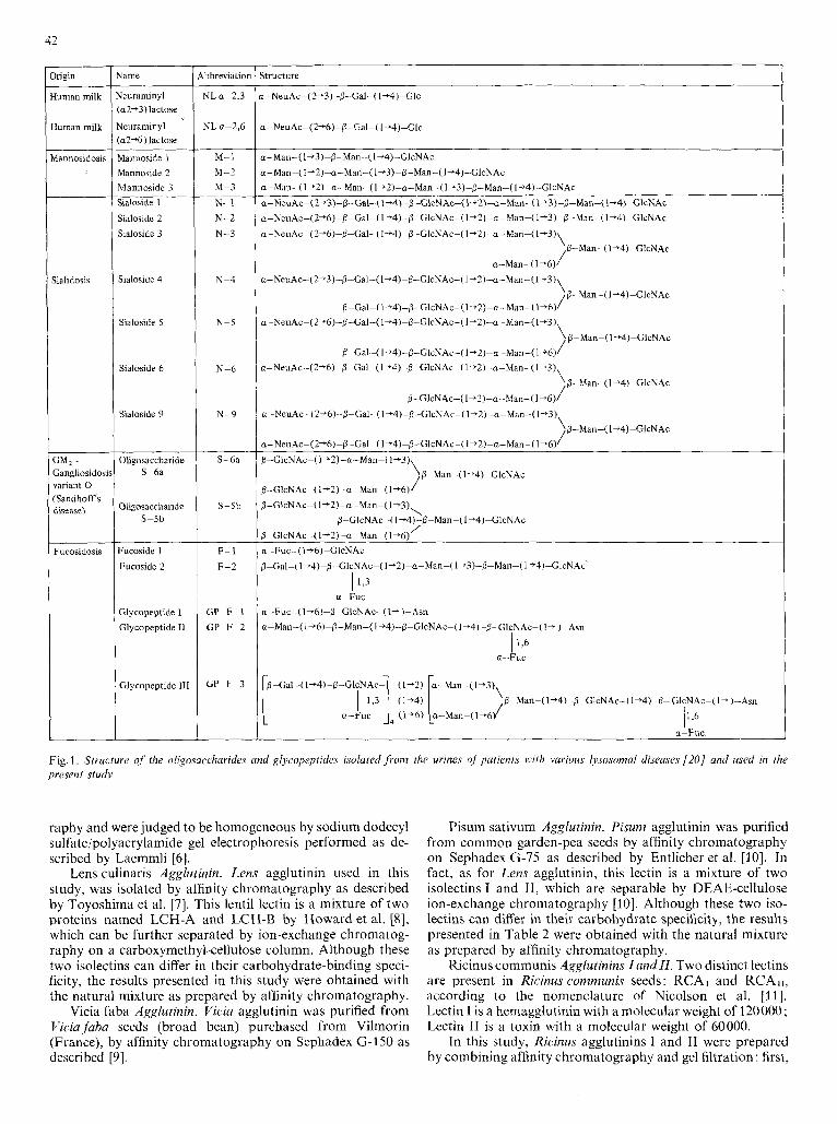
Fig. 1. Structure of the oligosaccharides and glycopeptides isolated from the urines of patients with various lysosomal diseases [ZO] and used in the present study
raphy and were judged to be homogeneous by sodium dodecyl sulfate/polyacrylamide gel electrophoresis performed as de- scribed by Laemmli [6].
Lens culinaris Agglutinin. Lens agglutinin used in this study, was isolated by affinity chromatography as described by Toyoshima et al. 171. This lentil lectin is a mixture of two proteins named LCH-A and LCH-B by Howard et al. [8], which can be further separated by ion-exchange chromatog- raphy on a carboxymethyl-cellulose column. Although these two isolectins can differ in their carbohydrate-binding speci- ficity, the results presented in this study were obtained with the natural mixture as prepared by affinity chromatography.
Vicia faba Agglutinin. Viciu agglutinin was purified from Viczufaba seeds (broad bean) purchased from Vilmorin (France), by affinity chromatography on Sephadex G-I 50 as described [9].
Pisum sativum Agglutinin. Pisunz agglutinin was purified from common garden-pea seeds by affinity chromatography on Sephadex G-75 as described by Entlicher et al. [lo]. In fact, as for Lens agglutinin, this lectin is a mixture of two isolectins I and 11, which are separable by DEAE-cellulose ion-exchange chromatography [lo]. Although these two iso- lectins can differ in their carbohydrate specificity, the results presented in Table 2 were obtained with the natural mixture as prepared by affinity chromatography.
Ricinus communis Agglutinins Zand 11. Two distinct lectins are present in Ricinuscommunis seeds: RCAl and RCAII, according to the nomenclature of Nicolson et al. [Il l . Lectin I is a hemagglutinin with a molecular weight of 120000; Lectin I1 is a toxin with a molecular weight of 60000.
In this study, Ricinus agglutinins I and I1 were prepared by combining affinity chromatography and gel filtration: first,
43
higin
2. Human erotransfernn
3. Human actotransferrin
2 . Human actotransferrin
D. Hen ivotransferrir.
Z . human Zohn’s ‘raction 1V
:. Bovine actotransferrin
G. Hen xomucoid
Abbrenatic
2P-hSTF
ZP-hLTF-
2P-hLTF-
SP-ovoTF
Structure
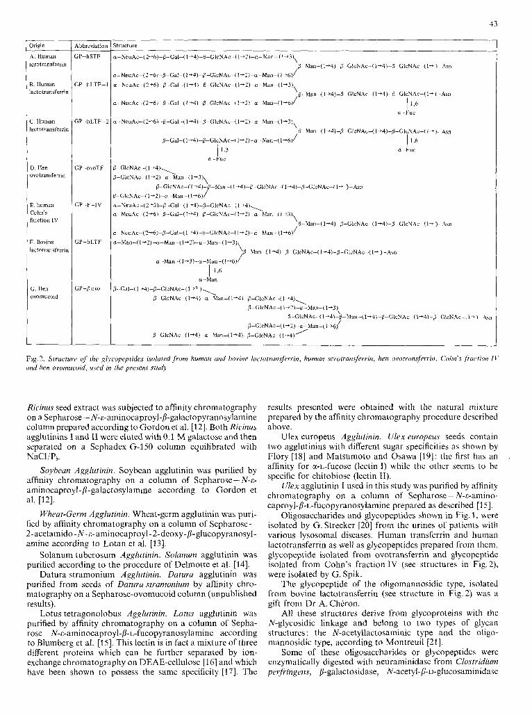
Fig. 2. StructurP of h e glycopeplides isolated from human and bovine lactotrunsferrin, humun serotransferrin, hen ovotrunsferrin, Cohn’s fraction I V und hen ovomucoid, used in the present study
Ricinus seed extract was subjecied to affinity chroinatography on a Sepharose - N-E-aminocaproyl-P-galactopyranosylamine column prepared according to Gordon et al. [12]. Both Ricinus agglutinins I and I1 were eluted with 0.1 M galactose and then separated on a Sephadex G-150 column equilibrated with NaCl/P,.
Soybean Agglutinin. Soybean agglutinin was purified by affinity chromatography on a column of Sepharose - N-6- aminocaproyl-[I-galactosylamine according to Gordon et al. [12].
Wheal-Germ Agglutinin. Wheat-germ agglutinin was puri- fied by affinity chromatography on a column of Sepharose- 2 - acetamido - N - c- aminocapro yl - 2 - deoxy - P- glucop yranosyl- amine according to Lotan et al. [13].
Solanum tuberosum Agglutinin. Solanum agglutinin was purified according to the procedure of Delmotte et al. [14].
Datura stramonium Agglutinin. Datura agglutinin was purified from seeds of Datzrru stramonium by affinity chro- matography on a Sepharose-ovomucoid column (unpublished results).
Lotus tetragonolobus Agglutinin. Lotus agglutinin was purified by affinity chromatography on a column of Sepha- rose - N-E-aminocaproyl-b-L-fucopyranosylamine according to Blumberg et al. [15]. This lectin is in Fact a mixture of three different proteins which can be further separated by ion- exchange chromatography on DEAE-cellulose [I61 and which have been shown to possess the same specificity [17]. The
results presented were obtained with the natural mixture prepared by the affinity chromatography procedure described above.
Ulex europeus Agglutinin. Ulex europrus seeds contain two agglutinins with different sugar specificities as shown by Flory [I81 and Matsumoto and Osawa [19]: the first has an affinity for a-L-fucose (lectin I) while the other seems to be specific for chitobiose (lectin 11).
Ulex agglutinin I used in this study was purified by affinity chromatography on a column of Sepharose - N-c-amino- caproyl-P-L-fucopyranosylamine prepared as described [I 51.
Oligosaccharides and glycopeptides shown in Fig. 1, were isolated by G. Strecker [20] from the urines of patients with various lysosomal diseases. Human transferrin and human lactotransferrin as well as glycopeptides prepared from them, glycopeptide isolated from ovotransferrin and glycopeptide isolated from Cohn’s fraction IV (see structures in Fig. 2), were isolated by G. Spik.
The glycopeptide of the oligomannosidic type, isolated from bovine lactotransferrin (see structure in Fig.2) was a gift from Dr A. Chkron.
All these structures derive from glycoproteins with the N-glycosidic linkage and belong to two types of glycan structures : the N-acetyllactosaminic type and the oligo- niannosidic type, according to Montreuil [21].
Some of these oligosaccharides or glycopeptides were cnzymatically digested with neuraminidase from Clostridium perfiingens, /3-galactosidase, N-acetyl-p-u-glucosaminidase
44
from Jack-bean meal and a-L-fucosidase from rat kidney (Boehringer) as previously described [22].
Hemagglutination Tests
Agglutination of human red blood cells, group Of, in NaCI/ Pi and inhibition of this hemagglutination by various oligosaccharides or glycopeptides was carried out in Linbro microtiter U-plates Titertek (Linbro Scientific Co, Hamden, USA) by a twofold serial dilution technique [19].
To each 0.05 ml of twofold serial dilution of sugar solu- tion, in NaC1/Pi, was added an equal volume of lectin solution, carefully diluted to contain four minimum hemagglutinating doses.
After incubation for 1 h at room temperature, 0.05 ml of a 3 % erythrocyte suspension in NaCI/Pi was added. The mixture was kept for 1 h at room temperature and then examined for agglutination.
Results were expressed as the minimum concentration (mM) required to completely inhibit one hemagglutining dose; account was taken of the threefold dilution caused by the addition of lectin and erythrocytes.
RESULTS AND DISCUSSION LECTINS WITH A SPECIFICITY FOR %-MANNOSE OR &-GLUCOSE
Concanavalin A
The results of the inhibition by various oligosaccharidic structures of hemagglutination by concanavalin A are pres- ented in Table 2 and can be summarized as follows.
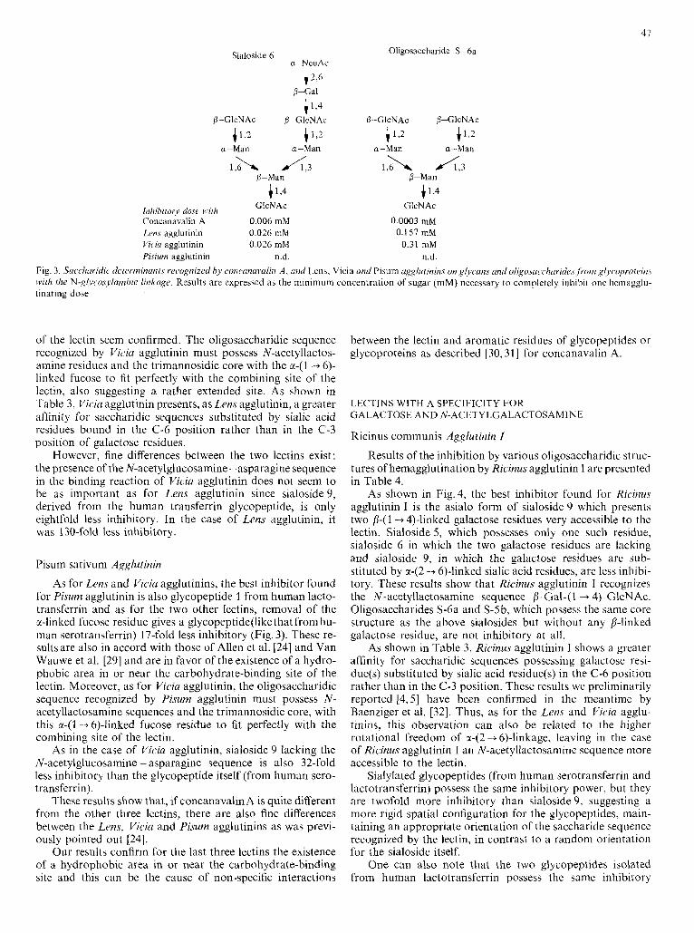
As shown on Fig. 3, concanavalin A presents a great affinity for the trimannosidic core substituted by two N-acetyl- P-glucosaminyl residues (oligosaccharide S-6a). The affinity is not decreased when this structure is modified by the addition of a p-(l -+ 4)-linked N-acetylglucosamine on the &linked mannose residue (oligosaccharide S-5b). On the contrary, the affinity is reduced when these N-acetylglucosamine resi- dues are substituted by galactosyl residues (sialosides 5 and 6, and the asialoglycopeptide from human serotransferrin) or by sialogalactosyl residues (sialoside 9, glycopeptide from human serotransferrin). Moreover, the presence of an a-(1 -+ 6)-linked mannose residue in a terminal non-reducing position enhances the affinity as demonstrated by the fact that sialoside 3 (Fig. 1) is 66-fold more inhibitory than sialoside 2.
On the other hand, mannoside 1 (Fig. 1) which possesses an exposed a-(1 -+ 3)-mannose, but which lacks the core struc- ture, is a poor inhibitor, even when this ~ - ( 1 --+ 3)-linked mannose is substituted by one or two E-(I-+ 2)-linked mannose residues (mannosides 2 and 3). However, the glycopeptide isolated from bovine lactotransferrin which possesses the complete trimannosidic structure with the a-( 1 -+ 3)-linked and a-(1 -+ 6)-linked mannose residues substituted by four additional a-linked mannose residues (oligomannosidic type structure) is a very good inhibitor of hemagglutination.
Substitution of the core a-(1 -+ 3)-linked mannose residue at position C-4, as in the glycopeptide from ovotransferrin, decreases the affinity of concanavalin A as compared to the asialoagalacto form of glycopeptide from serotransferrin (Fig. 3).
No difference can be found between the inhibitory power of a glycopeptide isolated from transferrin (Fig. 2A) and the oligosaccharidic structure derived from it (sialoside 9 in Fig. I) , suggesting that the N-acetylglucosamine - asparagine core
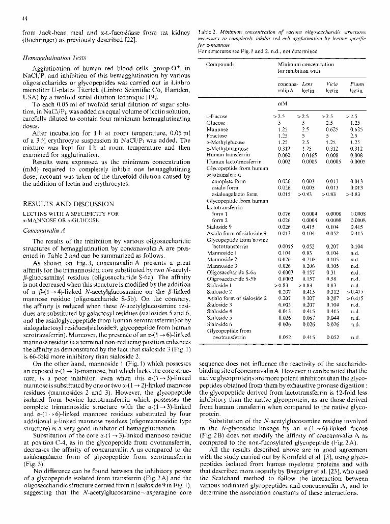
Table 2. Minimum concentration of various oligosuccharidic structures necessary to completely inhibit red cell agglutination hy lectins specific for a-mannose For structures see Fig. 1 and 2. n.d., not determined
Compounds Minimum concentration for inhibition with
concana- Lens Vicia Pisum valin A lectin lectin lectin
- __
L-Fucose Glucose Mannose Fructose a- Methylglucose a-Methylmannose Human transferrin Human lactotransferrin Glycopeptide from human serotransferrin
complete form asialo form asialoagalacto form
Glycopeptide from human lactotransferrin
form 1 form 2
Sialoside 9 Asialo form of sialoside 9 Glycopeptide from bovine
Mannoside 1 Mannoside 2 Mannoside 3 Oligosaccharide S-6a Oligosaccharide S-5b Sialoside 1 Sialoside 2 Asialo form of sialoside 2 Sialoside 3 Sialoside 4 Sialoside 5 Sialoside 6 Glycopeptide from
ovotransferrin
lactotransferrin
mM __ ~- -
>2.5 >2.5 5 5 1.25 2.5 1.25 5 1.25 2.5 0.312 1.25 0.002 0.0165 0.002 0.0005
0.026 0.003 0.026 0.003 0.015 >0.83
0.026 0.0004 0.026 0.0004 0.026 0.415 0.013 0.104
0.0015 0.052 0.104 0.83 0.026 0.210 0.026 0.206 0.0003 0.157 0.0003 0.157
0.207 0.415 0.207 0.207 0.003 0.207 0.013 0.415 0.026 0.087 0.006 0.026
>0.83 >0.83
0.052 0.415
- .
>2.5 >2.5 2.5 1.25 0.625 0.625 5 2.5 1.25 1.25 0.312 0.312 0.008 0.008 0.0005 0.0005
0.013 0.013 0.013 0.013
20.83 >0.83
0.0008 0.0008 0.0008 0.0008 0.104 0.415 0.052 0.415
0.207 0.104 0.104 n.d. 0.105 n.d. 0.105 n.d. 0.31 n.d. 0.58 n.d. 0.83 n.d. 0.312 >0.415 0.207 >0.415 0.104 n.d. 0.415 n.d. 0.044 n.d. 0.026 n.d.
0.052 n.d
sequence does not influence the reactivity of the saccharide- binding siteofconcanavalinA. However,it can be notedthat the native glycoproteins are more potent inhibitors than the glyco- peptides obtained from them by exhaustive pronase digestion : the glycopeptide derived from lactotransferrin is 12-fold less inhibitory than the native glycoprotein, as are those derived from human transferrin when compared to the native glyco- protein.
Substitution of the N-acetylglucosamine residue involved in the N-glycosidic linkage by an E-( 1 -+ 6)-linked fucose (Fig.2B) does not modify the affinity of concanavalin A as compared to the non-fucosylated glycopeptide (Fig. 2 A).
All the results described above are in good agreement with the study carried out by Kornfeld et al. [3], using glyco- peptides isolated from human myeloma proteins and with that described more recently by Baenziger et al. [23], who used the Scatchard method to follow the interaction between various iodinated glycopeptides and concanavalin A, and to determine the association constants of these interactions.
45
Our own results, as expressed in Table 2, can be com- pared to these association constants and, from our values, the affinity of a given saccharidic structure for concanavalin A immobilized on Sepharose 4B can be predicted.
Lens culinaris Agglutinin
The results of the inhibition by various oligosaccharidic structures of hemagglutination by Lens agglutinin are given in Table 2.
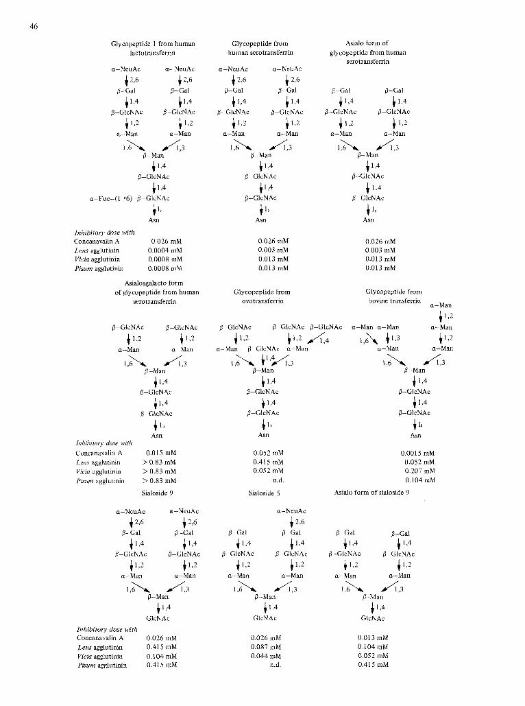
As shown in Fig. 3, the best inhibitor for Lens agglutinin is a glycopeptide isolated from human lactotransferrin (Fig. 2B). Another glycopeptide with an additional fucose residue (Fig. 2C) possesses the same inhibitory power.
Removal of all the fucose residues from these glyco- peptides with an x-L-fucosidase from rat kidney gives glyco- peptides about 10-fold less inhibitory, as is the glycopeptide isolated from human serotransferrin (Fig. 2A).
These results show the importance of the a-(1 -+ 6)-linked fucose, as a major determinant of the binding. The a-(1 + 3)- linked fucose on the external N-acetylglucosaniine residue, as found in glycopeptide 2 from human lactotransferrin, does not seem to play any role in this binding. On the other hand, the results confirm those found by Allen et al. [24] who dis- covered that 3-O-methyl-~-glucose and 3-O-benzyl-~-glucose are better inhibitors than glucose itself, suggesting the ex- istence of a hydrophobic area in or near the lectin-binding site, which can interact with the methyl or the benzyl groups. The methyl group provided by the a-(1 -+ 6)-linked fucose would fit perfectly with this hydrophobic area.
In the case of concanavalin A, results had also shown the existence of a hydrophobic region close to the carbo- hydrate-binding site that could interact specifically with the phenyl groups of /3-u-glucopyranosides [25], but our results show that the methyl group of L-fucose does not enhance the binding in this case. However, while the presence of an a-(1 -6)-linked fucose near the N-glycosidic linkage seems to be a necessary condition for a maximal recognition of a saccharidic sequence by Lens agglutinin, it is not a sufficient one. In fact, glycopeptides possessing this a-(1 + 6)-linked fucose residue are not inhibitory (glycopeptides I and I1 of Fig. I ) or are very poorly inhibitory (glycopeptide I11 of Fig. I). Moreover, the presence of terminal galactose residues seems to be necessary since in contrast to the asialo form, the asialo- agalacto form of the glycopeptide of human serotransferrin is not inhibitory at all. In fact, removal of terminal sialic acid residues alone from the glycopeptide of human serotransferrin has no effect on the inhibitory activity as compared to the native glycopeptide. On the basis of these results, it can be concluded that Lens agglutinin recognizes bi-antennary N- acetyllactosaminic-type glycopeptide with galactose residues in a terminal non-reducing position, the binding being greatly enhanced by the presence of an a-(1 -+ 6)-linked fucose residue near the N-glycosidic linkage. These results can be compared with those published by Kornfeld et al. [26] who found that the most potent inhibitor for Lens agglutinin is a glycopeptide isolated from myeloma IgG which possesses the same glycan structure [3] as glycopeptide 1 of human lactotransferrin. These authors did not emphasize the importance of the presence of the a-(1 -+ 6)-linked fucose near the N-glycosidic linkage for a maximal recognition by the lectin.
The glycopeptide isolated from bovine lactotransferrin which possesses the classical trimannosidic core substituted by four additional a-linked mannose residues (oligomanno- sidic-type structure) is 140-fold less inhibitory than glyco-
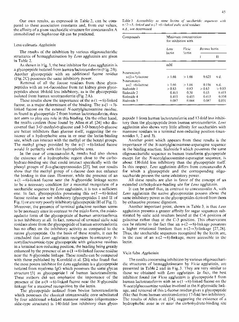
Table 3. Accessibility t o .some lectins of .saccharidic sequences wirlz x-i2+3)-linked and cc-/2-+h)-linked siulic ucid residues n.d., not determined
~
Compounds Minimum concentration for inhibition with
Lens Vicia Ric rnuJ leLtin lectin Iectin
I I1
mM .~ ~ ~~. ~~ ~.
Neurdminyl- r-(2+3)-lactose > 1.66 > 1.66 0.625 n.d.
Neuraminyl- x-( 2 4 6)-lactose > 1.66 > 1.66 0.156 n.d.
Sialoside 1 > 0.83 0.83 >OX3 > 0.83 Sialoside 2 0.415 0.31 0.83 0.415 Sialoside 4 0.415 0.415 0.415 0.104 Sialoside 5 0.087 0.044 0.087 0.051
peptide 1 from human lactotransferrin and 17-fold less inhibi- tory than the glycopeptide from human serotransferrin. Lens agglutinin also shows very little affinity for saccharides with mannose residues in a terminal non-reducing position (man- nosides 1, 2 and 3).
Another point which appears from these results is the importance of the N-acetylglucosamine-asparagine sequence in the binding reaction. Sialoside 9 which possesses the same oligosaccharidic sequence as human transferrin glycopeptide except for the N-acetylglucosamine-asparagine sequence, is about 130-fold less inhibitory than the glycopeptide itself. In this respect, Lens agglutinin differs from concanavalin A for which a glycopeptide and the corresponding oligo- saccharide possess the same inhibitory power.
All these results are also in favor of the concept of an extended carbohydrate-binding site for Lens agglutinin.
It can be noted that, in contrast to concanavalin A, with Lens agglutinin the native glycoproteins possess about the same inhibitory power as the glycopeptides derived from them by exhaustive pronase digestion.
Another important point, shown in Table 3, IS that Lens agglutinin presents a greater affinity for sugar sequences sub- stituted by sialic acid residues bound at the C-6 position of galactose rather than at the C-3 position. This observation can be related to the fact that an a42 + 6)-linkage possesses a higher rotational freedom than a-(2+3)-linkage [27,28]. Thus, the saccharidic sequences recognized by the lectin are, in the case of an a-(2-+6)-linkage, more accessible to the lectin.
Vicia faba Agglutinin
The results concerning inhibition by various oligosacchari- dic structures of hemagglutination by Viciu agglutinin, are presented in Table 2 and in Fig. 3. They are very similar to those we obtained with Lens agglutinin. In fact, the best inhibitor found for Viczu agglutinin is glycopeptide 1 from human lactotransferrin with an a-(1 -+6)-linked fucose on the N-acetylglucosamine residue involved in the N-glycosidic link- age, and removal of this L-fucose residue gives a glycopeptide (like that from human serotransferrin) 17-fold less inhibitory. The results of Allen et al. [24], suggesting the existence of a hydrophobic zone in or near the carbohydrate-binding site
46
Glycopeptide 1 from human lactotransferrin
a-NeuAc a-NeuAc
f 2,6 i 2 6
4 1 9 f L4
8-Gal P-Gal
0-GlcNAc 0-GlcNAc
+1,2 c 1 2
1 , h A 3
a-Man a-Man
0-Man
i 1,4
c l , 4
P-GlcNAc
a-Fuc-( 1+6)-P-GlcNAc
i 1, Asn
Inhibitory dose with Concanavalin A 0.026 mM Lens agglutinin 0.0004 mM Viciu agglutinin 0.0008 mM Pisum agglutinin 0.0008 mM
Asialoagalacto form of glycopeptide from human
serotransferrin
P-GIcNAc P-GlcNAc
4 1 2 4 1 2
,,A A . 3
4 L 4
+ 1)
a-Man a-Man
&Man f1 ,4
P-GlcNAc
P-GlcNAc
Asn Inhibitory dose with Concanavalin A 0.0 15 mM Lens agglutinin > 0.83 mM Viciu agglutinin > 0.83 mM Pisum agglutinin > 0.83 mM
Sialoside 9
0-Man
41,4 GlcNAc
Inhibitory dose with Concanavalin A 0.026 mM Lens agglutinin 0.41 5 mM Vicia agglutinin 0.104 mM Pisum agglutinin 0.415 mM
Glycopeptide from human serotransferrin
a-NeuAc a-Neu Ac
4 2 6 4 2 6
4 1,4 1 1 , 4
&Gal 0-Gal
8-GlcNAc P-GlcNAc c 1 2 4 1,2
l h A , 3
a-Man a-Man
&Man
4 1 9
1 1 9 P-GlcNAc
p-GlcNAc
c 1, Asn
0.026 mM 0.003 mM 0.013 mM 0.013 mM
Asialo form of glycopeptide from human
serotransferrin
0.026 mM 0.003 mM 0.013 mM 0.013 mM
Glycopeptide from Glycopeptide from ovotransferrin bovine trans ferrin a-Man c 1 2
+1>2 p-GlcNAc 8-GlcNAc 0-C-lcNAc a-Man a-Man a-Man
a-?.fan 1,6\
a-Man 4 12. + 1 J A 4
a-Man 0-ClcNAc a-Man
0.052 mM 0.41 5 mM 0.052 mM
n.d.
Sialoside 5
a-NeuAc
4 2,6
4 1 9 4 L4 &Gal P-Gal
0-GlcNAc P-GlcNAc
f 1,2 4 1 2
l , h A , 3
a-Man a-Man
!.-Man + 1.4 GlcNAc
0.026 mM 0.087 mM 0.044 mM
n.d.
&Man
i 1,4
1 1,4
8-GlcNAc
P-GlcNAc
c 1, Asn
0.001 5 mM 0.052 mM 0.207 mM 0.104 mM
Asialo form of sialoside 9
0.01 3 mM 0.104 mM 0.052 mM 0.41 5 mM
41
Oligosaccharide S-6a Sialoside 6 a NcuAc
{ 2 6
4 1,4
p-Gal
0-GlcNAc GlcNAc 0-GlcNAc p-GlcNAc
4 1 2 4 1 2 + 1 2 { 1,2 a-Man a-Man a-Man a-Man
GlcNAc Inhihitoq dosc wi/h Concanavalin A 0.006 mM Lm.r ‘igglutinin 0.026 mM Viciu agglutinin 0.026 mM Pisium agglutinin n.d.
1 , h A , 3 &Man
+1,4 GlcNAc
0.0003 mM 0.1 57 mM
0.31 mM n.d.
Fig. 3. Sacchuridic deternzinunts recognized by c.oi~ccirrn~a/in A , und Lens, Vicia nnd Pisum ugglutinins on glycuns and oligosucchciride.s,~r~in ~ l y o p r o l c i n s with Ihe N-g/ycosylarninc linkage. Results are expressed as the minimum concentration of sugar (mM) necessary to completely inhibii onc hcmagglu- tinating dose
of the lectin seem confirmed. The oligosaccharidic sequence recognized by Viciu agglutinin must possess N-acetyllactos- amine residues and the trimannosidic core with the a-(1 4 6)- linked fucose to fit perfectly with the combining site of the lectin, also suggesting a rather extended site. As shown in Table 3, VicW agglutinin presents, as Lens agglutinin, a greater affinity for saccharidic sequences substituted by sialic acid residues bound in the C-6 position rather than in the C-3 position of galactose residues.
However, fine differences between the two lectins exist : the presence of the N-acetylglucosamine-asparagine sequence in the binding reaction of Vicia agglutinin does not seem to be as important as for Lens agglutinin since sialoside 9, derived from the human transferrin glycopeptide, is only eightfold less inhibitory. In the case of Lens agglutinin, it was 130-fold less inhibitory.
Pisum sativum Agglutinin
As for Lens and Viciu agglutinins, the best inhibitor found for Pisum agglutinin is also glycopeptide 1 from human lacto- transferrin and as for the two other lectins, removal of the x-linked fucose residue gives a glycopeptide(1ikethat from hu- man serotransferrin) 17-fold less inhibitory (Fig. 3) . These re- sults are also in accord with those of Allen et al. [24] and Van Wauwe et al. [29] and are in favor of the existence of a hydro- phobic area in or near the carbohydrate-binding site of the lectin. Moreover, as for Viciu agglutinin, the oligosaccharidic sequence recognized by Pisum agglutinin must possess N- acetyllactosamine sequences and the trimannosidic core, with this a-(1 -+ 6)-linked fucose residue to fit perfectly with the combining site of the lectin.
As in the case of Viciu agglutinin, sialoside 9 lacking the N-acetylglucosamine - asparagine sequence is also 32-fold less inhibitory than the glycopeptide itself (from human sero- transferrin).
Thcse results show that, if concanavalin A is quite different from the other three Icctins, there are also fine differences between the Lens, Viciu and Pisum agglutinins as was previ- ously pointed out [24].
Our results confirm for the last three lectins the existence of a hydrophobic area in or near the carbohydrate-binding site and this can be the cause of non-specific interactions
between the lectin and aromatic residues of glycopeptides or glycoproteins as described [30,31] for concanavalin A.
LECTINS WITH A SPECIFICITY FOR GALACTOSE AND N-ACETYLGALACTOSAMINE
Ricinus communis Agglutinin I
Results of the inhibition by various oligosaccharidic struc- tures of hemagglutination by Ricinus agglutinin I are presented in Table 4.
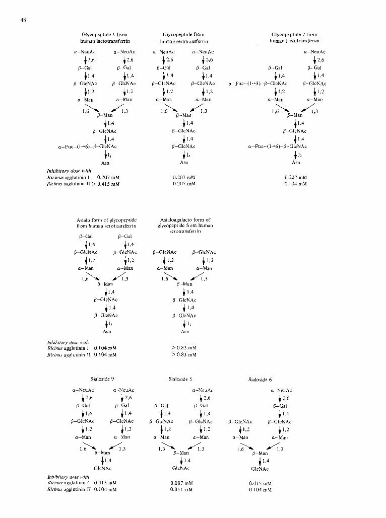
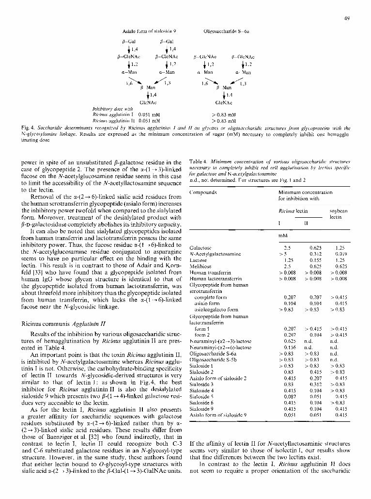
As shown in Fig.4, the best inhibitor found for Ricinus agglutinin I is the asialo form of sialoside 9 which presents two p-(1 --$4)-linked galactose residues very accessible to the lectin. Sialoside 5 , which possesses only one such residue, sialoside 6 in which the two galactose residues are lacking and sialoside 9, in which the galactose residues are sub- stituted by a-(2 -+ 6)-linked sialic acid residues, are less inhibi- tory. These results show that Ricinus agglutinin I recognizes the N-acetyllactosamine sequence [i-Gal-(l -+ 4)-GlcNAc. Oligosaccharides S-6a and S-Sb, which possess the same core structure as the above sialosides but without any /S-linked galactose residue, are not inhibitory at all.
As shown in Table 3 , Ricinus agglutinin I shows a greater affinity for saccharidic sequences possessing galactose resi- due(s) substituted by sialic acid residue(s) in the C-6 position rather than in the C-3 position. These results we preliminarily reported [4,S] have been confirmed in the meantimc by Baenziger et al. [32]. Thus, as for the Lens and Viciu agglu- tinins, this observation can also be related to the higher rotational freedom of a-(2 + 6)-linkage, leaving in the case of Ricinus agglutinin I an N-acetyllactosaniine sequence more accessible to the lectin.
Sialylated glycopeptides (from human serotransferrin and lactotransferrin) possess the same inhibitory power, but they are twofold more inhibitory than sialoside 9, suggesting a more rigid spatial configuration for the glycopeptides, main- taining an appropriate orientation of the saccharide sequence recognized by the lectin, in contrast to a random orientation for the sialoside itself.
One can also note that the two glycopeptides isolated from human lactotransferrin possess the same inhibitory
48
Glycopeptide 1 from human lactotransferrin
a-NeuAc a-NeuAc
4 2,6 4 2,6 8-Gal 8-Gal
1 1 4 )L4
4 1 2 4 1 2
8-GlcNAc 8-GlcNAc
a-Man a-Man
1 2 - 4 3 8-Man
+ L 4
4 1,
B-GlcNAc
4 1 9 a-Fuc-( 1+6)-P-GlcNAc
Asn Inhibitory dose with Ricinus agglutinin I 0.207 mM Ricinus agglutinin IT > 0.41 5 mM
Asialo form of glycopeptide from human serotransferrin
&Gal 8-Gal
+1,4 4 1 9 8-GlcNAc 8-GlcNAc
4 1 2 +1>2
1,6 \ A 3
a-Man a-Man
&Man
41,4
i 1 4
P-GlcNAc
P-GIcNAc
t Asn
Inhibitory dose with Ricinus agglutinin I 0.104 mM Rrcinus agglutinin I1 0.104 mM
Glycopeptide from human serotransferrin
a-NeuAc a-Neu Ac
1 2 5 4 2,6
) 1,4 )1,4
P-Gal 8-Cal
P-GlcNAc 8-GlcNAc
112 + 1 2 a-l.4an a-Man
0.207 mM 0.207 mM
Asialoagalacto form of glycopeptide from human
serotransferrin
8-GlcNAc 8-GlcNAc + 1 2 1 1 , 2
1 2 - x A3
a-Man a-Man
8-Man
1 1 , 4
4 1,4 8-GlcNAc
B-GlcNAc
+ Asn
> 0.83 mM > 0.83 mM
Glycopeptide 2 from human lactotransterrin
a-NeuAc
4 2,6
1 1,4 1 9 8-Gal &Gal
a-Fuc-( 1+3)-8-GlcNAc 8-GlcNAc + 1 2 + 1 2 a-Man a-Man
0.201 mM 0.104 mM
Sialoside 9 Sialoside 5 Sialoside 6
a-NeuAc a NeuAc a-NeuAc a-NeuAc
8-Gal p-Gal &Gal 8-Gal !%Gal i 2 6 i 2,6 1 2,6 4 2,6
4 1,4 4 1,4 + 1,4 4 1 9 4 1,4 0-GlcNAc 8-GlcNAc O-GICNAC P-GlcNAc O-GlcNAc P-GlcNAc + 1 2 4 1 2 + 1,2 + 1 2 + 1 2 i 1,2
a-Man a-Man a-Man a-Man a-Man a-Man
l b 4 3 &Man
Inhibitory dose with Ricinus agglutinin I 0.41 5 mM Ricinus agglutinin I1 0.104 niM
I,& 4 , 3 !.-Man
0.087 mM 0.051 mM
1 , b 4 3 0-Man 4 1,4 GlcNAc
0.41 5 mM 0.104mM
4Y
Asialo form of sialoside 9
0-Gal &Gal
Ohgosaccharide S-6a
4 1 4 +1,4
4 l , 2 4 1 2 4 1 2 p > 2
P-GlcNAc P-GlcNAc 0-GlcNAc 0-GlcNAc
a -Man a -Man a-Man a-Man
1,4 4 1 4 GlcNAc GlcNAc
Inhibitory dose with Ricinus agglutinin I 0.051 mM > 0.83 mM Ricinus agglutinin I I 0.051 mM > 0.83 mM
Fig. 4. Saccharidic determinants recognized by Ricinus agglutinins I utid I1 on glycans or oligosaccharidic structures ,from glycoproteins with the N-glycosylamine linkage. Results are expressed as the minimum concentration of sugar (mM) necessary to completely inhibit one hemagglu- tinating dose
power in spite of an unsubstituted /%galactose residue in the case of glycopeptide 2. The presence of the a-( 1 + 3)-linked fucose on the N-acetylglucosamine residue seems in this case to limit the accessibility of the N-acetyllactosamine sequence to the lectin.
Removal of the a-(2 + 6)-linked sialic acid residues from the human serotransferrin glycopeptide (asialo form) increases the inhibitory power twofold when compared to the sialylated form. Moreover, treatment of the desialylated product with P-D-galactosidase completely abolishes its inhibitory capacity.
It can also be noted that sialylated glycopeptides isolated from human transferrin and lactotransferrin possess the same inhibitory power. Thus, the fucose residue cc-(1+6)-linked to the N-acetylglucosamine residue conjugated to asparagine seems to have no particular effect on the binding with the lectin. This result is in contrast to those of Adair and Korn- feld [33] who have found that a glycopeptide isolated from human IgC whose glycan structure is identical to that of the glycopeptide isolated from human lactotransferrin, was about threefold more inhibitory than the glycopeptide isolated from human transferrin, which lacks the a-( 1 -+ 6)-linked fucose near the N-glycosidic linkage.
Ricinus communis Agglutinin ZZ
Results of the inhibition by various oligosaccharidic struc- tures of hemagglutination by Ricinus agglutinin I1 are pres- ented in Table 4.
An important point is that the toxin Ricinus agglutinin 11, is inhibited by N-acetylgalactosamine whereas Ricinus agglu- tinin I is not. Otherwise, the carbohydrate-binding specificity of lectin I1 towards N-glycosidic-derived structures is very similar to that of lectin I : as shown in Fig.4, the best inhibitor for Ricinus agglutinin I1 is also the desialylated sialoside 9 which presents two p-(1+ 4)-linked galactose resi- dues very accessible to the lectin.
As for the lectin I, Ricinus agglutinin I1 also presents a greater affinity for saccharidic sequences with galactose residues substituted by a-(2 + 6)-linked rather than by a- (2 + 3)-linked sialic acid residues. These results differ from those of Baenziger et al. [32] who found indirectly, that in contrast to lectin I, lectin I1 could recognize both C-3 and C-6 substituted galactose residues in an N-glycosyl-type structure. However, in the same study, these authors found that neither lectin bound to 0-glycosyl-type structures with sialic acid a-(2 + 3)-linked to the /3-Gal-(1 + 3)-GalNAc units.
Table 4. Minimum concentration of various oligosaccharidic structures necessnry to completely inhibif red cell agglutinuzion by Icciins specific for galactose and N-acetylgalactosamine n.d., not determined. For structures see Fig. 1 and 2
Compounds Minimum concentration for inhibition with
Ric inus lectin soybedn
I 11
-~ -
lectin -~
Galactose N-Acet ylgalactosamine Lactose Melibiose Human transferrin Human lactotransferrin Glycopeptide from human serotransferrin
complete form asialo form asialoagalacto form
Glycopeptide from human lactot ransferrin
form 1 form 2
Neuraminyl-(ct2+ 3)-lactose Neuraminyl-(a2~6)-lactose Oligosaccharide S-6a Oligosaccharide S-5b Sialoside 1 Sialoside 2 Asialo form of sialoside 2 Sialoside 3 Sialoside 4
Sialoside 6 Sialoside 9 Asialo form of sialoside 9
Sialoside 5
m M
2.5
1.25 2.5
> 0.008 > 0.008
> 5
0.207 0.104
> 0.83
0.207 0.207 0.625 0.156
> 0.83 > 0.83 > 0.83
0.83 0.415 0.83 0.415 0.087 0.43 5 0.415 0.051
0.625 0.312 0.155 0.625
> 0.008 > 0.008
0.207 0.104
> 0.83
> 0.41 5 0.104
n.d. n.d. > 0.83 > 0.83 > 0.83
0.415 0.207 0.312 0.104 0.051 0.104 0.104 0.051
1.25 0.019 1.25 0.625
> 0.008 > 0.008
> 0.41 5 0.41 5
> 0.83
> 0.415 > 0.415 n.d. n.d. n.d. n.d. > 0.83 > 0.83
> 0.83 > 0.83
> 0.83
0.41 5
0.41 5
0.41 5 0.41 5
If the affinity of lectin I1 for N-acetyllactosaminic structures seems very similar to those of isolectin I, our results show that fine differences between the two lectins exist.
In contrast to the lectin I, R i c k s agglutinin I1 does not seem to require a proper orientation of the saccharidic
SO
sequence recognized by the lectin, for sialoside 9 which possesses a random configuration as compared to the sialylated glycopeptide isolated from human transferrin is in fact two- fold more inhibitory.
It is also interesting to compare the inhibitory power of the glycopeptide from human serotransferrin to that of glyco- peptides l and 2 from human lactotransferrin. The substitu- tion of the N-acetylglucosamine residue taking part in the glycosylamine linkage by an a-(1 -+ 6)-linked fucose residue decreases the inhibitory power of this glycopeptide at least twofold. This result is also in contrast to that of Adair and Kornfeld [33] who have found that a glycopeptide isolated from human IgG was about four-fold more inhibitory than the glycopeptide isolated from human transferrin.
On the other hand, glycopeptide 2 from human lacto- transferrin is at least fourfold more inhibitory than glyco- peptide l from the same source. Here, in contrast to Ricinus agglutinin I, the a-(1 -+ 3)-linked fucose on the N-acetyl- glucosamine residue does not seem to limit the accessibility of the unsubstituted p-galactose residue to the lectin.
Differences between the two lectins towards glycopeptides with an 0-glycosidic linkage, bearing /%Gal-( 1 -+ 3)-GalNAc sequences have also been characterized by Baenziger et al. [32]. According to their results, Ricinus agglutinin 11 binds glyco- peptides containing this sequence with greater association constant than agglutinin I, and can bind glycopeptides with terminal N-acetylgalactosamine residues, while agglutinin I cannot.
Table 5. Minimum concentration of various oligosatchuriclic structures necessary to completely inhibit red blood celi agxlutination by lectins specific fbr N-ucetylglucosamine
Compounds Minimum concentration for inhibition with lectin from
wheat Solunum Datum germ
- ~~~~ -
N-Acelylglucosamine Chitobiose Chitotriose Human transferrin Human lactotransferrin Glycopeptide from human serotransferrin
complete form asialoagalacto form
Glycopeptide from human lactotransferrin
form 1 form 2
Glycopeptide from hen ovomucoid Glycopeptide from ovotransferrin Sialoside 2 Asialo form of sialoside 2 Sialoside 9 Asialo form of sialoside 9
mM
> 15 0.4 0.0375 0.008
> 0.008
> 0.415 > 0.415
> 0.415 > 0.415
> 0.415 > 0.415 > 0.415 > 0.415 > 0.415
0.65
> 15 1.85 0.01 5
> 0.008 > 0.008
> 0.415 > 0.415
0.207 0.207 0.104 0.415 0.41 5 0.41 5 0.207
> 0.415
> 15 > 1.85
0.19 > 0.008 > 0.008
0.415 > 0.415
0.207 0.207 0.026 0.052
> 0.415 > 0.415 > 0.415 > 0.415
Soybean Agglutinin
Results of the inhibition by various oligosaccharidic struc- tures of hemagglutination by soybean agglutinin are presented in Table 4.
The best inhibitor is N-acetylgalactosamine itself. Galac- tose and lactose are 70-fold less inhibitory than this sugar, and melibiose with a terminal non-reducing a-linked galac- tose is twofold more inhibitory than lactose.
These results are in good agreement with those of Pereira et al. [34] and Hammarstrom et al. [35] and confirm that soybean agglutinin presents the greatest affinity for N-acetyl- galactosamine residues.
Desialylated glycopeptides or oligosaccharides (asialo form of the glycopeptide from human serotransferrin, asialo form of sialoside 9) of the N-acetyllactosaminic type, pos- sessing exposed non-reducing p-(1 -+ 4)-llnked galactose resi- dues are slightly inhibitory as shown previously [36]. How- ever enzymatic removal of these galactose residues (the asialo- agalacto form of the glycopeptide from human serotrans- ferrin) results in a complete loss of their inhibitory power. This point is contrary to the results of Irimura et al. [36] who find that porcine thyroglobulin p-glycopeptide, from which both sialic acid and galactose residues have been enzymically removed, is as good an inhibitor for soybean agglutinin as the desialylated glycopeptide.
LECTINS WITH A SPECIFICITY FOR N-ACETYLGLUCOSAMINE
Wheat-Germ Agglutinin
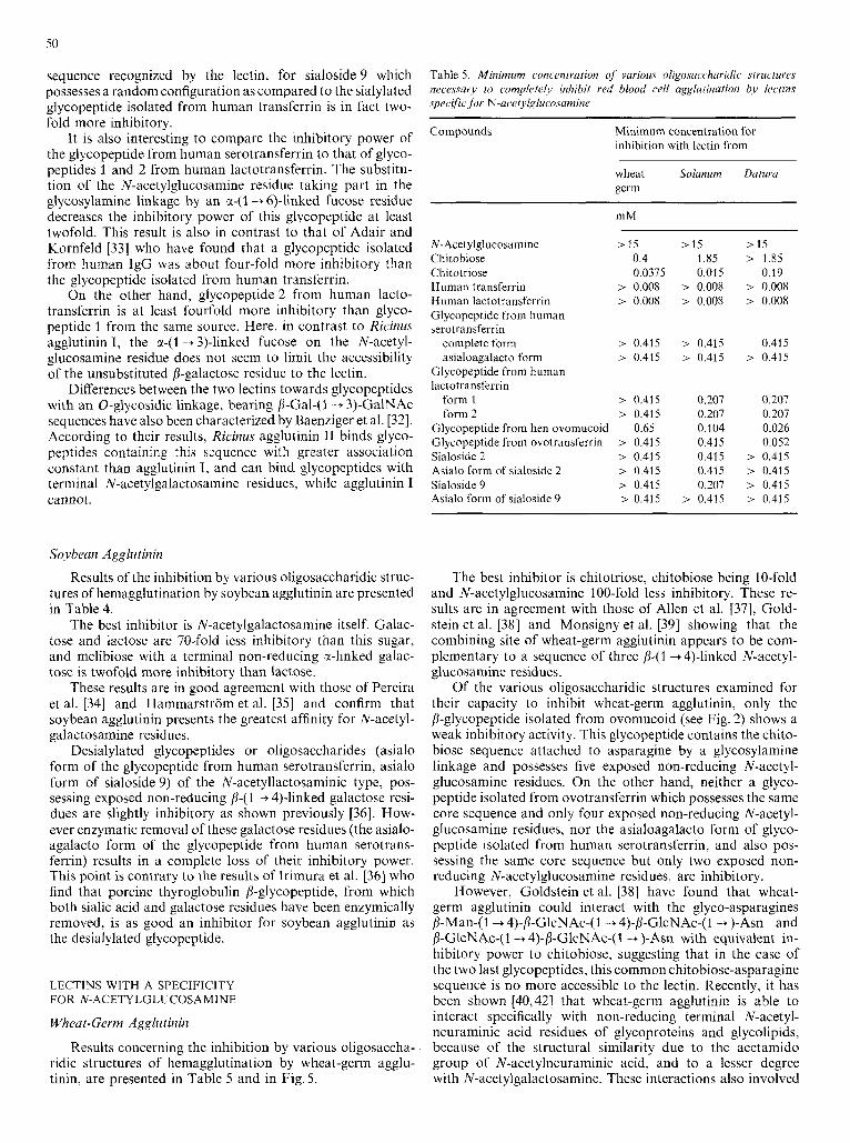
Results concerning the inhibition by various oligosaccha- ridic structures of hemagglutination by wheat-germ agglu- tinin, are presented in Table 5 and in Fig. 5.
The best inhibitor is chitotriose, chitobiose being 10-fold and N-acetylglucosamine 100-fold less inhibitory. These re- sults are in agreement with those of Allen et al. [37], Gold- stein et al. [38] and Monsigny et al. [39] showing that the combining site of wheat-germ agglutinin appears to be com- plementary to a sequence of three 8-(1+ 4)-linked N-acetyl- glucosamine residues.
Of the various oligosaccharidic structures examined for their capacity to inhibit wheat-germ agglutinin, only the b-glycopeptide isolated from ovomucoid (see Fig. 2) shows a weak inhibitory activity. This glycopeptide contains the chito- biose sequence attached to asparagine by a glycosylamine linkage and possesses five exposed non-reducing N-acetyl- glucosamine residues. On the other hand, neither a glyco- peptide isolated from ovotransferrin which possesses the same core sequence and only four exposed non-reducing N-acetyl- glucosamine residues, nor the asialoagalacto form of glyco- peptide isolated from human serotransferrin, and also pos- sessing the same core sequence but only two exposed non- reducing N-acetylglucosamine residues, are inhibitory.
However, Goldstein et al. [38] have found that wheat- germ agglutinin could interact with the glyco-asparagines /l-Man-( 1 -+ 4)-P-GlcNAc-( 1 -+ 4)-P-GlcNAc-( 1 -+ )-Am and /l-GlcNAc-( 1 --t 4)-P-GlcNAc-( 1 -+ )-Am with equivalent in- hibitory power to chitobiose, suggesting that in the case of the two last glycopeptides, this common chitobiose-asparagine sequence is no more accessible to the lectin. Recently, it has been shown [40,42] that wheat-germ agglutinin is able to interact specifically with non-reducing terminal N-acetyl- neuraminic acid residues of glycoproteins and glycolipids, because of the structural similarity due to the acetamido group of N-acetylneuraminic acid, and to a lesser degree with N-acetylgalactosamine. These interactions also involved
Chitobiose Chitotriose
P-GLcNAc O-C-lcNAc
i 1 , 4 +1,4
41,4
P-GIcNAc P-GIcNAc
P-C-ICNAC
Inhihitory c1o.sc with wheat-germ agglutinin 0.4 mM Solunum agglutinin 1.85 mM Datum agglutinin > 1.85 mM
0.0375 mM
0.01 5 mM
0.19 mM
Glycopeptide 1 from human lactotransferrin
a-Neu Ac a-NeuAc 4 2,6 i 2,6
4 1,4 i 13 6-GlcNAc p-GlcNAc
S-C-al &Gal
1 1 2 i 1,2
LA A 3
a-Man a-Man
p-Man
i 1,4 0-GlcNAc
4 1,4 a-Fuc-! 1+6)-p-GlcNAc
4 1, Asn
Glycopeptide from ovotransferrin
> 0.415 mM > 0.41 5 mM
0.207 mM 0.41 5 mM
0.207 mM 0.052 mM
Clycopeptide from hen ovomucoid &Gal 4 1,4
P-GIcNAc P-GlcNAc P-GIcNAc 4 1 9 a-Man
+ 1 ,9-G a-Man
4 1,4 +1,4 p-GlcNAc 0-GlcNAc 8- GIcNAc 0-GlcNAc
\ +1J + 1 , 5 4 4 a-Man 0-C-lcNAc a-Man
I& &Man
p-GlcNAc + 1,4
+1,4 P-GlcNAc +
Asn Inhibitory dose with wheat-germ agglutinin 0.65 mM Solanurn agglutinin 0.104 mM Datum agglutinin 0.026 mM
Fig. 5. Smcharidie detet-minun!s rrroxnized by uggluiinins from wfieai germ, Solanuin und Dat ura u,qg/urinins on glyc.an,s and oligosaccharida fiom sarious glycoproteins. Results arc expressed as the minimum concentration of sugar (mM) necessary t o completely inhibit one hemagglutinating dose
a charge effect and what Monsigny et al. [41] call an ‘avidity effect’ : glycoconjugates (glycoproteins, glycolipids or glyco- peptides) with a high density of terminal non-reducing N- acetylglucosamine or N-acetylneuraminic acid residues will interact with wheat-germ agglutinin. This may be the case in the interaction with the P-glycopeptide isolated from ovo- mucoid which possesses five external non-reducing N-acetyl- glucosamine residues.
On the other hand, glycopeptides or oligosaccharides such as the glycopeptide from human serotransferrin or sialoside 9, which possess only two external N-acetylneuraminic acid residues, or the asialoagalacto form of the glycopeptide from
human serotransferrin which possesses only two exposed non-reducing N-acetylglucosamine residues, are not inhibi- tory.
All these results show that the interactions between wheat- germ agglutinin and cell-surface glycoconjugates are very complex and have to be interpreted very carefully.
Solanum tuberosum Agglutinin
Results of the inhibition by various oligosaccharidic struc- tures of hemagglutination by Solanum agglutinin, are shown in Table 5 and in Fig. 5 .
52
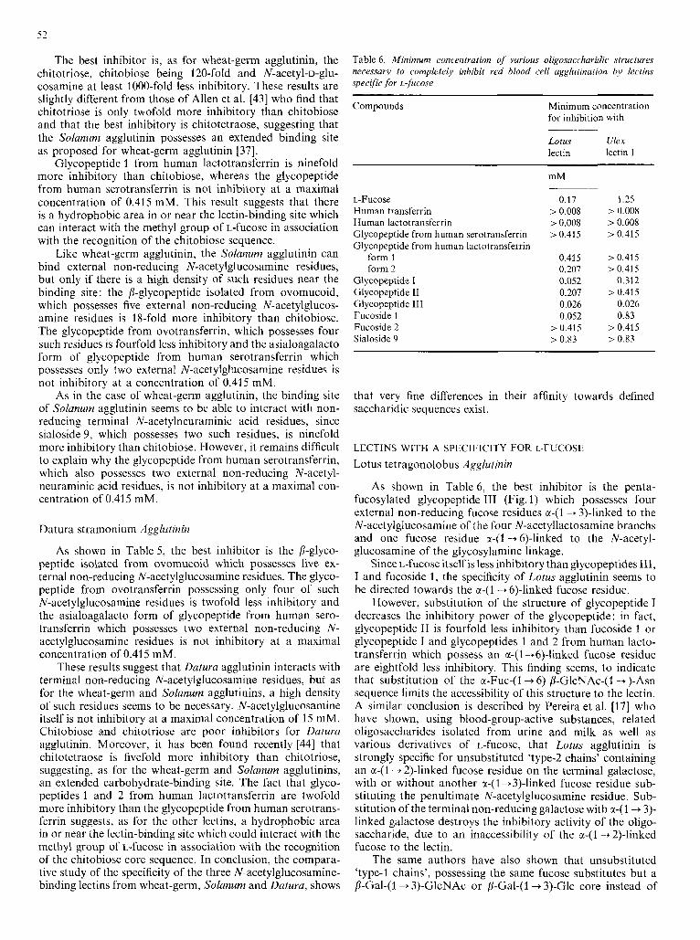
The best inhibitor is, as for wheat-germ agglutinin, the chitotriose, chitobiose being 120-fold and N-acetyl-D-glu- cosamine at least 1000-fold less inhibitory. These results are slightly different from those of Allen et al. [43] who find that chitotriose is only twofold more inhibitory than chitobiose and that the best inhibitory is chitotetraose, suggesting that the Solanum agglutinin possesses an extended binding site as proposed for wheat-germ agglutinin [37].
Glycopeptide 1 from human lactotransferrin is ninefold more inhibitory than chitobiose, whereas the glycopeptide from human serotransferrin is not inhibitory at a maximal concentration of 0.415 mM. This result suggests that there is a hydrophobic area in or near the lectin-binding site which can interact with the methyl group of L-fucose in association with the recognition of the chitobiose sequence.
Like wheat-germ agglutinin, the Solanum agglutinin can bind external non-reducing N-acetylglucosamine residues, but only if there is a high density of such residues near the binding site : the P-glycopeptide isolated from ovomucoid, which possesses five external non-reducing N-acetylglucos- amine residues is 18-fold more inhibitory than chitobiose. The glycopeptide from ovotransferrin, which possesses four such residues is fourfold less inhibitory and the asialoagalacto form of glycopeptide from human serotransferrin which possesses only two external N-acetylglucosamine residues is not inhibitory at a concentration of 0.415 mM.
As in the case of wheat-germ agglutinin, the binding site of Solanum agglutinin seems to be able to interact with non- reducing terminal N-acetylneuraminic acid residues, since sialoside 9, which possesses two such residues, is ninefold more inhibitory than chitobiose. However, it remains difficult to explain why the glycopeptide from human serotransferrin, which also possesses two external non-reducing N-acetyl- neuraminic acid residues, is not inhibitory at a maximal con- centration of 0.415 mM.
Datura stramonium Agglutinin
As shown in Table 5, the best inhibitor is the P-glyco- peptide isolated from ovomucoid which possesses five ex- ternal non-reducing N-acetylglucosamine residues. The glyco- peptide from ovotransferrin possessing only four of such N-acetylglucosamine residues is twofold less inhibitory and the asialoagalacto form of glycopeptide from human sero- transferrin which possesses two external non-reducing N- acetylglucosamine residues is not inhibitory at a maximal concentration of 0.415 mM.
These results suggest that Datura agglutinin interacts with terminal non-reducing N-acetylglucosamine residues, but as for the wheat-germ and Solanum agglutinins, a high density of such residues seems to be necessary. N-acetylglucosamine itself is not inhibitory at a maximal concentration of 15 mM. Chitobiose and chitotriose are poor inhibitors for Datura agglutinin. Moreover, it has been found recently [44] that chitotetraose is fivefold more inhibitory than chitotriose, suggesting, as for the wheat-germ and Solanum agglutinins, an extended carbohydrate-binding site. The fact that glyco- peptides l and 2 from human lactotransferrin are twofold more inhibitory than the glycopeptide from human serotrans- ferrin suggests, as for the other lectins, a hydrophobic area in or near the lectin-binding site which could interact with the methyl group of L-fucose in association with the recognition of the chitobiose core sequence. In conclusion, the compara- tive study of the specificity of the three N-acetylglucosamine- binding lectins from wheat-germ, Solanum and Datura, shows
Table 6. Minimum Concentration of various oligosaccharidic structures necessary to completely inhibit red bfood cell agglutination by lectins specific for L-jiucose
Compounds Minimum concentration for inhibition with
Lotus Ulex lectin lectin I
L-Fucose Human transferrin Human lactotransferrin Glycopeptide from human serotransferrin Glycopeptide from human lactotransferrin
form 1 form 2
Glycopeptide I Glycopeptide 11 Glycopeptide I11 Fucoside 1 Fucoside 2 Sialoside 9
mM
0.17 > 0.008 > 0.008 > 0.415
0.415 0.207 0.052 0.207 0.026 0.052
> 0.415 > 0.83
1.25 > 0.008 > 0.008 > 0.415
> 0.415 > 0.415
0.312 > 0.415
0.026 0.83
> 0.415 > 0.83
that very fine differences in their affinity towards defined saccharidic sequences exist.
LECTINS WITH A SPECIFICITY FOR L-FUCOSE
Lotus tetragonolobus Agglutinin
As shown in Table 6, the best inhibitor is the penta- fucosylated glycopeptide I11 (Fig. 1) which possesses four external non-reducing fucose residues M-(1 -+ 3)-linked to the N-acetylglucosamine of the four N-acetyllactosamine branchs and one fucose residue a-(I -+ 6)-linked to the N-acetyl- glucosamine of the glycosylamine linkage.
Since L-fucose itself is less inhibitory than glycopeptides 111, I and fucoside 1, the specificity of Lotus agglutinin seems to be directed towards the ~ ( 1 -+ 6)-linked fucose residue.
However, substitution of the structure of glycopeptide I decreases the inhibitory power of the glycopeptide: in fact, glycopeptide I1 is fourfold less inhibitory than fucoside 1 or glycopeptide I and glycopeptides 1 and 2 from human lacto- transferrin which possess an u-( 2 +6)-linked fucose residue are eightfold less inhibitory. This finding seems, to indicate that substitution of the a-Fuc-(I -+ 6)-/$GlcNAc-(l -+ )-Asn sequence limits the accessibility of this structure to the lectin. A similar conclusion is described by Pereira et al. [I71 who have shown, using blood-group-active substances, related oligosaccharides isolated from urine and milk as well as various derivatives of L-fucose, that Lotus agglutinin is strongly specific for unsubstituted ‘type-2 chains’ containing an a - ( l - + 2)-linked fucose residue on the terminal galactose, with or without another a-(1 +3)-linked fucose residue sub- stituting the penultimate N-acetylglucosamine residue. Sub- stitution of the terminal non-reducing galactose with a-(I -+ 3)- linked galactose destroys the inhibitory activity of the oligo- saccharide, due to an inaccessibility of the cr-(I-+ 2)-linked fucose to the lectin.
The same authors have also shown that unsubstituted ‘type-I chains’, possessing the same fucose substitutes but a p-Gal-( 1 -+ 3)-GlcNAc or /$Gal-( 1 -+ 3)-Glc core instead of
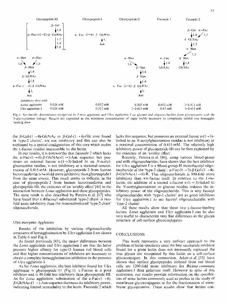
Glycopeptide 111
0-Gal
4 1 9 .-Fuc-( 1 +3)-fi-Gl~NA,
a-Man a-Man
I,& J1,3
+1>4
+ 1,
0-Man 4 1 9
0-GlcNAc
a-Fuc (1 + 6) p GlcNAc
Asn
Inhibitory dose with Lotus agglutinin 0.026 mM Ulex agglutinin 1 0.026 mM
Glycopeptide 1 Glycopeptide I1
a-Fuc-( 1+6)-O-GlcNAc
4 1, Asn
a-Man
1 2 - X P-Man
1,4 11 Man
i 1 , 4 a-Fuc-( 1+6)-p-GlcNAc +
Asn
0.052 mM 0.207 mM 0.3 12 mM > 0.41 5 mM
Fucoside I
a-Fuc
4 1,6 GlcNAc
0.052 mM 0.83 mM
Fucoside 2
$-Gal a-Fuc + 1,4 J 1,3
41’2
0-GlcNAc
a-Man
/1 ,3 P-?Aan
4 1 9 GlcNAc
> 0.41 5 mM > 0.41 5 mM
Fig. 6 . Swcliaridic d(,rcvnzinutirs rec.ognixd /iy Lotus ugglurinin and Ulcx agglutinin I on gljcnm and oligosacclitrri~~~’.~ , f io t i i gl~wp,oreirz.\ wiih the N-glycosylamine linkage. Results are expressed as the minimum concentration of sugar (mM) necessary to completely inhibit one hcmagglu- h a t i n g dose
the /l-Gal-( 1 -+ 4)-GlcNAc or /l-Gal-(I + 4)-Glc core found in ‘type-2 chains’, are not inhibitory and this can also be explained by a spatial configuration of this core which makes the L-fucose residue inaccessible to the lectin.
In our results, it is noteworthy that fucoside 2 which lacks the a-Fuc-( 1 + 6)-/l-GlcNAc-(I -+)-Am sequence but pos- sesses an external fucose a-(1 -+3)-linked to an N-acetyl- glucosamine residue, is not inhibitory at a maximal concen- tration of 0.41 5 mM. However, glycopeptide 2 from human lactotransferrin is twofold more inhibitory than glycopeptide 1 from the same source. This result seems to indicate, in the case of glycopeptide 2 from human lactotransferrin and glycopeptide 111, the existence of an ‘avidity effect’[41] in the interaction between Lotus agglutinin and these glycopeptides. The same result is also described by Pereira et al. 1171 who have found that a difucosyl substituted ‘type-2 chain’ is two- fold more inhibitory than the monosubstituted ‘type-2 chain’ oligosaccharide.
Ulex europeus Agglutinin
Results of the inhibition by various oligosaccharidic structures of hemagglutination by Ulex agglutinin I are shown in Table 6 and Fig. 6.
As found previously [45], the major differences between the Lotus agglutinin and Ulex agglutinin I are that the latter presents higher affinity for type-0 human red blood cells and that higher concentrations of inhibitors are necessary to obtain a complete hemagglutination inhibition in the presence of Ulex agglutinin I.
As for Lotus agglutinin, the best inhibitor found for Ulex agglutinin is glycopeptide I11 (Fig. 1). L-Fucose is a poor inhibitor and is 48-fold less inhibitory than glycopeptide 111. As for Lotus agglutinin, substitution of the a-Fuc-(I + 6)- b-GlcNAc-(l -+ )-Am sequence decreases its inhibitory power, indicating limited accessibility to the lectin. Fucoside 2 which
lacks this sequence, but possesses an external fucose a-(1 -+3)- linked to an N-acetylglucosamine residue is not inhibitory at a maximal concentration of 0.415 mM. The relatively high inhibitory power of glycopeptide 111 can be then explained by the existence of an ‘avidity effect’.
Recently, Pereira et al. [46], using various blood-group and milk oligosaccharides, have shown that the best inhibitor for Ulex agglutinin I is a blood-group-H monofucosyl oligo- saccharide of the ‘type-2 chain’: cc-Fuc-(I +2)-/3-Gal-(1 -+ 4)- D-GlcNAc-(l+ 6)-R. This oligosaccharide is 900-fold more inhibitory than a-L-fucose itself. In contrast to the L0tu.s lectin, the addition of a second L-fucose a-(1 -+3)-linked to the N-acetylglucosamine or glucose residue reduces the in- hibitory power of the oligosaccharide. This is why fucosyl oligosaccharides with ‘type-1 chains’ are as good inhibitors for Ulex agglutinin I as are fucosyl oligosaccharides with ‘type-2 chains’.
All these results show that these two L-fucose-binding lectins, Lotus agglutinin and Ulex agglutinin I can be also very useful to characterize very fine differences in the glycan structure of cell-surface glycoconjugates.
CONCLUSIONS
This work represents a very indirect approach to the problem of lectin specificity since the best saccharide inhibitor found for a given lectin does not necessarily represent the true glycan site recognized by this lectin on a cell-surface glycoconjugate. In this connection, Adair et al. [33] have shown that surface glycopeptides isolated from red blood cells are 1200-fold more inhibitory for Ricinus comrnunis agglutinin I than galactose itself. However in spite of this restriction, our results provide information on the specifici- ties of some lectins commonly used as probes in the study of membrane glycoconjugates or for the fractionation of mem- brane glycoproteins. These results show that lectins con-
54
sidered 'identical' in terms of monosaccharide specificity, possess the ability to recognize fine differences in more com- plex structures.
Moreover, our results show that different lectins are able to recognize different saccharidic sequences, but belonging to the same glycan structure. As these sequences are likely to be common to numerous glycoproteins, including cell- membrane glycoproteins, the use of some of the lectins as a tool in the study of cell-surface carbohydrates seems to be limited. The same conclusion was drawn by Nilsson et al. [47] who found that different lectins bind to the same cell-surface glycoproteins. Kimura et al. [48] also arrive at this con- clusion and show that commonly used lectins such as con- canavalin A, Lens and wheat-germ agglutinins and Ricinus agglutinin I react with a broad spectrum of lymphocyte cell- surface glycoproteins.
The relative affinity of a lectin for a given glycan struc- ture, as expressed in the different tables, can be compared with the true association constant of the lectin for this glycan : the lower is the minimum amount of the glycan necessary to inhibit hemagglutination by this lectin, the higher is the association constant of the lectin for this saccharide. More- over, from these results, the ability of a given lectin insolu- bilized on Sepharose to fractionate oligosaccharides or glyco- peptides containing the saccharide sequence recognized by the lectin can be predicted.
In addition, our results confirm previous data concerning the spatial configuration of the glycan moiety of glyco- proteins. In fact, many lectins (Lens and Viciu agglutinins, and Ricinus agglutinins I and 11) present a greater affinity for saccharidic sequences substituted by sialic acid residues bound in the a-(2 -+ 6) position of galactose rather than in the a-(2 -+ 3) position and this can be related to the high rota- tional freedom of a-(2 + 6)-linkage. Some lectins with an extended binding site, such as Lens, Viciu and Pisum agglu- tinins, react to a greater extent with their specific oligo- saccharidic sequence, when this sequence belongs to a glyco- peptide possessing the chitobiose-asparagine core than with the derived oligosaccharides lacking the B-GlcNAc-(l+ )-Am sequence. These results can be related to the fact that the chitobiose core gives to the whole glycan a more rigid con- formation as opposed to the random configuration of the derived oligosaccharide ; however, the presence of charges on asparagine could also be involved in the binding.
Finally, our results show that many lectins (from Lens, Vicia, Pisum, Solanum and Datura) seem to possess in or near their carbohydrate-binding site, a hydrophobic area such as that described for concanavalin A [25] and this could be the cause of non-specific hydrophobic interactions between the lectin and residues of glycopeptides or glycoproteins, as de- scribed for concanavalin A [30,31].
This work was supported in part by the Centre National de la Recherche Scientifique L. A. 217: Relations Structure-Fonction des Con- stituants Membranaires and R. C. P. 529: Glucides et GlycoconjuguPs), by the Institut National de la Sunti et de la Recherche Me'dicale (U 124: Unite de Recherches Ultrastructurules et Biochimiques sur les Cellules Normules et Cance'reuses) and by the Delegution Gendrale a la Recherche Scientifique et Technique (Action concerrbe: Canct'rogen&r et Pharma- cologie du Cancer, Contract 79-7-0669). We are indebted to Prof. Michel Monsigny for his gift of potato agglutinin, to Dr Andre Chtron for providing bovine milk lactotransferrin glycopeptide and to Mrs Renee Debray-Vandersyppe and Monique Benai'ssa for their skilful technical assistance. Thanks are due to Prof. Michel Monsigny and Prof. Nathan Sharon for helpful advice and critical reading of the manusript.
REFERENCES
1. Lis, H. & Sharon, N . (1977) in The Antigens (Sela, M., ed.) vol. 4,
2. Goldstein, I. J. & Hayes, C. E. (1978) Adv. Carbohydr. Chem. Bio-
3. Kornfeld, R. & Ferris, C. (1975) J . B i d . Chem. 250, 2614-2619. 4. Debray, H., Decout, D., Strecker, G., Montreuil, J . & Monsigny,
M. (1978) Proc. 9th Int. Symp. Curhohydr. Chem. 385-386. 5. Debray, H., Decout, D., Strecker, G., Montreuil, J . & Monsigny,
M. (1979) Protides Biol. Fluid,T Proc. Colloq. Brussels, 27,451 - 454. 6. Laemmli, U . K . (1970) Nature (Lond.) 227, 680-685. 7. Toyoshima, S., Osawa, T. & Tonomura, A. (1970) Biochim. Biophys.
8. Howard, I. K., Sage, H . J., Stein, M. D., Young, N. M., Leon, M.
9. Allen, H. J . & Johnson, E. A. Z. (1976) Biochim. Biophys. Acta,
10. Entlicher, G., Kostir, J. V. & Kocourek, J. (1970) Biochim. Biophys.
11. Nicolson, G. L., Blaustein, J. & Etzler, M. E. (1974) Biochemistry,
12. Gordon, J. A,, Blumberg, S., Lis, H. & Sharon, N. (1972) FEBS
13. Lotan, R., Gussin, A. E. S., Lis, H. & Sharon, N. (1973) Biochem.
14. Delmotte, F., Kieda, C. & Monsigny, M. (1975) FEBS Lett. 53,
15. Blumberg, S., Hildesheim, J., Yariv, J. & Wilson, K. J. (1972) Bio-
16. Kalb, A. J. (1968) Biochim. Biophys. Acta, 168, 532-536. 17. Pereira, M. E. A. & Kabat, E. A. (1974) Biochemistry, 13, 3184-
18. Flory, L. L. (1966) Vox Sang. 11, 137-156. 19. Matsumoto, I. & Osawa, T. (1970) Arch. Biochem. Biophys. 140,
20. Strecker, G. & Montreuil, J. (1979) Biochimie (Paris) 61, 1199-
21. Montreuil, J . (1975) Pure Appf. Chem. 42, 431 -477. 22. Spik, G., Bayard, B., Fournel, B., Strecker, G., Bouquelet, S. &
Montreuil, J . (1975)FEBSLett. 50,296-299. 23. Baenziger, J . U. & Fiete, D. (1979) J . Biol. Chem. 254, 2400-2407. 24. Allen, A. K., Desai, N. N. & Neuberger, A. (1976) Biochem. J . 155,
25. Loontiens, F. G., Van Wauwe, J . P., De Gussem, R. & De Bruyne,
26. Kornfeld, S., Rogers, J. & Gregory, W. (1971) J . Bid. Chem. 246,
27. Montreuil, J., Fournet, B., Spik, G. & Strecker, G. (1978) C.R.
28. Montreuil, J . & Vliegenthart, J. F. G. (1979) Proc. 5th Int. Symp.
29. Van Wauwe, J . P., Loontiens, F. G. & De Bruyne, C. K. (1975) Bio-
30. Davey, M. W., Huang, J. W., Sulkowski, E. & Carter, W. A. (1974)
31. Davey, M. W., Sulkowski, E. & Carter, W. A. (1976) Biochemistry,
32. Baenziger, J. U. ;t Fiete, D. (1979) J . Biol. Chem. 254, 9795-9799. 33. Adair, W. L. & Kornfeld, S. (1974) J . Biol. Chem. 24Y, 4696-4704. 34. Pereira, M. E. A,, Kabat, E. A. & Sharon, N. (1974) Carbohydr.
35. Hammarstrom, S., Murphy, L. A., Goldstein, I. J. & Etzler, M. E.
36. Irimura, T., Kawaguchi, T., Terao, T. & Osawa, T. (1975) Carbo-
37. Allen, A. K., Neuberger, A. & Sharon, N. (1973) Biochem. 1. 131,
38. Goldstein, I. J., Hammarstrom, S. & Sundblad, S. (1975) Biochim.
39. Monsigny, M., Delmotte, F. & Helene, C. (1978) Proc. Nut1 Acad.
pp. 429-529, Academic Press, New York.
chem. 35,127 - 340.
Acta, 221, 514-521.
A. & Dyckes, D. F. (1971) J . Bid. Chem. 246, 1590-1595.
444, 374- 385.
Acta, 221, 272-281.
13, 196-204.
Lett. 24, 193- 196.
Biophys. Res. Commun. 52, 656 - 662.
324- 330.
chim. Biophys. Aria, 264, 171 - 176.
3 192.
484-491.
1246.
127- 135.
C. K. (1973) Curbohydr. Res. 30, 51 -62.
6581 -6586.
Hehd. Seances Acad. Sci. Ser. D. Sri. Nut. 287, 837-840.
Glycoconjugutes, I , 35 - 78.
chim. Biophys. Actu, 379, 456-461.
J . Biol. Chem. 249,6354-6355.
IS, 704-713.
Res. 37, 89-102.
(1977) Biochemistry, 16, 2750- 2755.
hydr. Res. 39, 317-327.
155 - 162.
Biophjn. Acta, 405, 53-61.
Sci. USA, 75, 1324-1328.
55
40. Bhavanandan, V. P. & Katlic, A. W. (1979) J . Bid. Clzem. 254, 45. Allen, H. J., Johnson, E. A. Z. & Matta, K . L. (1977) lmmunol.
41. Monsigny, M., Roche, A. C., Sene, C., Maget-Dana, R. & Del- 46. Pereira, M. E. A,, Kisailus, E. C., Gruezo, F. Kahai, E. A. (1978)
42. Petcrs, R. P., Ebisu, S., Goldstein, I . J. & Flashner, M. (1979) Bio-
43. Allen, A. K. & Neuberger, A. (1973) Biochem. J . 135, 307-334. 44. Kilpatrick, D. C. & Yeoman, M. M. (1978) Biachrm. J . 175, 1151 -
4000-4008.
motte, F. (1980) Eur. J . Biochem. 104, 147-153.
chemistry, 18, 5505 - 551 1,
Commun. 6, 585 - 602.
Arch. Biochrm. Biophys. IHS, 108- 115. 47. Nilsson, S. F. & Waxdal, M . J. (1978) Biochemi.stry, 17, 903-910. 48. Kimura, A., Wigzell, H., Holmquist, G., Ersson, B. & Carlsson, P.
(1979) J . Exp. Med. 149, 473-484.
1153.
H. Debray*, D. Dccout, G. Strecker, G. Spik, and J . Montreuil, Laboratoire de Chimie Biologique, UniversitC des Sciences et Techniqucs de Lille I, Boire postale 36, F-59650 Villeneuve d’Ascq, France ~~ ~.
* To whom correspondcncc should be addressed.