Embed Size (px)
Citation preview
Korean J. Chem. Eng., 23(4), 669-671 (2006)
SHORT COMMUNICATION
669
†To whom correspondence should be addressed.
E-mail: [email protected]
Spectral dependency of Eu2+-activated silicate phosphors on the compositionfor LED application
Sang Hyeon Kim, Hyo Jin Lee, Kyeong Phil Kim and Jae Soo Yoo†
Department of Chemical Engineering, ChungAng University, Seoul 156-756, Korea(Received 27 April 2005 • accepted 2 February 2006)
Abstract−An Sr2SiO4-Ba2SiO4 material system doped by Eu2+ was studied for light emitting diodes (LEDs) applica-
tion. The main concern was the precise control of excitation and emission spectra for maximum light yield and color
coordinate, which was carried out by changing the composition of the alkaline earth ions in host lattice. The Sr2SiO4 :
Eu-Ba2SiO4 : Eu system was found to be excellent for white LED applications with excitation in the 380-465 nm region.
Especially, the yellow light intensity from (Sr,Ba)SiO4 : Eu phosphors was comparable to YAG : Ce phosphors in case
of blue LED excitation.
Key words: White LED, Phosphor, Silicate, Spectrum
INTRODUCTION
Recently, intensive attention has been given to the development
of white light emitting diodes (LEDs), because of their high effi-
ciency as well as longevity. Phosphor-converted white LEDs have
long been in the commercial markets. A structure widely reported
is the use of blue-emitting LED covered by a yellow emitting phos-
phor such as Y3Al5O12 : Ce (YAG:Ce), encapsulated in an epoxy
resin [Narendran et al., 2004]. They are used for the backlight of a
color cellular phone and the illumination lamp in an automobile. To
date, many kinds of phosphors have been selected and even many
new materials have been reported for the LED application [Starick
et al., 2003; Proceedings of Phosphor Global Summit, 2004]. For
the device application, it is very important to obtain the proper white
CIE (International Commission on Illumination) point for realizing
full color images as well as high efficiency. The white color coordi-
nate is very sensitive to the emission spectrum of the phosphors in
case of the phosphor-converted LED. Also, to obtain the maximum
quantum yield, the excitation spectrum of the phosphor needs to be
overlapped by the emission spec- trum from LED as much as pos-
sible. The emission spectrum of the LED, which is available and
high efficiency, shows peaks near 405 nm or 460 nm. Therefore,
much effort has gone into development of phosphors with the proper
emission (550-700 nm) and excitation (380-470 nm) wavelengths.
As of now, most phosphor-converted LEDs have used Y3Al5O12
(YAG) doped by Ce3+, which is excited by blue light (~460 nm) and
emits orange light (~560 nm). Besides Ce3+ ions, the ions of Eu2+,
Tb3+, and Mn2+ are known to have luminescent transitions suitable
for LED applications, but proper selection of the host lattice remains
a great challenge to phosphor engineers. The main factors respon-
sible for different spectral properties of given activator ions in differ-
ent host lattices are known to be covalency and crystal field around
activator ions.
Blasse et al. reported on the fluorescence of Eu2+-activated sili-
cates [Blasse et al., 1968]. The main factors controlling the excita-
tion and emission wavelengths of a given activator ion were the
host lattice and the alkaline-earth ion. Poort et al. [1996] examined
the luminescence of Eu2+ in silicate host lattices with alkaline earth
ions and reported that the spectrum peaks could be controlled by
the preferential orientation of a d orbital in the chain direction. Re-
cently, we reported that a very efficient blue-emitting silicate phos-
phor could be excited near 400 nm [Hong et al., 2003].
In this work, we focused on the variation of crystal fields by chang-
ing the ratio of the Ba/Sr in the Sr2SiO4-Ba2SiO4 material system.
The proper control of PLE (Photoluminescence Emission) and PL
(Photoluminescence) was the major concern of this work in that
the orange light would be maximized near 465-nm excitation.
EXPERIMENTAL
The starting materials for the phosphor were high purity BaCO3,
SrCO3, SiO2, and Eu2O3, all 99.99% pure. Stoichiometric amounts of
the starting materials were mixed thoroughly, and NH4Cl (99.99%)
was added as a flux. The mole fraction of the NH4Cl flux was taken
as an independent variable to maximize the light output. The weighed
materials were ball-milled for longer than 48 hours in isopropyl al-
cohol. After evaporating the alcohol in air, the samples were pre-
heated in a muffle furnace at 600 oC for 4 hrs. Then, they were heat-
treated in the reducing atmosphere (N2+2% H2) at a temperature
between 1,200 and 1,400 oC for several hours. The final reaction
time and temperature were optimized for maximum PL intensity.
After washing with DI (Distillation) water to remove the flux, the
samples were sintered again for 3 hr at 1,300 oC in N2+2% H2 to
improve crystallinity.
The phosphor powders were characterized by scanning electron
microscopy (SEM-Philips Model 515). The crystal structure was iden-
tified by X-ray diffractometry (XRD-Scintag, Model SDS 2000) us-
ing Cu Kα (λ= 1.540562). The excitation spectra were measured by
photoluminescence on an optical bench using scanned monochro-
matized excitation from a xenon lamp (ORC Model LH1751300),
while the emission spectra were measured at a particular excitation
670 S. H. Kim et al.
July, 2006
wavelength with a second monochromater and photomultiplier de-
tector.
RESULTS AND DISCUSSION
This work was undertaken in an attempt to study the spectral var-
iation in excitation as well as in emission spectra for LED applica-
tions. The emission and absorption spectra of the Eu2+ are broad
due to the transitions between the 4f7 ground state and the crystal
field components of the 4f65d excited state configuration. The emis-
sion varies from ultraviolet to red, depending on the covalency, the
size of the cation, and the strength of the crystal field in host lattices.
Here, we partially replaced strontium ions with the Ba ions in a stron-
tium ortho-silicate system doped by Eu2+ activators to investigate
the spectral variation. Since the ionic radius of Eu2+ (0.117 nm) is
similar to that of Sr2+ (0.113 nm), Sr salts are believed to be suitable
host lattices for Eu2+ activation. Also, we assumed that Eu2+ ions
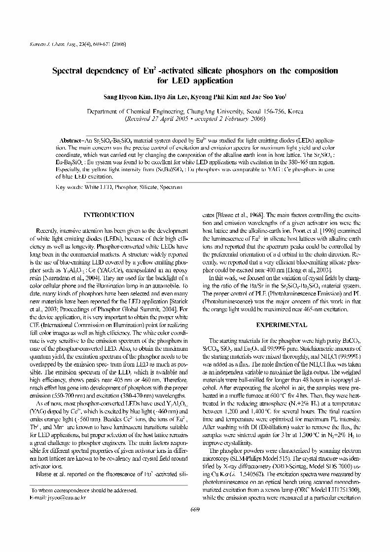
occupy Sr-site in host lattices. Typical X-ray diffraction patterns of
SrxBa2−xSiO4 : Eu phosphors in this work are shown in Fig. 1. As
all preparations are known to have a single-phase material [Barry,
1968], crystal planes were grown a little bit differently, depending
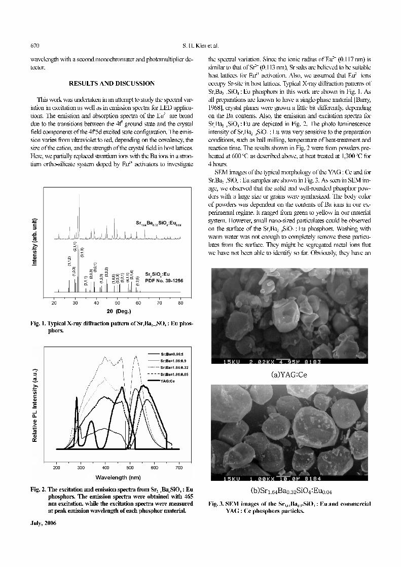
on the Ba contents. Also, the emission and excitation spectra for
SrxBa2−xSiO4 : Eu are depicted in Fig. 2. The photo luminescence
intensity of SrxBa2−xSiO4 : Eu was very sensitive to the preparation
conditions, such as ball milling, temperature of heat-treatment and
reaction time. The results shown in Fig. 2 were from powders pre-
heated at 600 oC as described above, at heat treated at 1,300 oC for
4 hours.
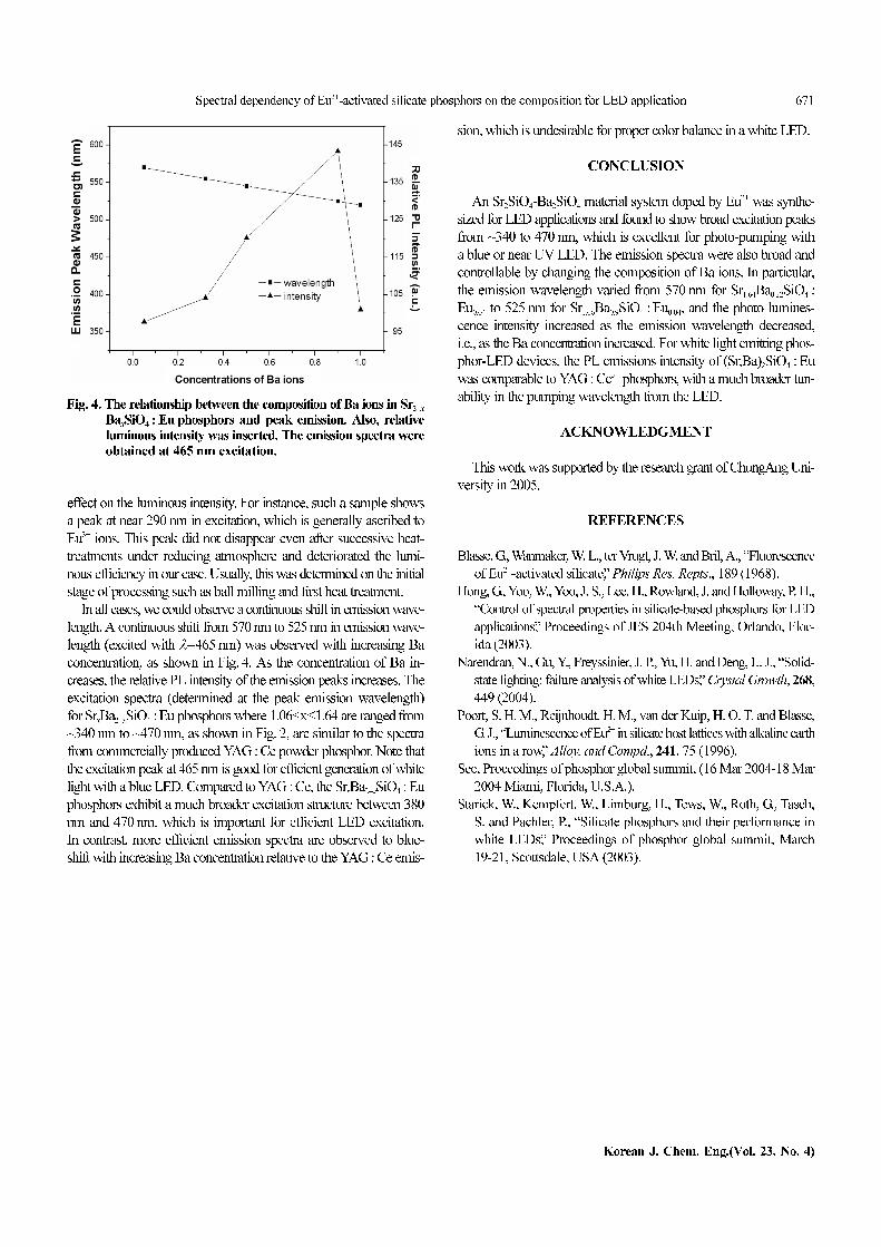
SEM images of the typical morphology of the YAG : Ce and for
SrxBa2−xSiO4 : Eu samples are shown in Fig. 3. As seen in SEM im-
age, we observed that the solid and well-rounded phosphor pow-
ders with a large size or grains were synthesized. The body color
of powders was dependent on the contents of Ba ions in our ex-
perimental regime. It ranged from green to yellow in our material
system. However, small nano-sized particulates could be observed
on the surface of the SrxBa2−xSiO4 : Eu phosphors. Washing with
warm water was not enough to completely remove these particu-
lates from the surface. They might be segregated metal ions that
we have not been able to identify so far. Obviously, they have an
Fig. 2. The excitation and emission spectra from Sr2−xBaxSiO4 : Eu
phosphors. The emission spectra were obtained with 465nm excitation, while the excitation spectra were measuredat peak emission wavelength of each phosphor material.
Fig. 1. Typical X-ray diffraction pattern of SrxBa2−xSO4 : Eu phos-
phors.
Fig. 3. SEM images of the Sr1.64Ba0.32SiO4 : Eu and commercialYAG : Ce phosphors particles.
Spectral dependency of Eu2+-activated silicate phosphors on the composition for LED application 671
Korean J. Chem. Eng.(Vol. 23, No. 4)
effect on the luminous intensity. For instance, such a sample shows
a peak at near 290 nm in excitation, which is generally ascribed to
Eu3+ ions. This peak did not disappear even after successive heat-
treatments under reducing atmosphere and deteriorated the lumi-
nous efficiency in our case. Usually, this was determined on the initial
stage of processing such as ball milling and first heat treatment.
In all cases, we could observe a continuous shift in emission wave-
length. A continuous shift from 570 nm to 525 nm in emission wave-
length (excited with λ=465 nm) was observed with increasing Ba
concentration, as shown in Fig. 4. As the concentration of Ba in-
creases, the relative PL intensity of the emission peaks increases. The
excitation spectra (determined at the peak emission wavelength)
for SrxBa2−xSiO4 : Eu phosphors where 1.06≤x≤1.64 are ranged from
~340 nm to ~470 nm, as shown in Fig. 2, are similar to the spectra
from commercially produced YAG : Ce powder phosphor. Note that
the excitation peak at 465 nm is good for efficient generation of white
light with a blue LED. Compared to YAG : Ce, the SrxBa2−xSiO4 : Eu
phosphors exhibit a much broader excitation structure between 380
nm and 470 nm, which is important for efficient LED excitation.
In contrast, more efficient emission spectra are observed to blue-
shift with increasing Ba concentration relative to the YAG : Ce emis-
sion, which is undesirable for proper color balance in a white LED.
CONCLUSION
An Sr2SiO4-Ba2SiO4 material system doped by Eu2+ was synthe-
sized for LED applications and found to show broad excitation peaks
from ~340 to 470 nm, which is excellent for photo-pumping with
a blue or near UV LED. The emission spectra were also broad and
controllable by changing the composition of Ba ions. In particular,
the emission wavelength varied from 570 nm for Sr1.64Ba0.32SiO4 :
Eu0.04 to 525 nm for Sr1.06Ba0.9SiO4 : Eu0.04, and the photo lumines-
cence intensity increased as the emission wavelength decreased,
i.e., as the Ba concentration increased. For white light emitting phos-
phor-LED devices, the PL emissions intensity of (Sr,Ba)2SiO4 : Eu
was comparable to YAG : Ce3+ phosphors, with a much broader tun-
ability in the pumping wavelength from the LED.
ACKNOWLEDGMENT
This work was supported by the research grant of ChungAng Uni-
versity in 2005.
REFERENCES
Blasse, G., Wanmaker, W. L., ter Vrugt, J. W. and Bril, A., “Fluorescence
of Eu2+-activated silicate,” Philips Res. Repts., 189 (1968).
Hong, G., Yoo, W., Yoo, J. S., Lee, H., Rowland, J. and Holloway, P. H.,
“Control of spectral properties in silicate-based phosphors for LED
applications,” Proceedings of JES 204th Meeting, Orlando, Flor-
ida (2003).
Narendran, N., Gu, Y., Freyssinier, J. P., Yu, H. and Deng, L. J., “Solid-
state lighting: failure analysis of white LEDs,” Crystal Growth, 268,
449 (2004).
Poort, S. H. M., Reijnhoudt, H. M., van der Kuip, H. O. T. and Blasse,
G. J., “Luminescence of Eu2+ in silicate host lattices with alkaline earth
ions in a row,” Alloy. and Compd., 241, 75 (1996).
See, Proceedings of phosphor global summit, (16 Mar 2004-18 Mar
2004 Miami, Florida, U.S.A.).
Starick, W., Kempfert, W., Limburg, H., Tews, W., Roth, G., Tasch,
S. and Pachler, P., “Silicate phosphors and their performance in
white LEDs,” Proceedings of phosphor global summit, March
19-21, Scottsdale, USA (2003).
Fig. 4. The relationship between the composition of Ba ions in Sr2−x
BaxSiO4 : Eu phosphors and peak emission. Also, relative
luminous intensity was inserted. The emission spectra wereobtained at 465 nm excitation.