Embed Size (px)
Citation preview

Contributions to Zoology, 84 (1) 1-12 (2015)
Spectral transmittance of the spectacle scale of snakes and geckos
Kevin van Doorn1, 2, Jacob G. Sivak1
1 School of Optometry and Vision Science, Faculty of Science, University of Waterloo, 200 University Avenue West, Waterloo, Ontario, Canada N2L 3G1 2 E-mail: [email protected]
Key words: Gekkonidae, keratin, Serpentes, Squamata
Abstract
The spectral transmittance of the optical media of the eye plays a substantial role in tuning the spectrum of light available for capture by the retina. Certain squamate reptiles, including snakes and most geckos, shield their eyes beneath a layer of transparent, cornified skin called the ‘spectacle’. This spectacle offers an added opportunity compared with eyelidded animals for tayloring the spectrum. In particular, the hard scale that cov-ers the surface of the spectacle provides a unique material, keratin, rarely found in vertebrate eyes, a material which may have unique spectral properties. To verify this, shed snake and gecko skins were collected and the spectral transmittance of spectacle scales was spectrophotometrically analyzed. The spec-tacle scale was found generally to behave as a highpass filter with a cut-off in the ultraviolet spectrum where taxonomic variation is mostly observed. The spectacle scales of colubrid and elapid snakes were found to exhibit higher cut-off wave-lengths than those of pythonids, vipers, and most boids. Gecko spectacle scales in turn exhibited exceptional spectral transmit-tance through the visual spectrum down into the UV-B. It is suggested that this is due to the absence of beta-keratins in their spectacle scale.
Contents
Introduction ........................................................................................ 1Material and methods ....................................................................... 2 Sample collection ........................................................................ 2 Spectrophotometry ...................................................................... 3 Thickness measurements ........................................................... 3 Analytical methods ..................................................................... 3Results .................................................................................................. 3 Snake spectacle scale transmittance ...................................... 3 Gecko spectacle scale transmittance ..................................... 3 Spectacle scale thickness .......................................................... 5 Relationship between spectacle scale thickness and λ50% ... 5Discussion ........................................................................................... 5 Spectacle scale transmittance: taxonomic variation ......... 5 Considering the spectacle scale’s role as an optical filter .. 7 Considering the spectacle scale’s role as mechanical
barrier ............................................................................................ 8 Gecko versus snake spectacle scales and a discussion of
keratin composition .................................................................... 8
Conclusion ........................................................................................... 9Acknowledgements ........................................................................... 9References ........................................................................................... 9
Introduction
The optical media of the eye play a crucial role in tun-ing the spectrum of light incident upon the retina. For example, tissues may filter out short wavelengths of the blue and ultraviolet (UV) ranges to increase image contrast or block harmful radiation, such as occurs with the yellow crystalline lenses of some squirrels (Walls, 1931; Chou and Cullen, 1984), squamate rep-tiles (Walls, 1942; Röll et al., 1996; Röll, 2000) and fishes (Walls and Judd, 1933; Kennedy and Milkman, 1956; Muntz, 1973). The spectral transmittance and absorption of vari-ous ocular media (i.e. the cornea, lens, neural retina, and aqueous and vitreous humours) have been studied in all vertebrate taxa (reviewed in Douglas and Mar-shall, 1999), although data on reptiles remains some-what limited (Ellingson et al., 1995; Bowmaker et al., 2005), and the reptilian spectacle, despite its unique position in the optics of squamate eyes, has received surprisingly little attention (Safer et al., 2007; Hart et al., 2012). The spectacle is a layer of transparent skin that cov-ers the eyes of many squamates, including all snakes and most geckos (Fig. 1; Walls, 1942). Despite being the primary window through which these animals see, very few studies have investigated the spectral properties of the spectacle. Hart et al. (2012) and Safer et al. (2007) respectively reported on the transmittance of hydrophi-id sea snake spectacles and rattlesnake spectacle scales, the former measuring in the visible and UV range while the latter focused on the infrared spectrum, which is not of visual relevance. Given the unusual na-ture of the reptilian spectacle as an extra layer in the

2 Van Doorn & Sivak – Spectral transmittance of the spectacle scale
optical apparatus of the eye which may further absorb or reflect wavelengths that are unnecessary for or del-eterious to an animal’s vision (e.g. due to chromatic aberration or scatter, Sivak, 1982; Sivak and Mandel-man, 1982), an investigation of its optical properties over a broad range of species may be beneficial to bet-ter understand its contribution to vision in squamates. Reptilian spectacles consist of soft tissues (dermal stroma, epidermal epithelia, and conjunctiva) and hard keratin (the stratum corneum, referred to as the ‘spec-tacle scale’). The dermal stroma of the spectacle is similar to the cornea with its lamellar arrangement of highly organized collagen fibers (Da Silva et al., 2014) and is thus likely to exhibit similar spectral properties. The spectacle scale however presents a unique mate-rial in the optics of the eye, as keratinizing epithelia are typically absent from vertebrate eyes (the few known exceptions being the ant- or termite-eating echidna (Tachyglossus Illiger, 1811), armadillo (Dasy-pus Linnaeus, 1758) and aardvark (Orycteropus G. Cu-vier, 1798), all of which are reported to possess kerati-nized corneas (Walls, 1942; Duke-Elder, 1958)). As a result of its unique composition, the spectacle scale itself may exhibit unique spectral properties and provide a unique opportunity in the evolution of ocular filtering. Previous research by van Doorn et al. (2014) has shown that the biochemical composition of specta-cle scales varies taxonomically, differing between spe-cies and particularly between families, as well as be-
tween snakes and geckos, the latter of which lack one whole class of keratin proteins (the beta-keratins) thought to otherwise be present in all squamate scales (Maderson, 1985; Landmann, 1986). Thus if keratins vary in their transmissive properties, one could theo-rize that the spectral transmittance of spectacle scales may vary between snake families and between snakes and geckos. The research presented here, a study of the spectral transmittance of shed snake and gecko specta-cle scales, provides evidence that this is the case.
Material and methods
The experiments described here consisted of spectro-photometric measurements of snake and gecko specta-cle scales collected from shed skins. Because the spec-tral transmittance of a material typically correlates with its thickness, spectacle scale thicknesses were also measured.
Sample collection
Spectacle scales from 43 species of snake (6 boids, 7 pythonids, 10 viperids, 3 elapids, and 17 colubrids) and 2 species of gekkonid gecko were investigated. These were collected from shed skins donated by pri-vate pet owners and zoos. The species investigated, including all specimens of particular species, along
Spectacle Scales
Spectacle Scales
Fig. 1. Shed gecko (left) and snake (right) skins showing the dorsal head region and indicating the spectacle scales. Compared with other scales which are translucent at best and may be pigmented, the spectacle scales are optically transparent.

3Contributions to Zoology, 84 (1) – 2015
with the species’ authorities are summarized in Table 1. Because moulting snakes frequently soak them-selves to soften the skin prior to shedding, the sheds were air dried upon collection and stored for up to 2 months in paper envelopes to prevent spoilage. When kept under such conditions, spectacle scales have been found to retain their spectral properties over very long periods, up to and including several years (van Doorn, unpubl. data).
Spectrophotometry
Spectacle scales were cut from the sheds and mounted with adhesive tape to a sample holder equipped with either an 8 mm aperture for larger scales or a 1 mm aperture for smaller scales. The sample holder was placed within a Varian Cary 500 UV-VIS-IR dual-beam spectrophotometer such that the scanning beam was passed through the scale from front to back (i.e. the beam was incident upon the outer surface). Meas-urements were made from 200 to 750 nm in 2 nm in-crements. Published reports of keratin’s transmittance in both dry and wet states (Bendit and Ross, 1961; Bruls et al., 1984) have shown that hydration has a mi-nor effect on transmittance and that it doesn’t change the overall profile of transmittance curves. This is likely due to the spectral properties of water itself, no-tably that it exhibits modest absorption of long wave-lengths (i.e. in the red range) and very short wave-lengths (i.e. in the deep UV range), as well as its capa-bility as a thin film to reduce optical scatter by ‘smoothing out’ surface irregularities of the material. As a result, all scans of shed spectacle scales in these experiments were performed dry, particularly because the long scan times resulted in hydrated scales drying out mid-scan anyway, which could lead to slight defor-mation of the scale and small spurious vertical shifts in spectral transmittance. The measurements from both the right and left eyes of each specimen were av-eraged unless the shed had only one usable spectacle scale, in which case the reported measurements con-sist of solely the one.
Thickness measurements
A gauge designed for measuring the thickness of hard contact lenses was used to measure the thickness of the spectacle scales. Some scales were unavailable for thickness measurements, including those of the 3 elap-ids, due to having been used in an unrelated experi-ment.
Analytical methods
The 50% cut-off wavelength (λ50%), the boundary be-neath which >50% of the incident light is attenuated (either by absorption, reflection or scatter), was deter-mined for each sample from the raw data and rounded to the nearest integer. To even the representation of species in the analyses, specimens were weighted 1/n, where n is the number of specimens of a particular species that were available to a given test (N.B.: n may be lower for thickness analyses than for transmittance analyses due to availability of the scales as noted above). To determine if λ50% and spectacle scale thick-ness vary between families, Kruskal-Wallis analysis of variance on ranks was performed. Dunn’s method of multiple comparisons was used to clarify which fami-lies differed from which. A correlation on ranks (Spearman’s Rho) of λ50% versus thickness was calcu-lated to determine how much the latter contributes to the former. All analyses were done with Statistica 11.
Results
Snake spectacle scale transmittance
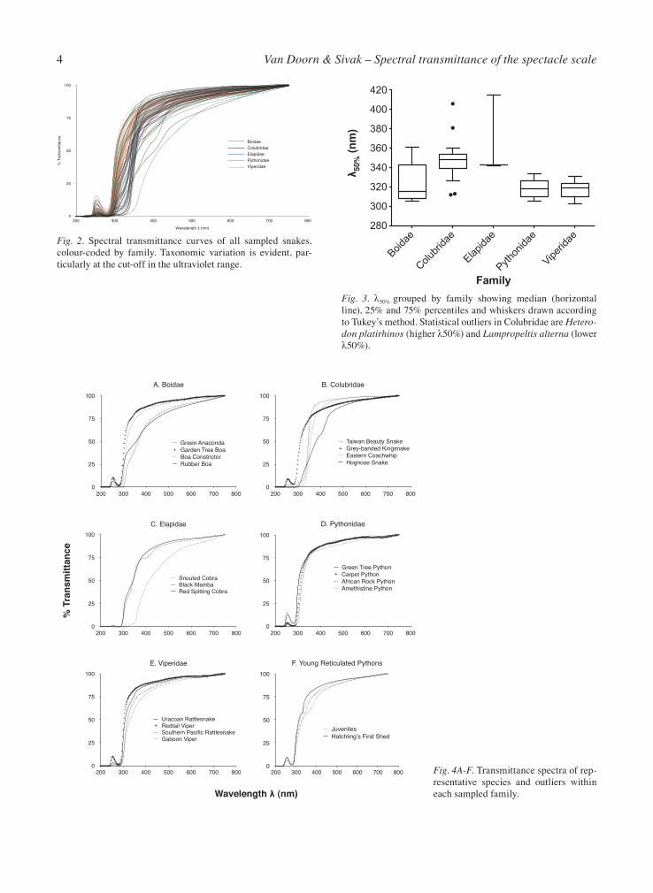
The spectral transmittance curves of all snake specta-cle scale samples are plotted in Fig. 2. In most species, the spectacle transmittance is relatively high from the far red to the near UV-A, although in many cases there is a slight reduction from long to short wavelengths. Most variation occurs within the UV range, with the cut-off wavelengths varying noticeably between species and families. The λ50% of individual sheds are reported in Table 1. The λ50% means, minima and maxima for each family are listed in Table 2 with a box plot shown in Fig. 3. Because Fig. 2 is too cluttered to allow proper evaluation of individual curves, the transmittance curves of a few representative species and outliers from each family are plotted in Figures 4A-E to aid with visual inspection. Boas generally have low λ50%’s as represented in Fig. 4A by Boa constrictor and the Garden Tree Boa. Two exceptions to this are the Green Anaconda and the Rub-ber Boa (the only erycine boa sampled), both of which exhibit higher cut-offs as well as a modest degree of at-tenuation of most wavelengths. The characteristic peak at 254 nm is also absent in the green anaconda. Colubrids, represented here mostly by North Amer-ican colubrine species, tend to exhibit higher λ50%’s similar to the Eastern Coachwhip and the Taiwan
4 Van Doorn & Sivak – Spectral transmittance of the spectacle scale
E. Viperidae
0
25
50
75
100
200 300 400 500 600 700 800
Uracoan RattlesnakeRedtail ViperSouthern Pacific RattlesnakeGaboon Viper
A. Boidae
0
25
50
75
100
200 300 400 500 600 700 800
Green AnacondaGarden Tree BoaBoa ConstrictorRubber Boa
B. Colubridae
0
25
50
75
100
200 300 400 500 600 700 800
Taiwan Beauty SnakeGrey-banded KingsnakeEastern CoachwhipHognose Snake
C. Elapidae
0
25
50
75
100
200 300 400 500 600 700 800
Snouted CobraBlack MambaRed Spitting Cobra
D. Pythonidae
0
25
50
75
100
200 300 400 500 600 700 800
Green Tree PythonCarpet PythonAfrican Rock PythonAmethistine Python
F. Young Reticulated Pythons
0
25
50
75
100
200 300 400 500 600 700 800
JuvenilesHatchling’s First Shed
Wavelength (nm)
% T
rans
mitt
ance
% T
rans
mitt
ance
0
25
50
75
100
Wavelength λ (nm)
200 300 400 500 600 700 800
BoidaeColubridaeElapidaePythonidaeViperidae
Boidae
Colubr
idae
Elapida
e
Python
idae
Viperid
ae280
300
320
340
360
380
400
420
Family
50
% (n
m)
Fig. 4A-F. Transmittance spectra of rep-resentative species and outliers within each sampled family.
Fig. 2. Spectral transmittance curves of all sampled snakes, colour-coded by family. Taxonomic variation is evident, par-ticularly at the cut-off in the ultraviolet range.
Fig. 3. λ50% grouped by family showing median (horizontal line), 25% and 75% percentiles and whiskers drawn according to Tukey’s method. Statistical outliers in Colubridae are Hetero-don platirhinos (higher λ50%) and Lampropeltis alterna (lower λ50%).
5Contributions to Zoology, 84 (1) – 2015
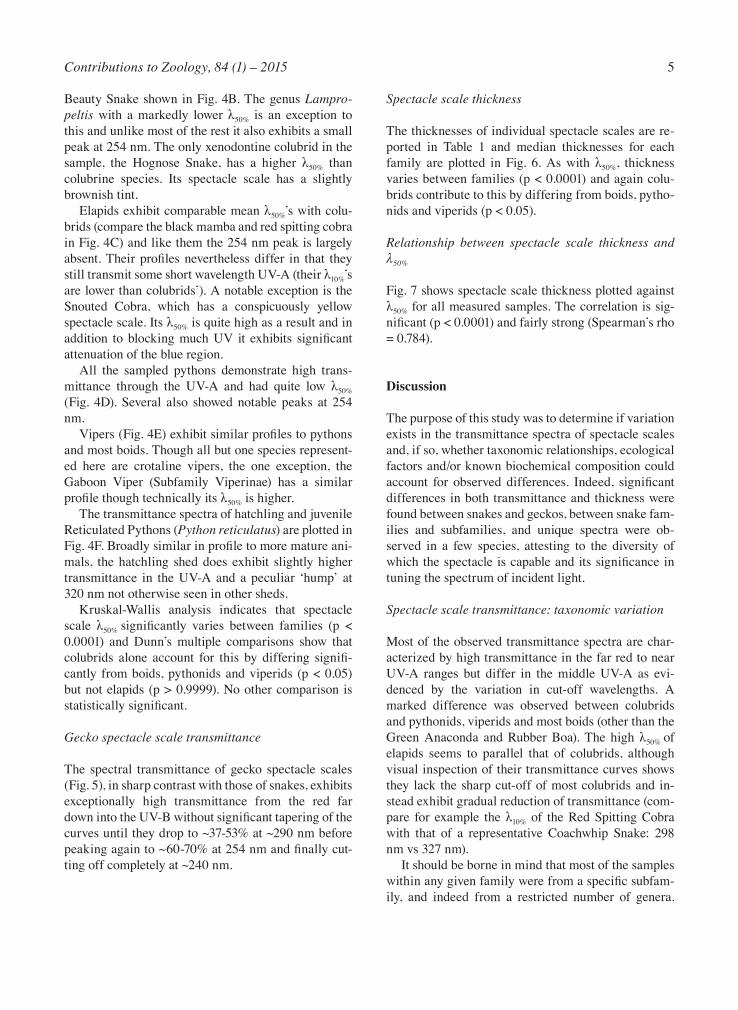
Beauty Snake shown in Fig. 4B. The genus Lampro-peltis with a markedly lower λ50% is an exception to this and unlike most of the rest it also exhibits a small peak at 254 nm. The only xenodontine colubrid in the sample, the Hognose Snake, has a higher λ50% than colubrine species. Its spectacle scale has a slightly brownish tint. Elapids exhibit comparable mean λ50%’s with colu-brids (compare the black mamba and red spitting cobra in Fig. 4C) and like them the 254 nm peak is largely absent. Their profiles nevertheless differ in that they still transmit some short wavelength UV-A (their λ10%’s are lower than colubrids’). A notable exception is the Snouted Cobra, which has a conspicuously yellow spectacle scale. Its λ50% is quite high as a result and in addition to blocking much UV it exhibits significant attenuation of the blue region. All the sampled pythons demonstrate high trans-mittance through the UV-A and had quite low λ50% (Fig. 4D). Several also showed notable peaks at 254 nm. Vipers (Fig. 4E) exhibit similar profiles to pythons and most boids. Though all but one species represent-ed here are crotaline vipers, the one exception, the Gaboon Viper (Subfamily Viperinae) has a similar profile though technically its λ50% is higher. The transmittance spectra of hatchling and juvenile Reticulated Pythons (Python reticulatus) are plotted in Fig. 4F. Broadly similar in profile to more mature ani-mals, the hatchling shed does exhibit slightly higher transmittance in the UV-A and a peculiar ‘hump’ at 320 nm not otherwise seen in other sheds. Kruskal-Wallis analysis indicates that spectacle scale λ50% significantly varies between families (p < 0.0001) and Dunn’s multiple comparisons show that colubrids alone account for this by differing signifi-cantly from boids, pythonids and viperids (p < 0.05) but not elapids (p > 0.9999). No other comparison is statistically significant.
Gecko spectacle scale transmittance
The spectral transmittance of gecko spectacle scales (Fig. 5), in sharp contrast with those of snakes, exhibits exceptionally high transmittance from the red far down into the UV-B without significant tapering of the curves until they drop to ~37-53% at ~290 nm before peaking again to ~60-70% at 254 nm and finally cut-ting off completely at ~240 nm.
Spectacle scale thickness
The thicknesses of individual spectacle scales are re-ported in Table 1 and median thicknesses for each family are plotted in Fig. 6. As with λ50%, thickness varies between families (p < 0.0001) and again colu-brids contribute to this by differing from boids, pytho-nids and viperids (p < 0.05).
Relationship between spectacle scale thickness and λ50%
Fig. 7 shows spectacle scale thickness plotted against λ50% for all measured samples. The correlation is sig-nificant (p < 0.0001) and fairly strong (Spearman’s rho = 0.784).
Discussion
The purpose of this study was to determine if variation exists in the transmittance spectra of spectacle scales and, if so, whether taxonomic relationships, ecological factors and/or known biochemical composition could account for observed differences. Indeed, significant differences in both transmittance and thickness were found between snakes and geckos, between snake fam-ilies and subfamilies, and unique spectra were ob-served in a few species, attesting to the diversity of which the spectacle is capable and its significance in tuning the spectrum of incident light.
Spectacle scale transmittance: taxonomic variation
Most of the observed transmittance spectra are char-acterized by high transmittance in the far red to near UV-A ranges but differ in the middle UV-A as evi-denced by the variation in cut-off wavelengths. A marked difference was observed between colubrids and pythonids, viperids and most boids (other than the Green Anaconda and Rubber Boa). The high λ50% of elapids seems to parallel that of colubrids, although visual inspection of their transmittance curves shows they lack the sharp cut-off of most colubrids and in-stead exhibit gradual reduction of transmittance (com-pare for example the λ10% of the Red Spitting Cobra with that of a representative Coachwhip Snake: 298 nm vs 327 nm). It should be borne in mind that most of the samples within any given family were from a specific subfam-ily, and indeed from a restricted number of genera.

6 Van Doorn & Sivak – Spectral transmittance of the spectacle scale
Some subfamilies represented here by a single species tend to demonstrate rather different transmittance spectra compared with the well represented subfamily. For example, among the colubrids, the xenodontine hognose snake showed the highest λ50% (mean: 382 nm, max: 406 nm), attenuating much of the UV-A spec-trum. Likewise among boids, the erycine Rubber Boa is second only to the Green Anaconda in its high λ50% (351 nm versus 361 nm, compared with a mean of 324 nm for the family as a whole), and the Gaboon Viper
Family Subfamily Mean λ50% (nm) Min λ50% Max λ50%
Boidae 323 305 361 Boinae 319 305 361 Erycinae (Charina bottae) 351 n/a n/aColubridae 347 312 406 Colubrinae 345 312 358 Xenodontinae 382 360 406 (Heterodon platirhinos) Elapidae 367 342 415Pythonidae 315 305 334Viperidae 317 303 331 Crotalinae 316 303 325 Viperinae (Bitis gabonica) 331 n/a n/a
Table 2. Mean spectacle scale λ50% for each snake family and subfamily as well as minima and maxima. Erycinae and Viperinae were represented by only one species, and Xenodontinae by 3 speci-mens of one species.
% T
rans
mitt
ance
0
25
50
75
100
Wavelength (nm)
200 300 400 500 600 700 800
Giant Day GeckoMarbled Gecko
% T
rans
mitt
ance
0200 300 400 500 600 700 800
Giant Day GeckoMarbled Gecko
% T
rans
mitt
ance
0200 300 400 500 600 700 800
Giant Day GeckoMarbled Gecko
Family
Thi
ckne
ss (
m)
Boidae
Colubrid
ae
Pythonid
ae
Viper
idae
0
20
40
60*
50
% (n
m)
0 20 40 60250
300
350
400
450
Spectacle Scale Thickness (µm)
Fig. 7. Correlation and regression plot of λ50% versus spectacle scale thickness. Data from all families are pooled and show a positive and significant correlation (Spearman s rho = 0.784, p < 0.05). A linear regression line is drawn to assist in visualizing the trend.
Fig. 5. Spectacle scale transmittance spectra in gekkonid geckos. Gecko spectacles exhibit exceptionally high transmit-tance through the visible and UV spectra, close to or at 100% until dropping somewhat in the UV-B, before peaking again at 254 nm in the UV-C.
Fig. 6. Plot of spectacle scale thicknesses grouped by family. The boxes show the median and 25th and 75th percentiles and the whiskers are drawn according to Tukey s method. Means are respectively 16 µm, 30 µm, 16 µm and 14 µm for Boidae, Colubridae, Pythonidae and Viperidae. Colubridae is seen to differ significantly from the other families in having a greater mean spectacle scale thickness (K-W p < 0.0001). In no other family does the highest value match or exceed the Colubridae mean (note that no elapids could be measured here).

7Contributions to Zoology, 84 (1) – 2015
has a higher λ50% than all crotaline vipers (331 nm ver-sus a mean of 317 nm). These findings warrant further investigation to determine if they are representative of their respective subfamilies. Spectacle scales’ λ50% correlates with their thick-ness although it’s unclear if thickness is the cause and λ50% the effect. The association may be indirect by vir-tue of both being characteristic of certain families, begging the question of why colubrid spectacle scales generally are thicker and have higher λ50% than other families, elapids and specific boids excluded. Also, ac-cording to the Spearman’s Rho of 0.784, λ50% and thickness are not perfectly related, so other factors may be at play here. To speculate on this, a considera-tion of the functional adaption of spectacle scale λ50% and thickness is called for.
Considering the spectacle scale’s role as an optical filter
Coloured ‘filters’ are present in the eyes of many spe-cies: the pigmented or iridescent corneas of numerous reef fish, the yellow lenses of some fish, squirrels and diurnal reptiles, the macula lutea of primates, and the photoreceptor oil droplets of birds and reptiles (Walls, 1942; Lythgoe, 1979; Douglas and Marshall, 1999; Hart, 2001). These are often associated with diurnal activity and are suggested to block harmful short wavelength radiation, to improve image contrast by removing shorter wavelengths that are more likely to scatter, or in the case of oil droplets to fine tune photo-receptor absorbance spectra to improve colour dis-crimination (reviewed in Douglas and Marshall, 1999). The spectacle scale’s contribution to overall transmit-tance of the eye is limited in most species to blocking the mid to far UV-A and UV-B, excepting the Snouted Cobra, Hognose Snake, and adult Spine-Bellied Sea Snake (Lapemis curtus (Shaw, 1802), Hart et al., 2012), all of which block much UV-A and some of the blue region of the spectrum. While the UV-A region spans a broad range from 315-400 nm, vision in this region will be restricted to the specific spectral absorbances of an animal’s retinal opsins. Several species of snake and gecko are known to possess UV-A sensitive cones (Loew, 1994; Elling-son et al., 1995; Loew et al., 1996; Sillman et al., 1997., 1999., 2001; Davies et al., 2009; Macedonia et al., 2009; Yang, 2010; Hart et al., 2012), suggesting that the visual perception of UV-A wavelengths is a com-mon trait throughout these taxa. The retinal absorb-ance spectra of four snakes included in this study have
been previously characterized (Thamnophis sirtalis (Sillman et al., 1997), Python regius (Sillman et al., 1999), Boa constrictor (Sillman et al., 2001), and Mas-ticophis flagellum (Macedonia et al., 2009)) with each found to possess a UV-sensitive opsin with a peak ab-sorbance near 360 nm, which is above the λ50% of all their spectacle scales but is remarkably close to it in the case of the Coachwhip Snake (max λ50% = 355 nm). T. sirtalis and the Coachwhip Snake are also known to possess yellow lenses (Walls, 1931) which likely pro-vide further UV blockage. The spectacle scale’s position as the initial optical filter may be advantageous in that it obviates the need for soft tissues vulnerable to intense radiation to per-form this function. However, only in colubrids, elapids, and the odd boid does the spectacle scale attenuate short wavelength UV, indicating that any ocular filtra-tion that might occur in the other boids and in vipers and pythons will nevertheless be accomplished by cel-lular tissues or the humours. While UV is implicated in cataract development and retinal damage (Sliney, 1986; Taylor, 1989; Gelatt et al., 2013), it has also been impli-cated in damage of the ocular surface itself, as in cer-tain cases of conjunctival neoplasms and keratitis (Wu et al., 2006; Gelatt et al., 2013). In humans, UV may also influence the development of pterygium which is characterized by anomalous growth of conjunctival tis-sue from the sclera or limbus over and into the corneal surface (Moran and Hollows, 1984). Mechanisms to minimize radiation-induced damage to the ocular sur-face should therefore be present in most species ex-posed to some amount of UV. For the species that bear it, the spectacle may be one such protective structure. One may hypothesize that the coachwhip snake’s sharp λ50% at ~350 nm may have been an adaptation to its di-urnal activities in arid habitats, but the evidence is cir-cumstantial as most colubrids in this study, diurnal or not, and regardless of habitat, have high cut-offs (genus Lampropeltis being the curious exception). Given that several of the vipers in this study (North and Central American rattlesnakes) are deserticolous and active di-urnally during some seasons (Landreth, 1973; Golan et al., 1982), it would have been compelling were their spectacle scales to exhibit high λ50% as a protective bar-rier to UV, but this is clearly not the case. A tangential, but interesting correlation in this regard is the presence of slit or near-slit pupils among the vipers, boas and pythons compared with the rounded pupils of all the sampled colubrids and elapids. Because the crystalline lenses of many colubrids (e.g. genera Masticophis, Col-uber, Elaphe) protrude through the pupil, a lower limit

8 Van Doorn & Sivak – Spectral transmittance of the spectacle scale
is set upon the constricted pupil diameter (Lampropeltis can constrict its pupil to rather small dimensions [see photo in Coborn, 1991: 251]). Vipers, boas and pythons are not limited in this regard and can constrict their pu-pils to smaller areas. While the pupil obviously plays no role in tuning the spectrum, it nevertheless regulates the absolute luminous flux within the eye, which may be protective in itself. An investigation of diurnally-active slit-pupilled colubrids (e.g. some members of subfamily Lycodontinae such as Oligodon ornatus Van Denburgh, 1909) may shed light on whether such a correlation ex-ists between pupil shape and spectacle transmittance. The conspicuous coloration of the snouted cobra’s yellow spectacle stands out in recalling the yellow lens-es and corneas of some diurnal terrestrial vertebrates, including the lenses of some snakes and geckos (Walls, 1931; Walls and Judd, 1933; Walls, 1942) and the lenses and corneas of certain fishes (Walls and Judd, 1933; Walls, 1942; Kennedy and Milkman, 1956; Muntz, 1973), which are suggested to function as barriers to UV and/or to increase retinal image contrast. The snouted cobra is not unique among snakes in possessing a yellow spectacle however as the adult Spine-Bellied Sea Snake’s spectacle blocks short wavelengths to a similar degree as the snouted cobra and, remarkably, to a much greater degree than the juvenile form of the spe-cies as reported by Hart et al. (2012). Because Hart et al. (2012) reported on the whole spectacle, dermis and scale together, it is unknown which layer accounts for the attenuation. The somewhat brown colouration of the hognose snake spectacle scale as observed in this study may also function as a modest filter. The chemical na-ture of spectacle scale colouration is not known, but may conceivably be related to its specific keratin iso-forms or fiber arrangement or it may be contributed by pigments deposited in the scale during keratogenesis or alternatively, it may result from staining by the animals’ substrate, such as by tannins or quinone pigments. Spectrophotometric measures and biochemical analy-ses on shed skins collected in the field or from captive animals kept on specific substrates would be valuable in determining the influence of environmental stains on spectacle scale pigmentation.
Considering the spectacle scale’s role as mechanical barrier
In addition to blocking more deep UV-A, a thicker spec-tacle scale will offer greater protection against physical injury during locomotion. Walls’ (1942) anecdote about observing ‘… the sadly scratched and dull appearance
of the spectacle of a garter snake inhabiting such an abrasive place as a stone wall’ is particularly relevant here; habitat and exposure of the eyes/spectacles due to morphology or method of locomotion may influence evolution of spectacle scale thickness and/or mechani-cal resistance. Another risk to eyes comes from prey or prey conspecifics disagreeing with the snakes’ inten-tions. This is well illustrated by Bonnet et al.’s (1999) account of a population of Island Tiger Snake (Notechis scutatus (Peters, 1861)) with a disproportionately high incidence of blindness caused by adult gulls protecting their nests. In this light, it is perhaps notable that colu-brids generally have thicker spectacle scales than vi-pers, boas and pythons. The colubrid species investi-gated in this study lack the vipers’ envenomation mech-anisms to subdue prey or deter predation, and they similarly lack the boas and pythons overall large size (though there is some overlap in body size, e.g. bull/pine snakes and Puerto Rican Boas). In regard to the gecko spectacle scale, it is perhaps not surprising that it is so thin since the two species in-vestigated in this study are arboreal insectivores. Unlike snakes who force their heads through abrasive substrate, geckos’ eyes rarely encounter anything more harmful than a small shoot or a leaf.
Gecko versus snake spectacle scales and a discussion of keratin composition
Compared with those of snakes, gecko spectacle scales exhibit extraordinarily high transmittance. Though thin-ner than snakes’ at 3-4 µm, they are not much thinner than a mojave rattlesnake’s (5 µm), yet the latter’s trans-mittance profile parallels those of other vipers, including the strong attenuation of UV-B and the much smaller peak at 254 nm. The Marbled Gecko (Gekko grossman-ni) is largely nocturnal, requiring little need for protec-tion from UV radiation. Indeed if UV is visually relevant to this species, the absence of UV filtration may be ad-vantageous to maximize photon capture. The diurnal Giant Day Gecko (Phelsuma madagascariensis gran-dis) in contrast will be exposed to as much UV as many diurnal snakes, yet its spectacle scale lets pass a tremen-dous dose of UV. The gecko spectacle scale appears quite simply to have evolved for maximal transmittance. To reiterate the notion that one must consider the whole eye’s spectral transmittance in evaluating an animal’s visual capabilities, it should be noted that despite this admission of UV through the gecko spectacle scale, the Giant Day Gecko’s retina is nevertheless well shielded (or benefitted by a contrast filter) by virtue of a yellow

9Contributions to Zoology, 84 (1) – 2015
lens (Tansley, 1961). It is not unique in this regard as many diurnal geckos possess yellow lenses (Röll et al., 1996; Röll and Schwemer, 1999; Röll, 2000, 2001). The spectral properties of a material are related to the chemical composition of that material, and specta-cle scales are known to vary in their keratin composi-tion according to family, subfamily, and even between conspecifics and between hatchlings and juveniles (van Doorn et al., 2014). The absence of beta-keratin in gecko spectacle scales (van Doorn et al., 2014) is per-haps most accountable for the observed differences be-tween snake and gecko spectacle transmittance. With their exceptionally high transmittance profiles that par-allel published alpha-keratin spectra (horse hair: Bendit and Ross, 1961; human stratum corneum: Bruls et al., 1984), gecko spectacle scales appear to exist at the high-est limit of what keratins can transmit. The spectacle scales of snakes, in contrast, attenuate shorter wave-lengths in the UV-A and particularly in the UV-B, and will even block or scatter longer wavelengths as evi-denced by their gradually tapering transmittance curves. Beta-keratin, for all its beneficial contributions to mechanical protection, does appear to limit spectral transmittance somewhat. It should be borne in mind that the measures reported here were on shed scales which will have been scratched and pitted during the routine activities of the animals (attesting to their pro-tective role!). This may account for some of the spectral attenuation with decreasing wavelength as optical scat-ter is inversely related to wavelength, but it is unlikely to account for the complete blockage of short wavelength UV-A, UV-B and the reduction or obliteration of the 254 nm peak in the UV-C (which though not biologi-cally relevant to earthbound animals nevertheless re-flects differences in the material). Another example of keratin’s influence on spectral transmittance may be seen in the reticulated python hatchling, whose first shed post-hatch, corresponding with the embryonic integument, exhibits a slightly dif-ferent transmittance profile, particular around 320 nm where the trace shows a ‘hump’ not otherwise seen in the juvenile or adult sheds. Though it wouldn’t be visu-ally relevant, it may reflect the different beta-keratin complement of the embryonic integument compared with more mature animals (van Doorn et al., 2014).
Conclusion
The contribution of the spectacle scale to the spectral properties of the eye varies significantly between taxa,
even down to the species-level in some cases. While its effect on the whole eye transmittance of some species may be insignificant (e.g. geckos, vipers, pythons, most boas), in others it may play a substantial role in tuning the visual spectrum (e.g. Snouted Cobra, Hognose Snake) or blocking harmful short wavelengths (e.g. colubrids with sharp cut-offs such as the Coachwhip Snake). Further research is warranted on other fami-lies and subfamilies of both snake and gecko of differ-ent ecologies. Biochemical analyses may be valuable in determining how keratin isoforms affect transmit-tance and to determine the nature of the colouration in some spectacle scales.
Acknowledgements
The authors are indebted to the generous donors of shed snake skins: Rob Caza, Little Ray’s Reptile Zoo in Ottawa, Ontario, and the Indian River Reptile Zoo in Indian River, Ontario. Our gratitude extends as well to Prof. Jeff Hovis of the University of Waterloo for his expertise of and assistance with all things spectrophotometric. We also thank two anonymous reviewers for their efforts in reviewing the manuscript and their helpful suggestions on improving it. This work was funded by the Nat-ural Sciences and Engineering Research Council of Canada (NSERC).
References
Bendit EG, Ross D. 1961. A technique for obtaining the ultra-violet absorption spectrum of solid keratin. Applied Spec-troscopy 15: 103-105.
Bonnet X, Bradshaw D, Shine R, Pearson D. 1999. Why do snakes have eyes? The (non-)effect of blindness in island tiger snakes (Notechis scutatus). Behavioral Ecology and Sociobiology 46: 267-272.
Bowmaker JK, Loew ER, Ott M. 2005. The cone photorecep-tors and visual pigments of chameleons. Journal of Com-parative Physiology A 191: 925-932.
Bruls WAG, Slaper H, van der Leun JC, Berrens L. 1984. Trans-mittance of human epidermis and stratum corneum as a function of thickness in the ultraviolet and visible wave-lengths. Photochemistry and Photobiology 40: 485-494.
Chou BR, Cullen AP. 1984. Spectral transmittance of the ocular media of the thirteen-lined ground squirrel (Spermophilus tridecemlineatus). Canadian Journal of Zoology 62: 825-830.
Coborn J. 1991. The atlas of snakes of the world. TFH Publica-tions.
Da Silva M-AO, Heegaard S, Wang T, Nyengaard JR, Bertelsen MF. 2014. The spectacle of the ball python (Python regius): A morphological description. Journal of Morphology 275: 489-496.
Davies WL, Cowing JA, Bowmaker JK, Carvalho LS, Gower DJ, Hunt DM. 2009. Shedding light on serpent sight: the visual pigments of henophidian snakes. Journal of Neuro-science 29: 7519-7525.

10 Van Doorn & Sivak – Spectral transmittance of the spectacle scale
Doorn KLH van, Sivak JG, Vijayan MM. 2014. Beta-keratin composition of the specialized spectacle scale of snakes and geckos. Canadian Journal of Zoology 92: 299-307.
Douglas RH, Marshall NJ. 1999. A review of vertebrate and invertebrate optical filters. Pp. 95-192 in: Archer SN, Djam-goz MBA, Loew ER, Partridge JC, and Vallerga S, eds, Adaptive Mechanisms in the Ecology of Vision. Dordrecht, Netherlands: Kluwer Academic Publishers.
Duke-Elder S. 1958. The Eye in Evolution. Henry Kimpton Publishing, London, UK.
Ellingson JM, Fleishman LJ, Loew ER. 1995. Visual pigments and spectral sensitivity of the diurnal gecko Gonatodes al-bogularis. Journal of Comparative Physiology A 177: 559-567.
Gelatt KN, Gilger BC, Kern TJ. 2013. Veterinary Ophthalmol-ogy 5th Ed. Ames, Iowa: Wiley-Blackwell.
Golan L, Radcliffe CW, Miller T, O’Connel B, Chiszar D. 1982. Prey trailing by the prairie rattlesnake (Crotalus viridis). Journal of Herpetology 16: 287-293.
Hart NS. 2001. The visual ecology of avian photoreceptors. Progress in Retinal and Eye Research 20: 675-703.
Hart NS, Coimbra JP, Collin SP, Westhoff G. 2012. Photorecep-tor types, visual pigments, and topographic specializations in the retinas of hydrophiid sea snakes. Journal of Compar-ative Neurology 520: 1246-1261.
Kennedy D, Milkman RD. 1956. Selective light absorption by the lenses of lower vertebrates, and its influence on spectral sensitivity. Biological Bulletin 111: 375-386.
Landmann L. 1986. The skin of reptiles: epidermis and dermis. Pp. 150-187 in: Bereiter-Hahn J, Matoltsy AG, Sylvia-Rich-ards K, eds, Biology of the Integument, Vertebrates 2. New York: Springer.
Landreth HF. 1973. Orientation and behavior of the rattlesnake, Crotalus atrox. Copeia 1: 26-31.
Loew ER. 1994. A third, ultraviolet-sensitive, visual pigment in the Tokay gecko (Gekko gecko). Vision Research 16: 811-818.
Loew ER, Govardovskii VI, Röhlick P, Szél Á. 1996. Micro-spectrophotometric and immunocytochemical identification of ultraviolet photoreceptors in geckos. Visual Neuroscience 13: 247-256.
Lythgoe JN. 1979. The Ecology of Vision. Oxford: Clarendon Press.
Macedonia JM, Lappin AK, Loew ER, Mcguire JA, Hamilton, PS, Plasman M, Brandt Y, Lemos-Espinal JA, Kemp DJ. 2009. Conspicuousness of Dickerson’s collared lizard (Cro-taphytus dickersonae) through the eyes of conspecifics and predators. Biological Journal of the Linnean Society 97: 749-765.
Maderson PFA. 1985. Some developmental problems of the rep-tilian integument. Pp. 523-598 in: Gans C, Billett F, Mader-son PFA, eds, Biology of the Reptilia, vol. 14 (editors). New York: John Wiley and Sons.
Moran TJ, Hollows FC. 1984. Pterygium and ultraviolet radia-tion: a positive correlation. British Journal of Ophthalmol-ogy 68: 343-346.
Muntz WRA. 1973. Yellow filters and absorption of light by the visual pigments of some Amazonian fishes. Vision Research 13: 2235-2254.
Röll B. 2000. Carotenoid and retinoid — two pigments in a gecko eye lens. Comparative Biochemistry and Physiology A 125: 105-112.
Röll B. 2001. Multiple origin of diurnality in geckos: evidence from eye lens crystallins. Naturwissenschaften 88: 293-296.
Röll B, Schwemer J. 1999. ι-crystallin and vitamin A2 isomers in lenses of diurnal geckos. Journal of Comparative Physi-ology A 185: 51-58.
Röll B, Amons R, de Jong WW. 1996. Vitamin A2 bound to cellular retinol-binding protein as ultraviolet filter in the eye lens of the gecko Lygodactylus picturatus. Journal of Bio-logical Chemistry 271: 10437-10440.
Safer AB, Grace MS, Kemeny GJ. 2007. Mid-infrared transmit-tance and reflection microscpectroscopy: analysis of a novel biological imaging system: the snake infrared-imaging pit organ. In: Molecular Spectroscopy: The Application Note-book 16-18.
Sillman AJ, Govardovskii WI, Röhlick P, Southard JA, Loew ER. 1997. The photoreceptors and visual pigments of the garter snake (Thamnophis sirtalis): a microspectrophoto-metric, scanning electron microscopic and immunocyto-chemical study. Journal of Comparative Physiology A 181: 89-101.
Sillman AJ, Carver JK, Loew ER. 1999. The photoreceptors and visual pigments in the retina of a boid snake, the ball python (Python regius). Journal of Experimental Biology 202: 1931-1938.
Sillman AJ, Johnson JL, Loew ER. 2001. Retinal photorecep-tors and visual pigments in Boa constrictor imperator. Jour-nal of Experimental Zoology 290: 359-365.
Sivak JG. 1982. The contribution of the crystalline lens to chro-matic and spherical aberration of the eye. Canadian Journal of Ophthalmology 44: 89-91.
Sivak JG, Mandelman T. 1982. Chromatic dispersion of the ocular media. Vision Research 22: 997-1003.
Sliney DH. 1986. Physical factors in cataractogenesis: ambient ultraviolet radiation and temperature. Investigative Ophthal-mology and Vision Science 27: 781-790.
Tansley K. 1961. The retina of a diurnal gecko, Phelsuma mad-agascariensis longinsulae. Pflüger’s Archiv für die gesamte Physiologie des Menschen und der Tiere 272: 262-269.
Taylor HR. 1989. The biological effects of UV-B on the eye. Photochemistry and Photobiology 50: 489-492.
Walls GL. 1931. The occurrence of colored lenses in the eyes of snakes and squirrels, and their probable significance. Co-peia 3: 125-127.
Walls GL. 1942. The Vertebrate Eye and its Adaptive Radia-tion. Hafner Publishing Company, New York, New York.
Walls GL, Judd HD. 1933. The intra-ocular colour-filters of ver-tebrates. British Journal of Ophthalmology 17: 641-675.
Wu J, Seregard S, Algvere PV. 2006. Photochemical damage of the retina. Survey of Ophthalmology 51: 461-481.
Yang CGY. 2010. Rod-like properties of small single cones: transmutated photoreceptors of garter snakes (Thamnophis proximus). MSc Thesis, University of Toronto, Canada.
Received: 2 June 2014Revised and accepted: 24 September 2014Published online: 12 December 2014Editor: J. van Rooijen

11Contributions to Zoology, 84 (1) – 2015
Table 1. Sampled species from which shed spectacle scales were collected and measured, including their 50% cut-off wavelengths (λ50%) and thicknesses. Thickness measurements are not available for some samples for reasons explained in the text.
Family Subfamily Species Common name λ50% Thickness (nm) (µm)
Gekkonidae Gekkoninae Gekko grossmanni Günther, 1994 Marbled gecko 243 4Gekkonidae Gekkoninae Phelsuma madagascariensis (Gray, 1831) Giant day gecko 246/266 3Boidae Boinae Boa constrictor Linnaeus, 1758 Boa Constrictor 317 20Boidae Boinae Boa constrictor Boa Constrictor 314 16Boidae Boinae Boa dumerili (Jan in Jan and Sordelli, 1860) Dumeril’s Boa 308 15Boidae Boinae Boa dumerili Dumeril’s Boa (juvenile) 308 14Boidae Boinae Corallus hortulanus (Linnaeus, 1758) Garden Tree Boa 305 Boidae Boinae Epicrates inornatus (Reinhardt, 1843) Puerto Rican Boa 318 12Boidae Boinae Eunectes murinus (Linnaeus, 1758) Green Anaconda 361 Boidae Erycinae Charina bottae (Blainville, 1835) Rubber Boa 351 18Colubridae Colubrinae Bogertophis subocularis (Brown, 1901) Transpecos Ratsnake 336 20Colubridae Colubrinae Drymarchon couperi (Holbrook, 1842) Indigo Snake 350 50Colubridae Colubrinae Elaphe guttata (Linnaeus, 1766) Corn snake 339 25Colubridae Colubrinae Elaphe guttata Corn snake (juvenile) 339 18Colubridae Colubrinae Elaphe obsoleta (Say in James, 1823) Black Ratsnake 347 19Colubridae Colubrinae Elaphe obsoleta Black Ratsnake 354 Colubridae Colubrinae Elaphe obsoleta Black Ratsnake 334 Colubridae Colubrinae Elaphe obsoleta Black Ratsnake 344 Colubridae Colubrinae Elaphe obsoleta lindheimeri Texas rat snake (leucistic) 355 38 (Baird and Girard, 1853) Colubridae Colubrinae Elaphe obsoleta lindheimeri Texas Rat snake (leucistic) 358 Colubridae Colubrinae Elaphe taeniurus Cope, 1861 Beauty snake 343 33Colubridae Colubrinae Elaphe taeniurus Beauty snake 347 30Colubridae Colubrinae Elaphe taeniurus Beauty Snake 347 31Colubridae Colubrinae Lampropeltis alterna (Brown, 1901) Grey-banded Kingsnake 313 19Colubridae Colubrinae Lampropeltis alterna Grey-banded Kingsnake 333 20Colubridae Colubrinae Lampropeltis alterna Grey-banded Kingsnake 312 14Colubridae Colubrinae Lampropeltis mexicana thayeri Thayer’s Kingsnake 326 20 Loveridge, 1924 Colubridae Colubrinae Lampropeltis triangulum hondurensis Honduran MIlksnake 342 K.L. Williams, 1978 Colubridae Colubrinae Masticophis flagellum flagellum (Shaw, 1802) Eastern Coachwhip 355 40Colubridae Colubrinae Masticophis flagellum flagellum Eastern Coachwhip 340 Colubridae Colubrinae Masticophis flagellum flagellum Eastern Coachwhip 350 50Colubridae Colubrinae Masticophis flagellum testaceus Western Coachwhip 354 (Say in James, 1823) Colubridae Colubrinae Pituophis catenifer (Blainville, 1835) Gopher Snake 348 46Colubridae Colubrinae Pituophis catenifer Gopher Snake 344 30Colubridae Colubrinae Pituophis melanoleucus Bullsnake 349 38Colubridae Colubrinae Pituophis melanoleucus (Daudin, 1803) Bullsnake 344 35Colubridae Colubrinae Pituophis melanoleucus Bullsnake 350 30Colubridae Colubrinae Pituophis melanoleucus Northern Pine Snake 350 Colubridae Colubrinae Pituophis melanoleucus Northern Pine Snake 351 50Colubridae Colubrinae Pituophis melanoleucus Southern Pine Snake 349 35Colubridae Colubrinae Pituophis melanoleucus lodingi Black Pine Snake 357 Blanchard, 1924 Colubridae Colubrinae Pituophis ruthveni Stull, 1929 Louisiana Pine Snake 348 Colubridae Colubrinae Spilotes pullatus (Linnaeus, 1758) Tiger Rat Snake 356 Colubridae Colubrinae Thamnophis sirtalis parietalis Red-sided garter snake 338 21 (Say in James, 1823) Colubridae Xenodontidae Heterodon platirhinos Hognose Snake 381 Latreille in Sonnini and Latreille, 1801 Colubridae Xenodontidae Heterodon platirhinos Hognose Snake (juvenile) 406 31Colubridae Xenodontidae Heterodon platirhinos Hognose Snake (juvenile) 360 25Elapidae Dendroaspis polylepis (Günther, 1864) Black Mamba 342

12 Van Doorn & Sivak – Spectral transmittance of the spectacle scale
cont. Table 1.
Family Subfamily Species Commonname λ50%Thickness (nm) (µm)
Elapidae Naja annulifera Peters, 1854 Snouted Cobra 415 Elapidae Naja pallida Boulenger, 1896 Red Spitting Cobra 343 Pythonidae Morelia amethistina (Schneider, 1801) Amethystine Python 310 16Pythonidae Morelia spilota (Lacépède, 1804) Carpet Python 313 Pythonidae Morelia spilota Carpet Python 312 19Pythonidae Morelia spilota Carpet Python 320 18Pythonidae Morelia viridis (Schlegel, 1872) Green Tree Python 305 13Pythonidae Python molurus bivittatus Kuhl, 1820 Burmese Python 308 20Pythonidae Python molurus bivittatus Burmese Python 329 10Pythonidae Python molurus bivittatus Burmese Python (juvenile) 319 15Pythonidae Python regius (Shaw, 1802) Ball Python 309 15Pythonidae Python reticulatus (Schneider, 1801) Reticulated Python (first shed) 312 15Pythonidae Python reticulatus Reticulated Python (juvenile) 326 14Pythonidae Python reticulatus Reticulated Python (juvenile) 318 13Pythonidae Python reticulatus Reticulated Python (juvenile) 334 Pythonidae Python sebae (Gmelin, 1788) Rock Python 322 Pythonidae Python sebae Rock Python 327 21Viperidae Crotalinae Agkistrodon bilineatus Günther, 1863 Mexican Mocassin 310 15Viperidae Crotalinae Agkistrodon bilineatus Mexican Mocassin 325 15Viperidae Crotalinae Bothrops neuwiedi Wagler, 1824 Jararaca Pintada 324 14Viperidae Crotalinae Crotalus basiliscus (Cope, 1864) Mexican West Coast Rattlesnake 317 Viperidae Crotalinae Crotalus durissus vegrandis Klauber, 1941 Uracoan Rattlesnake 310 15Viperidae Crotalinae Crotalus mitchellii pyrrhus (Cope, 1867) Southwestern Speckled 320 Rattlesnake Viperidae Crotalinae Crotalus oreganus helleri Meek, 1905 Southern Pacific Rattlesnake 319 Viperidae Crotalinae Crotalus scutulatus (Kennicott, 1861) Mojave Rattlesnake 307 5Viperidae Crotalinae Crotalus atrox Baird and Girard, 1853 Western Diamondback 320 Viperidae Crotalinae Trimeresurus erythrurus (Cantor, 1839) Redtail Viper 303 Viperidae Viperinae Bitis gabonica Gaboon Viper 331 22 (Duméril, Bibron and Duméril, 1854)