Embed Size (px)
Citation preview
Analytica Chimica Acta 428 (2001) 103–109
Spectrophotometric determination of drug partition coefficients indimyristoyl-l-a-phosphatidylcholine/water: a comparative study
using phase separation and liposome suspensions
Catarina Rodriguesa, Paula Gameiroa,∗, Salette Reisb,J.L.F.C. Limab, Baltazar de Castroa
a CEQUP/Departamento de Quımica, Faculdade de Ciências, Universidade do Porto, 4169-007 Porto, Portugalb CEQUP/Laboratório de Quımica-Fısica, Faculdade de Farmácia, Universidade do Porto, 4050-047 Porto, Portugal
Received 14 April 2000; received in revised form 19 September 2000; accepted 19 September 2000
Abstract
The partition coefficients (Kp) between lipid bilayers of dimyristoyl-l-a-phosphatidylcholine (DMPC) unilamellar lipo-somes and water were calculated for four drugs,chlordiazepoxide(a benzodiazepine),isoniazidandrifampicin(tuberculostaticdrugs) andgriseofulvin(an antifungal antibiotic), using spectrophotometric data and two different approaches. In one, spec-trophotometric data were obtained in liposome suspensions and the values ofKp were determined from second derivativeintensities. In the other, the aqueous and lipid phases were separated by filtering through a Centricon filter unit prior tospectrophotometric determination of drug concentration in each phase. Both methods provide precise and accurate values forthe partition coefficient of the drugs in water (Hepes)/DMPC media. None of them presents a clear advantage in the total timeneeded for the determinations, but for drugs which absorb near or below 275 nm, the need to eliminate liposome scatteringmay favour the phase separation method. © 2001 Elsevier Science B.V. All rights reserved.
Keywords:Partition coefficient; Unilamellar liposomes; DMPC; Derivative spectrophotometry
1. Introduction
Liposomes are spherical, self-closed structures thatmay consist of one or several concentric membranescomposed mainly by lipid molecules (usually phos-pholipids). Their sizes range from 20 nm to tens ofmicrometers, while the thickness of the membrane isaround 4 nm. They form spontaneously when the lipidsare dispersed in aqueous media and can entrap other
∗ Corresponding author. Tel.:+351-22-608-2889;fax: +351-22-608-2959.E-mail address:[email protected] (P. Gameiro).
substances either within their aqueous compartmentand/or within the membrane.
These organized structures are used not onlyas models for the more complex and metastablebiological membranes to help in the elucidation ofthe mechanism of action of several drugs, but alsoas drug delivery vehicles. The therapeutic and toxiceffects of drugs are strongly influenced by their lipidaffinity and the study of drug–membrane interactionis of vital importance in the study of absorption, dis-tribution, metabolism and elimination phenomena,toxic or therapeutic effects and bioaccumulation.
Numerous studies on the spectrophotometric deter-mination of liposome/water partition coefficients for
0003-2670/01/$ – see front matter © 2001 Elsevier Science B.V. All rights reserved.PII: S0003-2670(00)01209-5
104 C. Rodrigues et al. / Analytica Chimica Acta 428 (2001) 103–109
several drugs have appeared in the literature, sincethis parameter is important to predict drug behaviorin biological environments. However, direct applica-tion of this method has been hampered by the intensebackground signals caused by liposome light scatter-ing. To obviate this problem, in conventional partitionstudies, the aqueous and lipid phase are separated andthe latter is lysed; drug concentration in each phaseis then determined spectrophotometrically. The maindisadvantage of these methods lies with the separationprocedures, of which dialysis, filtration, centrifuga-tion and column chromatography have been reported[1–4]. Among the caveats of this approach, it must benoted that: (a) separations are normally time consum-ing, specially for large unilamellar vesicles (LUV) asthey are hard to settle upon centrifugation [3]; (b) thetemperature of the partition assay during phase sepa-ration must be kept constant [4]; and (c) phase separa-tion may disturb the equilibrium states of the samplesolutions [5–7].
The development of derivative spectrophotome-try made it possible to improve resolution by thesharpening of signals, at the cost of loss of back-ground signals. Furthermore, second derivative spec-trophotometry can eliminate parabolic backgroundfunctions, and derivatives of higher order can alsoeliminate background functions of higher order andcomplicate shape. Therefore, when a measurablespectral change occurs due to drug/lipid interaction,derivative spectrophotometry may be used to obtainpartition coefficients without phase separation, as thebackground signals due to liposome light scatteringcan normally be eliminated [8].
In this paper, we report the determination ofthe partition coefficients of four drugs,chlor-diazepoxide (a benzodiazepine), isoniazid andrifampicin (tuberculostatic drugs), andgriseofulvin(an antifungal antibiotic), between lipid bilayers ofdimyristoyl-l-a-phosphatidylcholine (DMPC) unil-amellar liposomes and water. The partition coefficientswere calculated from spectrophotometric data, butusing two different approaches. In one, second deriva-tive spectrophotometric data were obtained using lipo-some suspensions, whereas in the other, the aqueousand lipid phases were separated using a centrifugalfilter device, and drug concentrations were measuredin the aqueous solution and in an ethanolic solu-tion containing the lipid. The partition coefficients
water (Hepes)/DMPC obtained from the two differ-ent experimental procedures were found to be, withinexperimental error, identical.
2. Experimental
2.1. Material and methods
Chlordiazepoxidewas a gift from Hoffman-LaRoche. Isoniazid, rifampicin and griseofulvin,N-(2-hydroxyethyl)piperazine-N′-ethanesulfonic acid(Hepes) and dimyristoyl-l-a-phosphatidylcholine(DMPC) were from Sigma, and all other chemicalsfrom Merck (pro analysi). All lipid suspensions wereprepared with 10 mM Hepes buffer 0.1 M NaCl; pH7.4. Extruded liposomes were obtained by passingliposome suspensions through polycarbonate filtersof 100 nm pore size (Corning) in a 10 ml stainlesssteel extruder (Lipex) attached to a circulating waterbath.
In the studies involving phase separation, a Kubota6900 centrifuge and a centrifugal filter unit (Centri-con 30.000 units; Amicon) were used. For all drugs,1.5 ml of drug/liposome and liposome suspensionswere placed in the sample reservoir of different Cen-tricon tubes and then centrifuged at 5000 g. The fil-trate is removed and to the pellet is added buffer andthe suspension is recentrifuged. No detectable amountof drugs was found in the latter filtrate. Liposomalvesicles were resuspended in the original volume withHepes buffer. Aliquots were then taken for drug andlipid quantification.
Absorption spectra were recorded with a UNI-CAM UV-300 spectrophotometer equipped with aconstant-temperature cell holder. Absorption spectraof sample solutions (suspensions) were obtained bytwo methods. In one, they were measured againstthe corresponding reference solutions (suspensions),which had the same composition, but without drug. Inthe other, spectra of sample solutions (suspensions)and the corresponding reference solutions (suspen-sions) where measured against the buffer solution andthe latter was subtracted from the former. The finalcorrected absorption spectra were equal in both pro-cedures. All spectra were recorded at 37◦C in 1 cmcuvettes with a slit width of 2 nm. The spectral rangeswere from 225 to 400 nm forchlordiazepoxide, iso-
C. Rodrigues et al. / Analytica Chimica Acta 428 (2001) 103–109 105
niazid, andgriseofulvin, and from 375 to 600 nm forrifampicin. Second derivative spectra were calculatedusing the Savitzky–Golay method [9], in which thesecond-order polynomial convolution of 13 pointswas employed.
Size distribution of extruded liposomes was de-termined by quasi-elastic light scattering analysisusing a Malvern Instruments ZetaSizer 5000, andthe mean particle size of liposomes was found to be97.7±0.12 nm. Lipid concentration in vesicle suspen-sions was determined by phosphate analysis, using amodified version of the Fiske and Subbarow method[10].
2.2. Liposome and drug/liposome preparation
2.2.1. Incubation methodLiposomes were prepared by evaporation to dry-
ness of a lipid solution in chloroform with a stream ofargon; the film was then left under vacuum overnightto remove all traces of the organic solvent. The resul-tant dried lipid film was dispersed with Hepes bufferand the mixture was vortexed above the phase tran-sition temperature (37◦C) to produce multilamellarliposomes (MLV). Frozen and thawed MLVs wereobtained by repeating five times the cycle: freez-ing the vesicles in liquid nitrogen and thawing thesample in a water bath at 37◦C. Lipid suspensionswere equilibrated at 37◦C for 30 min and extruded10 times through polycarbonate filters, to form largeunilamellar vesicles (LUV) [11].
Sample solutions were prepared by adding aknown volume of drug, a suitable aliquot of vesiclesuspension and Hepes, whereas the correspondentreference solutions were prepared identically, butwithout drug. All suspensions were then vortexedfor 5 min and incubated at 37◦C for 30 min. Typi-cally, two sets of 10 1.5 ml vials were used in eachexperiment.
2.2.2. Encapsulation methodTwo identical dried lipid films were prepared by the
method described above. One of them was dispersedwith 3 ml of drug buffer solution, while the other wasdispersed with the same volume of Hepes buffer andused as reference. MLV were then freeze-thawed (fivecycles) and extruded through 100 nm polycarbonatefilters at 37◦C in order to form LUVs.
3. Results and discussion
Water/DMPC partition coefficients (Kp) were cal-culated from spectrophotometric data, but using twodifferent methods: one with and the other withoutphase separation. In the latter, absorption spectrawere recorded for liposome suspensions, and thevalues ofKp were calculated from second derivativespectrophotometry, whereas in the former, the valuesof Kp were calculated from direct absorption spec-tra of the aqueous and of the lysed lipid ethanolicsolutions, which were obtained after phase separa-tion. The concentration range over which Beer’s lawis obeyed, both for absorption and second deriva-tive spectrophotometry, and in aqueous and ethanolicsolutions, was determined for each drug and the ob-served ranges were: 6.7–40.4mM for chlordiazepox-ide; 105–211mM for isoniazid; 8.7–41.8mM forrifampicin; and 5.1–25.5mM for griseofulvin.
3.1. Determination of partition coefficients fromspectrophotometric data
The molar partition coefficient of drugs betweenlipid bilayer vesicles suspensions and aqueous solutionis defined as
Kp = (Cm/Ct)/[lipid]
(Cw/Ct)/[water](1)
in which C is the drug molar concentration, the sub-scripts ‘m’ and ‘w’ stand for drugs in lipidic and inaqueous media, and [lipid] and [water] represent lipidand water molar concentrations. Partition coefficientcan be determined from changes in drug absorbancecaused by binding to vesicles. As absorbance is pro-portional to solute concentration, at a specific wave-length, A = εmCm + εwCw, whereεm and εw arethe drug extinction coefficients in lipid bilayer andin water. Denoting the initial total drug concentrationby Ct, Ct = Cm + Cw, definingε = εm − εw, andrepresenting the difference between experimental ab-sorption and that obtained in the absence of liposomeby 1A, it is possible to write1A = A − εwCt. In-serting this expression in Eq. (1),1A can be relatedwith Kp through the following equation:
1A = KpεCt[lipid]
[water]+ Kp[lipid](2)
106 C. Rodrigues et al. / Analytica Chimica Acta 428 (2001) 103–109
Second derivative intensities, like absorbance, areproportional to solute concentration, as long as thebackground signal caused by the liposomes is en-tirely eliminated in the derivative spectrum. Denotingd2A/d2λ by D and d2ε/d2λ by E, 1D and Kp arerelated by an expression formally identical to (2),which can be written as
1D = KpECt[lipid]
[water]+ Kp[lipid](3)
The values of the partition coefficients,Kp, can be ob-tained by fitting Eqs. (2) and (3) to the experimentaldata (1D versus [L]) for a given drug concentration,using a nonlinear least-squares regression method.
3.2. Spectral changes induced by added liposomes
Analysis of absorption spectra reveals that back-ground signals due to liposomes are not alwayscompletely balanced in the sample and reference sus-pensions, despite the fact that they were preparedwith the same nominal vesicle concentration.
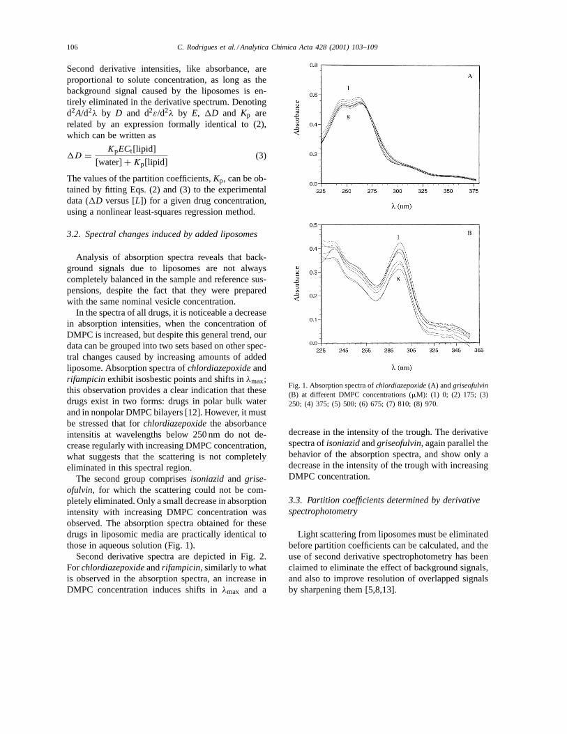
In the spectra of all drugs, it is noticeable a decreasein absorption intensities, when the concentration ofDMPC is increased, but despite this general trend, ourdata can be grouped into two sets based on other spec-tral changes caused by increasing amounts of addedliposome. Absorption spectra ofchlordiazepoxideandrifampicin exhibit isosbestic points and shifts inλmax;this observation provides a clear indication that thesedrugs exist in two forms: drugs in polar bulk waterand in nonpolar DMPC bilayers [12]. However, it mustbe stressed that forchlordiazepoxidethe absorbanceintensitis at wavelengths below 250 nm do not de-crease regularly with increasing DMPC concentration,what suggests that the scattering is not completelyeliminated in this spectral region.
The second group comprisesisoniazidand grise-ofulvin, for which the scattering could not be com-pletely eliminated. Only a small decrease in absorptionintensity with increasing DMPC concentration wasobserved. The absorption spectra obtained for thesedrugs in liposomic media are practically identical tothose in aqueous solution (Fig. 1).
Second derivative spectra are depicted in Fig. 2.Forchlordiazepoxideandrifampicin,similarly to whatis observed in the absorption spectra, an increase inDMPC concentration induces shifts inλmax and a
Fig. 1. Absorption spectra ofchlordiazepoxide(A) andgriseofulvin(B) at different DMPC concentrations (mM): (1) 0; (2) 175; (3)250; (4) 375; (5) 500; (6) 675; (7) 810; (8) 970.
decrease in the intensity of the trough. The derivativespectra ofisoniazidandgriseofulvin, again parallel thebehavior of the absorption spectra, and show only adecrease in the intensity of the trough with increasingDMPC concentration.
3.3. Partition coefficients determined by derivativespectrophotometry
Light scattering from liposomes must be eliminatedbefore partition coefficients can be calculated, and theuse of second derivative spectrophotometry has beenclaimed to eliminate the effect of background signals,and also to improve resolution of overlapped signalsby sharpening them [5,8,13].

C. Rodrigues et al. / Analytica Chimica Acta 428 (2001) 103–109 107
Fig. 2. Second derivative spectra ofchlordiazepoxide(A) andgriseofulvin (B) at different DMPC concentrations (mM): (1) 0;(2) 175; (3) 250; (4) 375; (5) 500; (6) 675; (7) 810; (8) 970.
In general, values ofKp can be calculated by fit-ting experimental data (1D versus [L]) obtained atany wavelength, to Eq. (3), but some care must betaken regarding the choice of wavelengths. To in-crease reproducibility and signal-to-noise ratio, themaximum-peak method is normally used for hetero-geneous samples [5,8,14], in which the1D values areobtained atλmax of the absorption spectra, as in thevicinity of absorption maxima small inaccuracies inwavelength reproducibility do not induce large vari-ability on 1D values [14]. Another precaution is tomake sure that the background signals are effectivelyeliminated specially at shorter wavelenghts where thescattering is higher.
Table 1Values of the wavelengths,λmax (nm), of the absorption maximaand of the wavelengths,λmax (nm),λmin (nm), of second derivativemaxima and minima forchlordiazepoxide, rifampicin, isoniazidand griseofulvin
Absorptionspectra
Second derivativespectra
λmax1 λmax2 λmin1 λmin2 λmax1 λmax2
Chlordiazepoxide 261 310 267 311 289 330Rifampicin 475 – 475 – 527 –Isoniazid 263 – 270 – 293 –Griseofulvin 295 – 296 – 275 317
The Kp values were obtained in the present workusing data from second derivative spectra, not only atλmin, but also data atλmax. The use of data for, atleast, two different wavelengths provides a check onthe effective elimination of scattering and on wave-length reproducibility, and in Table 1 are indicated thewavelengths used for each drug. The values were ob-tained by fitting Eq. (3) to data from at least two inde-pendent experiments (eight points), and at more thanone wavelength, and are included in Table 2. Further-more, for all drugs the values ofKp were found to beinsensitive to the wavelength used, and identical val-ues, within experimental error, were obtained for eachdrug, and this insensitivity provides evidence that thebackground signal has been effectively eliminated inthe spectral range used (Table 1).
Analysis of our data also points for caution in theuse of data in the spectral range below 275 nm, at leastfor the experimental conditions used in the presentwork. In fact, it was not possible to obtain preciseKpvalues (large standard deviations) from derivative in-tensities atλmax = 275 nm forgriseofulvin, less alonevalues similar to those obtained from data at longerwavelengths. A possible explanation for this anoma-lous behavior can be gained by assuming the existenceof random noise errors in this spectral region in whichliposome scattering is still large. As a final comment,and in support of this justification, it must be stressedthat light scattering as a source of additive noise in ab-sorption measurements, is more important at shorterwavelengths and will affect the weaker bands. Theband at 275 nm forgriseofulvincorresponds toλmaxin the derivative spectra, and thus, it is to be expectedsmall reproducibility in the derivative intensities. The
108 C. Rodrigues et al. / Analytica Chimica Acta 428 (2001) 103–109
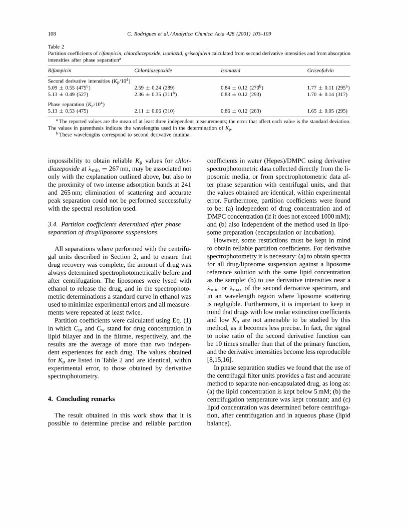
Table 2Partition coefficients ofrifampicin, chlordiazepoxide, isoniazid, griseofulvincalculated from second derivative intensities and from absorptionintensities after phase separationa
Rifampicin Chlordiazepoxide Isoniazid Griseofulvin
Second derivative intensities (Kp/104)5.09 ± 0.55 (475b) 2.59 ± 0.24 (289) 0.84± 0.12 (270b) 1.77 ± 0.11 (295b)5.13 ± 0.49 (527) 2.36± 0.35 (311b) 0.83 ± 0.12 (293) 1.70± 0.14 (317)
Phase separation (Kp/104)5.13 ± 0.53 (475) 2.11± 0.06 (310) 0.86± 0.12 (263) 1.65± 0.05 (295)
a The reported values are the mean of at least three independent measurements; the error that affect each value is the standard deviation.The values in parenthesis indicate the wavelengths used in the determination ofKp.
b These wavelengths correspond to second derivative minima.
impossibility to obtain reliableKp values forchlor-diazepoxideat λmin = 267 nm, may be associated notonly with the explanation outlined above, but also tothe proximity of two intense adsorption bands at 241and 265 nm; elimination of scattering and accuratepeak separation could not be performed successfullywith the spectral resolution used.
3.4. Partition coefficients determined after phaseseparation of drug/liposome suspensions
All separations where performed with the centrifu-gal units described in Section 2, and to ensure thatdrug recovery was complete, the amount of drug wasalways determined spectrophotometrically before andafter centrifugation. The liposomes were lysed withethanol to release the drug, and in the spectrophoto-metric determinations a standard curve in ethanol wasused to minimize experimental errors and all measure-ments were repeated at least twice.
Partition coefficients were calculated using Eq. (1)in which Cm andCw stand for drug concentration inlipid bilayer and in the filtrate, respectively, and theresults are the average of more than two indepen-dent experiences for each drug. The values obtainedfor Kp are listed in Table 2 and are identical, withinexperimental error, to those obtained by derivativespectrophotometry.
4. Concluding remarks
The result obtained in this work show that it ispossible to determine precise and reliable partition
coefficients in water (Hepes)/DMPC using derivativespectrophotometric data collected directly from the li-posomic media, or from spectrophotometric data af-ter phase separation with centrifugal units, and thatthe values obtained are identical, within experimentalerror. Furthermore, partition coefficients were foundto be: (a) independent of drug concentration and ofDMPC concentration (if it does not exceed 1000 mM);and (b) also independent of the method used in lipo-some preparation (encapsulation or incubation).
However, some restrictions must be kept in mindto obtain reliable partition coefficients. For derivativespectrophotometry it is necessary: (a) to obtain spectrafor all drug/liposome suspension against a liposomereference solution with the same lipid concentrationas the sample: (b) to use derivative intensities near aλmin or λmax of the second derivative spectrum, andin an wavelength region where liposome scatteringis negligible. Furthermore, it is important to keep inmind that drugs with low molar extinction coefficientsand low Kp are not amenable to be studied by thismethod, as it becomes less precise. In fact, the signalto noise ratio of the second derivative function canbe 10 times smaller than that of the primary function,and the derivative intensities become less reproducible[8,15,16].
In phase separation studies we found that the use ofthe centrifugal filter units provides a fast and accuratemethod to separate non-encapsulated drug, as long as:(a) the lipid concentration is kept below 5 mM; (b) thecentrifugation temperature was kept constant; and (c)lipid concentration was determined before centrifuga-tion, after centrifugation and in aqueous phase (lipidbalance).
C. Rodrigues et al. / Analytica Chimica Acta 428 (2001) 103–109 109
Taking into account these limitations, both methodswere shown to provide precise and accurate values forthe partition coefficient of several drugs with differenthydrolipophilic balance in water (Hepes)/DMPC sys-tem. None of the methods provides a clear advantagein the total time needed for the determinations, but fordrugs which absorb near or below 275 nm, the need toeliminate the liposome scattering may favor the phaseseparation method.
Acknowledgements
Partial financial support for this work was providedby “Fundação para a Ciência e Tecnologia” (FCT, Lis-boa) through “Programa Sapiens 99”. C. Rodriguesthanks FCT for a fellowship.
References
[1] M. Luxnat, H.J. Galla, Biochim. Biophys. Acta 856 (1986)274.
[2] G.V. Betageri, S.R. Dipali, J. Pharm. Pharmacol. 45 (1993)931.
[3] S.R. Dipali, S.B. Kulkarni, G.V. Betageri, J. Pharm.Pharmacol. 48 (1996) 1112.
[4] Y. Kaminoh, T. Inoue, S.-M. Ma, I. Ueda, S.H. Lin, Biochem.Biophys. Acta 946 (1988) 337.
[5] K. Kitamura, N. Imayoshi, T. Goto, H. Shiro, T. Mano, T.Nakay, Anal. Chim. Acta 304 (1995) 101.
[6] R. Welti, L.J. Mullikin, T. Yoshimura, G.M. Helmkamp Jr.,Biochemistry 23 (1984) 6086.
[7] A. Gursoy, B. Senyucel, J. Microencapsulation 14 (1997) 769.[8] G. Talsky, L. Mayring, H. Kreuzer, Angew. Chem. Int. Ed.
17 (1978) 785.[9] A. Savitzky, M.J.E. Golay, Anal. Chem. 36 (1964) 1611.
[10] C.H. Fiske, Y. Subbarow, J. Biol. Chem. 66 (1925) 375.[11] D.D. Lasic, Liposomes: From Physics to Applications,
Elsevier, Amsterdam, 1993, pp. 63–100.[12] K.A. Connors, Binding Constants: The Measurement of
Molecular Complex Stability, Wiley, New York, 1987,pp. 141–147.
[13] L. Sommer, Analytical Absorption Spectrophotometry in theVisible and Ultraviolet: The Principals (Stud. Anal. Chem.8), Elsevier, Amsterdam, 1989, pp. 186–199.
[14] K. Kitamura, N. Imayoshi, Anal. Sci. 8 (1992) 497.[15] T.C. O’Haver, G.L. Green, Anal. Chem. 48 (1976) 312.[16] T.R. Griffiths, K. King, H.A. Hubbard, Anal. Chim. Acta 143
(1982) 163.


![Liposome [GoR]](https://img.pdfslide.net/doc/110x75/54f49f044a795997318b4927/liposome-gor.jpg)