Embed Size (px)
Citation preview
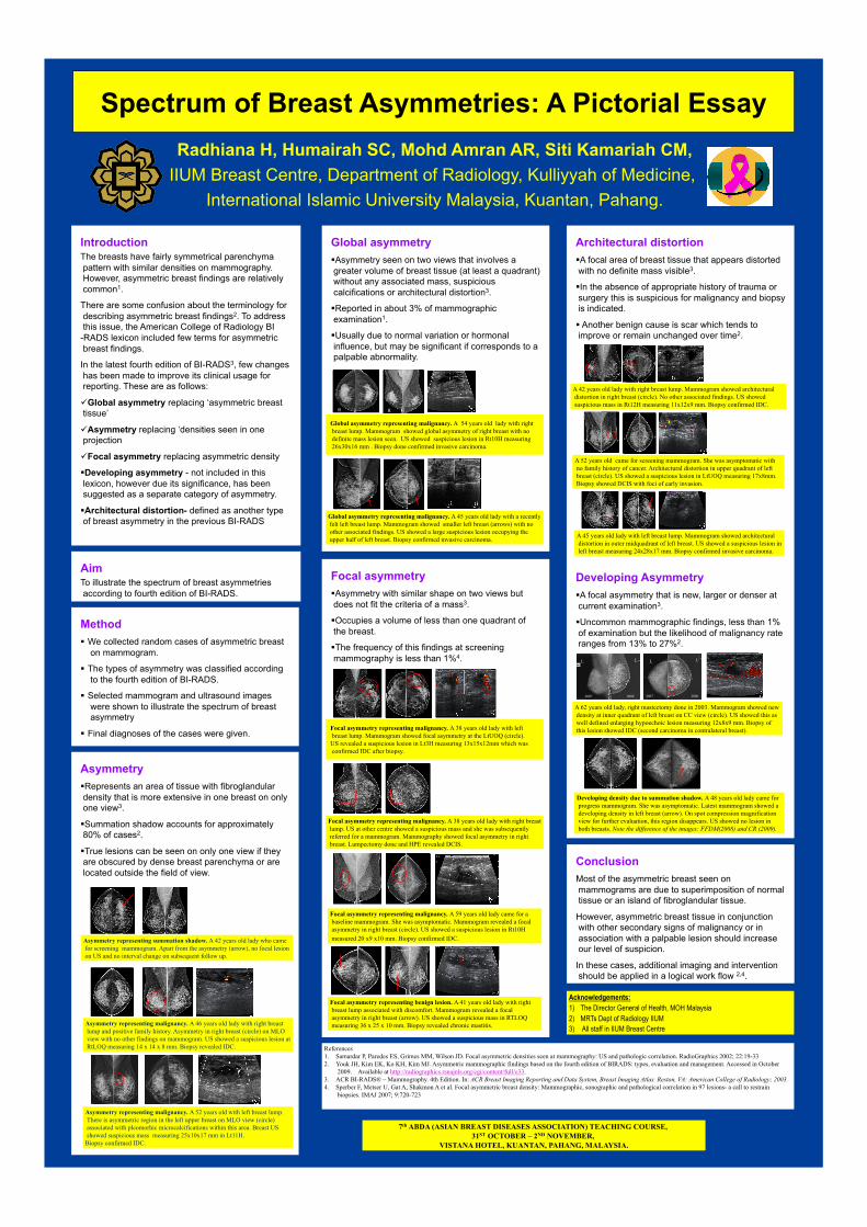
Spectrum of Breast Asymmetries: A Pictorial Essay Radhiana H, Humairah SC, Mohd Amran AR, Siti Kamariah CM,
IIUM Breast Centre, Department of Radiology, Kulliyyah of Medicine, International Islamic University Malaysia, Kuantan, Pahang.
Introduction The breasts have fairly symmetrical parenchyma pattern with similar densities on mammography. However, asymmetric breast findings are relatively common1.
There are some confusion about the terminology for describing asymmetric breast findings2. To address this issue, the American College of Radiology BI-RADS lexicon included few terms for asymmetric breast findings.
In the latest fourth edition of BI-RADS3, few changes has been made to improve its clinical usage for reporting. These are as follows:
Global asymmetry replacing ‘asymmetric breast tissue’
Asymmetry replacing ‘densities seen in one projection
Focal asymmetry replacing asymmetric density
Developing asymmetry - not included in this lexicon, however due its significance, has been suggested as a separate category of asymmetry.
Architectural distortion- defined as another type of breast asymmetry in the previous BI-RADS
Global asymmetry Asymmetry seen on two views that involves a greater volume of breast tissue (at least a quadrant) without any associated mass, suspicious calcifications or architectural distortion3.
Reported in about 3% of mammographic examination1.
Usually due to normal variation or hormonal influence, but may be significant if corresponds to a palpable abnormality.
Architectural distortion A focal area of breast tissue that appears distorted with no definite mass visible3.
In the absence of appropriate history of trauma or surgery this is suspicious for malignancy and biopsy is indicated.
Another benign cause is scar which tends to improve or remain unchanged over time2.
Developing Asymmetry A focal asymmetry that is new, larger or denser at current examination3.
Uncommon mammographic findings, less than 1% of examination but the likelihood of malignancy rate ranges from 13% to 27%2.
Aim To illustrate the spectrum of breast asymmetries according to fourth edition of BI-RADS.
Method We collected random cases of asymmetric breast
on mammogram.
The types of asymmetry was classified according to the fourth edition of BI-RADS.
Selected mammogram and ultrasound images were shown to illustrate the spectrum of breast asymmetry
Final diagnoses of the cases were given.
Conclusion Most of the asymmetric breast seen on mammograms are due to superimposition of normal tissue or an island of fibroglandular tissue.
However, asymmetric breast tissue in conjunction with other secondary signs of malignancy or in association with a palpable lesion should increase our level of suspicion.
In these cases, additional imaging and intervention should be applied in a logical work flow 2,4.
Focal asymmetry Asymmetry with similar shape on two views but does not fit the criteria of a mass3.
Occupies a volume of less than one quadrant of the breast.
The frequency of this findings at screening mammography is less than 1%4.
Asymmetry Represents an area of tissue with fibroglandular density that is more extensive in one breast on only one view3.
Summation shadow accounts for approximately 80% of cases2.
True lesions can be seen on only one view if they are obscured by dense breast parenchyma or are located outside the field of view.
Global asymmetry representing malignancy. A 54 years old lady with right breast lump. Mammogram showed global asymmetry of right breast with no definite mass lesion seen. US showed suspicious lesion in Rt10H measuring 26x30x16 mm . Biopsy done confirmed invasive carcinoma.
Focal asymmetry representing malignancy. A 38 years old lady with left breast lump. Mammogram showed focal asymmetry at the LtUOQ (circle). US revealed a suspicious lesion in Lt3H measuring 13x15x12mm which was confirmed IDC after biopsy.
Asymmetry representing summation shadow. A 42 years old lady who came for screening mammogram. Apart from the asymmetry (arrow), no focal lesion on US and no interval change on subsequent follow up.
Asymmetry representing malignancy. A 46 years old lady with right breast lump and positive family history. Asymmetry in right breast (circle) on MLO view with no other findings on mammogram. US showed a suspicious lesion at RtLOQ measuring 14 x 14 x 8 mm. Biopsy revealed IDC.
Asymmetry representing malignancy. A 52 years old with left breast lump. There is asymmetric region in the left upper breast on MLO view (circle) associated with pleomorhic microcalcifications within this area. Breast US showed suspicious mass measuring 25x10x17 mm in Lt11H. Biopsy confirmed IDC.
A 42 years old lady with right breast lump. Mammogram showed architectural distortion in right breast (circle). No other associated findings. US showed suspicious mass in Rt12H measuring 11x12x9 mm. Biopsy confirmed IDC.
A 52 years old came for screening mammogram. She was asymptomatic with no family history of cancer. Architectural distortion in upper quadrant of left breast (circle). US showed a suspicious lesion in LtUOQ measuring 17x8mm. Biopsy showed DCIS with foci of early invasion.
Global asymmetry representing malignancy. A 45 years old lady with a recently felt left breast lump. Mammogram showed smaller left breast (arrows) with no other associated findings. US showed a large suspicious lesion occupying the upper half of left breast. Biopsy confirmed invasive carcinoma.
Focal asymmetry representing malignancy. A 38 years old lady with right breast lump. US at other centre showed a suspicious mass and she was subsequently referred for a mammogram. Mammography showed focal asymmetry in right breast. Lumpectomy done and HPE revealed DCIS.
Focal asymmetry representing malignancy. A 59 years old lady came for a baseline mammogram. She was asymptomatic. Mammogram revealed a focal asymmetry in right breast (circle). US showed a suspicious lesion in Rt10H measured 20 x9 x10 mm. Biopsy confirmed IDC.
Developing density due to summation shadow. A 48 years old lady came for progress mammogram. She was asymptomatic. Latest mammogram showed a developing density in left breast (arrow). On spot compression magnification view for further evaluation, this region disappears. US showed no lesion in both breasts. Note the difference of the images: FFDM(2008) and CR (2009).
A 62 years old lady, right mastectomy done in 2003. Mammogram showed new density at inner quadrant of left breast on CC view (circle). US showed this as well defined enlarging hypoechoic lesion measuring 12x8x9 mm. Biopsy of this lesion showed IDC (second carcinoma in contralateral breast).
A 45 years old lady with left breast lump. Mammogram showed architectural distortion in outer midquadrant of left breast. US showed a suspicious lesion in left breast measuring 24x28x17 mm. Biopsy confirmed invasive carcinoma.
Focal asymmetry representing benign lesion. A 41 years old lady with right breast lump associated with discomfort. Mammogram revealed a focal asymmetry in right breast (arrow). US showed a suspicious mass in RTLOQ measuring 36 x 25 x 10 mm. Biopsy revealed chronic mastitis.
References 1. Samardar P, Paredes ES, Grimes MM, Wilson JD. Focal asymmetric densities seen at mammography: US and pathologic correlation. RadioGraphics 2002; 22:19-33 2. Youk JH, Kim EK, Ko KH, Kim MJ. Asymmetric mammographic findings based on the fourth edition of BIRADS: types, evaluation and management. Accessed in October
2009. Available at http://radiographics.rsnajnls.org/cgi/content/full/e33. 3. ACR BI-RADS® – Mammography. 4th Edition. In: ACR Breast Imaging Reporting and Data System, Breast Imaging Atlas. Reston, VA: American College of Radiology; 2003. 4. Sperber F, Metser U, Gat A, Shakmon A et al. Focal asymmetric breast density: Mammographic, sonographic and pathological correlation in 97 lesions- a call to restrain
biopsies. IMAJ 2007; 9:720-723
7th ABDA (ASIAN BREAST DISEASES ASSOCIATION) TEACHING COURSE, 31ST OCTOBER – 2ND NOVEMBER,
VISTANA HOTEL, KUANTAN, PAHANG, MALAYSIA.
Acknowledgements: 1) The Director General of Health, MOH Malaysia 2) MRTs Dept of Radiology IIUM 3) All staff in IIUM Breast Centre
R R
2007 2008
L LL L
2007 2008
2008 2009