Embed Size (px)
Citation preview
International Journal of Research in Medical Sciences | August 2015 | Vol 3 | Issue 8 Page 1889
International Journal of Research in Medical Sciences
Vijayan P et al. Int J Res Med Sci. 2015 Aug;3(8):1889-1894
www.msjonline.org pISSN 2320-6071 | eISSN 2320-6012
Research Article
Spectrum of malignant skin adnexal tumors – a single institution
study of 17 cases with clinicopathological correlation
Poornima Vijayan1*, Ramadas Nayak2, Laila M. Ilias1, Anupama Ponniah1
INTRODUCTION
Adnexal skin tumors aptly termed “troublesome tumors”
by Cotton D,1 pose a major diagnostic difficulty to both
the surgeon and the pathologist. The bewildering array of
differentiation they display and the ever-expanding list of
entities add further to the confusion.2 Tumors of the
pilosebaceous apparatus can occur as single-lineage
neoplasms or may manifest as complex proliferations
with multilineal differentiation patterns. Eccrine and
apocrine neoplasms present a bewildering array of
morphologies, which often defy precise classification.3
A large majority of skin adnexal tumors are benign and
for most part complete excision is curative. A malignant
counterpart of almost every Skin adnexal tumors has been
ABSTRACT
Background: Skin adnexal tumors are a rare, assorted group of tumors with differentiation towards hair follicle,
sebaceous glands or sweat glands. A vast majority of them are benign. But for every benign adnexal tumor, a
malignant counterpart exists. Many histological subtypes of these malignant tumors been described, but only in short
series or individual case reports. So, not much is known about their incidence or prognosis simply because of the
limited number of cases available for analysis. This study was undertaken to contribute towards this less traversed
area of dermatopathology.
Methods: In the present study, a total of 60 cases with a histopathological diagnosis of skin adnexal tumors were
studied. The slides and blocks were retrieved from the archives and were reviewed and were reclassified and subtyped
as per WHO classification of skin tumors, 2006.
Results: Among the 60 cases of adnexal tumors documented and reviewed over the four year study period, 17 cases
of malignant adnexal tumors were encountered. Of these, 10 (58%) were tumors with eccrine or apocrine
differentiation, 5 (29%) were of follicular differentiation and two (12%) were of sebaceous differentiation. Mammary
paget disease (MPD) was the most frequent malignant tumor encountered both overall and among the tumors with
eccrine and apocrine differentiation. Other tumors encountered in their order of frequency were Malignant
proliferating trichelemmal tumor, apocrine carcinoma, sebaceous carcinoma and extramammary paget disease,
trichelemmal carcinoma and eccrine carcinoma. These tumors were evaluated with regard to their age, site, gender
distribution, clinical characters and histopathological features.
Conclusion: Malignant adnexal tumors are extremely rare with indistinct clinical characteristics. They are locally
aggressive, and have the potential for nodal involvement and distant metastasis, with a poor clinical outcome. A high
index of suspicion is necessary to establish a diagnosis in most cases.
Keywords: Malignant adnexal skin tumors, Eccrine, Apocrine, Sebaceous, Trichelemmal
1Department of Pathology, MES Medical College, Malappuram district, Kerala, India 2Department of Pathology, Yenapoya Medical College, Mangalore, Karnataka, India
Received: 27 May 2015
Accepted: 05 July 2015
*Correspondence:
Dr. Poornima Vijayan,
E-mail: [email protected]
Copyright: © the author(s), publisher and licensee Medip Academy. This is an open-access article distributed under
the terms of the Creative Commons Attribution Non-Commercial License, which permits unrestricted non-commercial
use, distribution, and reproduction in any medium, provided the original work is properly cited.
DOI: http://dx.doi.org/10.18203/2320-6012.ijrms20150297

Vijayan P et al. Int J Res Med Sci. 2015 Aug;3(8):1889-1894
International Journal of Research in Medical Sciences | August 2015 | Vol 3 | Issue 8 Page 1890
described. These tumors are rare, locally aggressive, and
have the potential for nodal involvement and distant
metastasis, with a poor clinical outcome. Therefore,
establishing a diagnosis of malignancy in skin adnexal
tumors is important for therapeutic and prognostic
purposes.4
Many studies have been published on benign adnexal
tumors but studies on their malignant counterpart remain
far and few. Though, it is true that many short series and
case reports of individual and composite malignant
adnexal tumors have been reported in the western
literature, however, the fact remains that for many
adnexal carcinomas there are simple insufficient numbers
reported to develop much of an idea simply regarding
their prognosis.2 This study was undertaken so as to make
an attempt to make a contribution to this less traversed
area of dermatopathology.
METHODS
The present study was conducted retrospectively over a
period of five years, 2007 – 2012 in the Department of
Pathology at a tertiary centre in Karnataka, South India.
During this five year period, all excision biopsy
specimens which had a diagnosis of skin adnexal origin
was included in this study. This included both benign and
malignant adnexal tumors of the skin. A total of 60 cases
were retrieved and studied. The clinical details were
obtained from the hospital records and the requisition
form that was received in the department of pathology.
The slides and blocks were retrieved from the archives
and multiple serial sections were taken for each biopsy
and stained with routine haematoxylin and eosin stain.
The slides were reviewed and were reclassified and
subtyped as per WHO classification of skin tumors,
2006.2
RESULTS
The present study is a comprehensive analysis of
collective adnexal tumors of skin wherein 60 cases with
histopathological diagnosis of skin adnexal tumors were
studied retrospectively during a 5 year period. All cases
were reviewed and 17 cases of malignant adnexal skin
tumors were documented. These tumors have been
classified according to WHO classification2 and analyzed
with regard to their age, sex, site, clinical presentation as
well as their various histomorphologic patterns. Table 1
depicts the various malignant adnexal tumors
encountered in our study and their distribution according
to their differentiation.
Table 1: Malignant adnexal tumors.
Tumors Males Females Total
Tumors with follicular differentiation
Trichelemmal
Carcinoma 1 1 2
Malignant profilerating 1 2 3
trichelemmal tumor
Tumors with sebaceous differentiation
Sebaceous
Carcinoma - 2 2
Tumors with eccrine and apocrine differentiation
Eccrine
carcinoma 1 1
Mammary paget disease - 4 4
Apocrine carcinoma 3 3
Extramammary paget
Disease 1 1 2
Total 4 13 17
During the five year study period, a total of 60 cases of
skin adnexal tumors were reviewed, out of which 43
(71%) were benign and 17 (29%) were malignant. Out of
the 17 malignant cases, tumors with eccrine and apocrine
differentiation constituted 58% (10), 5 (29%) were of
follicular differentiation and two (12%) were of
sebaceous differentiation.
Pagets disease of the breast was the most common
malignant adnexal tumor constituting 4 cases, followed
by 3 cases of apocrine carcinoma and malignant
proliferating trichelemmal tumor. Two cases each of
trichelemmal carcinoma, sebaceous carcinoma and
extramammary pagets disease were encountered and
one case of eccrine carcinoma was also noted in our
study.
In most cases of malignant adnexal tumors in our study,
the age of incidence was the 6th and 7th decades, except
one case of eccrine carcinoma in a 45 year old male.
A definite female preponderance was noted in our
study.
Clinically, seven cases were correctly diagnosed as malignant adnexal tumor. Two malignant proliferating trichelemmal tumors were diagnosed as squamous cell carcinomas. Patients presented with recurrence in 2 cases of malignant proliferating trichelemmal tumor and one case of sebaceous carcinoma. All three cases of Apocrine carcinoma presented as axillary nodules. Tumor size was >5 cm in 8 tumors. Ulceration was noted in 2 cases of MPTT and one case of sebaceous carcinoma. All cases of Mammary Paget disease and extramammary pagets disease presented as eczematous and erythematous lesions. All 4 cases of MPD was associated with underlying invasive ductal carcinoma breast and one was associated with an in-situ component also. Neither cases of EMPD was associated with an internal malignancy. All tumors in our study were solitary lesions. Two cases of recurrent MPTT (Figure 1) and one case of recurrent trichelemmal carcinoma were treated with radical neck dissection (Figure 2 & 3) and lymphnode metastasis was noted in two cases (Figure 4 & 5). The salient clinical and histopathological features of the malignant tumors encountered in our study are enumerated in Tables 2 &
3.
Vijayan P et al. Int J Res Med Sci. 2015 Aug;3(8):1889-1894
International Journal of Research in Medical Sciences | August 2015 | Vol 3 | Issue 8 Page 1891
Table 2: Salient clinical features of malignant adnexal
skin tumors.
Clinical features Number
of cases Percentage
Number of lesions
Solitary 17 100%
Multiple - -
Size of lesion
<2cm 4 23%
2-5cm 5 29%
>5cm 8 47%
Other features
Tenderness 8 47%
Erythema/Eczema 6 35%
Ulceration 5 29%
Loss of circumscription 8 47%
Recurrence 3 17%
Asymmetry 7 41
Infiltrative borders 7 41%
Regional
Lymphadenopathy 3 17%
Table 3: Salient histopathological features of
malignant adnexal skin tumors.
Histopathology Number
of cases Percentage
Atypia 17 100%
Atypical Mitosis 17 100%
Pleomorphism 17 100%
Infiltrative borders 15 88%
Vascular invasion 10 58%
Lymphatic invasion 5 29%
Perineural invasion 4 23%
Lymph node metastasis 3 17%
Necrosis 7 41%
Lobular architecture 4 23%
Sheeting pattern 5 29%
Infiltrating cords and
trabeculae 0 0%
Papillary pattern 1 5%
Glandular pattern 1 5%
Pagetoid pattern 6 35%
Figure 1: Recurrent malignant proliferating
trichelemmal tumors over scalp in 65/ female.
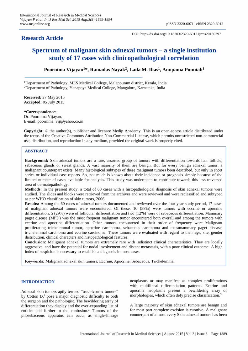
Figure 2: Radical neck dissection specimen in a case
of recurrent malignant proliferating trichelemmal
tumor.
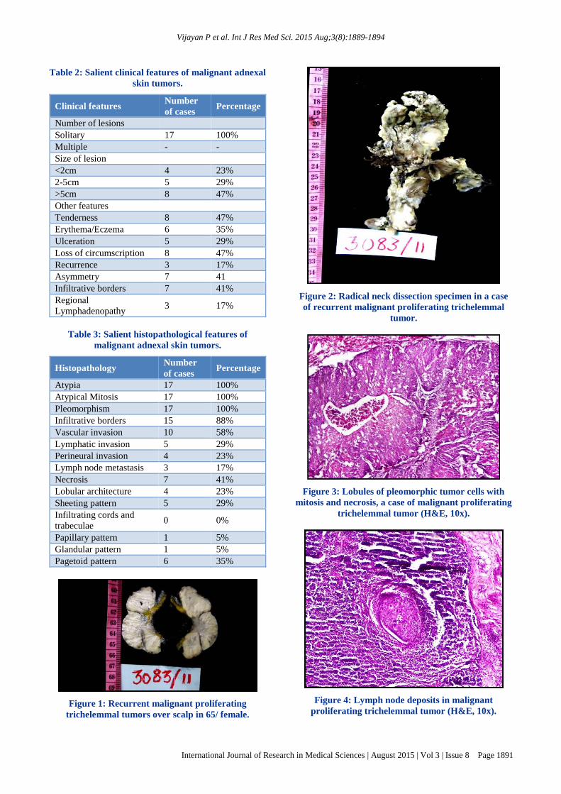
Figure 3: Lobules of pleomorphic tumor cells with
mitosis and necrosis, a case of malignant proliferating
trichelemmal tumor (H&E, 10x).
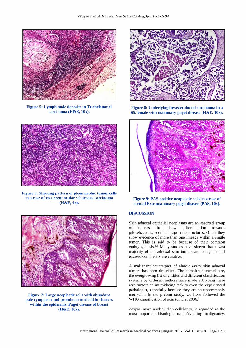
Figure 4: Lymph node deposits in malignant
proliferating trichelemmal tumor (H&E, 10x).
Vijayan P et al. Int J Res Med Sci. 2015 Aug;3(8):1889-1894
International Journal of Research in Medical Sciences | August 2015 | Vol 3 | Issue 8 Page 1892
Figure 5: Lymph node deposits in Trichelemmal
carcinoma (H&E, 10x).
Figure 6: Sheeting pattern of pleomorphic tumor cells
in a case of recurrent ocular sebaceous carcinoma
(H&E, 4x).
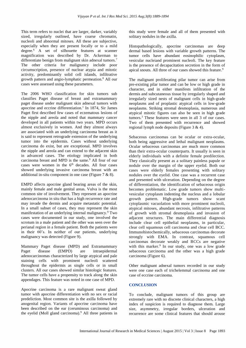
Figure 7: Large neoplastic cells with abundant
pale cytoplasm and prominent nucleoli in clusters
within the epidermis, Paget disease of breast
(H&E, 10x).
Figure 8: Underlying invasive ductal carcinoma in a
65/female with mammary paget disease (H&E, 10x).
Figure 9: PAS positive neoplastic cells in a case of
scrotal Extramammary paget disease (PAS, 10x).
DISCUSSION
Skin adnexal epithelial neoplasms are an assorted group
of tumors that show differentiation towards
pilosebaceous, eccrine or apocrine structures. Often, they
show evidence of more than one lineage within a single
tumor. This is said to be because of their common
embryogenesis.4,5 Many studies have shown that a vast
majority of the adnexal skin tumors are benign and if
excised completely are curative.
A malignant counterpart of almost every skin adnexal
tumors has been described. The complex nomenclature,
the evergrowing list of entities and different classification
systems by different authors have made subtyping these
rare tumors an intimidating task to even the experienced
pathologist, especially because they are so uncommonly
met with. In the present study, we have followed the
WHO classification of skin tumors, 2006.1
Atypia, more nuclear than cellularity, is regarded as the
most important histologic trait favouring malignancy.
Vijayan P et al. Int J Res Med Sci. 2015 Aug;3(8):1889-1894
International Journal of Research in Medical Sciences | August 2015 | Vol 3 | Issue 8 Page 1893
This term refers to nuclei that are larger, darker, variably
sized, irregularly outlined, have coarse chromatin,
nucleoli and abnormal mitoses. All these are subjective,
especially when they are present focally or to a mild
degree.6 A set of silhouette features at scanner
magnification was described by Dr. Ackerman to
differentiate benign from malignant skin adnexal tumors.7
The other criteria for malignancy include poor
circumscription, presence of nuclear atypia and mitotic
activity, predominantly solid cell islands, infiltrative
growth pattern and angio-lymphatic permeation.8 All our
cases were assessed using these parameters.
The 2006 WHO classification for skin tumors sub
classifies Paget disease of breast and extramammary
paget disease under malignant skin adnexal tumors with
apocrine and eccrine differentiation.1 In 1874, Sir James
Paget first described few cases of eczematous lesions of
the nipple and areola and noted that mammary cancer
developed in all patients within two years. MPD occurs
almost exclusively in women. And they almost always
are associated with an underlying carcinoma breast as it
is said to represent retrograde extension of the underlying
tumor into the epidermis. Cases without underlying
carcinoma do exist, but are exceptional. MPD involves
the nipple and areola and can extend to the adjacent skin
in advanced cases. The etiology implicated in both
carcinoma breast and MPD is the same.9 All four of our
cases were women in the 6th decades. All four cases
showed underlying invasive carcinoma breast with an
additional in-situ component in one case (Figure 7 & 8).
EMPD affects apocrine gland bearing areas of the skin,
mainly female and male genital areas. Vulva is the most
common site of involvement. They represent an apocrine
adenocarcinoma in situ that has a high recurrence rate and
may invade the dermis and acquire metastatic potential.
In a small subset of cases, they may represent the skin
manifestation of an underlying internal malignancy.9 Two
cases were documented in our study, one involved the
scrotum in a male patient and the other was seen over the
perianal region in a female patient. Both the patients were
in their 60’s. In neither of our patients, underlying
malignancy was detected (Figure 9).
Mammary Paget disease (MPD) and Extramammary
Paget disease (EMPD) are intraepidermal
adenocarcinomas characterized by large atypical and pale
staining cells with prominent nucleoli scattered
throughout the epidermis as single cells or in small
clusters. All our cases showed similar histologic features.
The tumor cells have a propensity to track along the skin
appendages. This feature was noted in one case of MPD.
Apocrine carcinoma is a rare malignant sweat gland
tumor with apocrine differentiation with no sex or racial
predeliction. Most common site is the axilla followed by
anogenital region. Variants of apocrine carcinoma have
been described on the ear (ceruminous carcinoma) and
the eyelid (Moll gland carcinoma).9 All three patients in
this study were female and all of them presented with
solitary nodules in the axilla.
Histopathologically, apocrine carcinomas are deep
dermal based lesions with variable growth patterns. The
tumor cells have abundant eosinophilic cytoplasm,
vesicular nucleiand prominent nucleoli. The key feature
is the presence of decapacitation secretion in the form of
apical snouts. All three of our cases showed this feature.9
The malignant proliferating pilar tumor can arise from
pre-existing pilar tumor and can be low or high grade in
character, and in either manifests infiltration of the
dermis and subcutaneous tissue by irregularly shaped and
irregularly sized nests of malignant cells in high-grade
neoplasms and of proplastic atypical cells in low-grade
neoplasms. Striking stromal desmoplasia, numerous and
atypical mitotic figures can also be seen in high grade
tumors.3 These features were seen in all 3 of our cases.
Two of them presented with recurrence and showed
regional lymph node deposits (Figure 3 & 4).
Sebaceous carcinomas can be ocular or extra-ocular,
both being aggressive and lethal malignant neoplasms.
Ocular sebaceous carcinomas are much more common
than their extra-ocular counterparts. It usually is seen in
elderly individuals with a definite female predilection.
They classically present as a solitary painless papule or
nodule over the margin of the upper eyelid. Both our
cases were elderly females presenting with solitary
nodules over the eyelid. One case was a recurrent case
and presented with ulceration. Depending on the degree
of differentiation, the identification of sebaceous origin
becomes problematic. Low grade tumors show multi-
vesicular cytoplasm indenting the nucleus and a lobular
growth pattern. High-grade tumors show scant
cytoplasmic vacuolation with more prominent nucleoli,
atypical mitoses, abundant necrosis, infiltrative patterns
of growth with stromal desmoplasia and invasion of
adjacent structures. The main differential diagnosis
include clear cell epithelial neoplasms, in particular,
clear cell squamous cell carcinoma and clear cell BCC.
Immunohistochemically, sebaceous carcinomas decorate
strongly with EMA. In contrast, squamous cell
carcinomas decorate weakly and BCCs are negative
with this marker.9 In our study, one was a low grade
sebaceous carcinoma and the other was a high grade
carcinoma (Figure 6).
Other malignant adnexal tumors recorded in our study
were one case each of trichelemmal carcinoma and one
case of eccrine carcinoma.
CONCLUSION
To conclude, malignant tumors of this group are
extremely rare with no discrete clinical characters, a high
index of suspicion is required to diagnose them. Large
size, asymmetry, irregular borders, ulceration and
recurrence are some clinical features that should arouse
Vijayan P et al. Int J Res Med Sci. 2015 Aug;3(8):1889-1894
International Journal of Research in Medical Sciences | August 2015 | Vol 3 | Issue 8 Page 1894
suspicion of malignancy. Histopathological features of
malignancy include loss of circumscription, nuclear
atypia, mitosis, pleomorphism, sheeting pattern,
infiltrative growth pattern, necrosis and lymphovascular
invasion. A thorough knowledge about the
histomorphological features is necessary for subtyping
these tumors based on their differentiation. Surgery with
wide excision margin and sometimes even local lymph
node resection may be necessary in these patients.
ACKNOWLEDGEMENTS
I wholeheartedly thank Dr. Radha Pai, our H.O.D. and all
my professors of KMC, Mangalore who have always
encouraged me throughout my tenure.
Funding: No funding sources
Conflict of interest: None declared
Ethical approval: The study was approved by the
Institutional Ethics Committee
REFERENCES
1. Cotton D. Troublesome tumors 1: Adnexal tumors
of the skin. J Clin Pathol.1991;44:543–8.
2. LeBoit P, Burg G, Weedon D, Sarasin A. Pathology
and genetics of skin tumors.Lyon: IARC Press;
2006.
3. Crowson AN, Margo CM, Mihm MC. Malignant
adnexal neoplasms. Mod Pathol. 2006;19:93-126.
4. Alsaad KO, Obaidat NA, Ghazarian D. Skin adnexal
neoplasms-part I: an approach to tumors of the
pilosebaceous unit. J Clin Pathol. 2007:60;129-44.
5. Alsaad KO, Obaidat NA, Ghazarian D. Skin adnexal
neoplasms-part 2: An approach to tumors of
cutaneous sweat glands. J Clin Pathol. 2007;60:145–
59.
6. Tirumalae R, Roopa MO. Benign vs. Malignant
Skin Adnexal Neoplasms: How Useful are
Silhouettes? Indian J Dermatol. 2013;58(1):30–3.
7. Ackerman AB. Differentiation of benign from
malignant neoplasms by silhouette. Am J
Dermatopathol. 1989;11:297–300.
8. Vaideeswar P, Madhiwale CV, Deshpande JR.
Malignant hidradenoma: A rare sweat gland tumor.
J Postgrad Med. 1999;45:56-7.
9. Requena L, Kutzner H, Hurt MA, Santa Cruz DJ,
Mehregan AH, Mengesha YM, et al. Appendageal
tumors – Malignant tumors with apocrine and
eccrine differentiation. In: LeBoit PE, Burg G,
Weedon D, Sarasin A, editors. Pathology and
genetics of skin tumors. World Health organization
classification of tumors. Lyon, France: IARC Press;
2006. p.127.
Cite this article as: Vijayan P, Nayak R, Ilias LM,
Ponniah A. Spectrum of malignant skin adnexal
tumors – a single institution study of 17 cases with
clinicopathological correlation. Int J Res Med Sci
2015;3(8):1889-94.