Embed Size (px)
Citation preview
University of South Florida University of South Florida
Scholar Commons Scholar Commons
Graduate Theses and Dissertations Graduate School
March 2019
Speeding Diagnosis and Saving Money Using Point of Care Speeding Diagnosis and Saving Money Using Point of Care
Ultrasound Rather Than MRI for Work-related MSK Injuries Ultrasound Rather Than MRI for Work-related MSK Injuries
Jared A. Jeffries University of South Florida, [email protected]
Follow this and additional works at: https://scholarcommons.usf.edu/etd
Part of the Medicine and Health Sciences Commons
Scholar Commons Citation Scholar Commons Citation Jeffries, Jared A., "Speeding Diagnosis and Saving Money Using Point of Care Ultrasound Rather Than MRI for Work-related MSK Injuries" (2019). Graduate Theses and Dissertations. https://scholarcommons.usf.edu/etd/7816
This Thesis is brought to you for free and open access by the Graduate School at Scholar Commons. It has been accepted for inclusion in Graduate Theses and Dissertations by an authorized administrator of Scholar Commons. For more information, please contact [email protected].
Speeding Diagnosis and Saving Money Using Point of Care Ultrasound Rather Than MRI for
Work-related MSK Injuries
by
Jared A. Jeffries
A thesis submitted in partial fulfillment of the requirements for the degree of
Master of Science in Public Health
Department of Occupational Medicine
College of Public Health
University of South Florida
Major Professor: Thomas Truncale, D.O., M.P.H.
Committee: Rachel Williams, M.D., M.S.P.H.
Alfred Mbah, Ph.D.
Date of Approval:
March 22, 2019
Keywords: Musculoskeletal injuries, Work injuries, POCUS, Musculoskeletal ultrasound
Copyright © 2019, Jared A. Jeffries
i
Table of Contents
List of Tables ii
Abstract iii
Chapter 1: Introduction 1
Chapter 2: Methods 5
Chapter 3: Results 7
Cases Where Ultrasound Alone or Ultrasound + Xray Could be Substituted for MRI 7
Potential Cost Savings if Ultrasound was Appropriately Substituted for MRI 11
Potential Time Savings if Ultrasound was Appropriately Substituted for MRI 11
Chapter 4: Discussion 12
Rationale for Study Inclusion and Exclusion Criteria 12
Representativeness of Study Data and Generalizability to the US Workforce 13
70% May Actually be an Underestimation 14
Conclusion 17
References 18
ii
List of Tables
Table 1: “Chart 14: Median days away from work and incidence rate due to injuries and illnesses
by nature, all ownerships, 2017” from Bureau of Labor Statistics 2017 Survey of Occupational
Injuries and Illnesses Chart Data 3
Table 2: Study Characteristics [Total # cases = 1482 (68% male)] 7
Table 3: Diagnoses which could be made only by MRI, versus by MRI or Xray organized by joint
of interest as well as # of cases that could be evaluated utilizing ultrasound + xray versus using
just ultrasound alone without missing any diagnoses found on MRI 8
Table 4: Number of cases that could be evaluated using ultrasound + xray without missing any
diagnoses made by MRI versus the number of cases that could be likely to change management
are excluded from the analysis 10
Table 5: % of MRI cases that could be evaluated with ultrasound alone, categorized by joint,
comparing this study to Parker et al. 14
iii
Abstract
This descriptive retrospective cohort study utilized a large workers comp insurer database. All
MRI's performed on peripheral joints during calendar year 2017 that were (a) 2 weeks after the initial
clinic visit, or (b) greater than 6 weeks after injury, but (c) not more than 3 months after the date of injury
were evaluated in this study. Individual diagnoses rendered on MRI reports for these cases were
categorized as to whether ultrasound alone or ultrasound + xray could adequately provide the same
diagnoses. Results showed that, ultrasound + xray would be able to provide all of the same diagnoses
compared to MRI in 54% of cases vs 33% of cases using ultrasound alone, highlighting the utility of
using ultrasound and xray together. The proportion of cases where ultrasound + xray could reasonably be
substituted for MRI increases to 70% overall when less severe diagnoses, considered not likely to change
management, were excluded from analysis. If point of care ultrasound was performed for all 1482 cases
with subsequent MRIs pursued in only 30% of cases, a cost savings between $456,186 and $331,698
would be realized, translating to $308 to $224 per patient. Additionally, if ultrasound + xray was
performed at the point of care during the first clinic visit for an injury, the definitive diagnoses could be
reached on average 33.3 days earlier. In total, these results suggest a significant proportion of
musculoskeletal workers comp injuries could be accurately and completely evaluated at the point of care
using ultrasound and xray together. This could yield greater provider and patient confidence in the
diagnosis and treatment plan as well as more expeditious accurate diagnoses leading to reductions in both
direct and indirect costs.
1
Chapter 1: Introduction
Ultrasound imaging has become routine in multiple specialties outside of Radiology including
Cardiology, OB, and Emergency Medicine. In the Orthopedic/Sports Medicine realm ultrasound seems to
be particularly useful with a recent paper showing 96% agreement between the findings of ultrasound
followed by MRI in evaluating extra-articular structures and pathology.1 In addition to this, a plethora of
musculoskeletal literature over the years describes the ability of ultrasound to evaluate structures and
diagnose many pathologies in the extremities with similar accuracy compared to MRI but it has an
advantage over MRI in that it can be done at the point of care.2 In the interest of brevity, only pathology
of the shoulder will be discussed in detail, though, similar evidence is available in the literature
highlighting the utility of ultrasound in diagnosing pathology in all peripheral joints of the body.
The greatest abundance of literature evaluating the utility of diagnostic ultrasound describes
shoulder pathology, where systematic reviews have shown equivalent or increased accuracy compared to
MRI in diagnosing full thickness and partial thickness rotator cuff tears.3,4 High accuracy in ultrasound
diagnosis has also been shown for other common shoulder pathologies including joint effusion, calcific
tendinosis, tendinopathy, biceps tendon tears and dislocations as well as moderate to high accuracy in
diagnosing subacromial/subdeltoid bursitis and impingement which are exceedingly common.4,5
Ultrasound attained moderate accuracy in diagnosis of rotator cuff muscle atrophy compared to MRI.6,7
A myriad of other shoulder related pathologies, such as pectoralis tears8,9, nerve compression by
vascular structures10, posterior labral tears or degeneration11, gout12, ganglion cysts13, and adhesive
capsulitis14,15 can be identified. Though these last few topics are currently underrepresented in the
literature, making definite claims regarding accuracy premature at this point, it becomes clear that
musculoskeletal ultrasound at the point of care has the ability to provide high quality diagnostic imaging,
and is able to do so expeditiously, at a low cost.
2
Ultrasound is also dynamic, meaning joints, muscles, and tendons can be seen moving, whereas
MRI is static. There is great value in being able to elicit and visualize subluxation, adhesion, friction, and
impingement while they are happening rather than relying on the presence of secondary indicators of
these pathologies on static images, if there even are any. Ultrasound has better resolution of
musculoskeletal structures outside of joints as well as nerves and blood vessels where color doppler and
compression can be utilized to analyze blood flow in real time. The exact location of pain can be quickly
examined and often reproduced using compression with the ultrasound transducer, and if the examiner is
unsure of possible pathology the patients unaffected arm or leg is available for comparison immediately.
Patients also prefer ultrasound examination to MRI. Furthermore, when imaging foreign bodies or tissue
near metal implants, MRI images become distorted, obscuring the adjacent tissue, whereas ultrasound
continues to provide high quality imaging.16,17,18,19,20
The high utility of ultrasound in evaluating musculoskeletal structures and ability to perform
these imaging exams at the point of care provides an opportunity to make ultrasound a routine part of
work-related musculoskeletal injury evaluation in the Occupational Medicine clinic in order to achieve
more cost-effective and expeditious diagnosis and treatment, helping to appropriately return workers to
full-duty sooner. This is especially pertinent considering in a 2014 report:
“OSHA estimates that work-related musculoskeletal disorders in the United States account for
over 600,000 injuries and illnesses (34 percent of all lost workdays reported to the Bureau of
Labor Statistics (BLS). These disorders now account for one out of every three dollars spent on
workers' compensation. It is estimated that employers spend as much as $20 billion a year on
direct costs for MSD-related workers' compensation, and up to five times that much for indirect
costs, such as those associated with hiring and training replacement workers.”21
The above quote refers to an estimation of both reported and non-reported occupational
musculoskeletal disorders. In 2017, Bureau of Labor Statistics injury reporting data showed a total of
349,050 occupational musculoskeletal injuries in the US, broken down as follows: shoulder 14.9%, leg
11.5%, arm 5.1%, multiple parts 5.4%, other 19.7%. Back and abdomen comprise the remaining share of
3
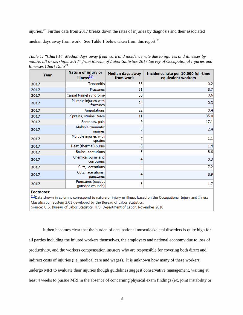
injuries.22 Further data from 2017 breaks down the rates of injuries by diagnosis and their associated
median days away from work. See Table 1 below taken from this report.23
Table 1: “Chart 14: Median days away from work and incidence rate due to injuries and illnesses by
nature, all ownerships, 2017” from Bureau of Labor Statistics 2017 Survey of Occupational Injuries and
Illnesses Chart Data23
It then becomes clear that the burden of occupational musculoskeletal disorders is quite high for
all parties including the injured workers themselves, the employers and national economy due to loss of
productivity, and the workers compensation insurers who are responsible for covering both direct and
indirect costs of injuries (i.e. medical care and wages). It is unknown how many of these workers
undergo MRI to evaluate their injuries though guidelines suggest conservative management, waiting at
least 4 weeks to pursue MRI in the absence of concerning physical exam findings (ex. joint instability or
4
deformity) due to the high cost of MRI.24 Anecdotally, it is often discussed among medical providers that
scheduling, performing, and receiving the report from an MRI routinely takes between 1-2 weeks; time
that is essentially wasted, contributing to the cost of the injury as the diagnosis and therefore appropriate
treatment plan is delayed.
The purpose of this study was to determine the proportion of peripheral joint MRI’s obtained in
workers compensation cases that could be substituted with a point of care ultrasound alone or ultrasound
+ xray since xray is often routinely performed on the first visit for most musculoskeletal injuries. Cost
and time savings by making this substitution in appropriate cases were also estimated assuming an
ultrasound could be performed in the office along with an xray on the first visit.
5
Chapter 2: Methods
This descriptive retrospective cohort study utilized a large workers compensation insurer database
which represents 39,000 small to medium sized companies from a wide range of industries in 12 different
states. All MRI's performed on peripheral joints during calendar year 2017 that were (a) at least 2 weeks
after the initial clinic visit, or (b) greater than 6 weeks after injury, but (c) not more than 3 months after
the date of injury were evaluated in this study.
A query was run in the insurer’s database to identify all claims where a non-contrast MRI of an
extremity was billed in 2017. The date of injury, first clinic visit, and MRI associated with those claims
were then cross-referenced to identify cases that met inclusion criteria. Diagnoses rendered on MRI
reports for these cases were transcribed into a spreadsheet and subsequently categorized as to whether
ultrasound alone or ultrasound + xray could adequately provide the same diagnoses using a coding system
we developed. The coded results were tabulated with percentages and 95% confidence intervals
calculated as appropriate.
In general, MRI and ultrasound were considered equivalent for imaging extra-articular soft tissue
structures such as nerve, tendon, muscle, ligament, bursa, synovium, adipose, etc. Exceptions to this rule
included structures known to be impossible or difficult to image with ultrasound such as the superior and
middle glenohumeral ligaments and many deep structures of the hip. MRI was always considered
superior to ultrasound in evaluating intra-articular structures (meniscus, labrum, ACL, PCL, cartilage).
MRI was considered superior to xray and ultrasound in evaluating diagnoses such as intra-articular
fractures and chondromalacia. Bone alignment, fractures, and osteoarthritis (OA)/degenerative changes
were considered to be both adequately imaged by MRI and xray, but not ultrasound.

6
Dates of the first clinic visit and dates MRI was performed were used to estimate time savings if
ultrasound had been performed on the first visit. Direct imaging cost savings were estimated using the
standard Florida state workers compensation fee schedule: $36 for a limited joint ultrasound study, $120
for a complete joint ultrasound study, $489 for a non-contrast joint MRI.
7
Chapter 3: Results
Cases Where Ultrasound Alone or Ultrasound + Xray Could be substituted for MRI
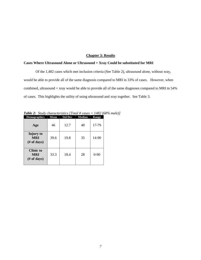
Of the 1,482 cases which met inclusion criteria (See Table 2), ultrasound alone, without xray,
would be able to provide all of the same diagnosis compared to MRI in 33% of cases. However, when
combined, ultrasound + xray would be able to provide all of the same diagnoses compared to MRI in 54%
of cases. This highlights the utility of using ultrasound and xray together. See Table 3.
Table 2: Study characteristics [Total # cases = 1482 (68% male)] Demographics Mean Std Dev Median Range
Age
46
12.7
48
17-79
Injury to
MRI
(# of days)
39.6
19.8
35
14-90
Clinic to
MRI
(# of days)
33.3
18.4
28
0-90
8
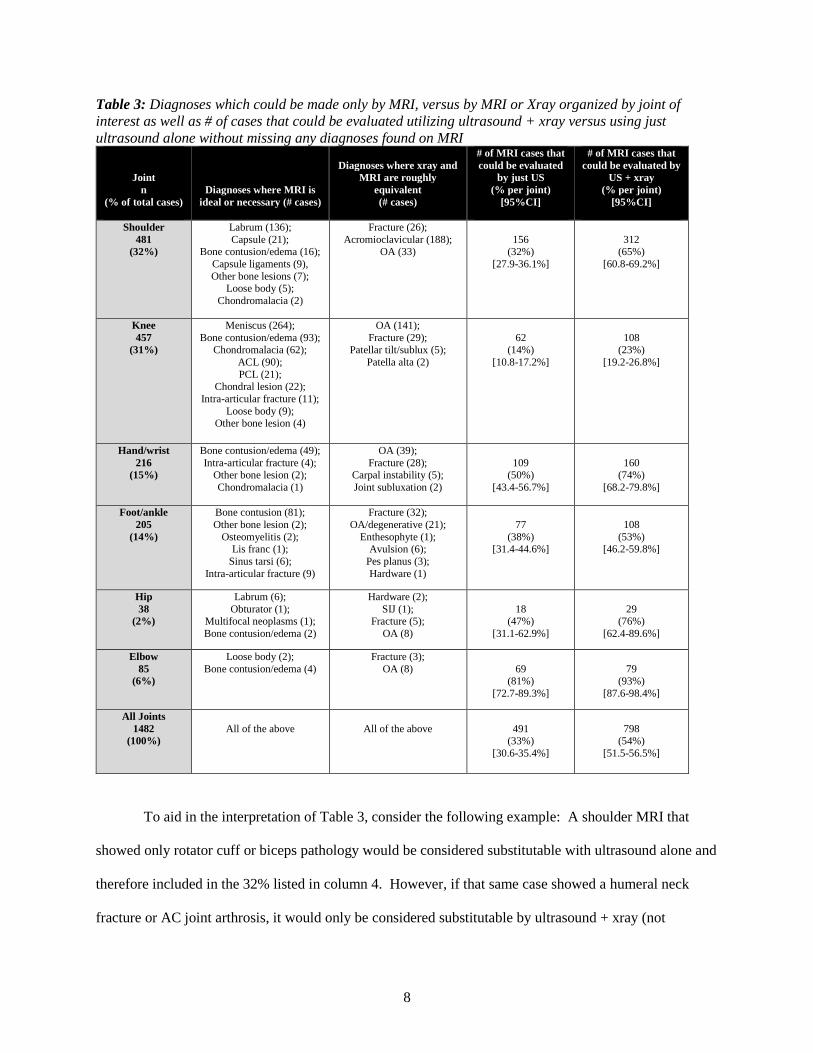
Table 3: Diagnoses which could be made only by MRI, versus by MRI or Xray organized by joint of
interest as well as # of cases that could be evaluated utilizing ultrasound + xray versus using just
ultrasound alone without missing any diagnoses found on MRI
Joint
n
(% of total cases)
Diagnoses where MRI is
ideal or necessary (# cases)
Diagnoses where xray and
MRI are roughly
equivalent
(# cases)
# of MRI cases that
could be evaluated
by just US
(% per joint)
[95%CI]
# of MRI cases that
could be evaluated by
US + xray
(% per joint)
[95%CI]
Shoulder
481
(32%)
Labrum (136);
Capsule (21); Bone contusion/edema (16);
Capsule ligaments (9),
Other bone lesions (7); Loose body (5);
Chondromalacia (2)
Fracture (26);
Acromioclavicular (188); OA (33)
156 (32%)
[27.9-36.1%]
312 (65%)
[60.8-69.2%]
Knee
457
(31%)
Meniscus (264);
Bone contusion/edema (93);
Chondromalacia (62);
ACL (90);
PCL (21); Chondral lesion (22);
Intra-articular fracture (11);
Loose body (9); Other bone lesion (4)
OA (141);
Fracture (29);
Patellar tilt/sublux (5);
Patella alta (2)
62
(14%)
[10.8-17.2%]
108
(23%)
[19.2-26.8%]
Hand/wrist
216
(15%)
Bone contusion/edema (49);
Intra-articular fracture (4); Other bone lesion (2);
Chondromalacia (1)
OA (39);
Fracture (28); Carpal instability (5);
Joint subluxation (2)
109 (50%)
[43.4-56.7%]
160 (74%)
[68.2-79.8%]
Foot/ankle
205
(14%)
Bone contusion (81);
Other bone lesion (2);
Osteomyelitis (2); Lis franc (1);
Sinus tarsi (6);
Intra-articular fracture (9)
Fracture (32);
OA/degenerative (21);
Enthesophyte (1); Avulsion (6);
Pes planus (3);
Hardware (1)
77
(38%) [31.4-44.6%]
108
(53%) [46.2-59.8%]
Hip
38
(2%)
Labrum (6);
Obturator (1); Multifocal neoplasms (1);
Bone contusion/edema (2)
Hardware (2);
SIJ (1); Fracture (5);
OA (8)
18 (47%)
[31.1-62.9%]
29 (76%)
[62.4-89.6%]
Elbow
85
(6%)
Loose body (2);
Bone contusion/edema (4)
Fracture (3);
OA (8)
69 (81%)
[72.7-89.3%]
79 (93%)
[87.6-98.4%]
All Joints
1482
(100%)
All of the above
All of the above
491 (33%)
[30.6-35.4%]
798 (54%)
[51.5-56.5%]
To aid in the interpretation of Table 3, consider the following example: A shoulder MRI that
showed only rotator cuff or biceps pathology would be considered substitutable with ultrasound alone and
therefore included in the 32% listed in column 4. However, if that same case showed a humeral neck
fracture or AC joint arthrosis, it would only be considered substitutable by ultrasound + xray (not
9
ultrasound alone), and subsequently included in the 65% listed in column 5. Lastly if that same case
included a labrum tear or glenohumeral ligament tear (which can only be imaged by MRI) it would not be
considered substitutable at all.
While the addition of xray findings to ultrasound findings dramatically increases the proportion
of cases that could be substituted/imaged completely without MRI (going from 33% to 54% overall), the
proportion of cases that could be substituted further increases to 70% overall when diagnoses which can
only be ascertained using MRI but are considered not likely to change management were excluded from
analysis (i.e. bone contusions/edema, chondromalacia, ACL and PCL sprains, meniscus degeneration,
labrum degeneration). In effect, only 30% of MRIs provided additional information that was likely to
change management which varied by joint as follows: shoulder 28%, knee 59%, hand/wrist 3%,
foot/ankle 10%, hip 21%, elbow 2%. See Table 4.
10
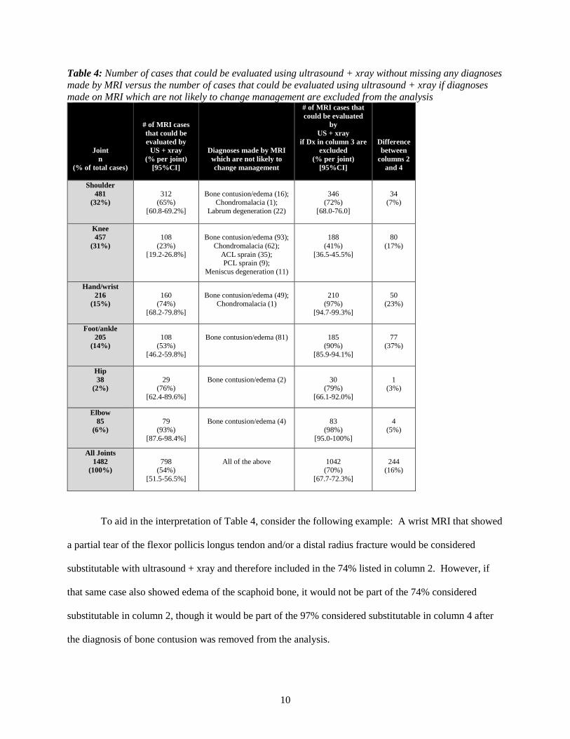
Table 4: Number of cases that could be evaluated using ultrasound + xray without missing any diagnoses
made by MRI versus the number of cases that could be evaluated using ultrasound + xray if diagnoses
made on MRI which are not likely to change management are excluded from the analysis
Joint
n
(% of total cases)
# of MRI cases
that could be
evaluated by
US + xray
(% per joint)
[95%CI]
Diagnoses made by MRI
which are not likely to
change management
# of MRI cases that
could be evaluated
by
US + xray
if Dx in column 3 are
excluded
(% per joint)
[95%CI]
Difference
between
columns 2
and 4
Shoulder
481
(32%)
312 (65%)
[60.8-69.2%]
Bone contusion/edema (16); Chondromalacia (1);
Labrum degeneration (22)
346 (72%)
[68.0-76.0]
34 (7%)
Knee
457
(31%)
108
(23%)
[19.2-26.8%]
Bone contusion/edema (93);
Chondromalacia (62);
ACL sprain (35); PCL sprain (9);
Meniscus degeneration (11)
188
(41%)
[36.5-45.5%]
80
(17%)
Hand/wrist
216
(15%)
160 (74%)
[68.2-79.8%]
Bone contusion/edema (49); Chondromalacia (1)
210 (97%)
[94.7-99.3%]
50 (23%)
Foot/ankle
205
(14%)
108 (53%)
[46.2-59.8%]
Bone contusion/edema (81)
185 (90%)
[85.9-94.1%]
77 (37%)
Hip
38
(2%)
29 (76%)
[62.4-89.6%]
Bone contusion/edema (2)
30 (79%)
[66.1-92.0%]
1 (3%)
Elbow
85
(6%)
79 (93%)
[87.6-98.4%]
Bone contusion/edema (4)
83 (98%)
[95.0-100%]
4 (5%)
All Joints
1482
(100%)
798 (54%)
[51.5-56.5%]
All of the above
1042 (70%)
[67.7-72.3%]
244 (16%)
To aid in the interpretation of Table 4, consider the following example: A wrist MRI that showed
a partial tear of the flexor pollicis longus tendon and/or a distal radius fracture would be considered
substitutable with ultrasound + xray and therefore included in the 74% listed in column 2. However, if
that same case also showed edema of the scaphoid bone, it would not be part of the 74% considered
substitutable in column 2, though it would be part of the 97% considered substitutable in column 4 after
the diagnosis of bone contusion was removed from the analysis.
11
Potential Cost Savings if Ultrasound was Appropriately Substituted for MRI
If point of care ultrasound was performed for all 1482 cases the total cost would range from
$53,352 (if all were limited joint studies) to $177,840 (if all were complete joint studies) versus $724,698
for MRIs. Total cost of imaging if ultrasound was performed in every case and MRI was additionally
performed for only the 30% of cases where it could provide a diagnosis which might change treatment
would therefore range from $268,512 to $393,000. Therefore, eliminating 70% of MRIs while
performing ultrasounds on every patient amounts to a cost savings ranging between $456,186 and
$331,698 for just the cases included in this study alone, which translates to a savings of $308 to $224 per
patient. Of note, the cost of xray was not factored into the cost savings calculations because it is a basic
imaging study which is required before MRI is approved by insurers and, therefore, it is assumed xray
was performed prior to all MRIs.
Potential Time Savings if Ultrasound was Appropriately Substituted for MRI
In terms of time savings, if ultrasound + xray was performed at the point of care during the first
clinic visit for an injury, the definitive diagnoses could be reached on average 33.3 days earlier (See Table
2), however, this does not include the additional time to get results back and review them with the patient
at a subsequent visit. It is difficult to estimate how this time savings would affect management, however,
it is reasonable to assume that a significant savings in indirect costs (ex. duty restrictions or time away
from work) might be realized.
12
Chapter 4: Discussion
Rationale for Study Inclusion and Exclusion Criteria
This study captures MRI data starting at 2 weeks post initial clinic visit with the thought that
MRIs performed within the first two weeks may represent cases with severe injuries where concerning
findings were present on physical exam (ex. joint instability, structural deformity, or extreme pain)
necessitating the most advanced and complete imaging immediately. MRIs performed within 2 weeks of
the initial clinic visit but greater than 6 weeks from the date of injury were included, assuming providers
were following guidelines where the elapsed time since the injury would be considered conservative
management without adequate improvement (hence why they presented in clinic over a month after
injury), and therefore MRI would be appropriate. MRIs performed after 3 months post injury were not
included because, in conversation/agreement with our Occupational Medicine colleagues, advanced
imaging at such a late date is often ordered in cases where a patient is paradoxically not progressing in
spite of little to no objective evidence to suggest an unhealed injury. Because it is the most complete
imaging modality, a negative MRI allows the provider to place the patient at maximal medical
improvement status and discharge the case where it will undergo arbitration and/or independent medical
evaluation. Because we did not conduct chart review of clinic notes, we would have no way to filter out
those types of cases.
13
Representativeness of Study Data and Generalizability to the US Workforce
According to the medical director of the workers compensation insurer where this study was
conducted, data from 2017 was representative of a typical year and was almost exactly on target with
recent previous years and hence the insurer’s predictions for total claims submitted and MRIs ordered. It
is therefore likely that the proportions of MRIs by joint included in this analysis are also representative of
a typical year. Given the insurer represents over 39,000 businesses in 12 states in a wide range of
industries, it is also likely that this sample is reasonably representative of the US workforce in general.
Though no previous papers have been published with regard to utilization of ultrasound vs. MRI
to evaluate musculoskeletal injuries in workers compensation cases, the results of this study do mirror a
paper by Parker et al. which described a study in the general population.25 In that paper, all
musculoskeletal MRIs in a radiology database performed over the course of one year (n = 3,621) were
analyzed in a similar fashion, revealing that 45.4% of primary diagnoses and 30.6% of total cases could
be evaluated completely using only ultrasound, which, upon extrapolation, they estimated could save
almost $7 billion in Medicare alone over the period of 2006-2020. The mean age and standard deviation
between the Parker et al. paper and this study are nearly the same (45.6 and 15.9 vs. 46 and 12.7
respectively). Because the Parker et al. paper included “all-comers” and the present study includes only
recent work injuries, the differences in population may explain the differences in percentage of cases
where ultrasound could be appropriately substituted for MRI, though the results are admittedly still quite
similar for multiple joints, suggesting the results of this study may be applicable to the broader fields of
Orthopedics and Sports Medicine outside of the Occupational Medicine/workers compensation realm.
See Table 5.
14
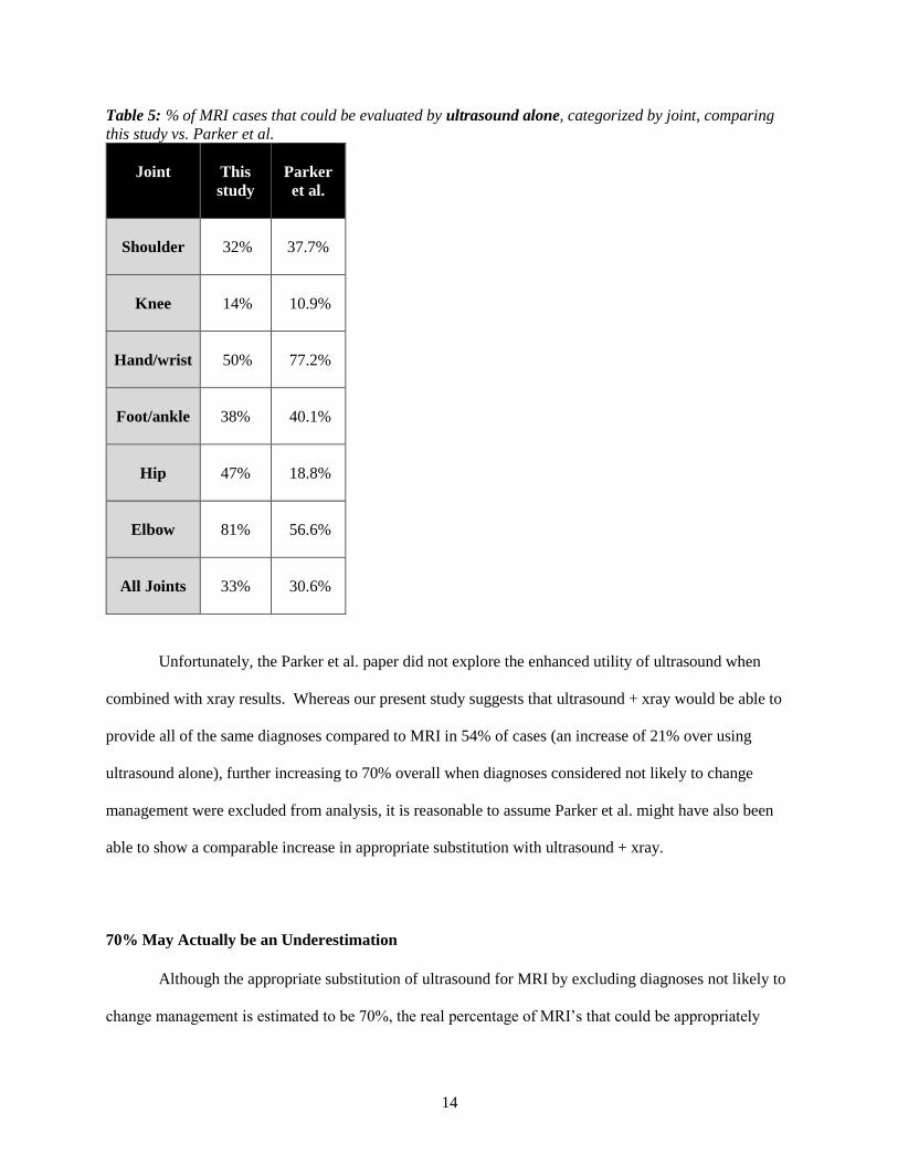
Table 5: % of MRI cases that could be evaluated by ultrasound alone, categorized by joint, comparing
this study vs. Parker et al.
Joint
This
study
Parker
et al.
Shoulder
32%
37.7%
Knee
14%
10.9%
Hand/wrist
50%
77.2%
Foot/ankle
38%
40.1%
Hip
47%
18.8%
Elbow
81%
56.6%
All Joints
33%
30.6%
Unfortunately, the Parker et al. paper did not explore the enhanced utility of ultrasound when
combined with xray results. Whereas our present study suggests that ultrasound + xray would be able to
provide all of the same diagnoses compared to MRI in 54% of cases (an increase of 21% over using
ultrasound alone), further increasing to 70% overall when diagnoses considered not likely to change
management were excluded from analysis, it is reasonable to assume Parker et al. might have also been
able to show a comparable increase in appropriate substitution with ultrasound + xray.
70% May Actually be an Underestimation
Although the appropriate substitution of ultrasound for MRI by excluding diagnoses not likely to
change management is estimated to be 70%, the real percentage of MRI’s that could be appropriately
15
substituted with ultrasound + xray is likely much higher than what we are able to estimate in this study
given the results of other recent papers on knee meniscus and shoulder labrum tears.
To this point, it is important to consider that multiple papers have documented the extremely high
prevalence of asymptomatic meniscus tears.26,27,28,29 Most practitioners would likely agree that a
diagnosis of knee meniscus tear on MRI (many of which may be incidental/correlate poorly with clinical
findings) often leads to an Orthopedic referral and arthroscopic surgery. The appropriateness of
arthroscopic surgery for MRI confirmed meniscus tears has recently been called into question by
randomized clinical trials showing no difference in outcomes compared to physical therapy, even after
two years of follow up provided the patients have no initial findings of knee instability, locking, are not
obese, and can weight bear well enough to participate in exercise.30,31,32,33,34,35,36 Furthermore, the size,
shape, and location of meniscus tears had no effect on outcomes. This then begs the question: How
useful is the information from an MRI if we should preferentially send patients to physical therapy as first
line treatment in the absence of physical exam findings that would necessitate MRI (i.e. knee instability,
locking, obesity, and inability to weight bear well enough to participate in exercise)?
Admittedly, a limitation of the present study is that physical exam findings were not reviewed and
therefore it is unknown how many cases included had a clinical presentation which would necessitate
MRI consistent with the aforementioned parameters. However, of the 264 cases where meniscus
pathology was found on MRI, and subsequently included in the 59% of knees examined that we
determined could not be substituted with ultrasound + xray (See Tables 3 & 4), only 4 had extrusions, 8
had bucket tears/flipped fragments/or prolapse, and 7 had intra-articular loose bodies. It may follow then
that a large proportion of those 264 knees with meniscal pathology (representing 17% of total cases
included in this study) might not have required MRI to achieve appropriate management, and instead
could have appropriately utilized the less costly and faster alternative of ultrasound + xray.
In an analogous line of inquiry, recent papers have highlighted the extremely high prevalence of
asymptomatic shoulder labral tears in the general population especially as age increases, although the
prevalence is also high in asymptomatic young athletes as well.37,38,39,40,41 In a randomized trial
16
comparing labral repair vs biceps tenodesis vs sham surgery there were no significant differences in
outcomes between groups.42 Additionally, surgeons seem to have a low level of agreement in how to treat
labral tears, and labral repairs in patients over 36 years old often fail.43,44 The latest guidelines conclude
that 2/3 of labral injuries improve adequately with therapy, and surgery should be reserved until after at
least 3 months of directed therapy has failed or in the presence of shoulder instability with significant
consideration for age as a predictor of outcome along with pathology of the biceps.45,46 So, as with
meniscus tears in the knee, we must then ask: How useful is the information from an MRI if we should
preferentially use conservative management in the absence of physical exam findings that would
necessitate MRI (i.e. shoulder instability, biceps pathology)?
In our present study, labral tears were seen on MRI in 113 cases (representing 7.6% of total cases
in the study) and subsequently included in the 28% of shoulder exams that we determined could not be
substituted with ultrasound + xray. Again, a limitation of the present study is that physical exam findings
were not reviewed and therefore it is unknown how many of the cases included had a clinical presentation
which would necessitate MRI, however, it seems likely that a large proportion of the labral tears found on
MRI might have been incidental, providing no benefit in a preferentially conservative management plan.
Of course, after performing a thorough history and physical exam the etiology of a patient’s pain
can be cryptic in some cases even with the addition of ultrasound to visualize extra-articular structures. In
the absence of physical exam findings such as joint instability or deformity that would necessitate an
MRI, and where the provider is unsure as to whether the source of a patient’s pain is intra-articular vs.
extra-articular, a diagnostic injection of anesthetic is a quick an easy way to rule this in or out at the point
of care.47,48 Therefore, having access to an ultrasound in the clinic can prove invaluable, both through
routine use of ultrasound diagnostic exams and subsequent ultrasound-guided diagnostic injections of
anesthetic to distinguish between intra- and extra-articular pathology induced pain for appropriate cases at
the point of care.
To be clear, we do not believe ultrasound could or should replace MRI because both have value
over the other in different scenarios. In evaluating MSK work injuries we believe ultrasound’s place is at
17
the point of care coupled with a good history and physical, helping to rule out red flags, and increasing
the likelihood of a correct diagnosis and subsequent optimal treatment plan so that workers return to duty
as quickly as possible, reducing indirect costs. It’s appropriate use at the point of care will also lead to
direct cost savings by reducing the number of unnecessary MRI’s ordered as subsequent specialist referral
and treatment for incidental findings.
Conclusion
The results of our present study suggest that the majority of musculoskeletal workers comp
injuries could be accurately and completely evaluated at the point of care using ultrasound and xray
together instead of MRI, and this number may be well in excess of 70%. This substitution, if
implemented appropriately, could yield greater provider and patient confidence in the diagnosis and
treatment plan as well as more expeditious accurate diagnoses leading to substantial reductions in both
direct and indirect costs.
Future directions in this area of research include confirmation of the results of this study with
other data sets which hopefully will correlate imaging findings with clinical findings, as well as head-to-
head trials of MRI vs ultrasound in evaluating MSK work injuries which may be able to highlight the
added value of dynamic imaging and power doppler use at the point of care. We also envision studies
which would use point of care ultrasound to follow injuries over time, tracking recovery and describing
its use in performing appropriate interventional procedures in the office for these patients. Our group is
currently conducting analyses similar to those performed in this study to describe of the utility of
ultrasound + xray in evaluating MSK work injuries over the acute post-injury period of 0 days to 2 weeks.
We will then use these data in aggregate to delineate patterns of injury, developing Bayesian conditional
probability network-based ultrasound scanning algorithms which are simplified to aid providers in point
of care injury evaluation, similar to the FAST scan in emergency medicine.
18
References
1. He, L., Delzell, P. & Schils, J. Comparison of MRI Findings After Musculoskeletal Ultrasound: An
Opportunity to Reduce Redundant Imaging. J. Am. Coll. Radiol. 15, 1116–1119 (2018).
2. Jacobson, J. A. Fundamentals of Musculoskeletal Ultrasound E-Book. (Elsevier Health Sciences,
2017).
3. Jesus, J. O. de, de Jesus, J. O., Parker, L., Frangos, A. J. & Nazarian, L. N. Accuracy of MRI, MR
Arthrography, and Ultrasound in the Diagnosis of Rotator Cuff Tears: A Meta-Analysis. American
Journal of Roentgenology 192, 1701–1707 (2009).
4. Henderson, R. E. A., Walker, B. F. & Young, K. J. The accuracy of diagnostic ultrasound imaging
for musculoskeletal soft tissue pathology of the extremities: a comprehensive review of the
literature. Chiropr. Man. Therap. 23, 31 (2015).
5. Zubler, V., Mamisch-Saupe, N., Pfirrmann, C. W. A., Jost, B. & Zanetti, M. Detection and
quantification of glenohumeral joint effusion: reliability of ultrasound. Eur. Radiol. 21, 1858–1864
(2011).
6. Strobel, K. et al. Fatty atrophy of supraspinatus and infraspinatus muscles: accuracy of US.
Radiology 237, 584–589 (2005).
7. Khoury, V., Cardinal, E. & Brassard, P. Atrophy and fatty infiltration of the supraspinatus muscle:
sonography versus MRI. AJR Am. J. Roentgenol. 190, 1105–1111 (2008).
8. Rehman, A. & Robinson, P. Sonographic evaluation of injuries to the pectoralis muscles. AJR Am. J.
Roentgenol. 184, 1205–1211 (2005).
9. Weaver, J. S. et al. Sonographic findings of pectoralis major tears with surgical, clinical, and
magnetic resonance imaging correlation in 6 patients. J. Ultrasound Med. 24, 25–31 (2005).
19
10. Carroll, K. W., Helms, C. A., Otte, M. T., Moellken, S. M. C. & Fritz, R. Enlarged spinoglenoid
notch veins causing suprascapular nerve compression. Skeletal Radiol. 32, 72–77 (2003).
11. Taljanovic, M. S. et al. Sonography of the glenoid labrum: a cadaveric study with arthroscopic
correlation. AJR Am. J. Roentgenol. 174, 1717–1722 (2000).
12. Thiele, R. G. & Schlesinger, N. Ultrasonography shows disappearance of monosodium urate crystal
deposition on hyaline cartilage after sustained normouricemia is achieved. Rheumatol. Int. 30, 495–
503 (2010).
13. Rutten, M. J. C. M., Matthieu J C, de Jong, M. D. F., Van loon, T. & Jager, G. J. Intratendinous
ganglion of the long head of the biceps tendon: US and MRI features (2010: 9b). European
Radiology 20, 2997–3001 (2010).
14. Lee, J. C., Sykes, C., Saifuddin, A. & Connell, D. Adhesive capsulitis: sonographic changes in the
rotator cuff interval with arthroscopic correlation. Skeletal Radiol. 34, 522–527 (2005).
15. Homsi, C., Bordalo-Rodrigues, M., da Silva, J. J. & Stump, X. M. G. R. G. Ultrasound in adhesive
capsulitis of the shoulder: is assessment of the coracohumeral ligament a valuable diagnostic tool?
Skeletal Radiol. 35, 673–678 (2006).
16. Jacobson, J. A. Musculoskeletal ultrasound and MRI: which do I choose? Semin. Musculoskelet.
Radiol. 9, 135–149 (2005).
17. Jacobson, J. A. Musculoskeletal ultrasound: focused impact on MRI. AJR Am. J. Roentgenol. 193,
619–627 (2009).
18. Sivan, M., Brown, J., Brennan, S. & Bhakta, B. A one-stop approach to the management of soft
tissue and degenerative musculoskeletal conditions using clinic-based ultrasonography.
Musculoskeletal Care 9, 63–68 (2011).
19. Finnoff, J. T. et al. American Medical Society for Sports Medicine (AMSSM) position statement:
interventional musculoskeletal ultrasound in sports medicine. PM R 7, 151–68.e12 (2015).
20. Hirahara, A. M. & Panero, A. J. A Guide to Ultrasound of the Shoulder, Part 1: Coding and
Reimbursement. Am. J. Orthop. 45, 176–182 (2016).
20
21. 1218-AB58 - 2014. PREVENTION OF WORK-RELATED MUSCULOSKELETAL DISORDERS
| Occupational Safety and Health Administration. (2019). Available at:
https://www.osha.gov/pls/oshaweb/owadisp.show_document?p_id=4481&p_table=UNIFIED_AGE
NDA. (Accessed: 16th March 2019)
22. Back injuries prominent in work-related musculoskeletal disorder cases in 2016 : The Economics
Daily: U.S. Bureau of Labor Statistics. (2018). Available at:
https://www.bls.gov/opub/ted/2018/back-injuries-prominent-in-work-related-musculoskeletal-
disorder-cases-in-2016.htm?view_full. (Accessed: 16th March 2019)
23. 2017 SURVEY OF OCCUPATIONAL INJURIES AND ILLNESSES CHART DATA. (2018).
Available at: https://www.bls.gov/iif/soii-chart-data-
2017.htm#BLStable_2018_23_13_0_0_footnotes. (Accessed: 16th March 2019)
24. Hegmann, K. T. ACOEM’s Occupational Medicine Practice Guidelines 3rd Ed. Vol 2&3. (American
College of Occupational and Environmental Medicine, 2011).
25. Parker, L. et al. Musculoskeletal imaging: medicare use, costs, and potential for cost substitution. J.
Am. Coll. Radiol. 5, 182–188 (2008).
26. Zanetti, M. et al. Patients with suspected meniscal tears: prevalence of abnormalities seen on MRI
of 100 symptomatic and 100 contralateral asymptomatic knees. AJR Am. J. Roentgenol. 181, 635–
641 (2003).
27. Boks, S. S., Vroegindeweij, D., Koes, B. W., Hunink, M. M. G. M. & Bierma-Zeinstra, S. M. A.
Magnetic resonance imaging abnormalities in symptomatic and contralateral knees: prevalence and
associations with traumatic history in general practice. Am. J. Sports Med. 34, 1984–1991 (2006).
28. Marom, N. & Mann, G. Asymptomatic Meniscal Tears. Sports Injuries 1–7 (2014).
doi:10.1007/978-3-642-36801-1_70-1
29. Englund, M. et al. Incidental meniscal findings on knee MRI in middle-aged and elderly persons. N.
Engl. J. Med. 359, 1108–1115 (2008).
21
30. van de Graaf, V. A. et al. Effect of Early Surgery vs Physical Therapy on Knee Function Among
Patients With Nonobstructive Meniscal Tears: The ESCAPE Randomized Clinical Trial. JAMA 320,
1328–1337 (2018).
31. Sihvonen, R. et al. Arthroscopic partial meniscectomy versus sham surgery for a degenerative
meniscal tear. N. Engl. J. Med. 369, 2515–2524 (2013).
32. Katz, J. N. et al. Surgery versus physical therapy for a meniscal tear and osteoarthritis. N. Engl. J.
Med. 368, 1675–1684 (2013).
33. Kise, N. J. et al. Exercise therapy versus arthroscopic partial meniscectomy for degenerative
meniscal tear in middle aged patients: randomised controlled trial with two year follow-up. Br. J.
Sports Med. 50, 1473–1480 (2016).
34. Yim, J.-H. et al. A comparative study of meniscectomy and nonoperative treatment for degenerative
horizontal tears of the medial meniscus. Am. J. Sports Med. 41, 1565–1570 (2013).
35. Østerås, H., Østerås, B. & Torstensen, T. A. Medical exercise therapy, and not arthroscopic surgery,
resulted in decreased depression and anxiety in patients with degenerative meniscus injury. Journal
of Bodywork and Movement Therapies 16, 456–463 (2012).
36. Herrlin, S. V. et al. Is arthroscopic surgery beneficial in treating non-traumatic, degenerative medial
meniscal tears? A five year follow-up. Knee Surg. Sports Traumatol. Arthrosc. 21, 358–364 (2013).
37. Schwartzberg, R. et al. High Prevalence of Superior Labral Tears Diagnosed by MRI in Middle-
Aged Patients With Asymptomatic Shoulders. Orthop J Sports Med 4, 2325967115623212 (2016).
38. Lansdown, D. A., Bendich, I., Motamedi, D. & Feeley, B. T. Imaging-Based Prevalence of Superior
Labral Anterior-Posterior Tears Significantly Increases in the Aging Shoulder. Orthop J Sports Med
6, 2325967118797065 (2018).
39. Fredericson, M. et al. Magnetic resonance imaging abnormalities in the shoulder and wrist joints of
asymptomatic elite athletes. PM R 1, 107–116 (2009).
40. Lesniak, B. P. et al. Glenohumeral findings on magnetic resonance imaging correlate with innings
pitched in asymptomatic pitchers. Am. J. Sports Med. 41, 2022–2027 (2013).
22
41. Miniaci, A., Mascia, A. T., Salonen, D. C. & Becker, E. J. Magnetic resonance imaging of the
shoulder in asymptomatic professional baseball pitchers. Am. J. Sports Med. 30, 66–73 (2002).
42. Hong, C.-K. & Su, W.-R. Sham surgery versus labral repair or biceps tenodesis for type II SLAP
lesions of the shoulder: a three-armed randomised clinical trial. Br. J. Sports Med. 51, 1780 (2017).
43. Wang, K. K., Yalizis, M., Hoy, G. A. & Ek, E. T. Current trends in the evaluation and treatment of
SLAP lesions: analysis of a survey of specialist shoulder surgeons. JSES Open Access 2, 48–53
(2018).
44. Provencher, M. T., McCormick, F., Dewing, C., McIntire, S. & Solomon, D. A prospective analysis
of 179 type 2 superior labrum anterior and posterior repairs: outcomes and factors associated with
success and failure. Am. J. Sports Med. 41, 880–886 (2013).
45. Hester, W. A., O’Brien, M. J., Heard, W. M. R. & Savoie, F. H. Current Concepts in the Evaluation
and Management of Type II Superior Labral Lesions of the Shoulder. Open Orthop. J. 12, 331–341
(2018).
46. Johannsen, A. M. & Costouros, J. G. A Treatment-Based Algorithm for the Management of Type-II
SLAP Tears. Open Orthop. J. 12, 282–287 (2018).
47. McFarland, E., Bernard, J., Dein, E. & Johnson, A. Diagnostic Injections About the Shoulder. J. Am.
Acad. Orthop. Surg. 25, 799–807 (2017).
48. Masala, S. et al. Diagnostic and therapeutic joint injections. Semin. Intervent. Radiol. 27, 160–171
(2010).