Embed Size (px)
Citation preview
Pregnancy
Sperm
Egg
Human Reproduction: Ovulation, Semen Production, and Fertilization

Sperm race to fertilize the egg
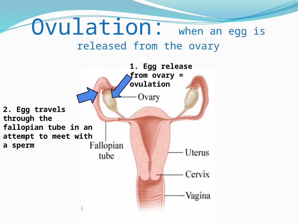
Ovulation: when an egg is released from the ovary
1. Egg release from ovary = ovulation
2. Egg travels through the fallopian tube in an attempt to meet with a sperm
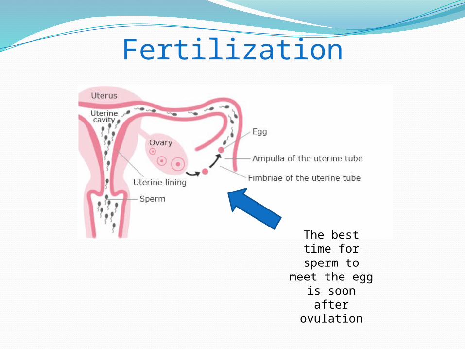
Fertilization
The best time for sperm to meet the egg is soon after
ovulation
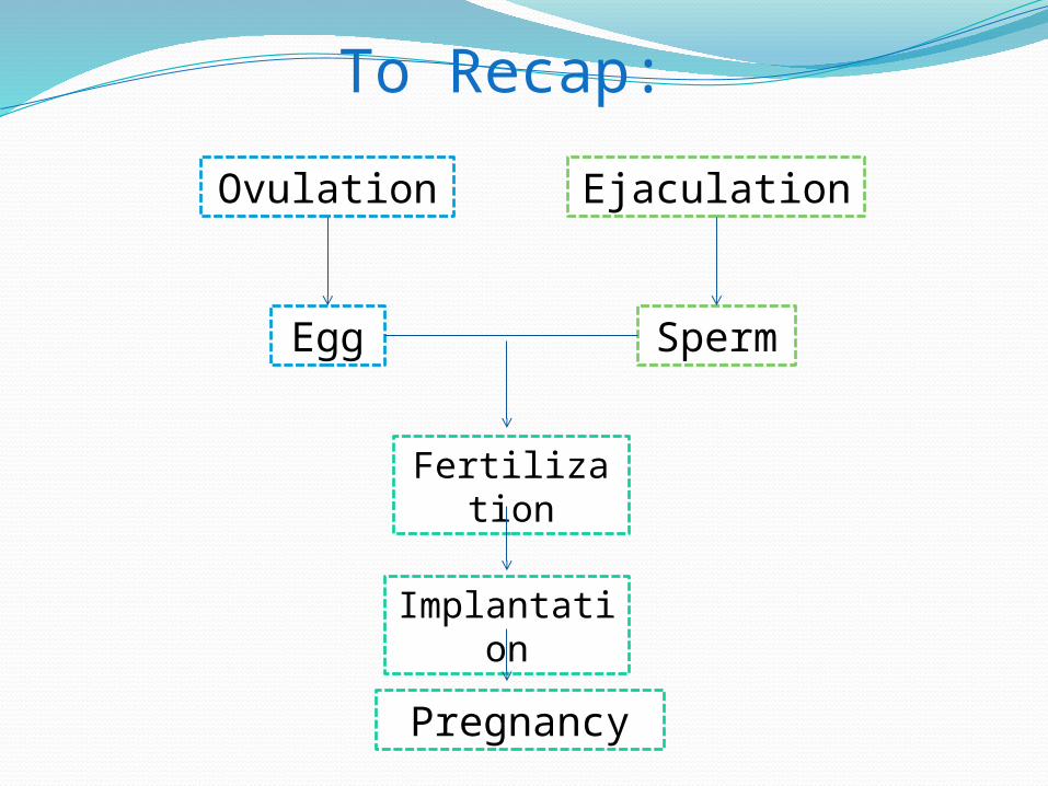
To Recap:
Ovulation
Egg
Ejaculation
Sperm
Fertilization
Implantation
Pregnancy
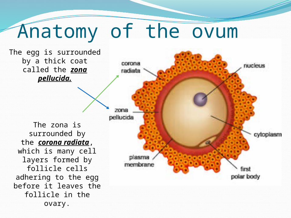
Anatomy of the ovumThe egg is surrounded
by a thick coat called the zona pellucida.
The zona is surrounded by the corona radiata,
which is many cell layers formed by
follicle cells adhering to the egg before it leaves the follicle in the ovary.
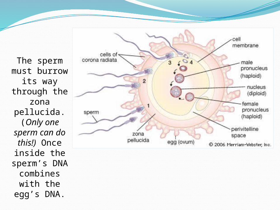
The sperm must burrow
its way through the
zona pellucida. (Only one
sperm can do this!) Once inside the
sperm’s DNA combines with the
egg’s DNA.
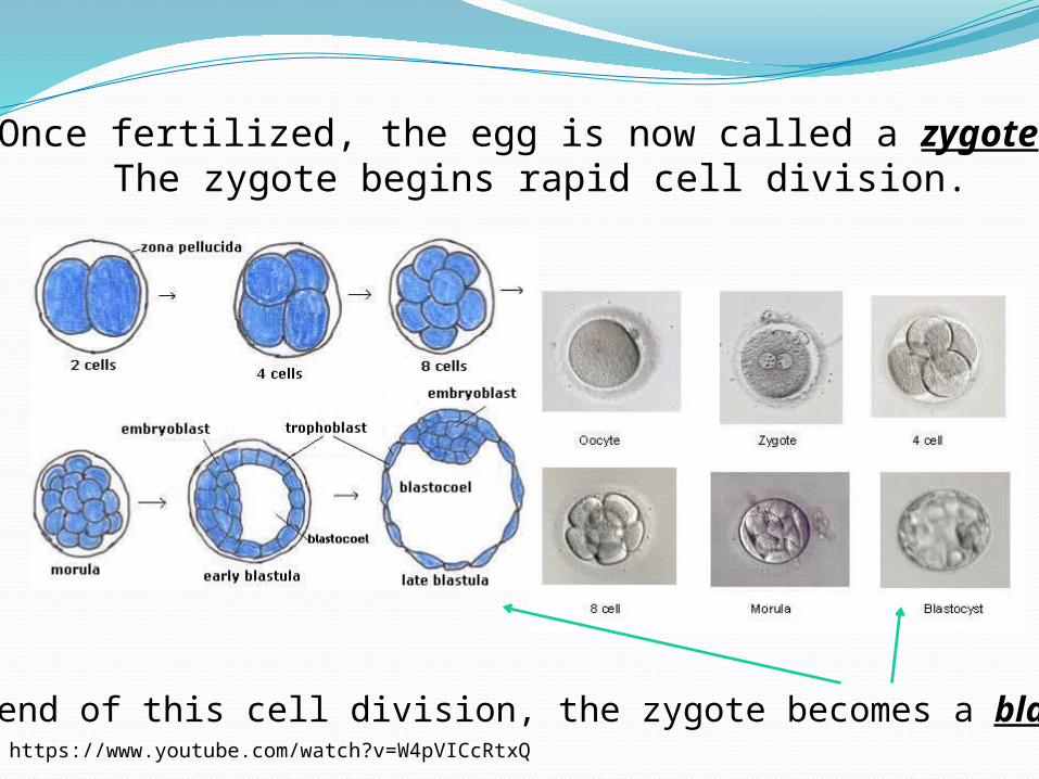
Once fertilized, the egg is now called a zygote. The zygote begins rapid cell division.
At the end of this cell division, the zygote becomes a blastocyst. https://www.youtube.com/watch?v=W4pVICcRtxQ
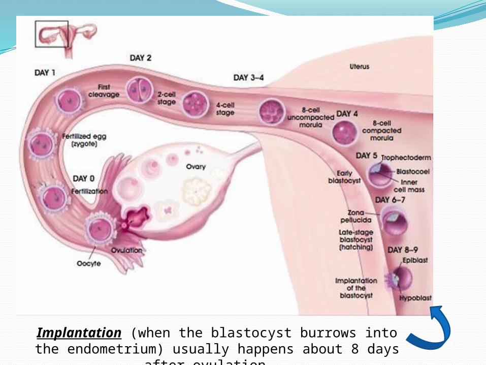
Implantation (when the blastocyst burrows into the endometrium) usually happens about 8 days after
ovulation.
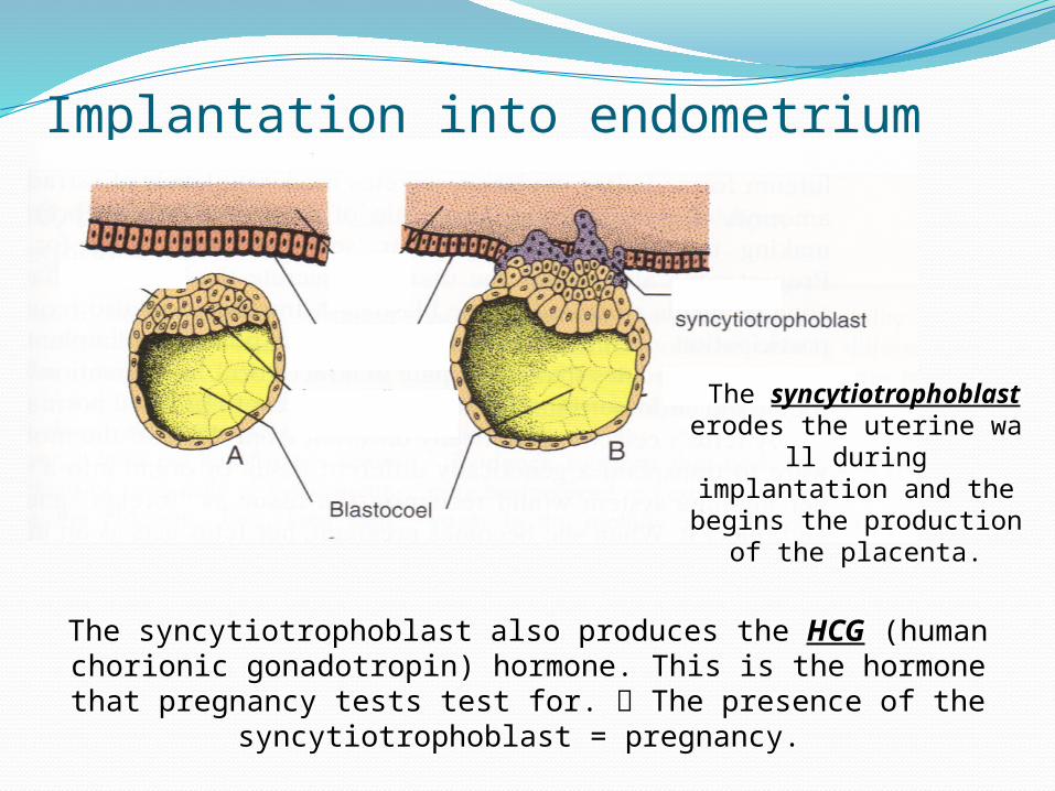
Implantation into endometrium
The syncytiotrophoblast
erodes the uterine wall during
implantation and the begins the production
of the placenta.
The syncytiotrophoblast also produces the HCG (human chorionic gonadotropin) hormone. This is the hormone that
pregnancy tests test for. The presence of the syncytiotrophoblast = pregnancy.
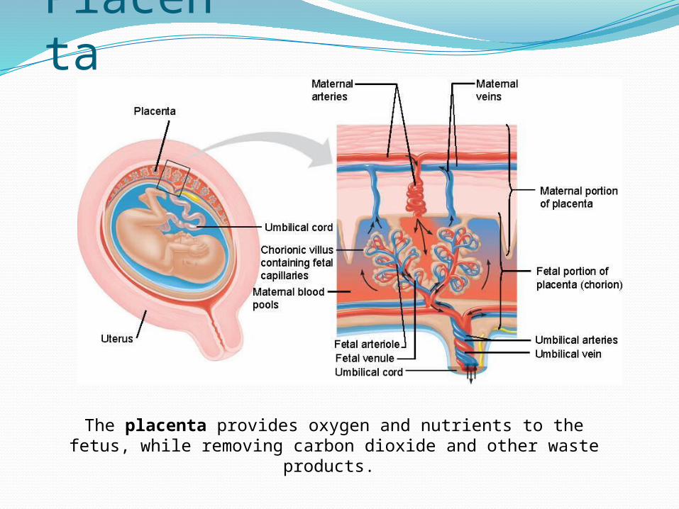
Placenta
The placenta provides oxygen and nutrients to the fetus, while removing carbon dioxide and other waste products.
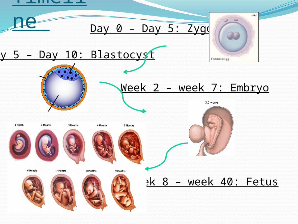
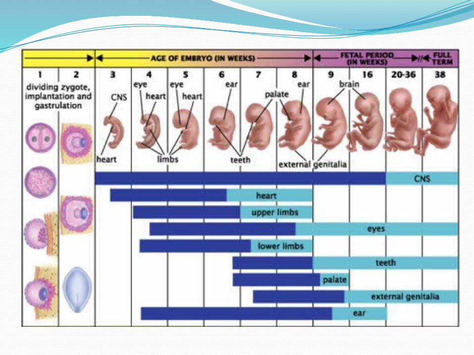
Timeline Day 0 – Day 5: Zygote
Day 5 – Day 10: Blastocyst
Week 2 – week 7: Embryo
Week 8 – week 40: Fetus
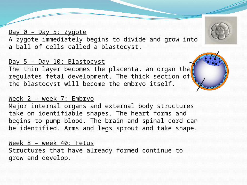
Day 0 – Day 5: ZygoteA zygote immediately begins to divide and grow into a ball of cells called a blastocyst.
Day 5 – Day 10: BlastocystThe thin layer becomes the placenta, an organ that regulates fetal development. The thick section of the blastocyst will become the embryo itself.
Week 2 – week 7: EmbryoMajor internal organs and external body structures take on identifiable shapes. The heart forms and begins to pump blood. The brain and spinal cord can be identified. Arms and legs sprout and take shape.
Week 8 – week 40: FetusStructures that have already formed continue to grow and develop.
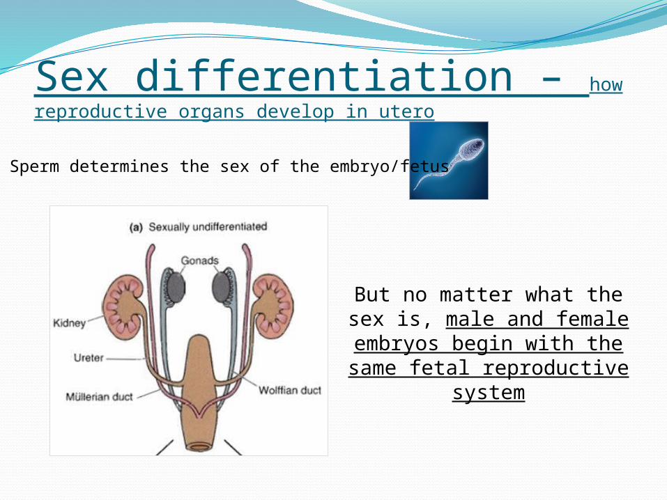
Sex differentiation – how reproductive organs develop in utero
Sperm determines the sex of the embryo/fetus
But no matter what the sex is, male and female embryos
begin with the same fetal reproductive system
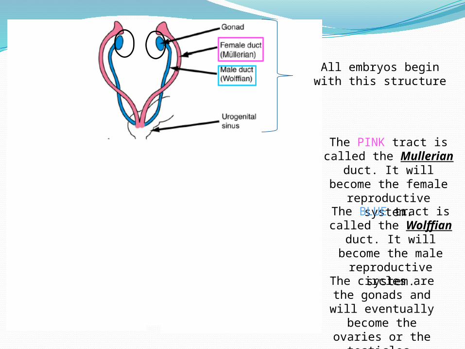
All embryos begin with this structure
The PINK tract is called the Mullerian duct. It will become
the female reproductive system.
The BLUE tract is called the Wolffian duct. It will become
the male reproductive system.The circles are the
gonads and will eventually become the ovaries or the
testicles.
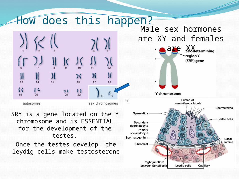
How does this happen?
SRY is a gene located on the Y chromosome and is ESSENTIAL for
the development of the testes.
Once the testes develop, the leydig cells make testosterone
Male sex hormones are XY and females are XX
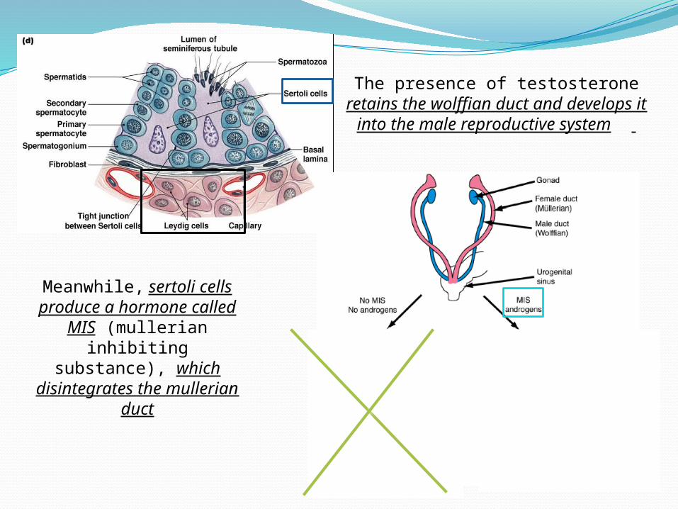
The presence of testosterone retains the wolffian duct and
develops it into the male reproductive system
Meanwhile, sertoli cells produce a hormone
called MIS (mullerian inhibiting substance),
which disintegrates the mullerian duct
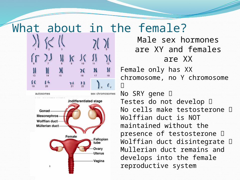
What about in the female?Male sex hormones are XY and females are XX
Female only has XX chromosome, no Y chromosome No SRY gene Testes do not develop No cells make testosterone Wolffian duct is NOT maintained without the presence of testosterone Wolffian duct disintegrate Mullerian duct remains and develops into the female reproductive system
So what does all of this mean?The male reproductive system REQUIRES activation in order to development. It needs the SRY gene and
testosterone.
The female system DOES NOT require activation. It will simply develop without the presence of
testosterone. The female reproductive system is the DEFAULT system.
It’s not that we all start as females in utero. We all start as the SAME- neither male nor female.
Symptoms of PregnancyMissed periodTender or swollen breastsNausea (with or without vomiting)Slight bleeding or “spotting”Increased urinationFatigueFood aversions or craving
The only way to REALLY determine pregnancy is to take a test.