Embed Size (px)
Citation preview
96:235-251, 2006. First published Apr 26, 2006; doi:10.1152/jn.01094.2005 J NeurophysiolAndrei I. Ivanov and Ronald L. Calabrese for Shared Release Sites Transmission Between Leech Interneurons: Evidence Spike-Mediated and Graded Inhibitory Synaptic
You might find this additional information useful...
64 articles, 33 of which you can access free at: This article cites http://jn.physiology.org/cgi/content/full/96/1/235#BIBL
including high-resolution figures, can be found at: Updated information and services http://jn.physiology.org/cgi/content/full/96/1/235
can be found at: Journal of Neurophysiologyabout Additional material and information http://www.the-aps.org/publications/jn
This information is current as of August 30, 2006 .
http://www.the-aps.org/.American Physiological Society. ISSN: 0022-3077, ESSN: 1522-1598. Visit our website at (monthly) by the American Physiological Society, 9650 Rockville Pike, Bethesda MD 20814-3991. Copyright © 2005 by the
publishes original articles on the function of the nervous system. It is published 12 times a yearJournal of Neurophysiology
on August 30, 2006
jn.physiology.orgD
ownloaded from
Spike-Mediated and Graded Inhibitory Synaptic Transmission Between LeechInterneurons: Evidence for Shared Release Sites
Andrei I. Ivanov and Ronald L. CalabreseDepartment of Biology, Emory University, Atlanta, Georgia
Submitted 17 October 2005; accepted in final form 29 March 2006
Ivanov, Andrei I. and Ronald L. Calabrese. Spike-mediated andgraded inhibitory synaptic transmission between leech interneurons:evidence for shared release sites. J Neurophysiol 96: 235–251, 2006.First published April 26, 2006; doi:10.1152/jn.01094.2005. Inhibitorysynaptic transmission between leech heart interneurons consist of twocomponents: graded, gated by Ca2� entering by low-threshold [low-voltage–activated (LVA)] Ca channels and spike-mediated, gated byCa2� entering by high-threshold [high-voltage–activated (HVA)] Cachannels. Changes in presynaptic background Ca2� produced byCa2� influx through LVA channels modulate spike-mediated trans-mission, suggesting LVA channels have access to release sites con-trolled by HVA channels. Here we explore whether spike-mediatedand graded transmission can use the same release sites and thus howCa2� influx by HVA and LVA Ca channels might interact to evokeneurotransmitter release. We recorded pre- and postsynaptic currentsfrom voltage-clamped heart interneurons bathed in 0 mM Na�/5 mMCa2� saline. Using different stimulating paradigms and inorganic Cachannel blockers, we show that strong graded synaptic transmissioncan occlude high-threshold/spike-mediated synaptic transmissionwhen evoked simultaneously. Suppression of LVA Ca currents di-minishes graded release and concomitantly increases the ability ofCa2� entering by HVA channels to release transmitter. Uncaging ofCa chelator corroborates that graded release occludes spike-mediatedtransmission. Our results indicate that both graded and spike-mediatedsynaptic transmission depend on the same readily releasable pool ofsynaptic vesicles. Thus Ca2�, entering cells through different Cachannels (LVA and HVA), acts to gate release of the same synapticvesicles. The data argue for a closer location of HVA Ca channels torelease sites than LVA Ca channels. The results are summarized in aconceptual model of a heart interneuron release site.
I N T R O D U C T I O N
At many synapses more than one kind of Ca channel isinvolved in neurotransmitter release and clusters of differentCa channel types may overlap and cooperate to trigger fusion(Meinrenken et al. 2003; Mintz et al. 1995; Reid et al. 1998;Wu and Shaggau 1994, 1997). On other hand, Ca2� enteringthe cell through different types of Ca channels may havedifferent effects on synaptic transmission. For example, in ratcalyx-type synapses, P/Q-type Ca channels trigger releasemore effectively than N- and R-type Ca channels, whichappear to be located more distantly from release sites (Wu et al.1998, 1999). Mouse cortical CA3–CA1 synapses appear to besimilarly organized (Qian and Noebels 2001).
Ca channel cooperativity in neurotransmitter release de-pends on the kind of Ca channels involved and the location ofthese channels with respect to active zones (Meinrenken et al.2002, 2003; but see Gentile and Stanley 2005). Ca channels at
calyx of Held synapses are clustered and located at differentdistances from the synaptic vesicle release trigger in differentactive zones, leading to variability in the release efficacy fromone active zone to another (Meinrenken et al. 2002). Involve-ment of different Ca channels in neurotransmitter release maychange during development (Fedchyshin and Wang 2005).Moreover, distant Ca channels that are not involved in Cadomain(s) that trigger release can nevertheless provide bulk(background) intracellular Ca2� that may modulate neurotrans-mitter release (Bertram et al. 1999).
Synapses may be either phasic (spike-mediated and fast) ortonic (non-spike-mediated and slow, or graded) and involvesynchronous and/or asynchronous release. At the same syn-apse, transmission may be both phasic (spike-mediated, syn-chronous) and tonic (graded, asynchronous) (Angstadt andCalabrese 1991; Ayali et al. 1998; Ivanov and Calabrese 2000,2006; Pan et al. 2001; Warzecha et al. 2003), and in some casesthe involvement of low-voltage–activated (LVA) Ca channelsin transmission is well documented (Angstadt and Calabrese1991; Ivanov and Calabrese 2000, 2006; Pan et al. 2001).Transmitter release in these neurons resembles catecholaminesecretion in chromaffin cells (Artalejo et al. 1994), whereactivation of different Ca channels leads to a different mode ofneurosecretion, and different Ca channels control catechol-amine secretion with different efficacies as a result of theirdifferent proximity to release sites. At other neuronal synapsesasynchronous (tonic, bulk Ca-dependent) release can often berecorded after spike-evoked (phasic, synchronous) release(Atluri and Regehr 1998) and be in a competitive relation withsynchronous release (Otsu et al. 2004).
In leeches, reciprocally inhibitory synaptic transmission be-tween heart interneurons is both spike-mediated [depends onhigh-voltage–activated (HVA) Ca channels] and graded (de-pends on two types of LVA Ca channels termed ICaS and ICaF)(Angstadt and Calabrese 1991; Ivanov and Calabrese 2000; Luet al. 1997; Olsen and Calabrese 1996). Previously we showedthat LVA Ca channels, which are widely distributed through-out heart interneurons, not only generate the plateau potentialthat drives the burst of action potential and mediate gradedtransmission, but also provide intracellular background Ca formodulation of spike-mediated transmission (Angstadt and Ca-labrese 1991; Arbas and Calabrese 1987; Ivanov and Calabrese2000, 2003; Lu et al. 1997; Olsen and Calabrese 1996).Moreover, we demonstrated in the companion paper that bothICaS and ICaF mediate release from the same release sites(Ivanov and Calabrese 2006). We also showed that manipulat-
Address for reprint requests and other correspondence: A. I. Ivanov, De-partment of Biology, Emory University, 1510 Clifton Road N.E., Atlanta, GA30322 (E-mail: [email protected]).
The costs of publication of this article were defrayed in part by the paymentof page charges. The article must therefore be hereby marked “advertisement”in accordance with 18 U.S.C. Section 1734 solely to indicate this fact.
J Neurophysiol 96: 235–251, 2006.First published April 26, 2006; doi:10.1152/jn.01094.2005.
2350022-3077/06 $8.00 Copyright © 2006 The American Physiological Societywww.jn.org
on August 30, 2006
jn.physiology.orgD
ownloaded from
ing intracellular Ca2� concentration with Ca chelators had astronger effect on graded synaptic transmission than on spike-mediated transmission and its plasticity, indicating either dif-ferent active zones for spike-mediated and graded synaptictransmission or a different location of HVA and LVA Cachannels in the same active zones (Ivanov and Calabrese2003). These findings led us to propose two questions that weaddress here. Are the vesicle pools and release sites for high-threshold (thus spike-mediated) and low-threshold (graded)transmission shared? To the extent that these vesicle pools andrelease sites are shared, does Ca2� entering by HVA and LVACa channels have equivalent access to the release trigger?
In this study, we recorded pre- and postsynaptic currentsfrom voltage-clamped heart interneurons bathed in 0 mMNa�/5 mM Ca2� saline. Through the use of different stimu-lating paradigms, inorganic Ca channels blockers, and photo-release of Ca2� and Ca chelator, we were able to show thatboth graded and spike-mediated synaptic transmission dependon transmitter release from the same release sites using thesame releasable pool of synaptic vesicles. Ca2� ions enteringcells through different Ca channels (low- and high-threshold)act to gate release the same synaptic vesicles. Our resultsfurther argue for a closer association of HVA Ca channels torelease sites than LVA Ca channels and are summarized in aconceptual model of a heart interneuron release site.
M E T H O D S
Animals
Adult leeches (H. medicinalis) were obtained from Leeches USAand Biopharm and maintained in artificial pond water (Leeches USA)at approximately 15°C.
Preparation
Leeches were anesthetized in cold saline, after which individualganglia (midbody ganglion 3 or 4) were dissected and pinned in clear,Sylgard-coated open bath recording/imaging chamber (RC-26,Warner Instrument) with a working volume of 150 �l. The sheath onthe ventral surface of the ganglion was removed with microscalpels.Ganglia were superfused continually with normal leech saline (Ni-cholls and Baylor 1968) containing (in mM) 115 NaCl, 4 KCl, 1.8CaCl2, 10 glucose, and 10 N�-2-hydroxyethylpiperazine-N�-2-ethane-sulfonic (HEPES) acid buffer, adjusted to pH 7.4 with NaOH or HCl.The preparation was mounted ventral side up on the stage of anOlympus BX50WI fluorescent microscope with an Olympus 40�/0.80W water immersion objective.
Electrophysiology
Heart interneurons were penetrated with thin-walled (1 mm OD,0.75 mm ID) borosilicate microelectrodes (A-M Systems). and iden-tified by the posterolateral position of their somata on the ventralsurface of the ganglion and by their characteristic pattern of rhythmicbursting. In all experiments, the recording microelectrode, insertedinto a postsynaptic cell, was filled with 4 M K-acetate, 20 mM KCl(unbuffered, pH 8.4). The “presynaptic” microelectrode was filledwith 1 M K-acetate, 1.5 M tetraethyl ammonium acetate (TEA-acetate), and 1.5 Cs-acetate (unbuffered, pH 7.9) to block outwardcurrents. Microelectrodes were coated along their shanks with Sylgard186 (Dow-Corning) and had resistances of 20–45 M� and timeconstants of 0.5–1.5 ms when capacity compensated.
Once the cells were penetrated with recording microelectrodes, forall experiments, the superfusate was immediately switched to Na�-
free/5 mM Ca2� saline containing (in mM) 110.0 N-methyl-D-gluca-mine (NMDG), 4.0 KCl, 5.0 CaCl2, 10.0 glucose, and 10.0 HEPESacid buffer, adjusted to pH 7.4 with KOH or HCl. In a few of theseexperiments, Ca2� in the saline was reduced to 2 mM with suitableosmotic adjustment of NMDG to 115.0 mM. In some cases, 150 �MCd2�, 1 mM Ni2�, or both were added to the saline.
In all experiments, the activity of the pre- and postsynaptic cell wasrecorded in voltage-clamp mode. Voltage-clamp recordings weremade with an Axoclamp-2A amplifier (Axon Instruments) in single-electrode voltage-clamp (SEVC) mode with a sampling rate of 5 kHz.In each case, the electrode potential was monitored on an oscilloscopeto ensure that the potential settled between current injection cycles.All recordings were referenced to a chlorided silver wire used toground the bath. All electrophysiological data were acquired, digi-tized, and stored on a Pentium IV (Intel) computer using pCLAMP7.0/8.0 software with a Digidata 1200 or 1320A interface from AxonInstruments.
All stimulus protocols were generated using the pCLAMP programCLAMPEX. The usual voltage-clamp protocol consisted of voltagepulses from a holding potential of �70 mV to various depolarizingvoltages, or from a different holding potential to a fixed depolarizingpotential. Various approaches were used, from single voltage pulses/steps to combined pulses/steps, and to trains of “artificial spikes,”which were copied from recordings of spontaneously active heartinterneurons, recorded in separate experiments. Software-controlledleak subtraction was implemented as previously described (Ivanovand Calabrese 2003). More details on all stimulus protocols used areprovided in RESULTS.
Ca imaging
In some experiments, we monitored changes in intracellular Ca2�
with the fluorescent indicator Calcium Orange (Molecular Probes). Inthese experiments, one cell (presynaptic) was iontophoretically filledwith Calcium Orange [see Ivanov and Calabrese (2000, 2003) fordetails of methods and indicator properties] and then repenetratedafter 5–15 min with a recording microelectrode. Changes of CalciumOrange fluorescence were continuously monitored and recorded withan ICCD-350f CCD camera (Video Scope International), connected tothe fluorescent microscope mentioned above, equipped with an Olym-pus U-MNG (exciter filter BP 530–550 nm, dichroic mirror DM 570nm, barrier filter BA 590 nm) filter set, 10% neutral density filter, andan Olympus 40�/0.80W water immersion objective and Axon Imag-ing Workbench 4.0 (AIW 4.0) software with a Digidata 2000 interface(Axon Instruments) on a Pentium III (Intel) computer. Intensifier gainand black (baseline) levels were adjusted to achieve minimal back-ground fluorescence, convenient visualization of the filled neuron, andsufficient dynamic range for monitoring fluorescence changes.
Our setup permits the acquisition of full-frame images of 640 �480 pixels size at a resolution of 0.379 �m2 for 1 pixel (395 � 295�m for full frame) with the Olympus 40�/0.80W water immersionobjective. Changes of fluorescence were recorded from the approxi-mate synaptic contact region of a heart interneuron (600–1,200 pixels,235–470 �m2), as described by Ivanov and Calabrese (2003). In allexperiments, the maximal available acquisition rate (video rate, 30Hz) was used, yielding a time resolution of 33 ms. Video signals wereaccumulated for 33 ms per image, without any kind of gating, usingthe camera’s DC mode.
To synchronize the acquisition of electrophysiological data and Cafluorescence recording, the Digidata 2000 and Digidata 1200/1320Awere connected using a DIO-3 cable interface (Axon Instruments) thatpermits one program to trigger the other. In our experiments, we usedpCLAMP 7.0/8.0 protocols to trigger data acquisition by Axon Im-aging Workbench, which in turn controlled the shutter for the imaginglamp.
236 A. I. IVANOV AND R. L. CALABRESE
J Neurophysiol • VOL 96 • JULY 2006 • www.jn.org
on August 30, 2006
jn.physiology.orgD
ownloaded from

UV photolysis of caged Ca2� chelator
In some experiments Ca imaging/electrophysiology was combinedwith UV photolysis. For experiments of this kind, the optical systemwas modified and used as described by Ivanov and Calabrese (2003).Briefly, for UV photolysis we used a 100-W mercury lamp, equippedwith a UV transmitting fused-silica condenser, an electronic shutter(Oriel Instruments), and a glass UV filter (U-360, Edmunds IndustrialOptics). The lamp was connected by a UV transmitting fused-silicafiber (core diameter 1,000 �m, numerical aperture 0.22; Oriel Instru-ments) to an “ablation laser unit” (Photonic Instruments), attached tothe microscope. The location and focusing of the spot of “uncaging”light were adjusted with controls on the ablation laser unit so the spotwas centered in the image plane and as close to the estimated centerof synaptic contact region as possible.
Diazo-2 (derived from BAPTA, “caged BAPTA”; MolecularProbes, diazo-2, tetrapotassium salt, “cell impermeant,” MW 710.86,cat no. D-3034) is a photoactivatable Ca2� scavenger; the nominal Kd
of diazo-2 for Ca2� changes on UV illumination from 2.2 �M toabout 80 nM (Adams and Tsien 1993; Delaney 2000); diazo-2photo-release Ca2� chelator on UV illumination was �360 nm.
To fill cells with diazo-2, the same techniques as for filling withCalcium Orange but different microelectrode solutions were used.Filling electrodes for diazo-2, contained (in mM): 5 Calcium Orange,40 diazo-2, and 40 KOH/HEPES, pH 7.2.
All protocols used for photo-release of caged Ca2� chelator weregenerated using the pCLAMP program CLAMPEX, which controlledthe shutter of the release lamp through the Digidata 1200/1320Aconnected to the shutter control unit. Typically, Ca2�/Ca2� chelatorwere photo-released for 800–1,600 ms during electrophysiologicaland Ca fluorescence data acquisition.
Data analysis
All stored data were analyzed on the same computer using thepCLAMP program CLAMPFIT and Origin 7.5 (OriginLab) software.Calcium fluorescence data are presented mainly as changes in fluo-rescence (�F/F), but in some cases as fluorescence (F); in this lattercase, the data are presented in units of absolute fluorescence on a scalefrom 0 to 255 fu (fluorescence units). Statistical analyses used one-way ANOVA, factorial ANOVA, and repeated-measures ANOVAwith post hoc comparisons made by Student’s t-test with Bonferronicorrection for multiple comparisons (Bonferroni test), linear regres-sion with 95% confidence interval, and linear correlation analysis, allperformed with Statistica 6 and Origin 7.5 software. Results arepresented/plotted as means � SD.
R E S U L T S
LVA channels are widely distributed throughout heart inter-neurons and serve to generate plateau potentials, support theformation of bursts of action potential during normal rhythmicactivity, mediate graded synaptic transmission, and provideintracellular background Ca2� for modulation of spike-medi-ated transmission (Angstadt and Calabrese 1991; Arbas andCalabrese 1987; Ivanov and Calabrese 2000, 2003; Lu et al.1997; Olsen and Calabrese 1996). Spike-mediated transmis-sion, on the other hand, is produced by HVA Ca channels ofunknown distribution (Ivanov and Calabrese 2000, 2003; Si-mon et al. 1994). These findings left unanswered two questionsaddressed in the present study. Are the vesicle pools andrelease sites for high-threshold (thus spike-mediated) and low-threshold (graded) transmission shared? To the extent thatthese vesicle pools and release sites are shared, does Ca2�
entering by HVA and LVA Ca channels have equivalent accessto the release trigger?
Do spike-mediated and graded synaptic transmission depletethe same pool of readily releasable vesicles inheart interneurons?
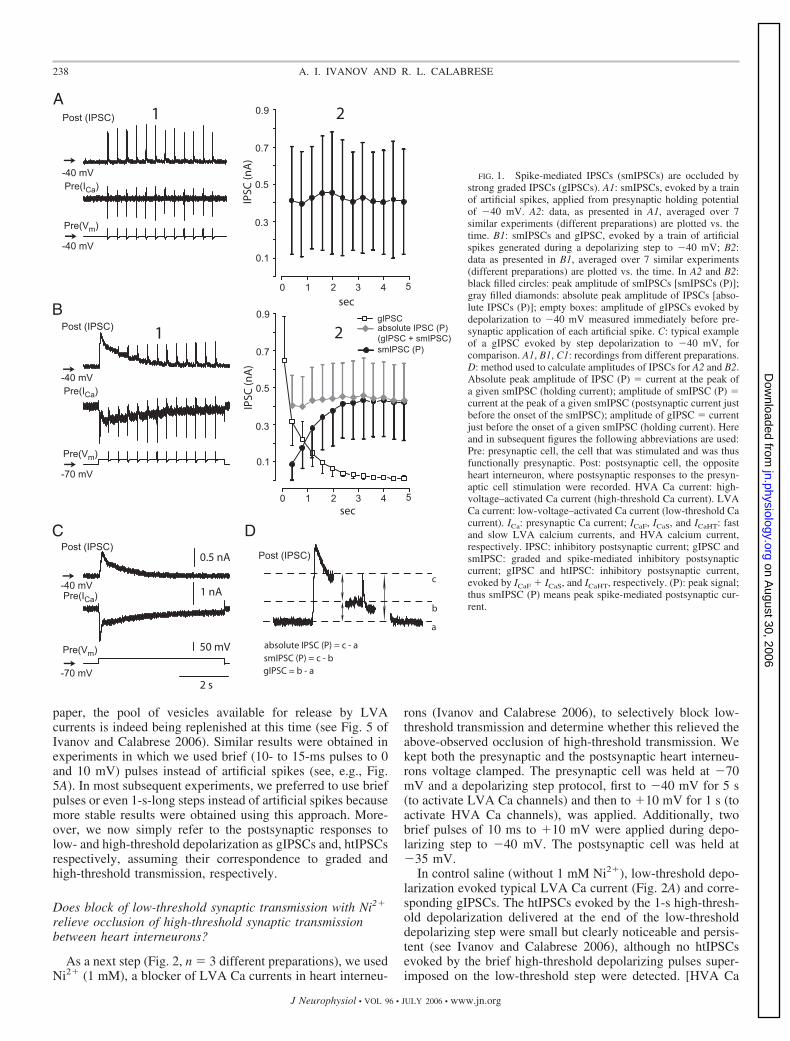
To determine whether vesicle pools and thus release sites areshared, we performed experiments designed to test whetherdepletion of releasable transmitter by low-threshold stimula-tion occluded release by high-threshold stimulation. In allexperiments presented in this paper, ganglia were bathed in 5mM Ca2�/0 mM Na� saline, and both pre- and postsynapticheart interneurons were voltage clamped with sharp microelec-trodes. To test for occlusion (Fig. 1, n 7), we elicitedlow-threshold (graded) transmission using depolarizing stepsfrom a holding potential of �70 to �40 mV that evoked thetwo kinetically distinct LVA Ca currents presynaptically (ICaFand ICaS) (Angstadt and Calabrese 1991) and the correspondingpostsynaptic responses (gIPSCF and gIPSCs) (Fig. 1C). Wealso elicited high-threshold transmission, using a train of “ar-tificial spikes” at 2.5 Hz from a holding potential of �40 mVthat evoked HVA Ca current presynaptically and the corre-sponding postsynaptic (spike-mediated) responses (smIPSCs).The train of artificial spikes evoked robust smIPSCs withtypical (compare with Ivanov and Calabrese 2003) stochasticvariations in their peak amplitude (Fig. 1A). Artificial spikeselicited during a 2-s depolarizing step to �40 mV from aholding potential of �70 mV, however, failed to elicit smIP-SCs during the intense graded transmission early in the step,but as the gIPSC waned (concomitantly with the inactivation ofLVA Ca current), they began to elicit smIPSCs comparable inamplitude to those elicited from �40 mV (Fig. 1B). The“absolute” peak amplitude of these IPSCs (i.e., amplituderelative to holding current; see Fig. 1D) was almost constant(with some weak tendency to increase during the depolarizingstep) and did not exceed the peak value of gIPSC [gIPSC (P)](Fig. 1, A2 and B2). The peak amplitude of smIPSCs (i.e.,amplitude relative to the baseline postsynaptic current recordedjust before the onset of any given smIPSC; see Fig. 1D),however, increased concomitantly with the decrease in thegIPSC (P). Although changes in peak amplitudes of smIPSCselicited from holding potential of �40 mV and “absolute” peakamplitude of IPSCs elicited during the depolarizing step to�40 mV did not depend on the time from the beginning oftrain/depolarization (P 1.0 and 0.99989, respectively, one-way ANOVA), changes in peak amplitude of smIPSCs elicitedduring the depolarization to �40 mV and in gIPSC werestrongly time dependent (P values of 0.000013 and 0.001,respectively, one-way ANOVA).
Thus it appears that strong graded synaptic transmission canocclude smIPSCs when they are simultaneously evoked. Thesimplest explanation for this occlusion is that HVA and LVACa currents evoke neurotransmitter release from common ves-icle pools and release sites. Depletion of the readily releasablevesicle pool by intense low-threshold Ca currents occludesrelease by Ca2� entering by high-threshold channels; inactiva-tion of the low-threshold currents diminished graded releaseand when accompanied by replenishment of the readily releas-able pool restores the ability of Ca2� entering by HVA Cachannels to release transmitter. As shown in the companion
237SHARED SITES FOR SPIKE-MEDIATED AND GRADED TRANSMISSION
J Neurophysiol • VOL 96 • JULY 2006 • www.jn.org
on August 30, 2006
jn.physiology.orgD
ownloaded from
paper, the pool of vesicles available for release by LVAcurrents is indeed being replenished at this time (see Fig. 5 ofIvanov and Calabrese 2006). Similar results were obtained inexperiments in which we used brief (10- to 15-ms pulses to 0and 10 mV) pulses instead of artificial spikes (see, e.g., Fig.5A). In most subsequent experiments, we preferred to use briefpulses or even 1-s-long steps instead of artificial spikes becausemore stable results were obtained using this approach. More-over, we now simply refer to the postsynaptic responses tolow- and high-threshold depolarization as gIPSCs and, htIPSCsrespectively, assuming their correspondence to graded andhigh-threshold transmission, respectively.
Does block of low-threshold synaptic transmission with Ni2�
relieve occlusion of high-threshold synaptic transmissionbetween heart interneurons?
As a next step (Fig. 2, n 3 different preparations), we usedNi2� (1 mM), a blocker of LVA Ca currents in heart interneu-
rons (Ivanov and Calabrese 2006), to selectively block low-threshold transmission and determine whether this relieved theabove-observed occlusion of high-threshold transmission. Wekept both the presynaptic and the postsynaptic heart interneu-rons voltage clamped. The presynaptic cell was held at �70mV and a depolarizing step protocol, first to �40 mV for 5 s(to activate LVA Ca channels) and then to �10 mV for 1 s (toactivate HVA Ca channels), was applied. Additionally, twobrief pulses of 10 ms to �10 mV were applied during depo-larizing step to �40 mV. The postsynaptic cell was held at�35 mV.
In control saline (without 1 mM Ni2�), low-threshold depo-larization evoked typical LVA Ca current (Fig. 2A) and corre-sponding gIPSCs. The htIPSCs evoked by the 1-s high-thresh-old depolarization delivered at the end of the low-thresholddepolarizing step were small but clearly noticeable and persis-tent (see Ivanov and Calabrese 2006), although no htIPSCsevoked by the brief high-threshold depolarizing pulses super-imposed on the low-threshold step were detected. [HVA Ca
FIG. 1. Spike-mediated IPSCs (smIPSCs) are occluded bystrong graded IPSCs (gIPSCs). A1: smIPSCs, evoked by a trainof artificial spikes, applied from presynaptic holding potentialof �40 mV. A2: data, as presented in A1, averaged over 7similar experiments (different preparations) are plotted vs. thetime. B1: smIPSCs and gIPSC, evoked by a train of artificialspikes generated during a depolarizing step to �40 mV; B2:data as presented in B1, averaged over 7 similar experiments(different preparations) are plotted vs. the time. In A2 and B2:black filled circles: peak amplitude of smIPSCs [smIPSCs (P)];gray filled diamonds: absolute peak amplitude of IPSCs [abso-lute IPSCs (P)]; empty boxes: amplitude of gIPSCs evoked bydepolarization to �40 mV measured immediately before pre-synaptic application of each artificial spike. C: typical exampleof a gIPSC evoked by step depolarization to �40 mV, forcomparison. A1, B1, C1: recordings from different preparations.D: method used to calculate amplitudes of IPSCs for A2 and B2.Absolute peak amplitude of IPSC (P) current at the peak ofa given smIPSC (holding current); amplitude of smIPSC (P) current at the peak of a given smIPSC (postsynaptic current justbefore the onset of the smIPSC); amplitude of gIPSC currentjust before the onset of a given smIPSC (holding current). Hereand in subsequent figures the following abbreviations are used:Pre: presynaptic cell, the cell that was stimulated and was thusfunctionally presynaptic. Post: postsynaptic cell, the oppositeheart interneuron, where postsynaptic responses to the presyn-aptic cell stimulation were recorded. HVA Ca current: high-voltage–activated Ca current (high-threshold Ca current). LVACa current: low-voltage–activated Ca current (low-threshold Cacurrent). ICa: presynaptic Ca current; ICaF, ICaS, and ICaHT: fastand slow LVA calcium currents, and HVA calcium current,respectively. IPSC: inhibitory postsynaptic current; gIPSC andsmIPSC: graded and spike-mediated inhibitory postsynapticcurrent; gIPSC and htIPSC: inhibitory postsynaptic current,evoked by ICaF � ICaS, and ICaHT, respectively. (P): peak signal;thus smIPSC (P) means peak spike-mediated postsynaptic cur-rent.
238 A. I. IVANOV AND R. L. CALABRESE
J Neurophysiol • VOL 96 • JULY 2006 • www.jn.org
on August 30, 2006
jn.physiology.orgD
ownloaded from
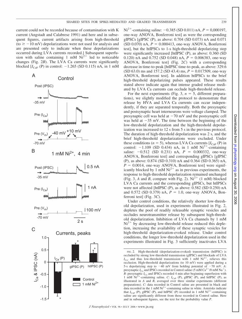
current could not be recorded because of contamination with Kcurrent (Angstadt and Calabrese 1991) and here and in subse-quent figures, current artifacts arising from high-threshold(to � �10 mV) depolarizations were not used for analysis andare presented only to indicate when these depolarizationsoccurred during LVA currents recorded.] Subsequent superfu-sion with saline containing 1 mM Ni2� led to noticeablechanges (Fig. 2B). The LVA Ca currents were significantlyblocked [ICaF (P) in control: �1.265 (SD 0.115) nA, in 1 mM
Ni2�-containing saline: �0.385 (SD 0.011) nA, P 0,000197,one-way ANOVA, Bonferroni test] as were the correspondinggIPSCs [gIPSC (P), as above: 0.704 (SD 0.073) nA and 0.071(SD 0.070) nA, P 0.000413, one-way ANOVA, Bonferronitest], but the htIPSCs to 1-s high-threshold depolarizing stepwere significantly increased [htIPSC (P), as above: 0.360 (SD0.120) nA and 0.752 (SD 0.048) nA, P 0.006383, one-wayANOVA, Bonferroni test] (Fig. 2C) with a correspondingdecrease in time-to-peak [htIPSC time-to-peak, as above: 329.6(SD 63.0) ms and 157.2 (SD 43.4) ms, P 0.017509, one-wayANOVA, Bonferroni test]. In addition htIPSCs to the briefhigh-threshold depolarizing pulses appeared. These resultsstated above indicate again that intense graded release medi-ated by LVA Ca currents can occlude high-threshold release.
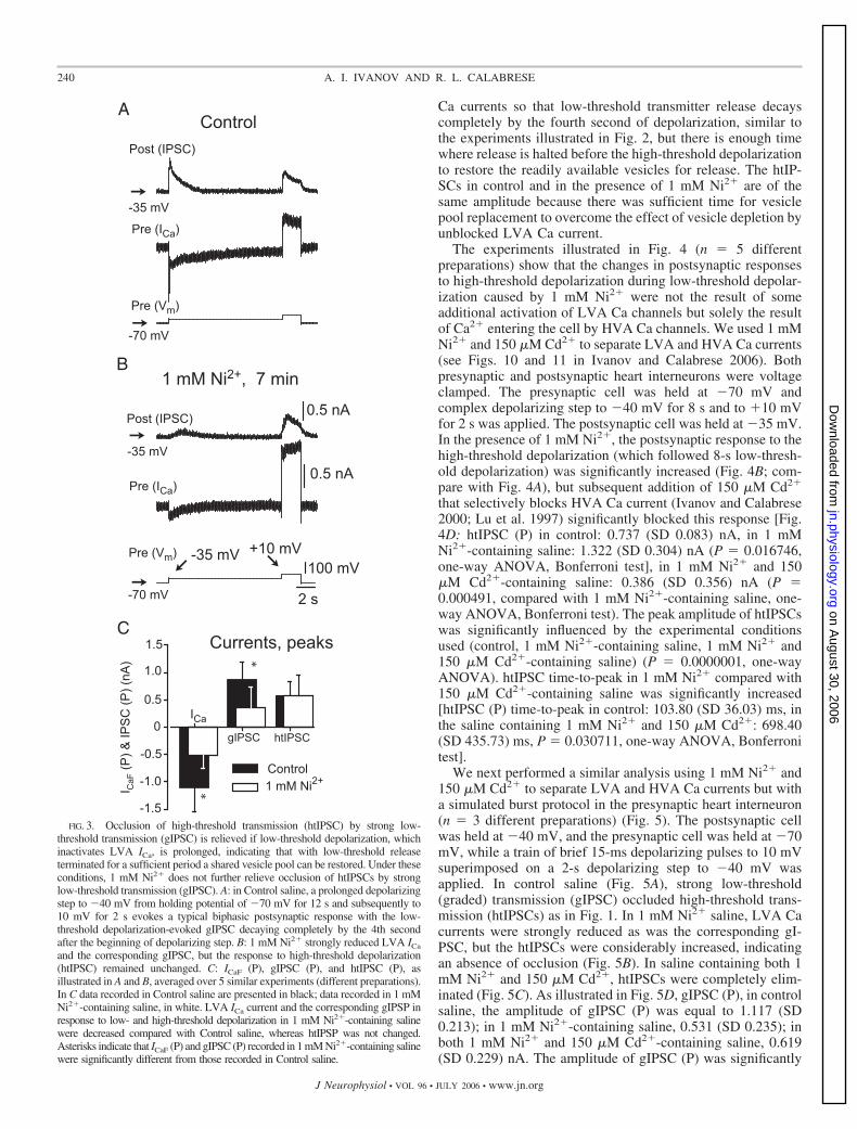
For the next experiments (Fig. 3, n 5, different prepara-tions), we slightly modified the protocol to demonstrate thatrelease by HVA and LVA Ca currents can occur indepen-dently, if they are separated temporally. Both the presynapticand postsynaptic heart interneurons were voltage clamped. Thepresynaptic cell was held at �70 mV and the postsynaptic cellwas held at �35 mV. The time between the beginning of thelow-threshold depolarization and the high-threshold depolar-ization was increased to 12 s from 5 s in the previous protocol.The duration of high-threshold depolarization was 2 s, and thebrief high-threshold depolarizations were excluded. Underthese conditions (n 5), whereas LVA Ca currents [ICaF (P) incontrol: �1.109 (SD 0.434) nA, in 1 mM Ni2�-containingsaline: �0.512 (SD 0.231) nA, P 0.000332, one-wayANOVA, Bonferroni test] and corresponding gIPSCs [gIPSC(P), as above: 0.874 (SD 0.310) nA and 0.364 (SD 0.365) nA,P 0.0014, one-way ANOVA, Bonferroni test] were signif-icantly blocked by 1 mM Ni2� as in previous experiments, theresponse to high-threshold depolarization remained unchanged(Fig. 3, A and B, compare with Fig. 2). Ni2� (1 mM) blockedLVA Ca currents and the corresponding gIPSCs, but htIPSCswere not affected [htIPSC (P), as above: 0.582 (SD 0.250) nAand 0.572 (SD 0.379) nA, P 1.0, one-way ANOVA, Bon-ferroni test] (Fig. 3C).
Under control conditions, the relatively shorter low-thresh-old depolarization, used in experiments illustrated in Fig. 2,depletes the pool of readily releasable synaptic vesicles andoccludes neurotransmitter release by subsequent high-thresh-old depolarization. Inhibition of LVA Ca channels by 1 mMNi2� by decreasing low-threshold release reduced this deple-tion, increasing the availability of these synaptic vesicles forhigh-threshold depolarization-evoked release. Under controlconditions, the longer low-threshold depolarization used in theexperiments illustrated in Fig. 3 sufficiently inactivates LVA
FIG. 2. High-threshold (depolarization-evoked) transmission (htIPSC) isoccluded by strong low-threshold transmission (gIPSC) and blockade of LVAICa, and thus low-threshold transmission with 1 mM Ni2�, relieves thisocclusion. High-threshold depolarizations (to 10 mV) were applied during a5-s depolarizing step to �40 mV from holding potential of �70 mV. A:presynaptic ICa and IPSCs recorded in Control saline (5 mM Ca2�/0 mM Na�).B: presynaptic ICa and IPSCs recorded 4 min after beginning superfusion with1 mM Ni2�-containing saline. C: ICaF (P), gIPSC (P), and htIPSC (P), asillustrated in A and B, averaged over three similar experiments (differentpreparations). C: data recorded in Control saline are presented in black anddata recorded in the 1 mM Ni2�-containing saline in white. Asterisks indicatethat ICaF (P), gIPSC (P), and htIPSC (P) recorded in 1 mM Ni2�-containingsaline are significantly different from those recorded in Control saline. Hereand in subsequent figures, see the text for the probability value P.
239SHARED SITES FOR SPIKE-MEDIATED AND GRADED TRANSMISSION
J Neurophysiol • VOL 96 • JULY 2006 • www.jn.org
on August 30, 2006
jn.physiology.orgD
ownloaded from
Ca currents so that low-threshold transmitter release decayscompletely by the fourth second of depolarization, similar tothe experiments illustrated in Fig. 2, but there is enough timewhere release is halted before the high-threshold depolarizationto restore the readily available vesicles for release. The htIP-SCs in control and in the presence of 1 mM Ni2� are of thesame amplitude because there was sufficient time for vesiclepool replacement to overcome the effect of vesicle depletion byunblocked LVA Ca current.
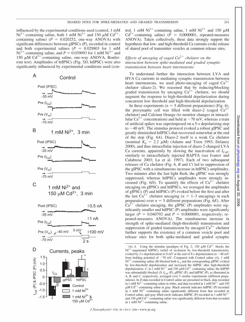
The experiments illustrated in Fig. 4 (n 5 differentpreparations) show that the changes in postsynaptic responsesto high-threshold depolarization during low-threshold depolar-ization caused by 1 mM Ni2� were not the result of someadditional activation of LVA Ca channels but solely the resultof Ca2� entering the cell by HVA Ca channels. We used 1 mMNi2� and 150 �M Cd2� to separate LVA and HVA Ca currents(see Figs. 10 and 11 in Ivanov and Calabrese 2006). Bothpresynaptic and postsynaptic heart interneurons were voltageclamped. The presynaptic cell was held at �70 mV andcomplex depolarizing step to �40 mV for 8 s and to �10 mVfor 2 s was applied. The postsynaptic cell was held at �35 mV.In the presence of 1 mM Ni2�, the postsynaptic response to thehigh-threshold depolarization (which followed 8-s low-thresh-old depolarization) was significantly increased (Fig. 4B; com-pare with Fig. 4A), but subsequent addition of 150 �M Cd2�
that selectively blocks HVA Ca current (Ivanov and Calabrese2000; Lu et al. 1997) significantly blocked this response [Fig.4D: htIPSC (P) in control: 0.737 (SD 0.083) nA, in 1 mMNi2�-containing saline: 1.322 (SD 0.304) nA (P 0.016746,one-way ANOVA, Bonferroni test], in 1 mM Ni2� and 150�M Cd2�-containing saline: 0.386 (SD 0.356) nA (P 0.000491, compared with 1 mM Ni2�-containing saline, one-way ANOVA, Bonferroni test). The peak amplitude of htIPSCswas significantly influenced by the experimental conditionsused (control, 1 mM Ni2�-containing saline, 1 mM Ni2� and150 �M Cd2�-containing saline) (P 0.0000001, one-wayANOVA). htIPSC time-to-peak in 1 mM Ni2� compared with150 �M Cd2�-containing saline was significantly increased[htIPSC (P) time-to-peak in control: 103.80 (SD 36.03) ms, inthe saline containing 1 mM Ni2� and 150 �M Cd2�: 698.40(SD 435.73) ms, P 0.030711, one-way ANOVA, Bonferronitest].
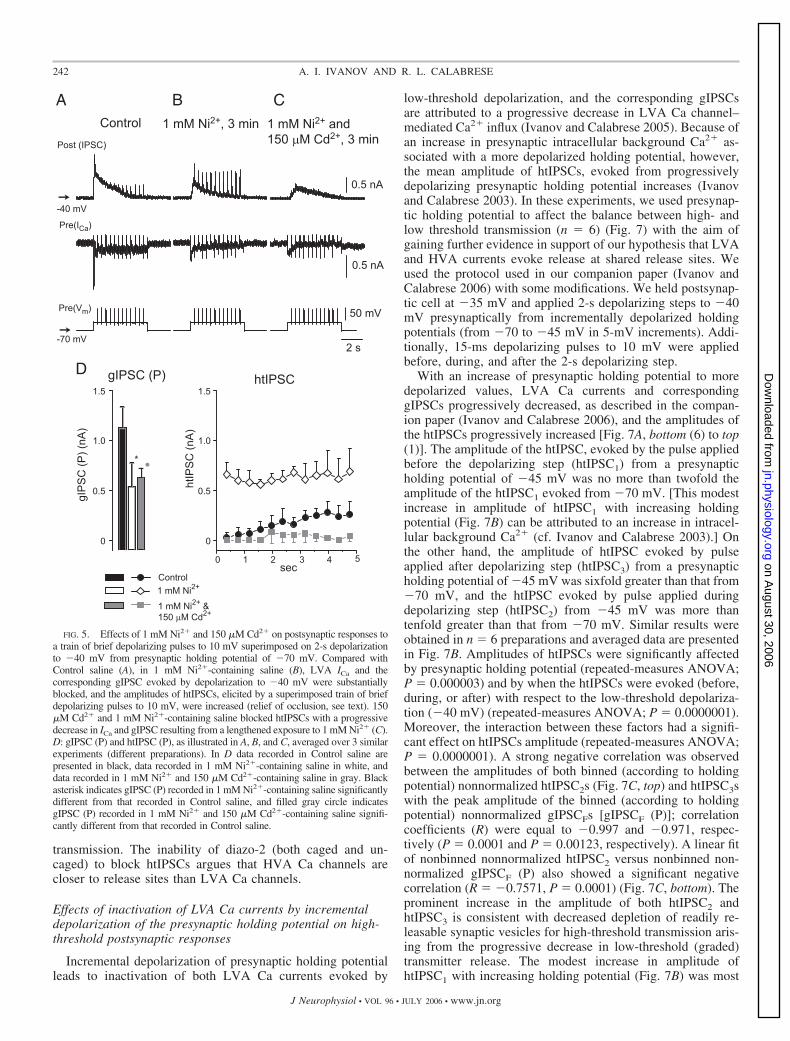
We next performed a similar analysis using 1 mM Ni2� and150 �M Cd2� to separate LVA and HVA Ca currents but witha simulated burst protocol in the presynaptic heart interneuron(n 3 different preparations) (Fig. 5). The postsynaptic cellwas held at �40 mV, and the presynaptic cell was held at �70mV, while a train of brief 15-ms depolarizing pulses to 10 mVsuperimposed on a 2-s depolarizing step to �40 mV wasapplied. In control saline (Fig. 5A), strong low-threshold(graded) transmission (gIPSC) occluded high-threshold trans-mission (htIPSCs) as in Fig. 1. In 1 mM Ni2� saline, LVA Cacurrents were strongly reduced as was the corresponding gI-PSC, but the htIPSCs were considerably increased, indicatingan absence of occlusion (Fig. 5B). In saline containing both 1mM Ni2� and 150 �M Cd2�, htIPSCs were completely elim-inated (Fig. 5C). As illustrated in Fig. 5D, gIPSC (P), in controlsaline, the amplitude of gIPSC (P) was equal to 1.117 (SD0.213); in 1 mM Ni2�-containing saline, 0.531 (SD 0.235); inboth 1 mM Ni2� and 150 �M Cd2�-containing saline, 0.619(SD 0.229) nA. The amplitude of gIPSC (P) was significantly
FIG. 3. Occlusion of high-threshold transmission (htIPSC) by strong low-threshold transmission (gIPSC) is relieved if low-threshold depolarization, whichinactivates LVA ICa, is prolonged, indicating that with low-threshold releaseterminated for a sufficient period a shared vesicle pool can be restored. Under theseconditions, 1 mM Ni2� does not further relieve occlusion of htIPSCs by stronglow-threshold transmission (gIPSC). A: in Control saline, a prolonged depolarizingstep to �40 mV from holding potential of �70 mV for 12 s and subsequently to10 mV for 2 s evokes a typical biphasic postsynaptic response with the low-threshold depolarization-evoked gIPSC decaying completely by the 4th secondafter the beginning of depolarizing step. B: 1 mM Ni2� strongly reduced LVA ICa
and the corresponding gIPSC, but the response to high-threshold depolarization(htIPSC) remained unchanged. C: ICaF (P), gIPSC (P), and htIPSC (P), asillustrated in A and B, averaged over 5 similar experiments (different preparations).In C data recorded in Control saline are presented in black; data recorded in 1 mMNi2�-containing saline, in white. LVA ICa current and the corresponding gIPSP inresponse to low- and high-threshold depolarization in 1 mM Ni2�-containing salinewere decreased compared with Control saline, whereas htIPSP was not changed.Asterisks indicate that ICaF (P) and gIPSC (P) recorded in 1 mM Ni2�-containing salinewere significantly different from those recorded in Control saline.
240 A. I. IVANOV AND R. L. CALABRESE
J Neurophysiol • VOL 96 • JULY 2006 • www.jn.org
on August 30, 2006
jn.physiology.orgD
ownloaded from
influenced by the experimental conditions used (control, 1 mMNi2�-containing saline, both 1 mM Ni2� and 150 �M Cd2�-containing saline) (P 0.020252, one-way ANOVA) withsignificant differences between gIPSCs (P), recorded in controland both experimental salines (P 0.029803 for 1 mMNi2�-containing saline, and P 0.039093 for 1 mM Ni2� and150 �M Cd2�-containing saline, one-way ANOVA, Bonfer-roni test). Amplitudes of htIPSCs (Fig. 5D, htIPSC) were alsosignificantly influenced by experimental conditions used (con-
trol, 1 mM Ni2�-containing saline, 1 mM Ni2� and 150 �MCd2�-containing saline) (P 0.0000001, repeated-measuresANOVA). Taken collectively, these data strongly support thehypothesis that low- and high-threshold Ca currents evoke releaseof shared pool of transmitter vesicles at common release sites.
Effects of uncaging of caged Ca2� chelator on theinteraction between spike-mediated and graded synaptictransmission between heart interneurons
To understand further the interaction between LVA andHVA Ca currents in mediating synaptic transmission betweenheart interneurons, we used photo-uncaging of caged Ca2�
chelator (diazo-2). We reasoned that by reducing/blockinggraded transmission by uncaging Ca2� chelator, we shouldaugment the response to high-threshold depolarization duringconcurrent low threshold and high-threshold depolarization.
In these experiments (n 5 different preparations) (Fig. 6),the presynaptic cell was filled with diazo-2 (caged Ca2�
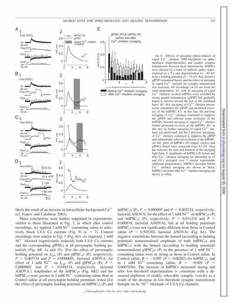
chelator) and Calcium Orange (to monitor changes in intracel-lular Ca2� concentration) and held at �70 mV, whereas a trainof artificial spikes was superimposed on a 5-s depolarizing stepto �40 mV. The stimulus protocol evoked a robust gIPSC andgreatly diminished htIPSCs that recovered somewhat at the endof the step (Fig. 6A). Diazo-2 itself is a weak Ca chelator(nominal Kd 2.2 �M) (Adams and Tsien 1993; Delaney2000), and thus intracellular injection of diazo-2 changed LVACa currents, apparently by slowing the inactivation of ICaF,similarly to intracellularly injected BAPTA (see Ivanov andCalabrese 2003; Lu et al. 1997). Each of two subsequentreleases of Ca chelator (Fig. 6, B and C) led to suppression ofthe gIPSC with a simultaneous increase in htIPSCs amplitudes.Two minutes after the last light flash, the gIPSC was stronglysuppressed, whereas htIPSCs amplitudes were strongly in-creased (Fig. 6D). To quantify the effects of Ca2� chelatoruncaging on gIPSCs and htIPSCs, we averaged the amplitudesof gIPSCs (P) and htIPSCs (P) evoked before the first and afterthe last Ca2� chelator uncaging (n 1–3 uncagings in eachpreparation) over n 5 different preparations (Fig. 6E). AfterCa2� chelator uncaging, the gIPSC (P) amplitudes were sig-nificantly smaller and htIPSC (P) amplitudes were significantlylarger (P 0.040792 and P 0.0000001, respectively; re-peated-measures ANOVA). The simultaneous increase instrength of spike-mediated (high-threshold) transmission andsuppression of graded transmission by uncaged Ca2� chelatorfurther supports the existence of a common vesicle pool andrelease sites for both spike-mediated and graded synaptic
FIG. 4. Using the stimulus paradigm of Fig. 2, 150 �M Cd2� blocks theNi2�-augmented htIPSCs (relief of occlusion by low-threshold transmission),evoked by 2-s depolarization to 0 mV at the end of 8-s depolarization to �40 mVfrom holding potential of �70 mV. Compared with Control saline (A), 1 mMNi2�-containing saline (B) blocked both ICa and the corresponding gIPSC evokedby low-threshold depolarization and increased the htIPSC after high-thresholddepolarization. C: in 1 mM Ni2� and 150 �M Cd2�-containing saline, the htIPSPwas substantially blocked. D: ICaF (P), gIPSC (P), and htIPSC (P), as illustrated inA, B, and C, respectively, averaged over 5 similar experiments (different prepa-rations). In D data recorded in Control saline are presented in black, data recordedin 1 mM Ni2�-containing saline in white, and data recorded in 1 mM Ni2� and 150�M Cd2�-containing saline in gray. Black asterisk indicates htIPSC (P) recordedin 1 mM Ni2�-containing saline significantly different from that recorded inControl saline, and gray filled circle indicates htIPSC (P) recorded in 1 mM Ni2�
and 150 �M Cd2�-containing saline was significantly different from that recordedin 1 mM Ni2�-containing saline.
241SHARED SITES FOR SPIKE-MEDIATED AND GRADED TRANSMISSION
J Neurophysiol • VOL 96 • JULY 2006 • www.jn.org
on August 30, 2006
jn.physiology.orgD
ownloaded from
transmission. The inability of diazo-2 (both caged and un-caged) to block htIPSCs argues that HVA Ca channels arecloser to release sites than LVA Ca channels.
Effects of inactivation of LVA Ca currents by incrementaldepolarization of the presynaptic holding potential on high-threshold postsynaptic responses
Incremental depolarization of presynaptic holding potentialleads to inactivation of both LVA Ca currents evoked by
low-threshold depolarization, and the corresponding gIPSCsare attributed to a progressive decrease in LVA Ca channel–mediated Ca2� influx (Ivanov and Calabrese 2005). Because ofan increase in presynaptic intracellular background Ca2� as-sociated with a more depolarized holding potential, however,the mean amplitude of htIPSCs, evoked from progressivelydepolarizing presynaptic holding potential increases (Ivanovand Calabrese 2003). In these experiments, we used presynap-tic holding potential to affect the balance between high- andlow threshold transmission (n 6) (Fig. 7) with the aim ofgaining further evidence in support of our hypothesis that LVAand HVA currents evoke release at shared release sites. Weused the protocol used in our companion paper (Ivanov andCalabrese 2006) with some modifications. We held postsynap-tic cell at �35 mV and applied 2-s depolarizing steps to �40mV presynaptically from incrementally depolarized holdingpotentials (from �70 to �45 mV in 5-mV increments). Addi-tionally, 15-ms depolarizing pulses to 10 mV were appliedbefore, during, and after the 2-s depolarizing step.
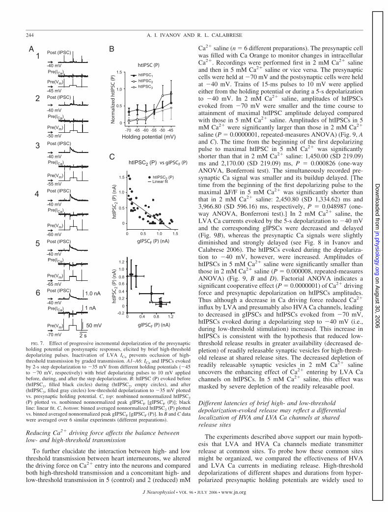
With an increase of presynaptic holding potential to moredepolarized values, LVA Ca currents and correspondinggIPSCs progressively decreased, as described in the compan-ion paper (Ivanov and Calabrese 2006), and the amplitudes ofthe htIPSCs progressively increased [Fig. 7A, bottom (6) to top(1)]. The amplitude of the htIPSC, evoked by the pulse appliedbefore the depolarizing step (htIPSC1) from a presynapticholding potential of �45 mV was no more than twofold theamplitude of the htIPSC1 evoked from �70 mV. [This modestincrease in amplitude of htIPSC1 with increasing holdingpotential (Fig. 7B) can be attributed to an increase in intracel-lular background Ca2� (cf. Ivanov and Calabrese 2003).] Onthe other hand, the amplitude of htIPSC evoked by pulseapplied after depolarizing step (htIPSC3) from a presynapticholding potential of �45 mV was sixfold greater than that from�70 mV, and the htIPSC evoked by pulse applied duringdepolarizing step (htIPSC2) from �45 mV was more thantenfold greater than that from �70 mV. Similar results wereobtained in n 6 preparations and averaged data are presentedin Fig. 7B. Amplitudes of htIPSCs were significantly affectedby presynaptic holding potential (repeated-measures ANOVA;P 0.000003) and by when the htIPSCs were evoked (before,during, or after) with respect to the low-threshold depolariza-tion (�40 mV) (repeated-measures ANOVA; P 0.0000001).Moreover, the interaction between these factors had a signifi-cant effect on htIPSCs amplitude (repeated-measures ANOVA;P 0.0000001). A strong negative correlation was observedbetween the amplitudes of both binned (according to holdingpotential) nonnormalized htIPSC2s (Fig. 7C, top) and htIPSC3swith the peak amplitude of the binned (according to holdingpotential) nonnormalized gIPSCFs [gIPSCF (P)]; correlationcoefficients (R) were equal to �0.997 and �0.971, respec-tively (P 0.0001 and P 0.00123, respectively). A linear fitof nonbinned nonnormalized htIPSC2 versus nonbinned non-normalized gIPSCF (P) also showed a significant negativecorrelation (R �0.7571, P 0.0001) (Fig. 7C, bottom). Theprominent increase in the amplitude of both htIPSC2 andhtIPSC3 is consistent with decreased depletion of readily re-leasable synaptic vesicles for high-threshold transmission aris-ing from the progressive decrease in low-threshold (graded)transmitter release. The modest increase in amplitude ofhtIPSC1 with increasing holding potential (Fig. 7B) was most
FIG. 5. Effects of 1 mM Ni2� and 150 �M Cd2� on postsynaptic responses toa train of brief depolarizing pulses to 10 mV superimposed on 2-s depolarizationto �40 mV from presynaptic holding potential of �70 mV. Compared withControl saline (A), in 1 mM Ni2�-containing saline (B), LVA ICa and thecorresponding gIPSC evoked by depolarization to �40 mV were substantiallyblocked, and the amplitudes of htIPSCs, elicited by a superimposed train of briefdepolarizing pulses to 10 mV, were increased (relief of occlusion, see text). 150�M Cd2� and 1 mM Ni2�-containing saline blocked htIPSCs with a progressivedecrease in ICa and gIPSC resulting from a lengthened exposure to 1 mM Ni2� (C).D: gIPSC (P) and htIPSC (P), as illustrated in A, B, and C, averaged over 3 similarexperiments (different preparations). In D data recorded in Control saline arepresented in black, data recorded in 1 mM Ni2�-containing saline in white, anddata recorded in 1 mM Ni2� and 150 �M Cd2�-containing saline in gray. Blackasterisk indicates gIPSC (P) recorded in 1 mM Ni2�-containing saline significantlydifferent from that recorded in Control saline, and filled gray circle indicatesgIPSC (P) recorded in 1 mM Ni2� and 150 �M Cd2�-containing saline signifi-cantly different from that recorded in Control saline.
242 A. I. IVANOV AND R. L. CALABRESE
J Neurophysiol • VOL 96 • JULY 2006 • www.jn.org
on August 30, 2006
jn.physiology.orgD
ownloaded from
likely the result of an increase in intracellular background Ca2�
(cf. Ivanov and Calabrese 2003).These conclusions were further supported in experiments,
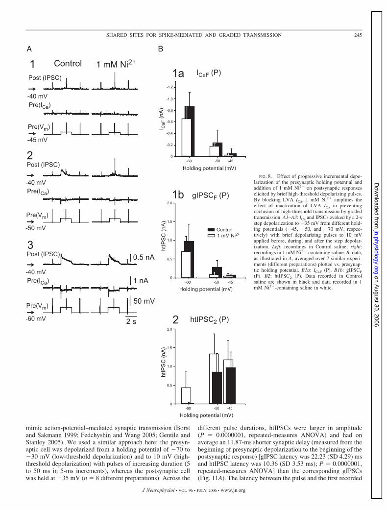
similar to those illustrated in Fig. 7, in which after controlrecordings, we applied 1 mM Ni2�-containing saline to selec-tively block LVA Ca currents (Fig. 9) (n 7). Controlrecordings were similar to Fig. 7 (Fig. 8A). As expected, 1 mMNi2� blocked (significantly reduced) both LVA Ca currentsand the corresponding gIPSCs at all presynaptic holding po-tentials (Fig. 8B, 1a and 1b). [For the effect of presynapticholding potential on ICaF (P) and gIPSCFs (P), respectively,P 0.005734 and P 0.0000001, factorial ANOVA; foreffect of 1 mM Ni2� on ICaF (P) and gIPSCFs (P), P 0.0000001 and P 0.048534, respectively, factorialANOVA.] Amplitudes of the htIPSC2s (Fig. 8B2) and thehtIPSC3s were greater in 1 mM Ni2�-containing saline than inControl saline at all presynaptic holding potentials tested [forthe effect of presynaptic holding potential on htIPSC2s (P) and
htIPSC3s (P), P 0.000005 and P 0.005734, respectively,factorial ANOVA; for the effect of 1 mM Ni2� on htIPSC2s (P)and htIPSC3s (P), respectively, P 0.011216 and P 0.000676, factorial ANOVA], but at all holding potentialshtIPSC1s were not significantly different from those in Controlsaline (P 0.505282, factorial ANOVA) (Fig. 8A). Thenegative correlations between the binned (according to holdingpotential) nonnormalized amplitude of both htIPSC2s andhtIPSC3s with the binned (according to holding potential)nonnormalized gIPSCFs (P) in the presence of 1 mM Ni2�-containing saline were as strong as those in Control saline. InControl saline, R �0.997 (P 0.00282) for htIPSC2s, andin 1 mM Ni2�-containing saline, R �0.999 (P 0.0007636). The increase of htIPSCs measured during andafter low-threshold depolarization is consistent with a de-creased depletion of readily releasable synaptic vesicles as aresult of the decrease in low-threshold synaptic transmissionbrought on by Ni2� blockade of LVA Ca channels.
FIG. 6. Effects of uncaging (photo-release) ofcaged Ca2� chelator (DM-Nitrophen) on spike-mediated (high-threshold) and graded synaptictransmission between heart interneurons. htIPSCswere elicited by a train of artificial spikes super-imposed on a 5-s step depolarization to �40 mVfrom a holding potential of �70 mV that elicited agIPSP (simulated burst), and the effect of uncagingof caged Ca2� chelator on synaptic transmissionwas assessed. All recordings (A–D) are from thesame preparation. A1: with no uncaging of cagedCa2� chelator, evoked htIPSCs were occluded bystrong graded transmission (gIPSP) but graduallybegan to recover toward the end of the simulatedburst. B1: first uncaging of Ca2� chelator prema-turely terminated the gIPSP and promoted recov-ery of the htIPSPs. C1: in this run, the previousuncaging of Ca2� chelator continued to suppressthe gIPSP and relieved some occlusion of thehtIPSPs. Second uncaging of caged Ca2� chelatorfurther promoted recovery of the htIPSPs. D1: inthis run, no further uncaging of caged Ca2� che-lator was performed, but the 2 previous uncagingsof Ca2� chelator continued to suppress the gIPSPand substantially relieved occlusion of the htIPSPs.A2–D2: plots of htIPSCs (P) (empty circles) andgIPSCs (black line), extracted from A1–D1. Graybar indicates the time and duration of the uncaginglight flash. E: amplitudes of htIPSCs (P) before andafter Ca2� chelator uncaging (as illustrated in A1and D1), averaged over 5 similar experiments(different preparations). htIPSCs recorded beforeCa2� chelator uncaging are shown in black;htIPSCs recorded after Ca2� chelator uncaging areshown in white.
243SHARED SITES FOR SPIKE-MEDIATED AND GRADED TRANSMISSION
J Neurophysiol • VOL 96 • JULY 2006 • www.jn.org
on August 30, 2006
jn.physiology.orgD
ownloaded from
Reducing Ca2� driving force affects the balance betweenlow- and high-threshold transmission
To further elucidate the interaction between high- and lowthreshold transmission between heart interneurons, we alteredthe driving force on Ca2� entry into the neurons and comparedboth high-threshold transmission and a concomitant high- andlow-threshold transmission in 5 (control) and 2 (reduced) mM
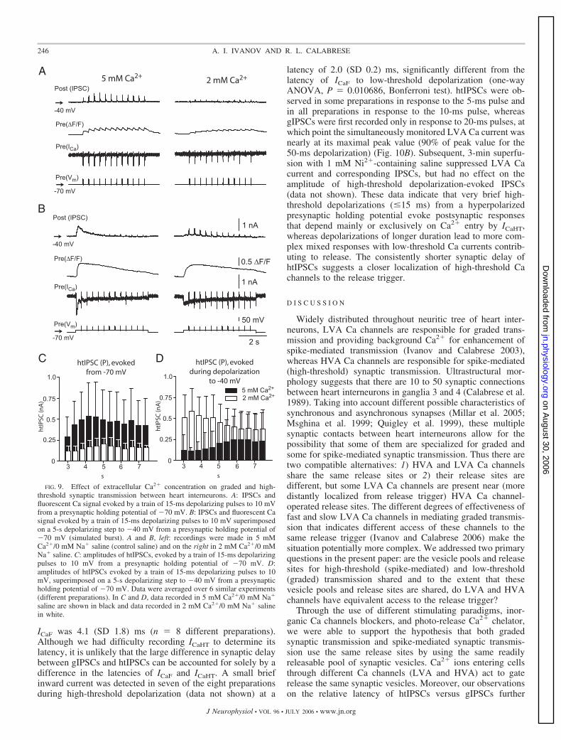
Ca2� saline (n 6 different preparations). The presynaptic cellwas filled with Ca Orange to monitor changes in intracellularCa2�. Recordings were performed first in 2 mM Ca2� salineand then in 5 mM Ca2� saline or vice versa. The presynapticcells were held at �70 mV and the postsynaptic cells were heldat �40 mV. Trains of 15-ms pulses to 10 mV were appliedeither from the holding potential or during a 5-s depolarizationto �40 mV. In 2 mM Ca2� saline, amplitudes of htIPSCsevoked from �70 mV were smaller and the time course toattainment of maximal htIPSC amplitude delayed comparedwith those in 5 mM Ca2� saline. Amplitudes of htIPSCs in 5mM Ca2� were significantly larger than those in 2 mM Ca2�
saline (P 0.0000001, repeated-measures ANOVA) (Fig. 9, Aand C). The time from the beginning of the first depolarizingpulse to maximal htIPSC in 5 mM Ca2� was significantlyshorter than that in 2 mM Ca2� saline: 1,450.00 (SD 219.09)ms and 2,170.00 (SD 219.09) ms, P 0.000826 (one-wayANOVA, Bonferroni test). The simultaneously recorded pre-synaptic Ca signal was smaller and its buildup delayed. [Thetime from the beginning of the first depolarizing pulse to themaximal �F/F in 5 mM Ca2� was significantly shorter thanthat in 2 mM Ca2� saline: 2,450.80 (SD 1,334.62) ms and3,966.80 (SD 596.16) ms, respectively, P 0.048987 (one-way ANOVA, Bonferroni test).] In 2 mM Ca2� saline, theLVA Ca currents evoked by the 5-s depolarization to �40 mVand the corresponding gIPSCs were decreased and delayed(Fig. 9B), whereas the presynaptic Ca signals were slightlydiminished and strongly delayed (see Fig. 8 in Ivanov andCalabrese 2006). The htIPSCs evoked during the depolariza-tion to �40 mV, however, were increased. Amplitudes ofhtIPSCs in 5 mM Ca2� saline were significantly smaller thanthose in 2 mM Ca2� saline (P 0.000008, repeated-measuresANOVA) (Fig. 9, B and D). Factorial ANOVA indicates asignificant cooperative effect (P 0.0000001) of Ca2� drivingforce and presynaptic depolarization on htIPSCs amplitudes.Thus although a decrease in Ca driving force reduced Ca2�
influx by LVA and presumably also HVA Ca channels, leadingto decreased in gIPSCs and htIPSCs evoked from �70 mV,htIPSCs evoked during a depolarizing step to �40 mV (i.e.,during low-threshold stimulation) increased. This increase inhtIPSCs is consistent with the hypothesis that reduced low-threshold release results in greater availability (decreased de-pletion) of readily releasable synaptic vesicles for high-thresh-old release at shared release sites. The decreased depletion ofreadily releasable synaptic vesicles in 2 mM Ca2� salineuncovers the enhancing effect of Ca2� entering by LVA Cachannels on htIPSCs. In 5 mM Ca2� saline, this effect wasmasked by severe depletion of the readily releasable pool.
Different latencies of brief high- and low-thresholddepolarization-evoked release may reflect a differentiallocalization of HVA and LVA Ca channels at sharedrelease sites
The experiments described above support our main hypoth-esis that LVA and HVA Ca channels mediate transmitterrelease at common sites. To probe how these common sitesmight be organized, we compared the effectiveness of HVAand LVA Ca currents in mediating release. High-thresholddepolarizations of different shapes and durations from hyper-polarized presynaptic holding potentials are widely used to
FIG. 7. Effect of progressive incremental depolarization of the presynapticholding potential on postsynaptic responses, elicited by brief high-thresholddepolarizing pulses. Inactivation of LVA ICa prevents occlusion of high-threshold transmission by graded transmission. A1–A6: ICa and IPSCs evokedby 2-s step depolarization to �35 mV from different holding potentials (�45to �70 mV, respectively) with brief depolarizing pulses to 10 mV appliedbefore, during, and after the step depolarization. B: htIPSC (P) evoked before(htIPSC1, filled black circles) during (htIPSC2, empty circles), and after(htIPSC3, filled gray circles) low-threshold depolarization to �35 mV plottedvs. presynaptic holding potential. C, top: nonbinned nonnormalized htIPSC2
(P) plotted vs. nonbinned nonnormalized peak gIPSCF [gIPSCF (P)]; blackline: linear fit. C, bottom: binned averaged nonnormalized htIPSC2 (P) plottedvs. binned averaged nonnormalized peak gIPSCF [gIPSCF (P)]. In B and C datawere averaged over 6 similar experiments (different preparations).
244 A. I. IVANOV AND R. L. CALABRESE
J Neurophysiol • VOL 96 • JULY 2006 • www.jn.org
on August 30, 2006
jn.physiology.orgD
ownloaded from
mimic action-potential–mediated synaptic transmission (Borstand Sakmann 1999; Fedchyshin and Wang 2005; Gentile andStanley 2005). We used a similar approach here: the presyn-aptic cell was depolarized from a holding potential of �70 to�30 mV (low-threshold depolarization) and to 10 mV (high-threshold depolarization) with pulses of increasing duration (5to 50 ms in 5-ms increments), whereas the postsynaptic cellwas held at �35 mV (n 8 different preparations). Across the
different pulse durations, htIPSCs were larger in amplitude(P 0.0000001, repeated-measures ANOVA) and had onaverage an 11.87-ms shorter synaptic delay (measured from thebeginning of presynaptic depolarization to the beginning of thepostsynaptic response) [gIPSC latency was 22.23 (SD 4.29) msand htIPSC latency was 10.36 (SD 3.53 ms); P 0.0000001,repeated-measures ANOVA] than the corresponding gIPSCs(Fig. 11A). The latency between the pulse and the first recorded
FIG. 8. Effect of progressive incremental depo-larization of the presynaptic holding potential andaddition of 1 mM Ni2� on postsynaptic responseselicited by brief high-threshold depolarizing pulses.By blocking LVA ICa, 1 mM Ni2� amplifies theeffect of inactivation of LVA ICa in preventingocclusion of high-threshold transmission by gradedtransmission. A1–A3: ICa and IPSCs evoked by a 2-sstep depolarization to �35 mV from different hold-ing potentials (�45, �50, and �70 mV, respec-tively) with brief depolarizing pulses to 10 mVapplied before, during, and after the step depolar-ization. Left: recordings in Control saline; right:recordings in 1 mM Ni2�-containing saline. B: data,as illustrated in A, averaged over 7 similar experi-ments (different preparations) plotted vs. presynap-tic holding potential. B1a: ICaF (P). B1b: gIPSCF
(P). B2: htIPSC2 (P). Data recorded in Controlsaline are shown in black and data recorded in 1mM Ni2�-containing saline in white.
245SHARED SITES FOR SPIKE-MEDIATED AND GRADED TRANSMISSION
J Neurophysiol • VOL 96 • JULY 2006 • www.jn.org
on August 30, 2006
jn.physiology.orgD
ownloaded from
ICaF was 4.1 (SD 1.8) ms (n 8 different preparations).Although we had difficulty recording ICaHT to determine itslatency, it is unlikely that the large difference in synaptic delaybetween gIPSCs and htIPSCs can be accounted for solely by adifference in the latencies of ICaF and ICaHT. A small briefinward current was detected in seven of the eight preparationsduring high-threshold depolarization (data not shown) at a
latency of 2.0 (SD 0.2) ms, significantly different from thelatency of ICaF to low-threshold depolarization (one-wayANOVA, P 0.010686, Bonferroni test). htIPSCs were ob-served in some preparations in response to the 5-ms pulse andin all preparations in response to the 10-ms pulse, whereasgIPSCs were first recorded only in response to 20-ms pulses, atwhich point the simultaneously monitored LVA Ca current wasnearly at its maximal peak value (90% of peak value for the50-ms depolarization) (Fig. 10B). Subsequent, 3-min superfu-sion with 1 mM Ni2�-containing saline suppressed LVA Cacurrent and corresponding IPSCs, but had no effect on theamplitude of high-threshold depolarization-evoked IPSCs(data not shown). These data indicate that very brief high-threshold depolarizations (�15 ms) from a hyperpolarizedpresynaptic holding potential evoke postsynaptic responsesthat depend mainly or exclusively on Ca2� entry by ICaHT,whereas depolarizations of longer duration lead to more com-plex mixed responses with low-threshold Ca currents contrib-uting to release. The consistently shorter synaptic delay ofhtIPSCs suggests a closer localization of high-threshold Cachannels to the release trigger.
D I S C U S S I O N
Widely distributed throughout neuritic tree of heart inter-neurons, LVA Ca channels are responsible for graded trans-mission and providing background Ca2� for enhancement ofspike-mediated transmission (Ivanov and Calabrese 2003),whereas HVA Ca channels are responsible for spike-mediated(high-threshold) synaptic transmission. Ultrastructural mor-phology suggests that there are 10 to 50 synaptic connectionsbetween heart interneurons in ganglia 3 and 4 (Calabrese et al.1989). Taking into account different possible characteristics ofsynchronous and asynchronous synapses (Millar et al. 2005;Msghina et al. 1999; Quigley et al. 1999), these multiplesynaptic contacts between heart interneurons allow for thepossibility that some of them are specialized for graded andsome for spike-mediated synaptic transmission. Thus there aretwo compatible alternatives: 1) HVA and LVA Ca channelsshare the same release sites or 2) their release sites aredifferent, but some LVA Ca channels are present near (moredistantly localized from release trigger) HVA Ca channel-operated release sites. The different degrees of effectiveness offast and slow LVA Ca channels in mediating graded transmis-sion that indicates different access of these channels to thesame release trigger (Ivanov and Calabrese 2006) make thesituation potentially more complex. We addressed two primaryquestions in the present paper: are the vesicle pools and releasesites for high-threshold (spike-mediated) and low-threshold(graded) transmission shared and to the extent that thesevesicle pools and release sites are shared, do LVA and HVAchannels have equivalent access to the release trigger?
Through the use of different stimulating paradigms, inor-ganic Ca channels blockers, and photo-release Ca2� chelator,we were able to support the hypothesis that both gradedsynaptic transmission and spike-mediated synaptic transmis-sion use the same release sites by using the same readilyreleasable pool of synaptic vesicles. Ca2� ions entering cellsthrough different Ca channels (LVA and HVA) act to gaterelease the same synaptic vesicles. Moreover, our observationson the relative latency of htIPSCs versus gIPSCs further
FIG. 9. Effect of extracellular Ca2� concentration on graded and high-threshold synaptic transmission between heart interneurons. A: IPSCs andfluorescent Ca signal evoked by a train of 15-ms depolarizing pulses to 10 mVfrom a presynaptic holding potential of �70 mV. B: IPSCs and fluorescent Casignal evoked by a train of 15-ms depolarizing pulses to 10 mV superimposedon a 5-s depolarizing step to �40 mV from a presynaptic holding potential of�70 mV (simulated burst). A and B, left: recordings were made in 5 mMCa2�/0 mM Na� saline (control saline) and on the right in 2 mM Ca2�/0 mMNa� saline. C: amplitudes of htIPSCs, evoked by a train of 15-ms depolarizingpulses to 10 mV from a presynaptic holding potential of �70 mV. D:amplitudes of htIPSCs evoked by a train of 15-ms depolarizing pulses to 10mV, superimposed on a 5-s depolarizing step to �40 mV from a presynapticholding potential of �70 mV. Data were averaged over 6 similar experiments(different preparations). In C and D, data recorded in 5 mM Ca2�/0 mM Na�
saline are shown in black and data recorded in 2 mM Ca2�/0 mM Na� salinein white.
246 A. I. IVANOV AND R. L. CALABRESE
J Neurophysiol • VOL 96 • JULY 2006 • www.jn.org
on August 30, 2006
jn.physiology.orgD
ownloaded from
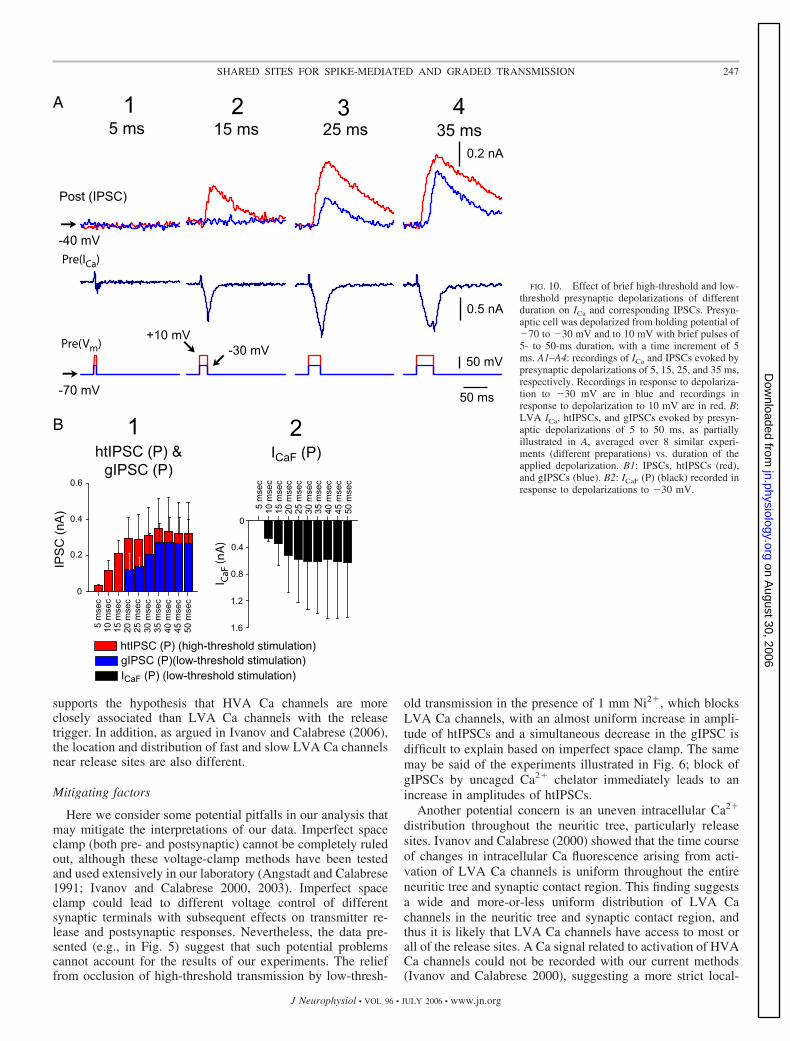
supports the hypothesis that HVA Ca channels are moreclosely associated than LVA Ca channels with the releasetrigger. In addition, as argued in Ivanov and Calabrese (2006),the location and distribution of fast and slow LVA Ca channelsnear release sites are also different.
Mitigating factors
Here we consider some potential pitfalls in our analysis thatmay mitigate the interpretations of our data. Imperfect spaceclamp (both pre- and postsynaptic) cannot be completely ruledout, although these voltage-clamp methods have been testedand used extensively in our laboratory (Angstadt and Calabrese1991; Ivanov and Calabrese 2000, 2003). Imperfect spaceclamp could lead to different voltage control of differentsynaptic terminals with subsequent effects on transmitter re-lease and postsynaptic responses. Nevertheless, the data pre-sented (e.g., in Fig. 5) suggest that such potential problemscannot account for the results of our experiments. The relieffrom occlusion of high-threshold transmission by low-thresh-
old transmission in the presence of 1 mm Ni2�, which blocksLVA Ca channels, with an almost uniform increase in ampli-tude of htIPSCs and a simultaneous decrease in the gIPSC isdifficult to explain based on imperfect space clamp. The samemay be said of the experiments illustrated in Fig. 6; block ofgIPSCs by uncaged Ca2� chelator immediately leads to anincrease in amplitudes of htIPSCs.
Another potential concern is an uneven intracellular Ca2�
distribution throughout the neuritic tree, particularly releasesites. Ivanov and Calabrese (2000) showed that the time courseof changes in intracellular Ca fluorescence arising from acti-vation of LVA Ca channels is uniform throughout the entireneuritic tree and synaptic contact region. This finding suggestsa wide and more-or-less uniform distribution of LVA Cachannels in the neuritic tree and synaptic contact region, andthus it is likely that LVA Ca channels have access to most orall of the release sites. A Ca signal related to activation of HVACa channels could not be recorded with our current methods(Ivanov and Calabrese 2000), suggesting a more strict local-
A
B
FIG. 10. Effect of brief high-threshold and low-threshold presynaptic depolarizations of differentduration on ICa and corresponding IPSCs. Presyn-aptic cell was depolarized from holding potential of�70 to �30 mV and to 10 mV with brief pulses of5- to 50-ms duration, with a time increment of 5ms. A1–A4: recordings of ICa and IPSCs evoked bypresynaptic depolarizations of 5, 15, 25, and 35 ms,respectively. Recordings in response to depolariza-tion to �30 mV are in blue and recordings inresponse to depolarization to 10 mV are in red. B:LVA ICa, htIPSCs, and gIPSCs evoked by presyn-aptic depolarizations of 5 to 50 ms, as partiallyillustrated in A, averaged over 8 similar experi-ments (different preparations) vs. duration of theapplied depolarization. B1: IPSCs, htIPSCs (red),and gIPSCs (blue). B2: ICaF (P) (black) recorded inresponse to depolarizations to �30 mV.
247SHARED SITES FOR SPIKE-MEDIATED AND GRADED TRANSMISSION
J Neurophysiol • VOL 96 • JULY 2006 • www.jn.org
on August 30, 2006
jn.physiology.orgD
ownloaded from
ization of a relatively small number of HVA channels torelease sites and very spatially restricted Ca domains near themouths of these channels (see also Ivanov and Calabrese2003).
Postsynaptic effects, such as changes in number and sensi-tivity of postsynaptic receptors and diffusion and removal oftransmitter from the cleft, might influence our recorded re-sponses. Nevertheless, the results presented in the paper, es-pecially the results of experiments that involved the block ofHVA and LVA channels (e.g., Figs. 5 and 8), uncaging ofcaged Ca2� chelator (Fig. 6), and changes in Ca2� drivingforce (Fig. 9) (as well results of Ivanov and Calabrese 2000,2003) indicate that the time course of postsynaptic responses isgoverned by transmitter release (i.e., by presynaptic Ca cur-rents and vesicle availability) and that any potential influenceof postsynaptic changes on synaptic transmission on the time-scale used here is small.
Association of Ca channels and the release trigger
The structural organization of active zones and thus theirfunctional properties vary from one synaptic connection toanother (Msghina et al. 1999; Poage and Meriney 2002; Satzleret al. 2002; Stanley 1997; Wachman et al. 2004). The spatialrelations between Ca channels and the readily releasable syn-aptic vesicles and their associated release trigger within anactive zone determine the release mode and reliability of agiven synaptic connection. There are thought to be three mainmodes of relation between Ca channels and the release trigger(Augustine 2001; see also Neher 1998).
1) Ca nanodomains arise from local diffusion from singleopen Ca channel located in intimate proximity to a releasetrigger (few nanometers). Activation of just a single Ca chan-nel can release acetylcholine at calyx-type presynaptic nerveterminal of the chick ciliary ganglion with the distance from Cachannel to release trigger being about 20 nm and the Ca2�
concentration near release trigger being about 10 �M (Stanley1993). Secretion at such a release site is triggered by Ca2� thatenters through a discrete, specifically associated cluster of Cachannels. In such a cluster the members nearest the releasetrigger are the most effective. Such an organization has theadvantage of allowing a molecular interaction between therelease mechanism and its triggering Ca channels (Gentile andStanley 2005; Stanley 1993, 1997).
2) Ca microdomains arise from multiple open Ca channels,clustered together but relatively distant from the release trig-ger. The involvement of multiple Ca channels, in some cases ofdifferent types, creating overlapping Ca2� influx to producemicrodomains gating transmitter release has been widely de-scribed (Fossier et al. 1993; Mintz et al. 1995; Qian andNoebels 2001; Reid et al. 1998; Wu and Shaggau 1994, 1997;Wu et al. 1998, 1999) and summarized and modeled byMeinrenken et al. (2002, 2003) for the rat calyx of Heldsynapse. The model assumes that the distance from a vesicle toCa channel clusters varies across multiple release sites of asingle calyx synapse from 30 to 300 nm, with Ca2� peakconcentrations from 0.5 to 40 �M. Such a topography leads torelease probability ranging from �0.01 to 1. One to a fewclusters can be present at an active zone, and one to a fewvesicles (release triggers) may be under control of one cluster.
3) Radial gradients of Ca2� are the result of Ca influxthrough Ca channels, randomly/uniformly distributed at �1�m from a release trigger. In chromaffin cells, where trans-mitter release is much slower than at synapses, Ca channelsand vesicles are not closely localized and radial diffusion ofCa2� from distant Ca channels activates release triggers (Chowet al. 1994; Marengo and Monck 2000; Neher 1998; Neher andAugustine 1992). The inhibitory synapses between heart inter-neurons appear to incorporate all three of these types oforganizations.
Proposed organization of Ca channels at release sites forthe synaptic connections between heart interneurons
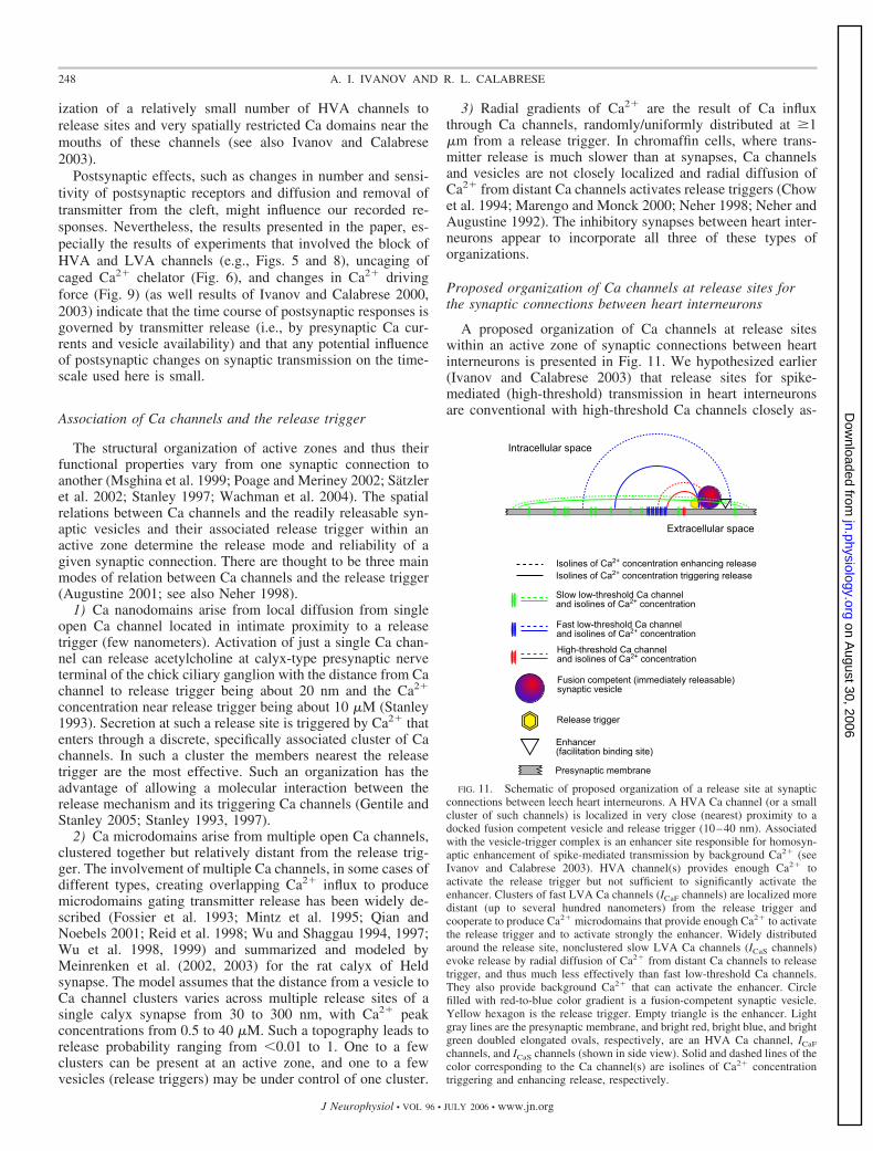
A proposed organization of Ca channels at release siteswithin an active zone of synaptic connections between heartinterneurons is presented in Fig. 11. We hypothesized earlier(Ivanov and Calabrese 2003) that release sites for spike-mediated (high-threshold) transmission in heart interneuronsare conventional with high-threshold Ca channels closely as-
FIG. 11. Schematic of proposed organization of a release site at synapticconnections between leech heart interneurons. A HVA Ca channel (or a smallcluster of such channels) is localized in very close (nearest) proximity to adocked fusion competent vesicle and release trigger (10–40 nm). Associatedwith the vesicle-trigger complex is an enhancer site responsible for homosyn-aptic enhancement of spike-mediated transmission by background Ca2� (seeIvanov and Calabrese 2003). HVA channel(s) provides enough Ca2� toactivate the release trigger but not sufficient to significantly activate theenhancer. Clusters of fast LVA Ca channels (ICaF channels) are localized moredistant (up to several hundred nanometers) from the release trigger andcooperate to produce Ca2� microdomains that provide enough Ca2� to activatethe release trigger and to activate strongly the enhancer. Widely distributedaround the release site, nonclustered slow LVA Ca channels (ICaS channels)evoke release by radial diffusion of Ca2� from distant Ca channels to releasetrigger, and thus much less effectively than fast low-threshold Ca channels.They also provide background Ca2� that can activate the enhancer. Circlefilled with red-to-blue color gradient is a fusion-competent synaptic vesicle.Yellow hexagon is the release trigger. Empty triangle is the enhancer. Lightgray lines are the presynaptic membrane, and bright red, bright blue, and brightgreen doubled elongated ovals, respectively, are an HVA Ca channel, ICaF
channels, and ICaS channels (shown in side view). Solid and dashed lines of thecolor corresponding to the Ca channel(s) are isolines of Ca2� concentrationtriggering and enhancing release, respectively.
248 A. I. IVANOV AND R. L. CALABRESE
J Neurophysiol • VOL 96 • JULY 2006 • www.jn.org
on August 30, 2006
jn.physiology.orgD
ownloaded from

sociated with a low-affinity secretory trigger (see Augustine etal. 1992; Llinas et al. 1995; Neher 1998; Stanley 1993, 1997)(possibly synaptotagmin 1) closely associated with synapticvesicle membranes (Sudhoff 2002, 2004; Sugita et al. 2002).This hypothesis is supported by our current findings. High-threshold synaptic transmission (spike-mediated transmissionand transmission evoked by brief or prolonged depolarizationsto �10 mV and higher) is insensitive to intracellularly injectedEGTA (Ivanov and Calabrese 2003) and is not very sensitive tocaged and uncaged diazo-2 (which is built on the fast Ca2�
chelator BAPTA) (Fig. 6; see also Ivanov and Calabrese 2003).These responses to fast and slow Ca2� chelators suggest a veryclose association (�40 nm) of the release trigger and high-threshold Ca channels (see Adler et al. 1991; Augustine et al.1992; Fedchyshin and Wang 2005; Meinrenken et al. 2002,2003; Neher 1998).
Such a close association of high-threshold Ca channels andthe release trigger may argue for direct binding interactionsbetween synaptic proteins and Ca channels that may be anessential determinant of synaptic transmission, although thenecessity of such coupling for synaptic transmission in inverte-brates is still unclear (Arien et al. 2003; Atlas 2001; Catterall1999; Jarvis and Zamponi 2001, 2005; Spafford and Zamponi2003; Spafford et al. 2003). A narrowly restricted localizationof a small number of HVA Ca channels to release sites isconsistent with our almost complete inability to record changesin intracellular Ca fluorescence in response to brief high-threshold depolarizations (see Ivanov and Calabrese 2000).The same conclusion follows from the much shorter synapticdelay for htIPSCs compared with the synaptic delay for gIPSCswith substantially smaller differences between latencies forICaHT and ICaF (Fig. 10).
Because synaptic modulation of spike-mediated transmis-sion depends on background Ca2� arising from LVA Cachannels but is independent of spiking activity (thus from Ca2�
entering by high-threshold Ca channels), the high-affinity fa-cilitation binding site (enhancer) appears to be localized moredistant from HVA Ca channels than the release trigger (Ivanovand Calabrese 2003), perhaps to prevent its saturation by Ca2�
entering the cell by HVA Ca channels. The separation of HVACa channels from the enhancer by a docked vesicle, suggestedfor activity-dependent facilitation by Zucker and colleagues(Tang et al. 2000; Zucker 1999; Zucker and Regehr 2002) andShahrezaei and Delaney (2004), is an attractive possibility. Wehave been unable yet to determine the cooperativity of HVAchannels in mediating release; i.e., how many HVA Ca chan-nels contribute to transmitter release. Nevertheless, HVA Cachannel–dependent release appears to be based on typicalnanodomains (Augustine 2001) with Ca channel(s) localized invery close proximity to docked/fusion ready vesicle.
The ability of ICaF to effectively occlude high-thresholdtransmission argues for release sites shared by LVA and HVAchannels. The sensitivity of ICaF-dependent release to both fastand slow Ca2� chelators (Ivanov and Calabrese 2003; presentfindings), the cooperativity of ICaF channels in triggering re-lease (Ivanov and Calabrese 2006), the longer synaptic delay(compared with HVA Ca channel–dependent synaptic trans-mission) suggests a more distant localization of ICaF channelclusters from release trigger than for the HVA Ca channels.This hypothesized localization—clustered ICaF channels withinactive zone at a distance from the release trigger—resembles
the clustering of Ca channels proposed for rat calyx of Heldsynapse where Ca2� microdomains are thought to triggerrelease (Meinrenken et al. 2002, 2003). Because of the massiveinflux of Ca2� by ICaF channels, Ca2� concentration at therelease enhancer is sufficient to evoke enhancement of high-threshold synaptic transmission. The release cooperativity ofabout 2 (see Fig. 6 in Ivanov and Calabrese 2006) for ICaFchannels does not necessarily mean that the clusters consists ofjust two Ca channels, but rather that not less than two ICaFchannels in any given cluster have to be open to evoketransmitter release. Wachman et al. (2004) found that in frogmotor nerve terminal there is a remarkably low probability ofa given Ca channel opening within an active zone after anaction potential. Thus Ca2� microdomains arising from clus-tered Ca channels appear to be responsible for ICaF channel–dependent transmitter release.
The lower effectiveness of slow low-threshold Ca channelsin transmitter release suggests that they are widely distributedthroughout active zone, nonclustered, and evoke release andenhancement of high-threshold synaptic transmission by radialdiffusion of Ca2� from relatively distant Ca channels to therelease trigger and the release enhancer, respectively (Ivanovand Calabrese 2006).
Such an organization of release sites appears optimal foreconomical reciprocal inhibitory synaptic transmission be-tween heart interneurons that increases the robustness of rhyth-mic bursting. A burst is initiated when LVA Ca currentsactivate supporting depolarization and spiking and produce agIPSC, which serves to terminate the burst of the oppositeheart interneuron. As the burst progresses, LVA Ca currentsinactivate, leading to a progressively decreasing gIPSC, andspike-mediated (HVA) synaptic transmission, which serves tokeep the opposite heart interneuron silent, increases; increasedbackground Ca2� resulting from LVA Ca currents enhancesthis transmission.
These considerations do not exclude the possibility that thereare release sites that are purely under control of LVA Cachannels or that there are release sites with different secretorytriggers, such as synaptotagmin 7, which has relatively highCa2� affinity and slow dynamics and seemingly subservesasynchronous release (Sudhoff 2002, 2004; Sugita et al. 2002).Such release sites must be in the minority, however, or theocclusion between high- and low-threshold synaptic transmis-sion would not be so complete.
Shared release sites for spike-mediated and graded transmis-sion in leech heart interneurons is inconsistent with the hy-pothesis of Matthews (2000) that low Ca2� affinity is associ-ated with sustained neurotransmitter release, whereas the brev-ity of Ca2� signals driven by action potentials allows for higherCa2� affinity and greater integration of local Ca2� signals. Inheart interneurons, coexisting Ca channels of different typesappear to be localized differently relative to the low-affinityrelease trigger and to the high-affinity enhancer at release sites,and thus there is the potential for both kinetic competitionbetween the release trigger and Ca2� buffers for Ca2� (Au-gustine et al. 1991) and the intercepting effect of Ca2� bufferson Ca2� that diffuses from distant Ca channels to the releasetrigger (Meinrenken et al. 2003). Ultimately, our data supportthe hypothesis of Jones (2003) that the role of a Ca channel inneurotransmitter release is determined less by gating kineticsthan by the channel location. The occurrence of multiple
249SHARED SITES FOR SPIKE-MEDIATED AND GRADED TRANSMISSION
J Neurophysiol • VOL 96 • JULY 2006 • www.jn.org
on August 30, 2006
jn.physiology.orgD
ownloaded from
calcium channel types may indicate that channels are special-ized for different functions (Jones 2003), but we have yet todetermine the specific functions of the different Ca channelstypes at release sites.
G R A N T S
This work was supported by National Institute of Neurological Disordersand Stroke Grant NS-24072.
R E F E R E N C E S
Adams SR and Tsien RY. Controlling cell chemistry with caged compounds.Annu Rev Physiol 55: 755–784, 1993.
Adler EM, Augustine GJ, Duffy SN, and Charlton MP. Alien intracellularcalcium chelators attenuate neurotransmitter release at the squid giantsynapse. J Neurosci 11: 1496–1507, 1991.
Angstadt JD and Calabrese RL. Calcium currents and graded synaptictransmission between heart interneurons of the leech. J Neurosci 11:746–759, 1991.
Arbas EA and Calabrese RL. Ionic conductances underlying the activity ofinterneurons that control heartbeat in the medicinal leech. Neurosci 7:3945–3952, 1987.
Arien H, Wiser O, Arkin IT, Leonov H, and Atlas D. Syntaxin 1Amodulates the voltage-gated L-type calcium channel (Cav1.2) in a cooper-ative manner. J Biol Chem 278: 29231–29239, 2003.
Artalejo CR, Adams ME, and Fox A. Three types of Ca2� channel triggersecretion with different efficacies in chromaffine cells. Nature 367: 72–76,1994.
Atlas D. Functional and physical coupling of voltage-sensitive calcium chan-nels with exocytotic proteins: ramifications for the secretion mechanism.J Neurochem 77: 972–985, 2001.
Atluri PP and Regehr WG. Delayed release of neurotransmitter from cere-bellar granule cells. J Neurosci 15: 8214–8227, 1998.
Augustine GJ. How does calcium trigger neurotransmitter release? Curr OpinNeurobiol 11: 320–326, 2001.
Augustine GJ, Adler EM, and Charlton MP. The calcium signal fortransmitter secretion from presynaptic nerve terminals. Ann NY Acad Sci635: 365–381, 1991.
Augustine GJ, Adler EM, Charlton MP, Hans M, Swandulla D, andZipser K. Presynaptic calcium signals during neurotransmitter release:detection with fluorescent indicators and other calcium chelators. J Physiol(Paris) 86: 129–134, 1992.
Ayali A, Johnson BR, and Harris-Warrick RM. Dopamine modulatesgraded and spike-evoked synaptic inhibition independently at single syn-apses in pyloric network of lobster. J Neurophysiol 79: 2063–2069, 1998.
Bertram R, Smith GD, and Sherman A. Modeling study of the effects ofoverlapping Ca2� microdomains on neurotransmitter release. Biophys J 76:735–750, 1999.
Borst JGG and Sakmann B. Effect of changes in action potential shape oncalcium currents and transmitter release in a calyx-type synapse of the ratauditory brainstem. Philos Trans R Soc Lond B Biol Sci 354: 347–355, 1999.
Calabrese RL, Angstadt JD, and Arbas EA. A neural oscillator based onreciprocal inhibition. In: Perspectives in Neural Systems and Behavior,edited by Carew TJ and Kelley DB. New York: Liss, 1989, p. 33–50.
Catterall WA. Interactions of presynaptic Ca2� channels and Snare proteinsin neurotransmitter release. Ann NY Acad Sci 868: 144–159, 1999.
Chow RH, Klingauf J, and Neher E. Time course of Ca2� concentrationtriggering exocytosis in neuroendocrine cells. Proc Natl Acad Sci USA 91:12765–12769, 1994.
Delaney KR. Uncaging calcium in neurons. In: Imaging Neurons. A Labora-tory Manual, edited by Yuste R, Lanni F, and Konnerth A. Cold SpringHarbor, NY: Cold Spring Harbor Laboratory Press, 2000.
Fedchyshin MJ and Wang LY. Developmental transformation of the releasemodality at the calyx of Held synapse. J Neurosci 25: 4131–4140, 2005.
Fossier P, Baux G, and Tauc L. Role of different types of Ca2� channels anda reticulum-like Ca2� pump in neurotransmitter release. J Physiol (Paris)87: 3–14, 1993.
Gentile L and Stanley EF. A unified model of presynaptic release site gatingby calcium channel domains. Eur J Neurosci 21: 278–282, 2005.
Ivanov AI and Calabrese RL. Intracellular Ca2� dynamics during spontane-ous and evoked activity of leech heart interneurons: low-threshold Cacurrents and graded synaptic transmission. J Neurosci 20: 4930–4943,2000.
Ivanov AI and Calabrese RL. Modulation of spike-mediated synaptic trans-mission by presynaptic background Ca2� in leech heart interneurons. JNeurosci 23: 1206–1218, 2003.
Ivanov AI and Calabrese RL. Graded inhibitory synaptic transmissionbetween leech interneurons: assessing the roles of two kinetically distinctlow-threshold Ca currents. J Neurophysiol 96: 218–234, 2006.
Jarvis SE and Zamponi GW. Interactions between presynaptic Ca2� chan-nels, cytoplasmic messengers and proteins of the synaptic vesicle releasecomplex. Trends Pharmacol Sci 22: 519–525, 2001.
Jarvis SE and Zamponi GW. Masters or slaves? Vesicle release machineryand the regulation of presynaptic calcium channels. Cell Calcium 37:483–488, 2005.
Jones SW. Calcium channels: unanswered questions. J Bioenerg Biomembr35: 461–475, 2003.
Llinas R, Sugimori M, and Silver RB. The concept of calcium concentrationmicrodomain in synaptic transmission. Neuropharmacology 34: 1443–1451,1995.
Lu J, Dalton JF IV, Stokes DR, and Calabrese RL. Functional role of Ca2�
currents in graded and spike-mediated synaptic transmission between leechheart interneurons. J Neurophysiol 77: 1779–1794, 1997.
Marengo FD and Monck JR. Development and dissipation of Ca2� gradientsin adrenal chromaffin cells. Biophys J 79: 1800–1820, 2000.
Matthews G. Vesicle fiesta at the synapse. Nature 406: 835–836, 2000.Meinrenken CJ, Borst JGG, and Sakmann B. Calcium secretion coupling at
calyx of Held governed by nonuniform channel-vesicle topography. JNeurosci 22: 1648–1667, 2002.
Meinrenken CJ, Borst JGG, and Sakmann B. Local routes revised: thespace and time dependence of the Ca2� signal for phasic transmitter releaseat the rat calyx of Held. J Physiol 547: 665–689, 2003.
Millar AG, Zucker RS, Ellis-Davies GCR, Charlton MP, and Atwood HL.Calcium sensitivity of neurotransmitter release differs at phasic and tonicsynapses. J Neurosci 25: 3113–3125, 2005.
Mintz IM, Sabatini BL, and Regehr WG. Calcium control of transmitterrelease at a cerebellar synapse. Neuron 15: 675–688, 1995.
Msghina M, Millar AG, Charlton MP, and Atwood HL. Calcium entryrelated to active zones and differences in transmitter release at phasic andtonic synapses. J Neurosci 19: 8419–8434, 1999.
Neher E. Vesicle pools and Ca2� microdomains: new tools for understandingtheir roles in neurotransmitter release. Neuron 20: 389–399, 1998.
Neher E and Augustine GJ. Calcium gradients and buffers in bovinechromaffin cells. J Physiol 450: 273–301, 1992.
Neher E and Zucker RS. Multiple calcium-dependent processes related tosecretion in bovine chromaffine cells. Neuron 10: 21–30, 1993.
Nicholls JG and Baylor DA. Specific modalities and receptive fields ofsensory neurons in the central nervous system of the leech. J Neurophysiol31: 740–756, 1968.
Olsen ØH and Calabrese RL. Activation of intrinsic and synaptic currents inleech heart interneurons by realistic waveforms. J Neurosci 16: 4958–4970,1996.
Otsu Y, Shahrezaei V, Li B, Raymond LA, Delaney KR, and Murphy TH.Competition between phasic and asynchronous release for recovered syn-aptic vesicles at developing hippocampal autaptic synapses. J Neurosci 24:420–433, 2004.
Pan ZH, Hu HJ, Perring P, and Angrade R. T-type Ca2� channels mediateneurotransmitter release in retinal bipolar cells. Neuron 32: 89–98, 2001.
Poage RE and Meriney SD. Presynaptic calcium influx, neurotransmitterrelease, and neuromuscular disease. Physiol Behav 77: 507–512, 2002.
Qian J and Noebels JL. Presynaptic Ca2� channels and neurotransmitterrelease at the terminal of a mouse cortical neuron. J Neurosci 21: 3721–3728, 2001.
Quigley PA, Msghina M, Govind CK, and Atwood HL. Visible evidence fordifferences in synaptic effectiveness with activity-dependent vesicular up-take and release of FM1-43. J Neurophysiol 81: 356–370, 1999.
Reid CA, Bekkers JM, and Clements JD. N- and P/Q-type Ca2� channelsmediate transmitter release with a similar cooperativity at rat hippocampalautapses. J Neurosci 18: 2849–2855, 1998.
Satzler K, Sohl LF, Bollmann JH, Borst JGG, Frotscher M, Sakmann B,and Lubke JHR. Three-dimensional reconstruction of a calyx of Held andits postsynaptic principal neuron in the medial nucleus of the trapezoid body.J Neurosci 22: 10567–10579, 2002.
Shahrezaei V and Delaney KR. Consequences of molecular-level Ca2�
channel and synaptic vesicle colocalization for the Ca2� microdomain andneurotransmitter exocytosis: a MonteCarlo study. Biophys J 87: 2352–2364,2004.
250 A. I. IVANOV AND R. L. CALABRESE
J Neurophysiol • VOL 96 • JULY 2006 • www.jn.org
on August 30, 2006
jn.physiology.orgD
ownloaded from
Simon TW, Schmidt J, and Calabrese RL. Modulation of high-thresholdtransmission between heart interneurons of the medicinal leech by FMRF-NH2. J Neurophysiol 71: 454–466, 1994.
Spafford JD, Munno DW, van Nierop P, Feng ZP, Jarvis SE, Callin WJ,Smit AB, Zamponi GW, and Syed NI. Calcium channel structural deter-minants of synaptic transmission between identified invertebrate neurons.J Biol Chem 278: 4258–4267, 2003.
Spafford JD and Zamponi GW. Functional interactions between presynapticcalcium channels and the neurotransmitter release machinery. Curr OpinNeurobiol 13: 308–314, 2003.
Stanley EF. Single calcium channels and acetylcholine release at a presynapticnerve terminal. Neuron 11: 1007–1011, 1993.
Stanley EF. The calcium channel and the organization of the presynaptictransmitter release face. Trends Neurosci 20: 404–409, 1997.
Sudhof TC. Synaptotagmins: why so many? J Biol Chem 277: 7629–7632,2002.
Sudhof TC. The synaptic vesicle cycle. Annu Rev Neurosci 27: 509–547, 2004.Sugita S, Shin OH, Han W, Lao Y, and Sudhof TC. Synaptotagmins from
a hierarchy of exocytotic Ca2� sensors with distinct Ca2� affinities. EMBOJ 21: 270–280, 2002.
Tang Y, Schlumpberger T, Kim T, Lueker M, and Zucker RS. Effects ofmobile buffers on facilitation: experimental and computational studies.Biophys J 78: 2735–2751, 2000.
Wachman ES, Poage RE, Stiles JR, Farkas DL, and Meriney SD.Spatial distribution of calcium entry evoked by single action poten-tial within the presynaptic active zone. J Neurosci 24: 2877–2885, 2004.
Warzecha AK, Kurtz R, and Egelhaaf M. Synaptic transfer of dynamicmotion information between identified neurons in the visual system of theblowfly. Neuroscience 119: 1103–1112, 2003.
Wu LG, Borst JGG, and Sakmann B. R-type Ca2� currents evoke trans-mitter release at a rat central synapse. Proc Natl Acad Sci USA 95:4720–4725, 1998.
Wu LG and Saggau P. Pharmacological identification of two types ofpresynaptic voltage-dependent calcium channels at CA3–CA1 synapses ofthe hippocampus. Neuroscience 14: 5613–5622, 1994.
Wu LG and Saggau P. Presynaptic inhibition of elicited neurotransmitterrelease. Trends Neurosci 20: 204–212, 1997.
Wu LG, Westenbroek RE, Borst JGG, Catterall WA, and Sakmann B.Calcium channel types with distinct presynaptic localization couple differ-entially to transmitter release in single calyx-type synapses. J Neurosci 19:726–736, 1999.
Zucker RS. Calcium and activity-dependent synaptic plasticity. Curr OpinNeurobiol 9: 305–313, 1999.
Zucker RS and Regehr WG. Short-term synaptic plasticity. Annu RevPhysiol 64: 355–405, 2002.
251SHARED SITES FOR SPIKE-MEDIATED AND GRADED TRANSMISSION
J Neurophysiol • VOL 96 • JULY 2006 • www.jn.org
on August 30, 2006
jn.physiology.orgD
ownloaded from


![spike-timing dependent plasticityprocesses. Similarly, there is a large menagerie of models of synaptic plasticity [19]. We consider two well-established models: Gerstner’s Spike](https://img.pdfslide.net/doc/110x75/604fc6cb5438ef3c0a569f78/spike-timing-dependent-plasticity-processes-similarly-there-is-a-large-menagerie.jpg)


![THE EFFECT OF SYNAPTIC DEPRESSION ON MODEL INHIBITORY … · phenomenological models [2], [36] have been able to capture the essence of synaptic transmission between pairs of neurons](https://img.pdfslide.net/doc/110x75/5f3df958df75e7017103e764/the-effect-of-synaptic-depression-on-model-inhibitory-phenomenological-models-2.jpg)