Embed Size (px)
Citation preview
Dottorato in Scienze Mediche Specialistiche
DOTTORATO DI RICERCA IN MEDICINA DEL SONNO
XXIV Ciclo
SPINAL CORD INJURY:
ASSESSMENT OF AUTONOMIC STATE-DEPENDENT CONTROL OF CARDIOVASCULAR SYSTEM AND
BODY CORE TEMPERATURE
Presentata da: Dr. Pietro Guaraldi
Coordinatore Dottorato: Relatori:
Prof. Pietro Cortelli Prof. Pietro Cortelli
Dr.ssa Federica Provini
Alma Mater Studiorum – Università di Bologna
Anno 2012

Alma Mater Studiorum – Università di Bologna
Anno 2012

DOTTORATO DI RICERCA IN SCIENZE MEDICHE SPECIALISTICHE
Progetto n°2 “MEDICINA DEL SONNO”
XXIV Ciclo
Settore Concorsuale: 06/D6 - NEUROLOGIA
Settore Scientifico Disciplinare di afferenza: MED 26
SPINAL CORD INJURY:
ASSESSMENT OF AUTONOMIC STATE-DEPENDENT CONTROL OF CARDIOVASCULAR SYSTEM AND
BODY CORE TEMPERATURE
Presentata da: Dr. Pietro Guaraldi
(n° matr. 376057)
Coordinatore Dottorato: Relatori:
Prof. Pietro Cortelli Prof. Pietro Cortelli
Dr.ssa Federica Provini
Alma Mater Studiorum – Università di Bologna
Esame finale anno 2012


ad Achira (Chiara)
“To whom I owe the leaping delight
That quickens my senses in our wakingtime
And the rhythm that governs the repose of our sleepingtime,
the breathing in unison”
T. S. Eliot

INDEX OF CONTENTS
INTRODUCTION 1
Section ISLEEP STRUCTURE IN SPINAL CORD INJURY PATIENTS
Section II ANALYSIS OF CARDIOVASCULAR PARAMETERS
Autonomic control of the cardiovascular system during wakefulness
The circadian rhythm of blood pressure and heart rate in spinal cord injury patients
Section III BODY CORE TEMPERATURE REGULATION IN SPINAL CORD INJURY PATIENTS
CONCLUSIONS
BIBLIOGRAPHY
APPENDIX
Section I
Section II.1
Section II.2
Section III
5
19
41
55
67
69
79
86
101
106

LIST OF ABBREVIATIONS
∆ (delta): Change of any variable quantity
∆BcT: changes in BcT
∆DBP: changes in DBP
∆HR: changes in HR
∆SBP: changes in SBP
ACR: acrophase
AD: autonomic dysreflexia
AHI: apnea hypopnea index
AMP: amplitude
ANOVA: analysis of variance
ANS: autonomic nervous system
ASIA: American Spinal Injury AssociationBcT: body core temperature
BMI: body mass index
BP: blood pressure
C4-7: cervical SCI
CF: cold face
CHD: coronary heart disease
CNT: control subjects
CPG: central pattern generators
CVD: cardiovascular disease
D2-12: thoracic SCI
DB: deep breathing
DBP: diastolic blood pressure
EEG: electroencephalogram
HF: high frequency
HR: heart rate
HRV: heart rate variability
HUTT: head-up tilt test
IE: isometric exercise
LF: low frequency
MET: metabolic equivalent
N1: stage 1 NREM sleep
N2: stage 2 NREM sleep
N3: stage 3 NREM sleep
NREM: non REM
NREM: non-rapid eye-movement sleep
OSA: obstructive sleep apnea
OV: overshoot
PLMS: periodic limb movement during sleep
PS: parasympathetic
PSA: power spectral analysis
PSG: polysomnographic
REM: rapid eye-movement sleep
SBP: systolic blood pressure
SCI: spinal cord injury
SE: sleep efficiency
SNA: sympathetic nerve activity
SRBD: sleep related breathing disorders
SSR: sympathetic skin response
SWS: slow wave sleep
TP: Total Power
TPR: total peripheral resistance
TST: total sleep time
V-PSG: Video-Polysomnography
VLF: very low frequency
VM: Valsalva manoeuvre
VR: Valsalva ratio
W: wake
INTRODUCTION
“The frog instantly dies when the spinal cord is pierced, and previous to this it lived without head, heart or any bowels or skin, and here, therefore, it would seem lies the foundation of movement and life.”
Leonardo da Vinci (Quaderni d’Anatomica, Vol. V)

INTRODUCTION
Scientific Background and Object of the Study:Few survivable injuries have as much impact on a patient’s life as acute spinal cord trauma and its associated human and social cost. Spinal cord injury (SCI) results not only in paralysis; but it is also associated with a range of autonomic dysregulation that can interfere with cardiovascular, bladder, bowel, temperature, and sexual function. The entity of the autonomic dysfunction is related to the level and severity of injury to descending autonomic (sympathetic) pathways [1-3].
For many years there was limited awareness of these issues and the attention given to them by the scientific and medical community was scarce. Yet, even if a new system to document the impact of SCI on autonomic function has recently been proposed [4], the current standard of assessment of SCI (American Spinal Injury Association (ASIA) examination) evaluates motor and sensory pathways, but not severity of injury to autonomic pathways [5].
Beside the severe impact on quality of life, autonomic dysfunction in persons with SCI is associated with increased risk of cardiovascular disease and mortality [3,6,7]. Therefore, obtaining information regarding autonomic function in persons with SCI is pivotal and clinical examinations and laboratory evaluations to detect the presence of autonomic dysfunction and quantitate its severity are mandatory.
Furthermore, previous studies demonstrated that there is an intimate relationship between the autonomic nervous system and sleep from anatomical, physiological, and neurochemical points of view [8]. Although, even if previous epidemiological studies demonstrated that sleep problems are common in SCI [9-12], so far only limited polysomnographic (PSG) data are available [13,14]. Finally, until now, circadian and state dependent autonomic regulation of blood pressure (BP), heart rate (HR) and body core temperature (BcT) were never assessed in SCI patients.
Aim of the current study was to establish the association between the autonomic control of the cardiovascular function, thermoregulation, sleep parameters and increased cardiovascular risk in SCI patients.
Study Population and Main Parameters:Thanks to the collaboration with the Spinal Unit of Azienda Ospedaliera Carreggi, Florence, ITALY, we assessed 5 cervical SCI with tetraplegia (C4-C7 lesions), 7 thoracic SCI with paraplegia (D2-D12 lesions). All patients underwent the following studies:
I. Nocturnal cardio-respiratory monitoring for detection of sleep related breathing disorders;
II. Assessment of daytime autonomic control of the cardiovascular system through cardiovascular reflexes;
III. Assessment of heart rate variability (HRV) during supine rest and head-up tilt;
IV. Evaluation of Sleep structure and efficiency by means of video-PSG;
V. Assessment of circadian rhythm and state and time dependent regulation of BP and HR;
VI. Assessment of circadian rhythm and state and time dependent regulation of BcT.
Introduction
1

Patients results were compared to controls, whose number varied according to the different assessments.
Inclusion Criteria:• Male sex;
• Complete spinal cord lesion (Frankel A);
• Clinically stable (at least 6 months after the spinal cord lesion);
• Absence of symptoms or signs of diabetes, cardiorespiratory disease or of other pathological conditions that might affect autonomic cardiovascular control;
• Patient provided informed consent to participate to the study.
Study ProtocolPatients and controls were admitted to hospital and studied following a 3-days protocol (Fig 0.1). Subjects were admitted on Monday morning; in the afternoon they received an enema to prepare for rectal temperature monitoring and at night they underwent nocturnal cardio-respiratory monitoring for detection of sleep related breathing disorders;.
On the following morning, between 8:00 am and 11:00 am, subjects underwent cardiovascular reflexes tests in a temperature controlled (23±1°C) clinical investigation room according to standard procedures [15].
After the assessment of cardiovascular reflexes (between 11:00 am and 12:00 am), arterial BP, HR, BcT and sleep-wake cycle were continuously monitored for 24 hours under controlled conditions (recording ending at 11:00 am of the following day).
Fig 0.1: study protocol
Day 1Mon
Day 2Tue
Day 3Wed
- stanza con variabili fisiche controllate;
- monitoraggio continuo di pressione arteriosa, frequenza cardiaca, respiro, ciclo sonno-veglia con sistema di acquisizione del segnale COLLEAGUE (1000 Hz/canale)
- monitoraggio della T° corporea interna (rettale) mediante mini-logger (ogni 2 minuti)
1:00 pm: Enema
2:00 pm: Nocturnal cardio-respiratory monitoring
9:00 am: Cardiovascular
reflexes
11:00 am: Circadian rhythm of BP and HR and BcT
11:00 am: Stop recording
Guaraldi P. Introduction
2
Thesis StructureThe thesis is organized in Sections originating from the original study protocol.
In each module it is specified the included population of patients and controls, the methods of data collection and analysis, results, discussion and conclusions.
The sections are structured as follow:
I. Sleep structure in spinal cord injury patients
II. Analysis of cardiovascular parameters1. Autonomic control of the cardiovascular system during wakefulness
2. The circadian rhythm of blood pressure and heart rate in spinal cord injury patients
III. Body core temperature regulation in spinal cord injury patients
In the Appendix figures, which were reduced in size to facilitate reading in each section, are reported in higher magnification, together with additional supplementary material.
The thesis is written in English to allow international diffusion and to favour its publication on peer reviewed medical journal.
Introduction
3
Bibliography:1. Claydon VE, Krassioukov AV (2006) Orthostatic hypotension and autonomic pathways after spinal cord injury. Journal of
neurotrauma 23 (12):1713-1725.
2. Furlan JC, Fehlings MG, Shannon P, Norenberg MD, Krassioukov AV (2003) Descending vasomotor pathways in humans: correlation between axonal preservation and cardiovascular dysfunction after spinal cord injury. Journal of neurotrauma 20 (12):1351-1363.
3. Krassioukov A, Claydon VE (2006) The clinical problems in cardiovascular control following spinal cord injury: an overview. Progress in brain research 152:223-229.
4. Alexander MS, Biering-Sorensen F, Bodner D, Brackett NL, Cardenas D, Charlifue S, Creasey G, Dietz V, Ditunno J, Donovan W, Elliott SL, Estores I, Graves DE, Green B, Gousse A, Jackson AB, Kennelly M, Karlsson AK, Krassioukov A, Krogh K, Linsenmeyer T, Marino R, Mathias CJ, Perkash I, Sheel AW, Schilero G, Schurch B, Sonksen J, Stiens S, Wecht J, Wuermser LA, Wyndaele JJ (2009) International standards to document remaining autonomic function after spinal cord injury. Spinal Cord 47 (1):36-43.
5. Marino RJ, Barros T, Biering-Sorensen F, Burns SP, Donovan WH, Graves DE, Haak M, Hudson LM, Priebe MM (2003) International standards for neurological classification of spinal cord injury. J Spinal Cord Med 26 Suppl 1:S50-56.
6. Myers J, Lee M, Kiratli J (2007) Cardiovascular disease in spinal cord injury: an overview of prevalence, risk, evaluation, and management. Am J Phys Med Rehabil 86 (2):142-152.
7. Garshick E, Kelley A, Cohen SA, Garrison A, Tun CG, Gagnon D, Brown R (2005) A prospective assessment of mortality in chronic spinal cord injury. Spinal Cord 43 (7):408-416.
8. Cortelli P, Lombardi C (2007) Autonomic Dysfunctions in Sleep Disorders. In: Culebras A (ed) Sleep disorders and neurologic diseases. vol 2, 2nd edn. Informa Healthcare, New York, pp 337-348.
9. Biering-Sorensen F, Biering-Sorensen M (2001) Sleep disturbances in the spinal cord injured: an epidemiological questionnaire investigation, including a normal population. Spinal Cord 39 (10):505-513.
10. Norrbrink Budh C, Hultling C, Lundeberg T (2005) Quality of sleep in individuals with spinal cord injury: a comparison between patients with and without pain. Spinal Cord 43 (2):85-95.
11. Jensen MP, Hirsh AT, Molton IR, Bamer AM (2009) Sleep problems in individuals with spinal cord injury: frequency and age effects. Rehabil Psychol 54 (3):323-331.
12. Biering-Sorensen F, Jennum P, Laub M (2009) Sleep disordered breathing following spinal cord injury. Respir Physiol Neurobiol 169 (2):165-170.
13. Adey WR, Bors E, Porter RW (1968) EEG sleep patterns after high cervical lesions in man. Arch Neurol 19 (4):377-383.
14. Scheer FA, Zeitzer JM, Ayas NT, Brown R, Czeisler CA, Shea SA (2006) Reduced sleep efficiency in cervical spinal cord injury; association with abolished night time melatonin secretion. Spinal Cord 44 (2):78-81.
15. Mathias CJ, Bannister R (1999) Investigation of autonomic disorders. In: Mathias CJ, Bannister R (eds) Autonomic failure : a textbook of clinical disorders of the autonomic nervous system. 4th edn. Oxford University Press, Oxford; New York, pp 169-195.
Guaraldi P. Introduction
4

Section I
SLEEP STRUCTURE IN SPINAL CORD INJURY PATIENTS
“Something for the rag and bone manOver my dead bodySomething big is gonna happenOver my dead body Someone saw someone's daughterOver my dead bodyThis is how I ended up sucked inOver my dead bodyI'm gonna go to sleepAnd let this wash all over meWe don't really want a monster taking overTip toeing, tying downWe don't want the loonies takin' overTip toeing, tying down our armsMay pretty horsesCome to you as you sleepI'm gonna go to sleepAnd let this wash over me”
Radiohead(Go to Sleep, Hail to the Thief, 2003)

Section I: SLEEP STRUCTURE IN SPINAL CORD INJURY PATIENTS:
A 24-h study under bed rest controlled conditions
Background:Previous cross-sectional descriptive studies and epidemiological reviews [1-4] demonstrated that sleep problems are common in spinal cord injury (SCI). These studies, which are based on sleep scales and self-administered questionnaire, demonstrated that patients with SCI, in comparison to the normal population, have greater difficulty falling asleep, have more frequent awakenings, sleep subjectively less well, sleep more hours, take more and longer naps, and snore more [4]. The elevated 15–40% prevalence of sleep disorders in SCI individuals [1,3,5,6] is higher than the ~4% prevalence typically reported in middle-aged men [7] and were related to: a) direct consequences of the injury, like pain and muscle spasm; b) concomitant sleep disorders, such as sleep related breathing disorders (SRBD) or periodic limb movement during sleep (PLMS); or c) to less specific associated condition as anxiety and depression.
Most of the study on sleep in SCI patients focused on SRBD demonstrating an increased incidence of sleep apnea [5,8-13]. Back in 1995, McEvoy et al. demonstrated, by means of a overnight cardio-respiratory monitoring on 40 tetraplegic patients, that the prevalence of SRBD is more than twice that observed in normal population [8]. Eleven out of 40 patients had an apnea hypopnea index (AHI) >15 per hour. More than 80% of apneas were obstructive or mixed, with no sustained hypoventilation. The AHI was found to correlate with neck circumference and sleeping position, but not with respiratory function, level of lesion or dose of antispastic medications. Further studies confirmed that SCI and tetraplegics in particular, have a higher incidence of SRBD [4,14,15], but provided conflicting results on the mechanisms contributing to sleep apnea [13]. The pathophysiology of SRBD in SCI is yet not fully understood. Several different predisposing factors may be involved: circumstances related to the injury, like paralyzed intercostal and abdominal muscles, impaired activation of diaphragm (in lesions above C5), or generic risk factors for SRBD, such as obesity, neck circumference, increased upper airways resistance and supine sleep posture. Furthermore, medications, such as benzodiazepines and baclofen, having relaxant effect on breathing muscle and upper airways together with a depressant effect on the central nervous system, may be involved [8-12,16-18].
Despite the previously mentioned epidemiological studies and the plethora of studies on SRBD, so far only limited data are available on sleep architecture in SCI. In 1968 Adey et al. [19] assessed by means of polysomnography (PSG) 8 upper cervical and 2 upper thoracic SCI patients in the early post-lesion period and 6 upper cervical and 1 lower thoracic SCI patients in a late post-lesion period. SCI patients in early post lesion period had a reduced total sleep time (TST) and an abnormal representation of sleep phases (a reduction in slow wave sleep (SWS) & REM sleep). Instead, SCI patients in the late post lesion period, demonstrated only a high prevalence of light sleep. No relation between age at onset and sleep distortion was found, but the patient with thoracic SCI, had a proportion of non REM (NREM) sleep and REM sleep similar to controls. Similarly, in a more recent polysomnographic case-controlled preliminary study by Scheer et al. [20] on a small sample of patients (3 cervical and 2 thoracic SCI), tetraplegics had a significantly lower (83%) sleep efficiency (SE) compared to thoracic SCI (93%). The SE of thoracic SCI was not different from healthy control subjects (94%). Moreover, there was no difference in the proportion of the different sleep stages, but cervical SCI had a significantly increased REM-onset latency compared to subjects with thoracic SCI.
Section I
5

Objective: Aim of the study was to assess with a 24-h Video-Polysomnography (V-PSG) the sleep structure in a population of 5 cervical SCI with tetraplegia, 7 thoracic SCI with paraplegia and 7 age and sex matched control subjects.
Materials and Methods:
SubjectsFive cervical SCI with tetraplegia (C4-7) and 7 thoracic SCI with paraplegia (D2-12) and 7 control subjects (CNT) underwent a 24-h V-PSG under bed-rest controlled conditions (Table I.1). One thoracic SCI (Pt 12) and all controls prolonged the V-PSG recording for further 24-h. Prior to data collection, all subjects were questioned regarding SRBD symptoms, and underwent a clinical examination to exclude pathological conditions that might affect sleep-wake cycle. All patients (excluding patient 1 and 4) and all controls were assessed for sleep related breathing disorders by means of a previous nocturnal cardio-respiratory monitoring.
The investigation conformed with the principles outlined in the Declaration of Helsinki [21]. The protocol was approved by the Institutional Review Board of the University of Bologna and all participants provided informed consent.
Study protocolWake-sleep cycle were continuously monitored for 24-h by an ambulatory polygraphic recorder (Albert Grass Heritage®, Colleague TM PSG Model PSG16P-1, Astro-Med, Inc, West Warwick, RI, USA or Neurofax Electroencephalograph EEG-1200, Nihon-Kohden Corp., Tokyo, Japan) recording electroencephalogram (EEG: C3-A2, C4-A1, O2-A1), right and left electrooculogram, electrocardiogram, and electromyogram of the mylohyoideus, left and right anterior tibialis muscles and thoraco-abdominal breathing. Simultaneous video monitoring was performed throughout the recording to assist in off-line data interpretation. During the study subjects were allowed to sleep ad libitum, living in a temperature (24±1°C) and humidity (40-50%) controlled room, lying in bed except when eating, in a light-dark schedule (light-off period: 11:00 pm-7:00 am). The subjects were placed on a 1.800 kcal/day diet divided into three meals (8:00 am, 12:00 am, 6:00 pm) and three snacks (10:00 am, 4:00 pm, 11:00 pm). From midnight preceding the monitoring, subjects were instructed to avoid alcohol and caffeinated beverages and to abstain from smoking.
Data analysesPatients were considered “underweight” if BMI was below 18,5; “normoweight” if BMI was between 18,5 and 24,9; “overweight” if BMI was between 25 and 29,9; “obese” if BMI was 30 or greater.
Sleep-related breathing disorders were diagnosed according to the criteria of the American Academy of Sleep Medicine Task Force [22] and rated as “mild” if the AHI was between 5 and 15; “moderate”, if AHI was between15 and 30 or “severe” if the AHI was greater than 30.
The 24-h sleep-wake cycle starting from 11:00 am on the first day of PSG recording was visually scored in 30 s epochs according to the standardized criteria of Rechtschaffen and Kales [23] as light (stages N1 and N2) NREM sleep, deep (stage N3) NREM sleep, and REM sleep. The total sleep time (TST), the sleep efficiency (SE: time spent asleep/time in bed x 100), the duration (min) and the percentage (%) of TST of each NREM sleep stage (N1, N2, N3) and of REM sleep, were calculated for every subject over the light-off period (from 11:00 pm to 7:00 am).
Guaraldi P. Sleep structure in spinal cord injury patients
6

The arousal index (AI: number of arousals/hour of sleep), the periodic limb movements in sleep index (PLMS-I: number of PLMS/hour of sleep) and the PLMS-arousal index (PLMS-AI: number of PLM associated with arousal/hour of sleep) were computed over the light-off period according to the scoring rules of the Sleep Disorders Atlas Task Force of the American Sleep Disorders Association [24] and the American Academy of Sleep Medicine Manual for the scoring of Sleep [25] respectively. An AI>15 and a PLMS-I>5 were considered as abnormal.
Wake-sleep fragmentation was determined by calculating the frame shifts index indicating the number of 30 s sleep stage shifts occurring every 15 minutes throughout the light-off period (from 11:00 pm to 7:00 am).
Statistical analysisData from sleep variables not normally distributed, were compared between groups using the Kruskal–Wallis test. All statistical analyses were performed with IBM SPSS Statistics version 20.0 and a p<0,05 was considered significant.
Table I.1 Patients clinical characteristics and medical therapy Table I.1 Patients clinical characteristics and medical therapy Table I.1 Patients clinical characteristics and medical therapy Table I.1 Patients clinical characteristics and medical therapy Table I.1 Patients clinical characteristics and medical therapy Table I.1 Patients clinical characteristics and medical therapy Table I.1 Patients clinical characteristics and medical therapy Table I.1 Patients clinical characteristics and medical therapy Table I.1 Patients clinical characteristics and medical therapy Table I.1 Patients clinical characteristics and medical therapy Pt n°
Group Lesion Site
Age(yrs)
Dis Hx
(yrs)
BMI SRB symptoms Therapy / dayTherapy / dayTherapy / day
1 C4-7 C4 32 15 24 habitual snoring, apneas, daytime sleepiness
- baclofen 100 mg- tamsulosin hydrochl 0,4 mg- baclofen 100 mg- tamsulosin hydrochl 0,4 mg- baclofen 100 mg- tamsulosin hydrochl 0,4 mg
2 C4-7 C4-C5 44 11 32 nil - risperidone 2mg- bisacodyl 10 mg- triazolam 125 mcg
- lactulose
3 C4-7 C5-C6 60 25 23 habitual snoring, apneas
- dantrolene sodium 75 mg- valproic acid 500 mg- lorazepam 2,5 mg
- baclofen 75 mg- terazosin 5 mg
4 C4-7 C5-C6 16 2 23 nil - sertraline hydrochl 50 mg5 C4-7 C6-C7 54 1 22 habitual snoring,
apneas, daytime sleepiness
- dihydroergotamine 2mg/ml 15- tamsulosin hydrochl 0,4 mg- desmopressin 60 mcg- dalteparin sodium 5000 U
- gabapentin 600 mg- baclofen 50 mg- oxybutynin 10 mg
- citalopram 20 mg- lansoprazole 15 mg
6 D2-12 D2-D3 24 4 18 nil - baclofen 75 mg- pregabalin 300 mg- trospium chloride 40 mg
- lansoprazole 30 mg- clomipramine 25 mg
7 D2-12 D4 59 1 29 habitual snoring in supine position
- hydroclorotiazide 25 mg- carvedilol 12,5 mg- amlodipine 5 mg
- ramipril 5 mg - amiloride 2,85 mg- citalopram 10 mg
8 D2-12 D4-D5 34 1 20 nil - baclofen 75 mg- oxybutynin 15 mg- pregabalin 150 mg
- tizanidine 3 mg
9 D2-12 D7-D8 26 0,5 23 nil - oxybutynin 10 mg- etidronic acid 1200 mg
11 D2-12 D9 61 1 23 habitual snoring, apneas
- triazolam 125 mcg- paroxetine 20 mg- enoxaparin sodium 6000 U
- oxybutynin 15 mg- diltiazem 30 mg- lansoprazole 30 mg
- tizanidine 18 mg
12 D2-12 D10 31 3 31 nil - intrathecal baclofen 0,5 mg- intrathecal morphine 1,5 mg
13 D2-12 D12 44 1,5 30 habitual snoring in supine position
- nil
BMI=body mass index; Dis Hx=disease history; Pt n°=patient number; SRB symptoms=symptoms of sleep related breathing disorders; yrs=years.BMI=body mass index; Dis Hx=disease history; Pt n°=patient number; SRB symptoms=symptoms of sleep related breathing disorders; yrs=years.BMI=body mass index; Dis Hx=disease history; Pt n°=patient number; SRB symptoms=symptoms of sleep related breathing disorders; yrs=years.BMI=body mass index; Dis Hx=disease history; Pt n°=patient number; SRB symptoms=symptoms of sleep related breathing disorders; yrs=years.BMI=body mass index; Dis Hx=disease history; Pt n°=patient number; SRB symptoms=symptoms of sleep related breathing disorders; yrs=years.BMI=body mass index; Dis Hx=disease history; Pt n°=patient number; SRB symptoms=symptoms of sleep related breathing disorders; yrs=years.BMI=body mass index; Dis Hx=disease history; Pt n°=patient number; SRB symptoms=symptoms of sleep related breathing disorders; yrs=years.BMI=body mass index; Dis Hx=disease history; Pt n°=patient number; SRB symptoms=symptoms of sleep related breathing disorders; yrs=years.BMI=body mass index; Dis Hx=disease history; Pt n°=patient number; SRB symptoms=symptoms of sleep related breathing disorders; yrs=years.BMI=body mass index; Dis Hx=disease history; Pt n°=patient number; SRB symptoms=symptoms of sleep related breathing disorders; yrs=years.
Results:
Subjects characteristicsPatients clinical characteristics and medical therapy are listed in Table I.1. All subjects were males. Cervical SCI ranged from C4 to C7. Thoracic SCI ranged from T2 to T12. Mean age±SD was 41±18 years for tetraplegics, 41±15 years for paraplegics and 46±11 years for controls (Table I.2). All patients had a complete Frankel A spinal cord lesion and they were clinically stable (the traumatic event occurred more than 6 months before the
Section I
7
time of the study). All SCI patients had a traumatic lesion, with the exception of patient 7 who suffered a spinal cord ischemic lesion. Cervical SCI had a disease history of 11±10 years (min-max=1-25), while in thoracic SCI ranged from 0,5 to 5 years with a mean of 2±1 years.
There were no age statistically significant differences between groups (Table I.2).
Among the 5 cervical SCI patients 4 were normoweight and 1 patient (Pt 2) was obese. In the thoracic SCI, 1 patient was underweight, 3 normoweight, 2 overweight and 1 obese. In the controls 1 patient was normoweight, 2 were overweight and 4 obese. Even if controls compared to SCI patient had a higher BMI, there were no statistically significant differences in BMI between groups (Table I.2).
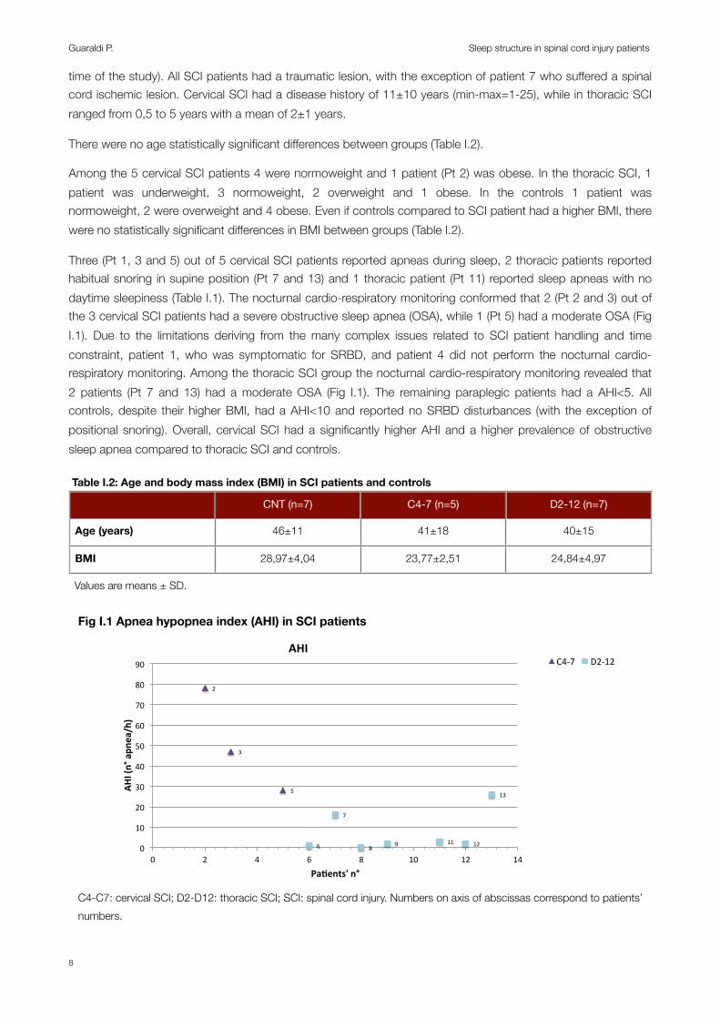
Three (Pt 1, 3 and 5) out of 5 cervical SCI patients reported apneas during sleep, 2 thoracic patients reported habitual snoring in supine position (Pt 7 and 13) and 1 thoracic patient (Pt 11) reported sleep apneas with no daytime sleepiness (Table I.1). The nocturnal cardio-respiratory monitoring conformed that 2 (Pt 2 and 3) out of the 3 cervical SCI patients had a severe obstructive sleep apnea (OSA), while 1 (Pt 5) had a moderate OSA (Fig I.1). Due to the limitations deriving from the many complex issues related to SCI patient handling and time constraint, patient 1, who was symptomatic for SRBD, and patient 4 did not perform the nocturnal cardio-respiratory monitoring. Among the thoracic SCI group the nocturnal cardio-respiratory monitoring revealed that 2 patients (Pt 7 and 13) had a moderate OSA (Fig I.1). The remaining paraplegic patients had a AHI<5. All controls, despite their higher BMI, had a AHI<10 and reported no SRBD disturbances (with the exception of positional snoring). Overall, cervical SCI had a significantly higher AHI and a higher prevalence of obstructive sleep apnea compared to thoracic SCI and controls.
Table I.2: Age and body mass index (BMI) in SCI patients and controls Table I.2: Age and body mass index (BMI) in SCI patients and controls Table I.2: Age and body mass index (BMI) in SCI patients and controls Table I.2: Age and body mass index (BMI) in SCI patients and controls
CNT (n=7) C4-7 (n=5) D2-12 (n=7)
Age (years) 46±11 41±18 40±15
BMI 28,97±4,04 23,77±2,51 24,84±4,97
Values are means ± SD.Values are means ± SD.Values are means ± SD.Values are means ± SD.
Fig I.1 Apnea hypopnea index (AHI) in SCI patients
C4-C7: cervical SCI; D2-D12: thoracic SCI; SCI: spinal cord injury. Numbers on axis of abscissas correspond to patients’ numbers.
2"
3"
5"
6"
7"
8" 9" 11" 12"
13"
0"
10"
20"
30"
40"
50"
60"
70"
80"
90"
0" 2" 4" 6" 8" 10" 12" 14"
AHI$(n°$apn
ea/h)$
Pa/ents'$n°$
AHI$
1"
2"
3"
4"
5"
6"
7" 8"
9" 11"
12"
12,5"
13" 14"
15" 16" 17"18"
19" 20"14,5"
15,5"
16,5"
17,5"
18,5"19,5"
20,5"
0"
10"
20"
30"
40"
50"
60"
70"
80"
0" 5" 10" 15" 20" 25"
Pa#ents')n°)
Arousal)Index)
C4.7" D2.12" CNT"
1" 2"
3"
4" 5" 6" 7" 8" 9"
11"
12"
12,5"
13" 14" 15" 16"
17"
18"19"
20"14,5" 15,5"16,5"
17,5"18,5"
19,5"20,5"0"
20"
40"
60"
80"
100"
120"
140"
160"
180"
0" 5" 10" 15" 20" 25"
Pa#ents')n°)
PLMS)Index)
C4.7" D2.12" CNT"
1"
2"
3"
4" 5"
6"
7"
8"
9"
11"
12"
12,5"
13"14"
15"
16"17"
18"19"
20"14,5" 15,5"
16,5" 17,5"18,5"
19,5"
20,5"
0,00"
10,00"
20,00"
30,00"
40,00"
50,00"
60,00"
70,00"
0" 5" 10" 15" 20" 25"
Pa#ents')n°)
Sleep)Fragmenta#on)Index)
C4.7" D2.12" CNT"
ArousalIndex
PLMSIndex
Sleep Fragm.
Index
Guaraldi P. Sleep structure in spinal cord injury patients
8

Sleep parametersSleep variables over the night-time (from 11:00 pm to 7:00 am) are shown in Table I.3, Fig I.2, Fig I.3 and Fig I.4.There were no significant differences in TST and SE between the three groups (Table I.3).
Cervical SCI compared to controls had a significantly lower amount of N1 NREM sleep (p=0,01), while no differences were observed between thoracic SCI and CNT (Table I.3). Cervical and thoracic SCI had a significantly higher amount of N2 NREM sleep compared to controls (p=0,008 and p=0,000, respectively) while the amount of stage 3 NREM seep was significantly reduced in tetraplegics and paraplegics compared to controls (p=0,039 and p=0,002, respectively) (Table I.3). Noteworthy, 1 cervical SCI patient (Pt 2) and two thoracic SCI (Pt 11 and 12) did not reach stage 3 NREM sleep during the night-time. REM sleep was equally present in all the 3 groups, but one cervical SCI (Pt 2) and one thoracic SCI (Pt 8) presented no REM sleep stages during the light-off period (Fig I.3). The analysis of the percentages of sleep phases out of TST (Fig I.2) confirmed a higher prevalence of N2 NREM sleep and a reduced percentage of SWS in cervical and thoracic SCI compared to controls.
Table I.3: sleep parameters over the night-time in SCI patients and controls Table I.3: sleep parameters over the night-time in SCI patients and controls Table I.3: sleep parameters over the night-time in SCI patients and controls Table I.3: sleep parameters over the night-time in SCI patients and controls CNT (n=14) C4-7 (n=5) D2-12 (n=8)
AHI <10* 51±25 7±10*
TST (min) 345±84 388±97 386±92
Sleep Efficiency (%) 72±17 74±17 71±16
N1 (min) 22±20* 5±5 9±5
N2 (min) 157±48*,§ 275±56 283±62
N3 (min) 89±30*,§ 49±28 34±42
REM (min) 80±26 59±39 59±34
N° of Arousals 56±34* 177±95 115±117
Arousal index 10±5 32±24 18±18
N° PLMS 29±48 176±393 210±312
PLMS index 5±7 25±56 38±60N° of arousals related to leg movement 9±13 22±43 34±61Arousal related to leg movement index 2±2 3±6 6±10
N° of phase shift 99±24 135±102 152±58
Phase shift index 18±6 25±23 25±14Values are means ± SD. =p<0,05 vs. cervical SCI. §=p<0,05 vs. thoracic SCI.Values are means ± SD. =p<0,05 vs. cervical SCI. §=p<0,05 vs. thoracic SCI.Values are means ± SD. =p<0,05 vs. cervical SCI. §=p<0,05 vs. thoracic SCI.Values are means ± SD. =p<0,05 vs. cervical SCI. §=p<0,05 vs. thoracic SCI.
The total number of arousal was significantly higher in cervical SCI compared to controls (p=0,022) but no statistically significant differences were observed between thoracic SCI and controls, nor in the arousal index between the 3 groups (Table I.3, Fig I.4).
The analysis wake-sleep fragmentation (based on the number of phase shift and phase shift index) over the night-time-period, due marked intra-subjects variability, disclosed no statistically significant differences between the 3 groups (Table I.3; Fig I.4).
Only 1 cervical SCI (Pt 3), 2 thoracic SCI (Pt 11 and 12) and 2 controls (subjects 17 and 19) had a PLMS Index > 5 (Fig I.5). The PLMS index was markedly elevated in Pt 3, 11 and 12 (PLMS-I=124; 66; 80, respectively) while it was 13 and 9,5 in subjects 17 and 19 (Fig I.5). Due to this intra-subjects variability, despite the mean value of PLMS and PLMS-I appeared higher in SCI subjects, no statistically significant differences were observed between the 3 groups (Table I.3; Fig I.4).
Section I
9

Fig I.2: Percentage of TST occupied by NREM and REM sleep phases in SCI patients and controls
Fig I.1 Percentage of sleep phases in SCI patients and controls
*
**
**
19
27
27
*=p<0,05C4-7: cervical SCI; CNT: controls; D2-12: thoracic SCI; SCI: spinal cord injury, N1: stage 1 NREM sleep; N2: stage 2 NREM sleep; N3: stage 3 NREM sleep; REM: REM sleep; SCI: spinal cord injury;TST: total sleep time.
Fig I.3: minutes of NREM and REM sleep phases in SCI patients and controls
N1
N2
N3
REM
1"
2" 3"
4"
5"6"
7"8"
9"11"
12"
12,5"13"
14"
15"16"
17"
18"
19"
20"14,5"
15,5"
16,5"
17,5"
18,5"
19,5"
20,5"
0"
10"
20"
30"
40"
50"
60"
70"
80"
90"
0" 5" 10" 15" 20" 25"
N1#(m
in)#
Subjects'#n°#
N1#
C4.7" D2.12" CNT"
1"2"
3"4"
5"
6"
7"
8"
9"
11"
12"
12,5"
13"
14"
15"
16"
17"
18" 19"
20"
14,5"
15,5"
16,5"
17,5"
18,5"
19,5" 20,5"
0"
50"
100"
150"
200"
250"
300"
350"
400"
450"
0" 5" 10" 15" 20" 25"
N2#(m
in)#
Subjects'#n°#
N2#
C4.7" D2.12" CNT"
1"
2"
3"
4" 5"
6"
7" 8"9"
11" 12"12,5"
13"
14"
15"
16"
17"
18"
19"
20"
14,5"
15,5"16,5"
17,5"
18,5"
19,5"
20,5"
0"
20"
40"
60"
80"
100"
120"
140"
160"
0" 5" 10" 15" 20" 25"
N3#(m
in)#
Subjects'#n°#
N334#
C4.7" D2.12" CNT"
1"
2"
3"
4"
5"
6"7"
8"
9"
11"
12"
12,5"
13"
14"
15"
16"17"
18"
19"
20"
14,5"
15,5"
16,5"
17,5" 18,5"
19,5"
20,5"
0"
20"
40"
60"
80"
100"
120"
140"
0" 5" 10" 15" 20" 25"
REM$(m
in)$
Subjects'$n°$
REM$
C4.7" D2.12" CNT"
4"
5" 6"
7"8" 9"
11"
12"
13"
14"
15"
16"
17"18"
19"
20"
21"
22"23"
24"
25"
27"
28"
29"
30"
31"
32"
33"
34"
35"
36"
37"
5" 10" 15" 20" 25" 30" 35" 40"Pa#ents')n°)
C4-7" D2-12" CNT" Numbers on axis of abscissas correspond to subjects’ numbers. C4-7: cervical SCI; CNT: controls; D2-12: thoracic SCI; SCI: spinal cord injury..
Guaraldi P. Sleep structure in spinal cord injury patients
10

Fig I.4: arousals, periodic limb movement and phase shifts in SCI patients and controls
Arousals
Periodic Limb Movement of Sleep
Fase shifts and sleep fragmentation
Fig I.2 Arousals, periodic limb movement and sleep fragmentation in SCI patients and controls
*
18
25
18
*=p<0,05. C4-7: cervical SCI; CNT: controls; D2-12: thoracic SCI; SCI: spinal cord injury, PLMS: periodic limb movement
during sleep.
Section I
11
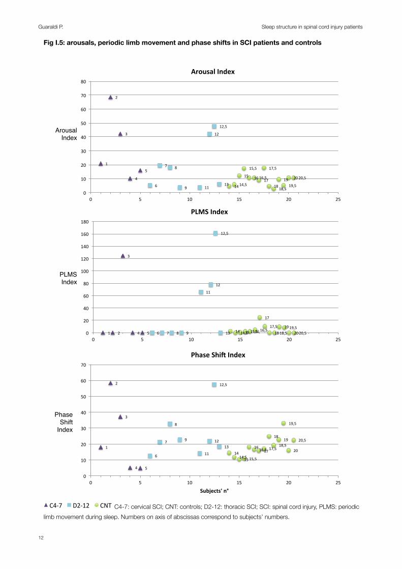
Fig I.5: arousals, periodic limb movement and phase shifts in SCI patients and controls
1"
2"
3"
4"
5"
6"
7" 8"
9" 11"
12"
12,5"
13" 14"
15" 16" 17"18"
19" 20"14,5"
15,5"
16,5"
17,5"
18,5"19,5"
20,5"
0"
10"
20"
30"
40"
50"
60"
70"
80"
0" 5" 10" 15" 20" 25"
Pa#ents')n°)
Arousal)Index)
C4.7" D2.12" CNT"
1" 2"
3"
4" 5" 6" 7" 8" 9"
11"
12"
12,5"
13" 14" 15" 16"
17"
18"19"
20"14,5" 15,5"16,5"
17,5"18,5"
19,5"20,5"0"
20"
40"
60"
80"
100"
120"
140"
160"
180"
0" 5" 10" 15" 20" 25"
Pa#ents')n°)
PLMS)Index)
C4.7" D2.12" CNT"
ArousalIndex
PLMSIndex
Phase Shift
Index
1"
2"
3"
4" 5"
6"
7"
8"
9"
11"
12"
12,5"
13"14"
15"
16"17"
18"19"
20"14,5" 15,5"
16,5" 17,5"18,5"
19,5"
20,5"
0"
10"
20"
30"
40"
50"
60"
70"
0" 5" 10" 15" 20" 25"
Subjects'*n°*
Phase*Shi1*Index*
C4.7" D2.12" CNT"
4"
5" 6"
7"8" 9"
11"
12"
13"
14"
15"
16"
17"18"
19"
20"
21"
22"23"
24"
25"
27"
28"
29"
30"
31"
32"
33"
34"
35"
36"
37"
5" 10" 15" 20" 25" 30" 35" 40"Pa#ents')n°)
C4-7" D2-12" CNT" C4-7: cervical SCI; CNT: controls; D2-12: thoracic SCI; SCI: spinal cord injury, PLMS: periodic limb movement during sleep. Numbers on axis of abscissas correspond to subjects’ numbers.
Guaraldi P. Sleep structure in spinal cord injury patients
12
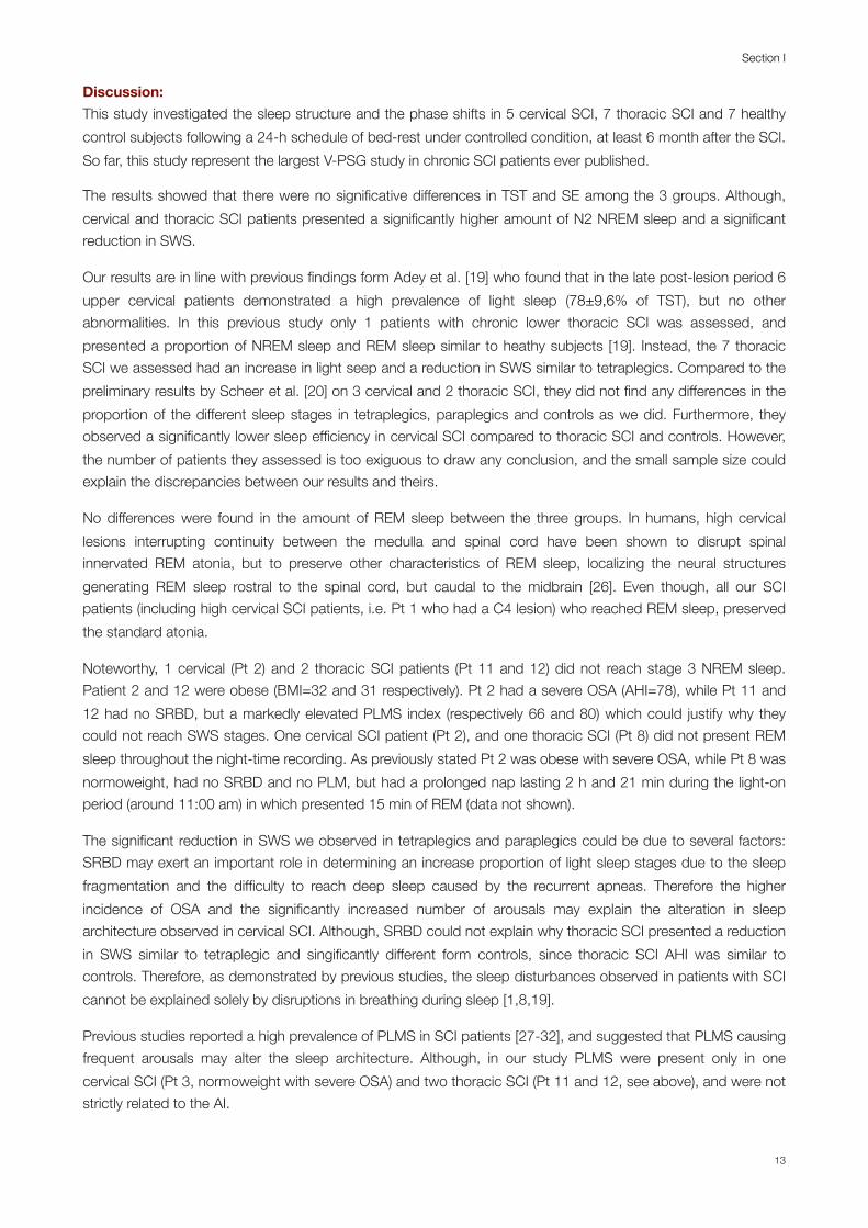
Discussion: This study investigated the sleep structure and the phase shifts in 5 cervical SCI, 7 thoracic SCI and 7 healthy control subjects following a 24-h schedule of bed-rest under controlled condition, at least 6 month after the SCI. So far, this study represent the largest V-PSG study in chronic SCI patients ever published.
The results showed that there were no significative differences in TST and SE among the 3 groups. Although, cervical and thoracic SCI patients presented a significantly higher amount of N2 NREM sleep and a significant reduction in SWS.
Our results are in line with previous findings form Adey et al. [19] who found that in the late post-lesion period 6 upper cervical patients demonstrated a high prevalence of light sleep (78±9,6% of TST), but no other abnormalities. In this previous study only 1 patients with chronic lower thoracic SCI was assessed, and presented a proportion of NREM sleep and REM sleep similar to heathy subjects [19]. Instead, the 7 thoracic SCI we assessed had an increase in light seep and a reduction in SWS similar to tetraplegics. Compared to the preliminary results by Scheer et al. [20] on 3 cervical and 2 thoracic SCI, they did not find any differences in the proportion of the different sleep stages in tetraplegics, paraplegics and controls as we did. Furthermore, they observed a significantly lower sleep efficiency in cervical SCI compared to thoracic SCI and controls. However, the number of patients they assessed is too exiguous to draw any conclusion, and the small sample size could explain the discrepancies between our results and theirs.
No differences were found in the amount of REM sleep between the three groups. In humans, high cervical lesions interrupting continuity between the medulla and spinal cord have been shown to disrupt spinal innervated REM atonia, but to preserve other characteristics of REM sleep, localizing the neural structures generating REM sleep rostral to the spinal cord, but caudal to the midbrain [26]. Even though, all our SCI patients (including high cervical SCI patients, i.e. Pt 1 who had a C4 lesion) who reached REM sleep, preserved the standard atonia.
Noteworthy, 1 cervical (Pt 2) and 2 thoracic SCI patients (Pt 11 and 12) did not reach stage 3 NREM sleep. Patient 2 and 12 were obese (BMI=32 and 31 respectively). Pt 2 had a severe OSA (AHI=78), while Pt 11 and 12 had no SRBD, but a markedly elevated PLMS index (respectively 66 and 80) which could justify why they could not reach SWS stages. One cervical SCI patient (Pt 2), and one thoracic SCI (Pt 8) did not present REM sleep throughout the night-time recording. As previously stated Pt 2 was obese with severe OSA, while Pt 8 was normoweight, had no SRBD and no PLM, but had a prolonged nap lasting 2 h and 21 min during the light-on period (around 11:00 am) in which presented 15 min of REM (data not shown).
The significant reduction in SWS we observed in tetraplegics and paraplegics could be due to several factors: SRBD may exert an important role in determining an increase proportion of light sleep stages due to the sleep fragmentation and the difficulty to reach deep sleep caused by the recurrent apneas. Therefore the higher incidence of OSA and the significantly increased number of arousals may explain the alteration in sleep architecture observed in cervical SCI. Although, SRBD could not explain why thoracic SCI presented a reduction in SWS similar to tetraplegic and singificantly different form controls, since thoracic SCI AHI was similar to controls. Therefore, as demonstrated by previous studies, the sleep disturbances observed in patients with SCI cannot be explained solely by disruptions in breathing during sleep [1,8,19].
Previous studies reported a high prevalence of PLMS in SCI patients [27-32], and suggested that PLMS causing frequent arousals may alter the sleep architecture. Although, in our study PLMS were present only in one cervical SCI (Pt 3, normoweight with severe OSA) and two thoracic SCI (Pt 11 and 12, see above), and were not strictly related to the AI.
Section I
13

A further possible explanation of the observed alteration in the sleep pattern in SCI patients involves melatonin [20]. Since the neural pathway for the endogenous production of melatonin passes through the cervical spinal cord [33,34] and patients with cervical SCI have an absence of the normal endogenous melatonin secretion at night [20,33] it was suggested that the alteration in night time melatonin secretion could chronically reduced sleep quality in cervical SCI patients [20]. Even if we believe that the disturbance in melatonin secretion may play a role in determining the disturbed sleep in cervical SCI patients, this hypothesis could not explain the sleep alteration we observed in paraplegics, whose melatonin secretion is supposed to be unaffected.
Therefore, other possible mechanisms may be involved. These mechanisms could be either the direct consequence of the injury or due to concomitant conditions or non specific factors.
Interestingly, thermoregulatory and hypnic mechanisms are thought to be linked [15]. Dependent on the site and extent of the lesion, individuals with a spinal cord injury (SCI) depend on the region above the lesion for thermoregulation [35,36] and may present a thermal dysfunction [35,37,38]. Therefore, it is tempting to speculate that the impaired thermoregulatory function in SCI patient may explain the sleep alteration we observed in cervical and thoracic SCI (see Section III). Although, the reverse could be also possible, being the reduced SWS to interfere with body temperature changes.
Further studies will be necessary to shed light on the possible pathogenetic mechanism of the reduction of SWS in cervical and thoracic SCI. Ideally, studies should be designed to investigate the effects of various interventions (e.g. treatment of OSA, PLMS or correction of thermal dysfunction) on normalization of sleep pattern in individuals with SCI.
LimitationsThe main limitation of our study is that during V-PSG, due to the limitations deriving from the many complex issues related to SCI patients handling, we could record only a single respiratory channel and no SO2. Therefore the occurrence of apneas during the V-PSG could not be assessed. Although, all subjects were questioned about SRBD disturbances during history taking and underwent a nocturnal cardio-respiratory monitoring (excluding Pt 1 and 4 and all controls). Furthermore, for ethical reasons we could not discontinue medications such as benzodiazepines and baclofen, which have a depressant effect on the central nervous system, increase the frequency and severity of SDB [39-41] but reduce PLMS [42].
Conclusions:This study represent the largest V-PSG study in chronic SCI patients ever published and demonstrate a compromised sleep in patients with chronic cervical and thoracic SCI, characterized by an augmented proportion N2 NREM sleep and reduced SWS compared to controls. No statistical differences in any sleep parameter were found between cervical and thoracic SCI patients. While the sleep disturbances in cervical SCI could be related to a significantly higher AHI, and possibly to an alteration in melatonin secretion, the pathogenesis of the sleep disorder in thoracic SCI is jet to be determined.
Guaraldi P. Sleep structure in spinal cord injury patients
14

Patient 10Several case reports have described PLMS in relation to various spinal cord lesion, including traumatic SCI [27-32,43]. The temporal relationship between the lesion occurrence and the PLMS presentation, and the reduction of PLMS in relation to the amelioration of the clinical conditions suggested a causal relationship between the two events. Furthermore, occurrence of PLMS in SCI led to formulation of pathogenetic mechanisms pointing to a possible participation of the Central Pattern Generators (CPG) and the flexor afferent reflex in the physiopathology of PLMS as discussed by Paulus and Schomburg [43,44].
Interesting we had the chance to assess a patient (Pt 10) with a D8 Brown-Séquard syndrome. He was male, 47 years old, with a BMI=22,99. He was on Baclofen 50 mg/die and oxibutinin 30 mg/die.
Patient 10 was not included in the study (who was limited to complete Frankel A SCI patients) but underwent V-PSG, which demonstrated the occurrence of spontaneous leg movement during sleep on both legs, which did not fulfill the criteria for PLMS.
We believe that assessing by means of formal V-PSG further patients with Brown-Séquard syndromes may shed light on the intriguing physiopathology of PLMS.
Section I
15
Bibliography:1. Biering-Sorensen F, Biering-Sorensen M (2001) Sleep disturbances in the spinal cord injured: an epidemiological
questionnaire investigation, including a normal population. Spinal Cord 39 (10):505-513.
2. Norrbrink Budh C, Hultling C, Lundeberg T (2005) Quality of sleep in individuals with spinal cord injury: a comparison between patients with and without pain. Spinal Cord 43 (2):85-95.
3. Jensen MP, Hirsh AT, Molton IR, Bamer AM (2009) Sleep problems in individuals with spinal cord injury: frequency and age effects. Rehabil Psychol 54 (3):323-331.
4. Biering-Sorensen F, Jennum P, Laub M (2009) Sleep disordered breathing following spinal cord injury. Respir Physiol Neurobiol 169 (2):165-170.
5. Burns SP, Little JW, Hussey JD, Lyman P, Lakshminarayanan S (2000) Sleep apnea syndrome in chronic spinal cord injury: associated factors and treatment. Arch Phys Med Rehabil 81 (10):1334-1339.
6. Hyyppa MT, Kronholm E (1989) Quality of sleep and chronic illnesses. Journal of clinical epidemiology 42 (7):633-638.
7. Young T, Palta M, Dempsey J, Skatrud J, Weber S, Badr S (1993) The occurrence of sleep-disordered breathing among middle-aged adults. The New England journal of medicine 328 (17):1230-1235.
8. McEvoy RD, Mykytyn I, Sajkov D, Flavell H, Marshall R, Antic R, Thornton AT (1995) Sleep apnoea in patients with quadriplegia. Thorax 50 (6):613-619.
9. Short DJ, Stradling JR, Williams SJ (1992) Prevalence of sleep apnoea in patients over 40 years of age with spinal cord lesions. J Neurol Neurosurg Psychiatry 55 (11):1032-1036.
10. Klefbeck B, Sternhag M, Weinberg J, Levi R, Hultling C, Borg J (1998) Obstructive sleep apneas in relation to severity of cervical spinal cord injury. Spinal Cord 36 (9):621-628.
11. Cahan C, Gothe B, Decker MJ, Arnold JL, Strohl KP (1993) Arterial oxygen saturation over time and sleep studies in quadriplegic patients. Paraplegia 31 (3):172-179.
12. Flavell H, Marshall R, Thornton AT, Clements PL, Antic R, McEvoy RD (1992) Hypoxia episodes during sleep in high tetraplegia. Arch Phys Med Rehabil 73 (7):623-627.
13. Tran K, Hukins C, Geraghty T, Eckert B, Fraser L (2009) Sleep-disordered breathing in spinal cord-injured patients: a short-term longitudinal study. Respirology 15 (2):272-276.
14. Ayas NT, Epstein LJ, Lieberman SL, Tun CG, Larkin EK, Brown R, Garshick E (2001) Predictors of loud snoring in persons with spinal cord injury. J Spinal Cord Med 24 (1):30-34.
15. Stockhammer E, Tobon A, Michel F, Eser P, Scheuler W, Bauer W, Baumberger M, Muller W, Kakebeeke TH, Knecht H, Zach GA (2002) Characteristics of sleep apnea syndrome in tetraplegic patients. Spinal Cord 40 (6):286-294.
16. Bonekat HW, Andersen G, Squires J (1990) Obstructive disordered breathing during sleep in patients with spinal cord injury. Paraplegia 28 (6):392-398.
17. Atkinson G, Davenne D (2007) Relationships between sleep, physical activity and human health. Physiology & behavior 90 (2-3):229-235.
18. Star AM, Osterman AL (1988) Sleep apnea syndrome after spinal cord injury. Report of a case and literature review. Spine (Phila Pa 1976) 13 (1):116-117.
19. Adey WR, Bors E, Porter RW (1968) EEG sleep patterns after high cervical lesions in man. Arch Neurol 19 (4):377-383.
20. Scheer FA, Zeitzer JM, Ayas NT, Brown R, Czeisler CA, Shea SA (2006) Reduced sleep efficiency in cervical spinal cord injury; association with abolished night time melatonin secretion. Spinal Cord 44 (2):78-81.
Guaraldi P. Sleep structure in spinal cord injury patients
16

21. Williams JR (2008) The Declaration of Helsinki and public health. Bulletin of the World Health Organization 86 (8):650-652.
22. Sleep-related breathing disorders in adults: recommendations for syndrome definition and measurement techniques in clinical research. The Report of an American Academy of Sleep Medicine Task Force (1999). Sleep 22 (5):667-689.
23. Rechtschaffen A, Kales A (1968) A manual of standardized terminology, techniques and scoring system for sleep stages of human subjects. . National Institutes of Health publication,, vol no 204. U. S. National Institute of Neurological Diseases and Blindness, Neurological Information Network, Bethesda, MD.
24. EEG arousals: scoring rules and examples: a preliminary report from the Sleep Disorders Atlas Task Force of the American Sleep Disorders Association (1992). Sleep 15 (2):173-184.
25. Iber C, Ancoli-Israel S, Chesson A, Quan S (eds) (2007) The AASM manual for the scoring of sleep and associated events: rules terminology and technical specifications. 1st edn. American Academy of Sleep Medicine, Westchester, IL
26. Smith HR (2008) The basic neurology of sleep. In: Smith HR, Comella C, Högl B (eds) Sleep medicine. Cambridge University Press, Cambridge ; New York, pp 1-8.
27. Yokota T, Hirose K, Tanabe H, Tsukagoshi H (1991) Sleep-related periodic leg movements (nocturnal myoclonus) due to spinal cord lesion. J Neurol Sci 104 (1):13-18.
28. Dickel MJ, Renfrow SD, Moore PT, Berry RB (1994) Rapid eye movement sleep periodic leg movements in patients with spinal cord injury. Sleep 17 (8):733-738.
29. Lee MS, Choi YC, Lee SH, Lee SB (1996) Sleep-related periodic leg movements associated with spinal cord lesions. Mov Disord 11 (6):719-722.
30. de Mello MT, Lauro FA, Silva AC, Tufik S (1996) Incidence of periodic leg movements and of the restless legs syndrome during sleep following acute physical activity in spinal cord injury subjects. Spinal Cord 34 (5):294-296.
31. Brown LK, Heffner JE, Obbens EA (2000) Transverse myelitis associated with restless legs syndrome and periodic movements of sleep responsive to an oral dopaminergic agent but not to intrathecal baclofen. Sleep 23 (5):591-594.
32. Telles SC, Alves RC, Chadi G (2011) Periodic limb movements during sleep and restless legs syndrome in patients with ASIA A spinal cord injury. J Neurol Sci 303 (1-2):119-123.
33. Zeitzer JM, Ayas NT, Shea SA, Brown R, Czeisler CA (2000) Absence of detectable melatonin and preservation of cortisol and thyrotropin rhythms in tetraplegia. J Clin Endocrinol Metab 85 (6):2189-2196.
34. Moore RY (1996) Neural control of the pineal gland. Behavioural brain research 73 (1-2):125-130.
35. Mathias CJ, Frankel HL (2002) Autonomic disturbances in spinal cord lesions. In: Mathias CJ, Bannister R (eds) Autonomic failure : a textbook of clinical disorders of the autonomic nervous system. 4th edn. Oxford University Press, Oxford ; New York, pp 494–513.
36. Guttmann L, Silver J, Wyndham CH (1958) Thermoregulation in spinal man. J Physiol 142 (3):406-419.
37. Sawka MN, Latzka WA, Pandolf KB (1989) Temperature regulation during upper body exercise: able-bodied and spinal cord injured. Medicine and science in sports and exercise 21 (5 Suppl):S132-140.
38. Boot CR, Binkhorst RA, Hopman MT (2006) Body temperature responses in spinal cord injured individuals during exercise in the cold and heat. International journal of sports medicine 27 (8):599-604.
Section I
17

39. Khan S, Plummer M, Martinez-Arizala A, Banovac K (2007) Hypothermia in patients with chronic spinal cord injury. J Spinal Cord Med 30 (1):27-30.
40. Dolly FR, Block AJ (1982) Effect of flurazepam on sleep-disordered breathing and nocturnal oxygen desaturation in asymptomatic subjects. The American journal of medicine 73 (2):239-243.
41. Taasan VC, Block AJ, Boysen PG, Wynne JW (1981) Alcohol increases sleep apnea and oxygen desaturation in asymptomatic men. The American journal of medicine 71 (2):240-245.
42. Bensmail D, Quera Salva MA, Roche N, Benyahia S, Bohic M, Denys P, Bussel B, Lofaso F (2006) Effect of intrathecal baclofen on sleep and respiratory function in patients with spasticity. Neurology 67 (8):1432-1436.
43. Bensmail D, Marquer A, Roche N, Godard AL, Lofaso F, Quera-Salva MA (2012) Pilot study assessing the impact of intrathecal baclofen administration mode on sleep-related respiratory parameters. Arch Phys Med Rehabil 93 (1):96-99.
44. Vetrugno R, Provini F, Plazzi G, Cortelli P, Montagna P (2005) Propriospinal myoclonus: a motor phenomenon found in restless legs syndrome different from periodic limb movements during sleep. Mov Disord 20 (10):1323-1329.
45. Paulus W, Schomburg ED (2006) Dopamine and the spinal cord in restless legs syndrome: does spinal cord physiology reveal a basis for augmentation? Sleep medicine reviews 10 (3):185-196.
Guaraldi P. Sleep structure in spinal cord injury patients
18
Section II
ANALYSIS OF CARDIOVASCULAR PARAMETERS
‘‘Spinal cord injury is a ferocious assault on the body that leaves havoc in its wake. Paralysis is certainly part of its legacy, but there are other equally devastating consequences including autonomic dysfunction: compromised cardiovascular, bowel, bladder, and sexual function. Treatments and cures for these losses would greatly improve the quality of life for all of us living with spinal cord injury. I am hopeful that the multi-faceted and collaborative approach to spinal cord repair ... means that there will be effective therapies for autonomic dysfunction in the not too distant future.’’
Christopher Reeve, September 30, 2004.


Section II.1: Autonomic control of the cardiovascular system during wakefulness
Background:Normal functioning of the autonomic nervous system is critically dependent on integrity of the spinal cord, as the entire sympathetic outflow (T1-L2/3) and the sacral parasympathetic outflow travel and synapse within the spinal cord, before supplying various target organs (Fig II.1.1).
Fig II.1.1: schematic outline of the major autonomic pathways controlling the circulation From Mathias C and Low D (2012) Autonomic Disturbances in Spinal Cord Injuries. In: Robertson D (ed) Primer on the autonomic nervous system. pp 505-509.
Schematic outline of the major autonomic pathways controlling the circulation. The major afferent input into the central nervous system is through the glossopharyngeal (CR,9) and vagus (CR,10) nerves by activation of baroreceptors in the carotid sinus and aortic arch. Chemoreceptors and low pressure receptors also influence the efferent outflow. The latter consists of the cranial parasympathetic (PS) outflow to the heart via the vagus nerves, and the sympathetic outflow from the thoracic and upper lumbar segments of the spinal cord. Activation of visceral, skin, and muscle receptors, in addition to cerebral stimulation influences the efferent outflow. In high spinal cord lesions, therefore, the input from chemoreceptors and baroreceptors is preserved along with the vagal efferent outflow, but there is no connection between the brain and the rest of the sympathetic outflow. The spinal sympathetic outflow may be activated through a range of afferents (visual, skin, muscle). This occurs through isolated spinal cord reflexes, not controlled by cerebral pathways, as seen normally.
X. AUTONOMIC DISORDERS
104. AUTONOMIC DISTURBANCES IN SPINAL CORD INJURIES506
0.6 mg IV
20 min post atropine(0.6 mg IV)
6 hr post atropine(0.6 mg IV)
FIGURE 104.2 (a) The effect of disconnecting the respirator (as required for aspirating the airways) on the blood pressure (BP) and heart rate (HR) of a recently injured tetraplegic patient (C4/5 lesion) in spinal shock, 6 hours after the last dose of intravenous atropine. Sinus brady-cardia and cardiac arrest, also observed in the electrocardiograph, were reversed by reconnection, intravenous atropine and external cardiac mas-sage (from Frankel et al. Lancet 1975;ii:1183–1185). (b) The effect of tra-cheal suction, 20 minutes after atropine in the same patient. Disconnection from the respirator and tracheal suction did not lower either heart rate or blood pressure. (from Mathias Eur J Int Care Med 1976;2:147–156).
FIGURE 104.3 (a) Blood pressure (BP) and heart rate (HR) in a tet-raplegic patient before and after head-up tilt, in the early stages of reha-bilitation, where there were few muscle spasms and minimal autonomic dysreflexia (from Mathias and Frankel Handbook of Clinical Neurology 1992;17:435–56). (b) Blood pressure (BP) and heart rate (HR) in a chronic tetraplegic patient before, during and after head-up tilt to 45°. Blood pressure promptly falls but with partial recovery, which in this case is linked to skeletal muscle spasms (S) inducing spinal sympathetic activ-ity. Some of the later oscillations may be due to the rise in plasma renin, which was measured where there were interruptions in the intra-arterial record. In the later phases of head-up tilt, skeletal muscle spasm occur more frequently and further elevate the blood pressure. On return to the horizontal, blood pressure rises rapidly above the previous level and then returns slowly to horizontal levels Heart rate usually moves in the opposite direction, except during muscle spasms when there is an initial increase. (from Mathias and Frankel Ann Rev Physiol 1999;50:577–592).
FIGURE 104.1 Schematic outline of the major autonomic pathways control-ling the circulation. The major afferent input into the central nervous system is through the glossopharyngeal (CR,9) and vagus (CR,10) nerves by activation of baroreceptors in the carotid sinus and aortic arch. Chemoreceptors and low pressure receptors also influence the efferent outflow. The latter consists of the cranial parasympathetic (PS) outflow to the heart via the vagus nerves, and the sympathetic outflow from the thoracic and upper lumbar segments of the spinal cord. Activation of visceral, skin, and muscle receptors, in addition to cerebral stimulation influences the efferent outflow. In high spinal cord lesions, therefore, the input from chemoreceptors and baroreceptors is preserved along with the vagal efferent outflow, but there is no connection between the brain and the rest of the sympathetic outflow. The spinal sympathetic outflow may be activated through a range of afferents (visual, skin, muscle). This occurs through isolated spinal cord reflexes, not controlled by cerebral pathways, as seen normally.
Therefore spinal cord injury (SCI) results not only in paralysis; but it is also associated with a range of autonomic dysregulation that can interfere with cardiovascular, bladder, bowel, temperature, and sexual function. The entity of the autonomic dysfunction is related to the level and severity of injury to descending autonomic (sympathetic) pathways [1-3]. For the sake of the comprehension, we can consider three types of SCI lesions that offer typical autonomic reactions (tetraplegics, paraplegics at T6 and paraplegics at T10) (Fig II.1.2):
1. In cervical transection, the entire part of the sympathetic outflow, together with the sacral parasympathetic outflow, is totally disconnected from cerebral control (Fig II.1.2 a), causing an autonomic malfunction which may affect the cardiovascular, thermoregulatory, sudomotor, gastrointestinal, urinary and reproductive systems [4]. Conversely, all somatic and visceral afferents coming from below the lesion (starting from T1–T2 dermatomes) can reflexively activate the whole sympathetic column. Postural challenges cause an immediate fall in both systolic and diastolic blood pressure (SBP, DBP), due to the lack of rise in total peripheral resistance (TPR) [5,6], which is inversely related to a rapid rise in heart rate (HR), due to withdrawal of vagal tone in response to unloading of baroreceptor afferents [5-8]. Similar cardiovascular findings are observed during a static maximum inspiratory breath-hold [9] or the Valsalva manoeuvre (VM) [10]. After the VM, the blood pressure (BP) overshoot relative to baseline is absent [10]. Pressor stimuli that either originate in, or are modulated by, the brain (either cerebral or cervical afferents) will have no effect [11]. Pressor stimuli below the lesion will activate sympathetic reflex along the isolated spinal cord that can result in autonomic dysreflexia (AD).
19
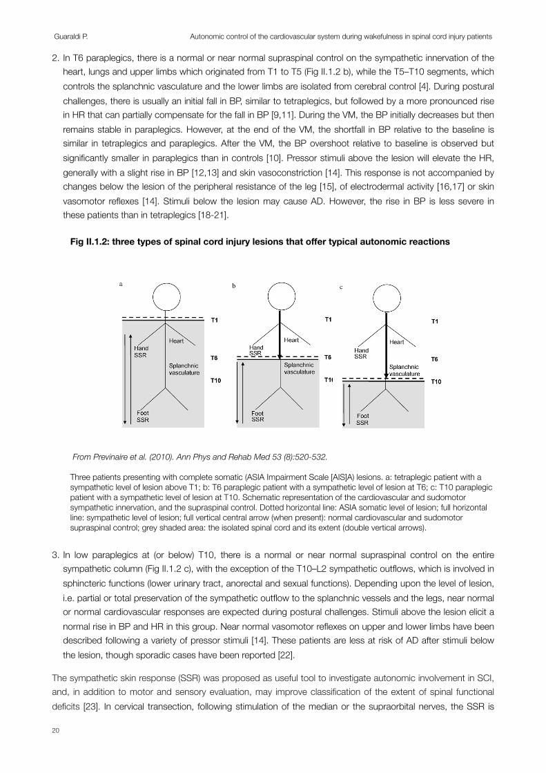
2. In T6 paraplegics, there is a normal or near normal supraspinal control on the sympathetic innervation of the heart, lungs and upper limbs which originated from T1 to T5 (Fig II.1.2 b), while the T5–T10 segments, which controls the splanchnic vasculature and the lower limbs are isolated from cerebral control [4]. During postural challenges, there is usually an initial fall in BP, similar to tetraplegics, but followed by a more pronounced rise in HR that can partially compensate for the fall in BP [9,11]. During the VM, the BP initially decreases but then remains stable in paraplegics. However, at the end of the VM, the shortfall in BP relative to the baseline is similar in tetraplegics and paraplegics. After the VM, the BP overshoot relative to baseline is observed but significantly smaller in paraplegics than in controls [10]. Pressor stimuli above the lesion will elevate the HR, generally with a slight rise in BP [12,13] and skin vasoconstriction [14]. This response is not accompanied by changes below the lesion of the peripheral resistance of the leg [15], of electrodermal activity [16,17] or skin vasomotor reflexes [14]. Stimuli below the lesion may cause AD. However, the rise in BP is less severe in these patients than in tetraplegics [18-21].
3. In low paraplegics at (or below) T10, there is a normal or near normal supraspinal control on the entire sympathetic column (Fig II.1.2 c), with the exception of the T10–L2 sympathetic outflows, which is involved in sphincteric functions (lower urinary tract, anorectal and sexual functions). Depending upon the level of lesion, i.e. partial or total preservation of the sympathetic outflow to the splanchnic vessels and the legs, near normal or normal cardiovascular responses are expected during postural challenges. Stimuli above the lesion elicit a normal rise in BP and HR in this group. Near normal vasomotor reflexes on upper and lower limbs have been described following a variety of pressor stimuli [14]. These patients are less at risk of AD after stimuli below the lesion, though sporadic cases have been reported [22].
The sympathetic skin response (SSR) was proposed as useful tool to investigate autonomic involvement in SCI, and, in addition to motor and sensory evaluation, may improve classification of the extent of spinal functional deficits [23]. In cervical transection, following stimulation of the median or the supraorbital nerves, the SSR is
Fig II.1.2: three types of spinal cord injury lesions that offer typical autonomic reactions
From Previnaire et al. (2010). Ann Phys and Rehab Med 53 (8):520-532.
Three patients presenting with complete somatic (ASIA Impairment Scale [AIS]A) lesions. a: tetraplegic patient with a sympathetic level of lesion above T1; b: T6 paraplegic patient with a sympathetic level of lesion at T6; c: T10 paraplegic patient with a sympathetic level of lesion at T10. Schematic representation of the cardiovascular and sudomotor sympathetic innervation, and the supraspinal control. Dotted horizontal line: ASIA somatic level of lesion; full horizontal line: sympathetic level of lesion; full vertical central arrow (when present): normal cardiovascular and sudomotor supraspinal control; grey shaded area: the isolated spinal cord and its extent (double vertical arrows).
1.1.4. Bedside assessmentsBedside assessments have, so far, little clinical application,
except for the skin axon reflex vasodilatation (SkARV) test[46]. The spoon test for assessing sweating [4], the observationof piloerection [54], and the pupillary reflex dilation in responseto a noxious stimulus of the upper thoracic dermatomes [2] mayneed reappraising.
1.2. Methods
PubMed was searched for articles related to cardiovascularand sudomotor tests that have been used in SCI patients. Thesearch strategies combined terms for ‘‘spinal cord injury’’ and
‘‘autonomic testing’’, respectively ‘‘spinal cord injury ORtetraplegia OR quadriplegia OR paraplegia’’ and ‘‘cold pressortest OR Valsalva manoeuvre OR postural challenge ORrespiratory challenge OR tilt test OR sympathetic skinresponses OR sweating’’. The main author screened all titlesand abstracts identified and acquired the full-text publication ofall potentially eligible studies. The bibliographies of allretrieved studies were screened for additional relevant articles.
To be eligible, studies had to:
! provide detailed descriptions of the neurological status of theindividuals, in particular the level and severity of the lesion;
[(Fig._1)TD$FIG]
Fig. 1. Three patients presenting with complete somatic (ASIA Impairment Scale [AIS] A) lesions. a: tetraplegic patient with a sympathetic level of lesion above T1;b: T6 paraplegic patient with a sympathetic level of lesion at T6; c: T10 paraplegic patient with a sympathetic level of lesion at T10. Schematic representation of the
cardiovascular and sudomotor sympathetic innervation, and the supraspinal control. Dotted horizontal line: ASIA somatic level of lesion; full horizontal line:
sympathetic level of lesion; full vertical central arrow (when present): normal cardiovascular and sudomotor supraspinal control; grey shaded area: the isolated spinalcord and its extent (double vertical arrows).
J.G. Previnaire et al. / Annals of Physical and Rehabilitation Medicine 53 (2010) 520–532522
Guaraldi P. Autonomic control of the cardiovascular system during wakefulness in spinal cord injury patients
20

absent in the hands and feet [1,24-29]. In T6 paraplegics following stimulation of the median nerve, the SSR is absent in the feet (see discussion below). The palmar SSR can be elicited if the T4–T5 spinal cord segment is located above the lesion level [1,24-29]. In low paraplegics on median nerve stimulation, plantar SSR are rarely found in T10–T12 patients, but more consistently found with lesion below T12 [1,24,26,27,29].
Beside the severe impact on quality of life, cardiovascular dysfunction is the leading cause of morbidity and mortality in SCI individuals [30,31]. This most commonly manifests as neurogenic shock (in acute SCI) [3]; supine hypotension [3]; orthostatic hypotension (OH: marked decreases in blood pressure in the upright position) [1,32]; AD (profound and life-threatening blood pressure increases following afferent stimulation below the lesion) [3]; ECG abnormalities and arrhythmia [33], particularly during AD [25,34]; and abnormal cardiovascular responses to exercise [26]. Therefore laboratory evaluations to detect the presence of autonomic dysfunction and quantitate its severity are recommended.
Although assessment of autonomic function through standardized battery of clinical tests [35] is always preferable, it require specialized equipment, may be invasive, and, thus, may be difficult to perform routinely in clinical practice. The assessment of heart rate variability (HRV) instead, is a simple and reliable measurement of sympathetic and parasympathetic cardiac control. It is noninvasive, requires relatively short (10–15 minutes) recording periods in a rested state with no positional changes, and can be performed with easily accessible devices. Furthermore it is highly reproducible in persons with SCI [36].
HRV may be evaluated by a number of methods. Assessment of HRV in the frequency domain identifies three main spectral components: very low frequency (VLF), low frequency (LF), and high frequency (HF) components. High-frequency (HF, ~0.25 Hz) component of HRV represents cardiac vagal control [37-39]. LF HRV is more controversial but is generally accepted to be due to oscillations in vagal outflow generated through the baroreflex and driven by sympathetically induced LF blood pressure variability [37,40-42]. Therefore LF power is a measure of both sympathetic (mainly) and parasympathetic activities. Consequently LF/HF ratio is a measure of sympathovagal balance. Total Power (TP) is the sum of the three components and is considered a global measure of HRV.
Previous studies on HRV in SCI demonstrated that LF component was missing or significantly decreased in cervical SCI as compared to controls and patients with thoracic SCI in both supine and upright positions. Normalized HF power was significantly less in patients with thoracic SCI group as compared to cervical SCI in the supine position and tended to be greater in the cervical SCI group than in the thoracic SCI and control groups in the upright position [43]. LF/HF power ratio in cervical SCI compared to control and thoracic SCI groups was found to be either significantly larger [44] or lower [43] or even normal [7].
Objective: Aim of our study was to assess the autonomic control of cardiovascular reflexes and HRV during supine rest (baseline) and head-up tilt test (HUTT) in chronic SCI patients.
Materials and Methods:
SubjectsFive cervical SCI with tetraplegia (C4-7), 7 thoracic SCI with paraplegia (D2-12) (Table II.1.1) and 26 age and sex matched control subjects (CNT) underwent assessment of cardiovascular reflexes and HRV while supine and tilted. Prior to data collection, all subjects underwent clinical examination. None of the subjects reported
Section II.1
21
symptoms or showed signs of diabetes, cardiorespiratory disease or of other pathological conditions that might affect autonomic cardiovascular control. The investigation conformed with the principles outlined in the Declaration of Helsinki [45]. The protocol was approved by the Institutional Review Board of the University of Bologna, and all participants provided informed consent.
Study protocolAll subjects were assessed in the morning, between 8 am and 12 am, in a quiet, temperature controlled clinical investigation room (23±1°C). Before the tests participants were allowed to have a light breakfast but had to abstain from smoking or drinking alcohol or caffeinated beverages from the night before the study. For ethical reasons, patients were allowed to use their usual medication, but all of them delayed their morning dose until after the experiment. Only one patient (Pt 12), who was on intrathecal baclofen and morphine could not postpone his treatment. Patients and controls were asked not to sleep or talk during the study.
All participants underwent HUTT, deep breathing (DB) and cold face (CF) under controlled laboratory conditions. Paraplegics and controls performed also VM and isometric exercise (IE) test.
SBP and DBP (Portapres model 2, TNO-TPD Biomedical Instrumentation), heart rate (Grass 7P511 and Light Work Station for digital RR quantification), oronasal and abdominal breathing (Grass DC preamplifier 7P1) were monitored continuously. Finger pressure detected by Portapres can be measured between 10 and 300 mmHg and the internal accuracy of the instrument is 1.5% of full scale.
After 30 min of supine rest, the HUTT (10 min at 65°), VM (40 mmHg for 15 sec), DB (6 breaths/min), CF test (60 sec of application of cold compresses at 0-1°C to the forehead), and IE (30% of maximal effort for 5 min) were performed using standard procedures [35]. The correct execution of tests was checked automatically by an electronic device which displays time and execution of the manoeuvre. Tests were repeated until they were performed with an error < 5% with respect to the expected procedure. The manoeuvres were carried out in the sequence described, allowing a period of rest to reach basal BP and HR values in between investigations. The results of each test were automatically obtained by means of home-developed software. The mean values of SBP, DBP and HR during the last minute of HUTT were compared to the baseline (obtained calculating the mean value of the last five minutes of supine rest preceding HUTT). During the Valsalva manoeuvre, the following indices of autonomic activity were considered: the ratio between HR in phases II and IV (VR) and the overshoot during phase IV (OV=difference between the highest SBP after the expiratory effort and the basal value). During deep breathing, the sinus arrhythmia (calculated in beats per minute using the ten longest R-R intervals during expiration and the ten shortest R-R intervals during inspiration) and the inspiratory-expiratory ratio (I/E=ratio between the mean of higher HR values during ten deep inspirations and the mean of the lower HR values during expirations) were calculated. During the cold face test, the changes (∆) with respect to the basal value of SBP, DBP and HR after 60 sec of application of cold compresses (0-1°C) to the forehead were computed. During sustained handgrip, the changes with respect to the basal value of SBP, DBP and HR were calculated after 5 min of isometric effort.
Spectral analysis of heart rate variabilityAutoregressive power spectral analysis (PSA) of HRV during supine rest and HUTT was performed using the dedicated HRV Analysis Software 1.1 for Windows developed by the Biomedical Signal Analysis Group, Department of Applied Physics, University of Kuopio, Finland (http:// venda.uku.fi/research/biosignal) [46] according to standard procedures [47]. We considered a high-frequency (HF), respiratory-linked component
Guaraldi P. Autonomic control of the cardiovascular system during wakefulness in spinal cord injury patients
22
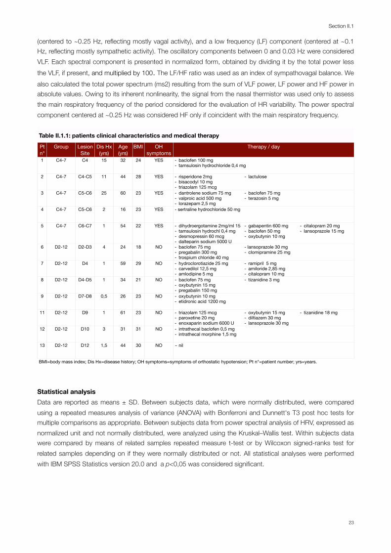
(centered to ~0.25 Hz, reflecting mostly vagal activity), and a low frequency (LF) component (centered at ~0.1 Hz, reflecting mostly sympathetic activity). The oscillatory components between 0 and 0.03 Hz were considered VLF. Each spectral component is presented in normalized form, obtained by dividing it by the total power less the VLF, if present, and multiplied by 100. The LF/HF ratio was used as an index of sympathovagal balance. We also calculated the total power spectrum (ms2) resulting from the sum of VLF power, LF power and HF power in absolute values. Owing to its inherent nonlinearity, the signal from the nasal thermistor was used only to assess the main respiratory frequency of the period considered for the evaluation of HR variability. The power spectral component centered at ~0.25 Hz was considered HF only if coincident with the main respiratory frequency.
Table II.1.1: patients clinical characteristics and medical therapyTable II.1.1: patients clinical characteristics and medical therapyTable II.1.1: patients clinical characteristics and medical therapyTable II.1.1: patients clinical characteristics and medical therapyTable II.1.1: patients clinical characteristics and medical therapyTable II.1.1: patients clinical characteristics and medical therapyTable II.1.1: patients clinical characteristics and medical therapyTable II.1.1: patients clinical characteristics and medical therapyTable II.1.1: patients clinical characteristics and medical therapyTable II.1.1: patients clinical characteristics and medical therapy
Pt n°
Group Lesion Site
Dis Hx (yrs)
Age(yrs)
BMI OH symptoms
Therapy / dayTherapy / dayTherapy / day
1 C4-7 C4 15 32 24 YES - baclofen 100 mg- tamsulosin hydrochloride 0,4 mg- baclofen 100 mg- tamsulosin hydrochloride 0,4 mg- baclofen 100 mg- tamsulosin hydrochloride 0,4 mg
2 C4-7 C4-C5 11 44 28 YES - risperidone 2mg- bisacodyl 10 mg- triazolam 125 mcg
- lactulose
3 C4-7 C5-C6 25 60 23 YES - dantrolene sodium 75 mg- valproic acid 500 mg- lorazepam 2,5 mg
- baclofen 75 mg- terazosin 5 mg
4 C4-7 C5-C6 2 16 23 YES - sertraline hydrochloride 50 mg
5 C4-7 C6-C7 1 54 22 YES - dihydroergotamine 2mg/ml 15- tamsulosin hydrochl 0,4 mg- desmopressin 60 mcg- dalteparin sodium 5000 U
- gabapentin 600 mg- baclofen 50 mg- oxybutynin 10 mg
- citalopram 20 mg- lansoprazole 15 mg
6 D2-12 D2-D3 4 24 18 NO - baclofen 75 mg- pregabalin 300 mg- trospium chloride 40 mg
- lansoprazole 30 mg- clomipramine 25 mg
7 D2-12 D4 1 59 29 NO - hydroclorotiazide 25 mg- carvedilol 12,5 mg- amlodipine 5 mg
- ramipril 5 mg - amiloride 2,85 mg- citalopram 10 mg
8 D2-12 D4-D5 1 34 21 NO - baclofen 75 mg- oxybutynin 15 mg- pregabalin 150 mg
- tizanidine 3 mg
9 D2-12 D7-D8 0,5 26 23 NO - oxybutynin 10 mg- etidronic acid 1200 mg
11 D2-12 D9 1 61 23 NO - triazolam 125 mcg- paroxetine 20 mg- enoxaparin sodium 6000 U
- oxybutynin 15 mg- diltiazem 30 mg- lansoprazole 30 mg
- tizanidine 18 mg
12 D2-12 D10 3 31 31 NO - intrathecal baclofen 0,5 mg- intrathecal morphine 1,5 mg
13 D2-12 D12 1,5 44 30 NO - nil
BMI=body mass index; Dis Hx=disease history; OH symptoms=symptoms of orthostatic hypotension; Pt n°=patient number; yrs=years.BMI=body mass index; Dis Hx=disease history; OH symptoms=symptoms of orthostatic hypotension; Pt n°=patient number; yrs=years.BMI=body mass index; Dis Hx=disease history; OH symptoms=symptoms of orthostatic hypotension; Pt n°=patient number; yrs=years.BMI=body mass index; Dis Hx=disease history; OH symptoms=symptoms of orthostatic hypotension; Pt n°=patient number; yrs=years.BMI=body mass index; Dis Hx=disease history; OH symptoms=symptoms of orthostatic hypotension; Pt n°=patient number; yrs=years.BMI=body mass index; Dis Hx=disease history; OH symptoms=symptoms of orthostatic hypotension; Pt n°=patient number; yrs=years.BMI=body mass index; Dis Hx=disease history; OH symptoms=symptoms of orthostatic hypotension; Pt n°=patient number; yrs=years.BMI=body mass index; Dis Hx=disease history; OH symptoms=symptoms of orthostatic hypotension; Pt n°=patient number; yrs=years.BMI=body mass index; Dis Hx=disease history; OH symptoms=symptoms of orthostatic hypotension; Pt n°=patient number; yrs=years.BMI=body mass index; Dis Hx=disease history; OH symptoms=symptoms of orthostatic hypotension; Pt n°=patient number; yrs=years.
Statistical analysisData are reported as means ± SD. Between subjects data, which were normally distributed, were compared using a repeated measures analysis of variance (ANOVA) with Bonferroni and Dunnett's T3 post hoc tests for multiple comparisons as appropriate. Between subjects data from power spectral analysis of HRV, expressed as normalized unit and not normally distributed, were analyzed using the Kruskal–Wallis test. Within subjects data were compared by means of related samples repeated measure t-test or by Wilcoxon signed-ranks test for related samples depending on if they were normally distributed or not. All statistical analyses were performed with IBM SPSS Statistics version 20.0 and a p<0,05 was considered significant.
Section II.1
23
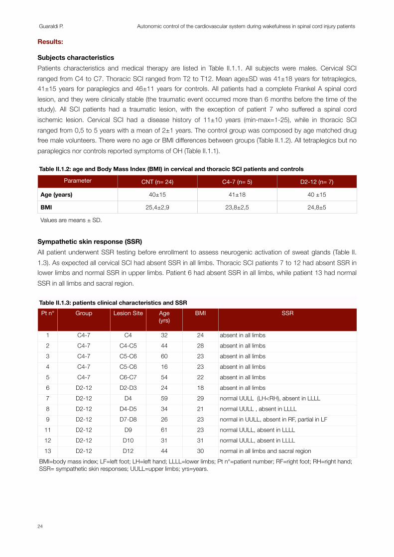
Results:
Subjects characteristicsPatients characteristics and medical therapy are listed in Table II.1.1. All subjects were males. Cervical SCI ranged from C4 to C7. Thoracic SCI ranged from T2 to T12. Mean age±SD was 41±18 years for tetraplegics, 41±15 years for paraplegics and 46±11 years for controls. All patients had a complete Frankel A spinal cord lesion, and they were clinically stable (the traumatic event occurred more than 6 months before the time of the study). All SCI patients had a traumatic lesion, with the exception of patient 7 who suffered a spinal cord ischemic lesion. Cervical SCI had a disease history of 11±10 years (min-max=1-25), while in thoracic SCI ranged from 0,5 to 5 years with a mean of 2±1 years. The control group was composed by age matched drug free male volunteers. There were no age or BMI differences between groups (Table II.1.2). All tetraplegics but no paraplegics nor controls reported symptoms of OH (Table II.1.1).
Table II.1.2: age and Body Mass Index (BMI) in cervical and thoracic SCI patients and controls Table II.1.2: age and Body Mass Index (BMI) in cervical and thoracic SCI patients and controls Table II.1.2: age and Body Mass Index (BMI) in cervical and thoracic SCI patients and controls Table II.1.2: age and Body Mass Index (BMI) in cervical and thoracic SCI patients and controls
Parameter CNT (n= 24) C4-7 (n= 5) D2-12 (n= 7)
Age (years) 40±15 41±18 40 ±15
BMI 25,4±2,9 23,8±2,5 24,8±5
Values are means ± SD.Values are means ± SD.Values are means ± SD.Values are means ± SD.
Sympathetic skin response (SSR)All patient underwent SSR testing before enrollment to assess neurogenic activation of sweat glands (Table II.1.3). As expected all cervical SCI had absent SSR in all limbs. Thoracic SCI patients 7 to 12 had absent SSR in lower limbs and normal SSR in upper limbs. Patient 6 had absent SSR in all limbs, while patient 13 had normal SSR in all limbs and sacral region.
Table II.1.3: patients clinical characteristics and SSRTable II.1.3: patients clinical characteristics and SSRTable II.1.3: patients clinical characteristics and SSRTable II.1.3: patients clinical characteristics and SSRTable II.1.3: patients clinical characteristics and SSRTable II.1.3: patients clinical characteristics and SSRPt n° Group Lesion Site Age
(yrs)BMI SSR
1 C4-7 C4 32 24 absent in all limbs2 C4-7 C4-C5 44 28 absent in all limbs3 C4-7 C5-C6 60 23 absent in all limbs4 C4-7 C5-C6 16 23 absent in all limbs5 C4-7 C6-C7 54 22 absent in all limbs6 D2-12 D2-D3 24 18 absent in all limbs7 D2-12 D4 59 29 normal UULL (LH<RH), absent in LLLL8 D2-12 D4-D5 34 21 normal UULL , absent in LLLL9 D2-12 D7-D8 26 23 normal in UULL, absent in RF, partial in LF11 D2-12 D9 61 23 normal UULL, absent in LLLL12 D2-12 D10 31 31 normal UULL, absent in LLLL13 D2-12 D12 44 30 normal in all limbs and sacral region
BMI=body mass index; LF=left foot; LH=left hand; LLLL=lower limbs; Pt n°=patient number; RF=right foot; RH=right hand; SSR= sympathetic skin responses; UULL=upper limbs; yrs=years.BMI=body mass index; LF=left foot; LH=left hand; LLLL=lower limbs; Pt n°=patient number; RF=right foot; RH=right hand; SSR= sympathetic skin responses; UULL=upper limbs; yrs=years.BMI=body mass index; LF=left foot; LH=left hand; LLLL=lower limbs; Pt n°=patient number; RF=right foot; RH=right hand; SSR= sympathetic skin responses; UULL=upper limbs; yrs=years.BMI=body mass index; LF=left foot; LH=left hand; LLLL=lower limbs; Pt n°=patient number; RF=right foot; RH=right hand; SSR= sympathetic skin responses; UULL=upper limbs; yrs=years.BMI=body mass index; LF=left foot; LH=left hand; LLLL=lower limbs; Pt n°=patient number; RF=right foot; RH=right hand; SSR= sympathetic skin responses; UULL=upper limbs; yrs=years.BMI=body mass index; LF=left foot; LH=left hand; LLLL=lower limbs; Pt n°=patient number; RF=right foot; RH=right hand; SSR= sympathetic skin responses; UULL=upper limbs; yrs=years.
Guaraldi P. Autonomic control of the cardiovascular system during wakefulness in spinal cord injury patients
24
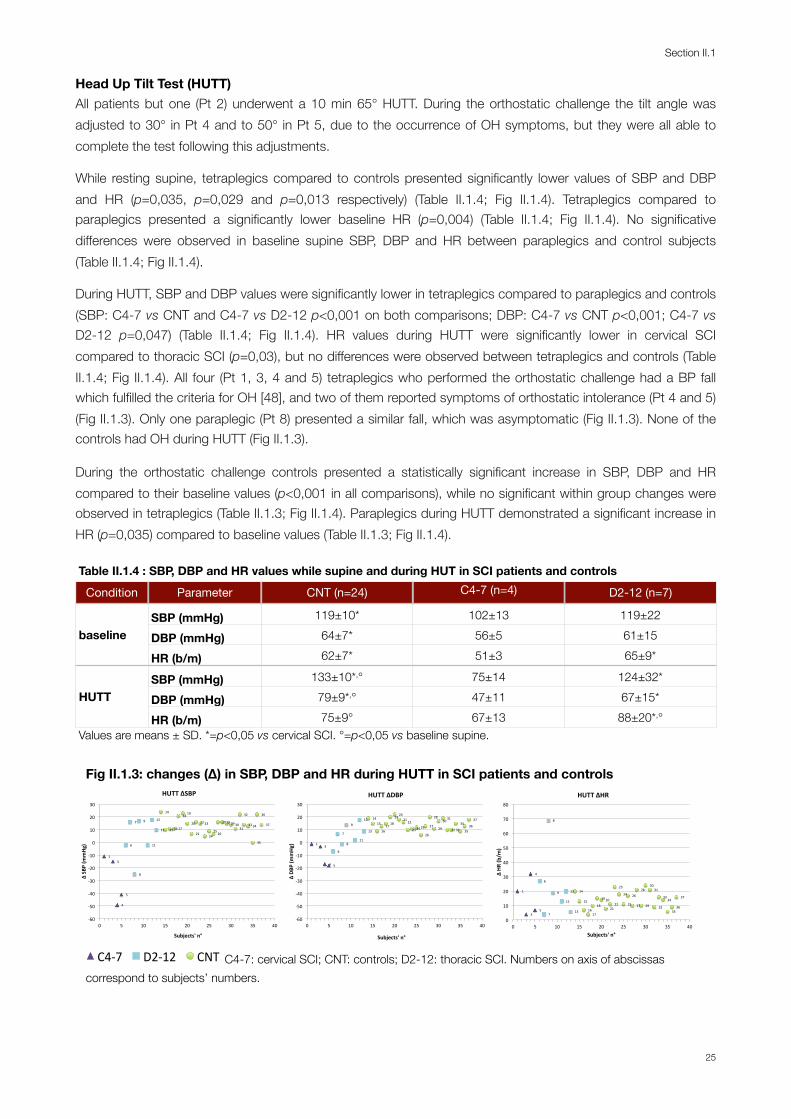
Head Up Tilt Test (HUTT)All patients but one (Pt 2) underwent a 10 min 65° HUTT. During the orthostatic challenge the tilt angle was adjusted to 30° in Pt 4 and to 50° in Pt 5, due to the occurrence of OH symptoms, but they were all able to complete the test following this adjustments.
While resting supine, tetraplegics compared to controls presented significantly lower values of SBP and DBP and HR (p=0,035, p=0,029 and p=0,013 respectively) (Table II.1.4; Fig II.1.4). Tetraplegics compared to paraplegics presented a significantly lower baseline HR (p=0,004) (Table II.1.4; Fig II.1.4). No significative differences were observed in baseline supine SBP, DBP and HR between paraplegics and control subjects (Table II.1.4; Fig II.1.4).
During HUTT, SBP and DBP values were significantly lower in tetraplegics compared to paraplegics and controls (SBP: C4-7 vs CNT and C4-7 vs D2-12 p<0,001 on both comparisons; DBP: C4-7 vs CNT p<0,001; C4-7 vs D2-12 p=0,047) (Table II.1.4; Fig II.1.4). HR values during HUTT were significantly lower in cervical SCI compared to thoracic SCI (p=0,03), but no differences were observed between tetraplegics and controls (Table II.1.4; Fig II.1.4). All four (Pt 1, 3, 4 and 5) tetraplegics who performed the orthostatic challenge had a BP fall which fulfilled the criteria for OH [48], and two of them reported symptoms of orthostatic intolerance (Pt 4 and 5) (Fig II.1.3). Only one paraplegic (Pt 8) presented a similar fall, which was asymptomatic (Fig II.1.3). None of the controls had OH during HUTT (Fig II.1.3).
During the orthostatic challenge controls presented a statistically significant increase in SBP, DBP and HR compared to their baseline values (p<0,001 in all comparisons), while no significant within group changes were observed in tetraplegics (Table II.1.3; Fig II.1.4). Paraplegics during HUTT demonstrated a significant increase in HR (p=0,035) compared to baseline values (Table II.1.3; Fig II.1.4).
Table II.1.4 : SBP, DBP and HR values while supine and during HUT in SCI patients and controls Table II.1.4 : SBP, DBP and HR values while supine and during HUT in SCI patients and controls Table II.1.4 : SBP, DBP and HR values while supine and during HUT in SCI patients and controls Table II.1.4 : SBP, DBP and HR values while supine and during HUT in SCI patients and controls Table II.1.4 : SBP, DBP and HR values while supine and during HUT in SCI patients and controls Condition Parameter CNT (n=24) C4-7 (n=4) D2-12 (n=7)
baselineSBP (mmHg) 119±10* 102±13 119±22
baseline DBP (mmHg) 64±7* 56±5 61±15baseline
HR (b/m) 62±7* 51±3 65±9*
HUTTSBP (mmHg) 133±10*,° 75±14 124±32*
HUTT DBP (mmHg) 79±9*,° 47±11 67±15*HUTT
HR (b/m) 75±9° 67±13 88±20*,°Values are means ± SD. *=p<0,05 vs cervical SCI. °=p<0,05 vs baseline supine.Values are means ± SD. *=p<0,05 vs cervical SCI. °=p<0,05 vs baseline supine.Values are means ± SD. *=p<0,05 vs cervical SCI. °=p<0,05 vs baseline supine.Values are means ± SD. *=p<0,05 vs cervical SCI. °=p<0,05 vs baseline supine.Values are means ± SD. *=p<0,05 vs cervical SCI. °=p<0,05 vs baseline supine.
Fig II.1.3: changes (∆) in SBP, DBP and HR during HUTT in SCI patients and controls
1"3"
4"
5"
6"
7"
8"
9"
11"
12"
13"
14"
15"16"17"
18"19"
20"
21"
22"23"
24"25"
26"
27"28"29"30"31"
32"
33"34"
35"
36"
37"
,60"
,50"
,40"
,30"
,20"
,10"
0"
10"
20"
30"
0" 5" 10" 15" 20" 25" 30" 35" 40"
Δ"SB
P"(m
mHg
)"
Subjects'"n°"
HUTT"ΔSBP""
1"3"
4" 5"
6"
7"
8"
9"
11"
12"
13"
14"15"
16"17"
18"
19"20"
21"22"
23"24"25"
26"
27"
28"
29"
30"31"
32"33"
34"
35"36"
37"
,60"
,50"
,40"
,30"
,20"
,10"
0"
10"
20"
30"
0" 5" 10" 15" 20" 25" 30" 35" 40"
Δ"DB
P"(m
mHg
)"
Subjects'"n°"
HUTT"ΔDBP"
1"
3"
4"
5"
6"
7"
8"
9"
11"
12"
13"
14"
15"
16"17"
18"
19"20"
21"22"
23"
24"
25"
26"
27"
28"
29"
30"31"
32"
33"34"
35"36"
37"
0"
10"
20"
30"
40"
50"
60"
70"
80"
0" 5" 10" 15" 20" 25" 30" 35" 40"
Δ"HR
"(b/m
)"
Subjects'"n°"
HUTT"ΔHR"
4"
5" 6"
7"8" 9"
11"
12"
13"
14"
15"
16"
17"18"
19"
20"
21"
22"23"
24"
25"
27"
28"
29"
30"
31"
32"
33"
34"
35"
36"
37"
5" 10" 15" 20" 25" 30" 35" 40"Pa#ents')n°)
C4-7" D2-12" CNT" C4-7: cervical SCI; CNT: controls; D2-12: thoracic SCI. Numbers on axis of abscissas correspond to subjects’ numbers.
Section II.1
25
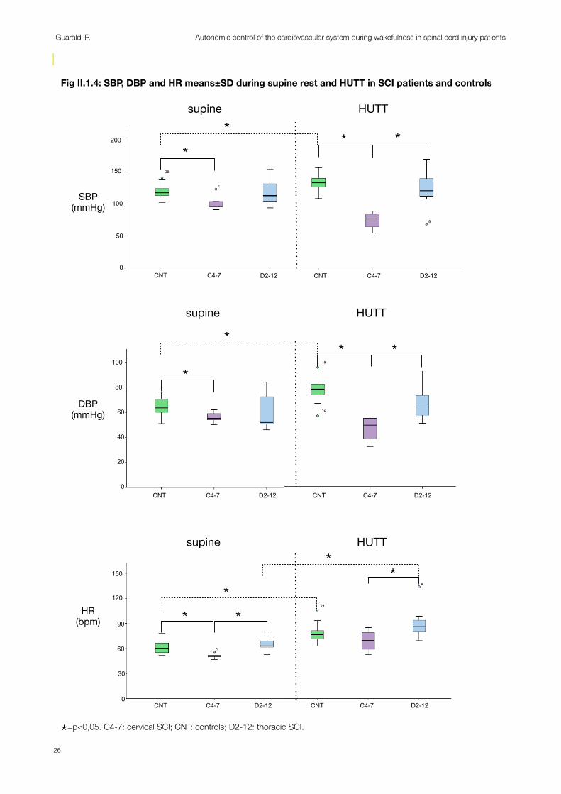
Fig II.1.4: SBP, DBP and HR means±SD during supine rest and HUTT in SCI patients and controls
!
*
!
* *
!
*supine HUTT
!
*
!
* **
!
*
* **
supine HUTT
supine HUTT
SBP(mmHg)
DBP(mmHg)
HR(bpm)
CNT C4-7 D2-12 CNT C4-7 D2-12
CNT C4-7 D2-12 CNT C4-7 D2-12
CNT C4-7 D2-12 CNT C4-7 D2-12
200
100
150
50
0
100
60
80
40
20
0
150
90
120
60
0
28
19
26
23
*
30
*=p<0,05. C4-7: cervical SCI; CNT: controls; D2-12: thoracic SCI.
Guaraldi P. Autonomic control of the cardiovascular system during wakefulness in spinal cord injury patients
26
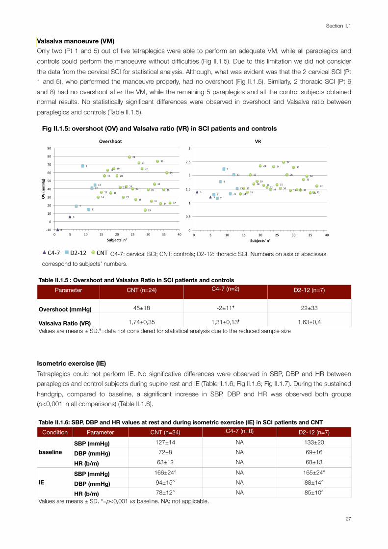
Valsalva manoeuvre (VM) Only two (Pt 1 and 5) out of five tetraplegics were able to perform an adequate VM, while all paraplegics and controls could perform the manoeuvre without difficulties (Fig II.1.5). Due to this limitation we did not consider the data from the cervical SCI for statistical analysis. Although, what was evident was that the 2 cervical SCI (Pt 1 and 5), who performed the manoeuvre properly, had no overshoot (Fig II.1.5). Similarly, 2 thoracic SCI (Pt 6 and 8) had no overshoot after the VM, while the remaining 5 paraplegics and all the control subjects obtained normal results. No statistically significant differences were observed in overshoot and Valsalva ratio between paraplegics and controls (Table II.1.5).
Fig II.1.5: overshoot (OV) and Valsalva ratio (VR) in SCI patients and controls
1"
5"
7"
9"
11"
12"13"
14"
15"
16"
17"
18"
19"
20"
21"
22"
23"
24"
25"
26"
27"
28"
29"
30"
31"
32"
33"
34"
35"
36"
37"
,10"
0"
10"
20"
30"
40"
50"
60"
70"
80"
90"
0" 5" 10" 15" 20" 25" 30" 35" 40"
OV#(m
mHg
)#
Subjects'#n°#
Overshoot#
1"
5"6"
7"
8"
9"
11"
12"
13"
14"
15"16"
17"
18"19"
20"
21"22"
23"
24"
25"26"
27"
28"
29"
30"
31"32"
33"34"
35"36"
37"
0"
0,5"
1"
1,5"
2"
2,5"
3"
0" 5" 10" 15" 20" 25" 30" 35" 40"
Subjects'*n°*
VR*
4"
5" 6"
7"8" 9"
11"
12"
13"
14"
15"
16"
17"18"
19"
20"
21"
22"23"
24"
25"
27"
28"
29"
30"
31"
32"
33"
34"
35"
36"
37"
5" 10" 15" 20" 25" 30" 35" 40"Pa#ents')n°)
C4-7" D2-12" CNT" C4-7: cervical SCI; CNT: controls; D2-12: thoracic SCI. Numbers on axis of abscissas correspond to subjects’ numbers.
Table II.1.5 : Overshoot and Valsalva Ratio in SCI patients and controls Table II.1.5 : Overshoot and Valsalva Ratio in SCI patients and controls Table II.1.5 : Overshoot and Valsalva Ratio in SCI patients and controls Table II.1.5 : Overshoot and Valsalva Ratio in SCI patients and controls Parameter CNT (n=24) C4-7 (n=2) D2-12 (n=7)
Overshoot (mmHg) 45±18 -2±11✝ 22±33
Valsalva Ratio (VR) 1,74±0,35 1,31±0,13✝ 1,63±0,4Values are means ± SD.✝=data not considered for statistical analysis due to the reduced sample sizeValues are means ± SD.✝=data not considered for statistical analysis due to the reduced sample sizeValues are means ± SD.✝=data not considered for statistical analysis due to the reduced sample sizeValues are means ± SD.✝=data not considered for statistical analysis due to the reduced sample size
Isometric exercise (IE) Tetraplegics could not perform IE. No significative differences were observed in SBP, DBP and HR between paraplegics and control subjects during supine rest and IE (Table II.1.6; Fig II.1.6; Fig II.1.7). During the sustained handgrip, compared to baseline, a significant increase in SBP, DBP and HR was observed both groups (p<0,001 in all comparisons) (Table II.1.6).
Table II.1.6: SBP, DBP and HR values at rest and during isometric exercise (IE) in SCI patients and CNTTable II.1.6: SBP, DBP and HR values at rest and during isometric exercise (IE) in SCI patients and CNTTable II.1.6: SBP, DBP and HR values at rest and during isometric exercise (IE) in SCI patients and CNTTable II.1.6: SBP, DBP and HR values at rest and during isometric exercise (IE) in SCI patients and CNTTable II.1.6: SBP, DBP and HR values at rest and during isometric exercise (IE) in SCI patients and CNTCondition Parameter CNT (n=24) C4-7 (n=0) D2-12 (n=7)
baselineSBP (mmHg) 127±14 NA 133±20
baseline DBP (mmHg) 72±8 NA 69±16baseline
HR (b/m) 63±12 NA 68±13
IESBP (mmHg) 166±24° NA 165±24°
IE DBP (mmHg) 94±15° NA 88±14°IE
HR (b/m) 78±12° NA 85±10°Values are means ± SD. °=p<0,001 vs baseline. NA: not applicable.Values are means ± SD. °=p<0,001 vs baseline. NA: not applicable.Values are means ± SD. °=p<0,001 vs baseline. NA: not applicable.Values are means ± SD. °=p<0,001 vs baseline. NA: not applicable.Values are means ± SD. °=p<0,001 vs baseline. NA: not applicable.
Section II.1
27
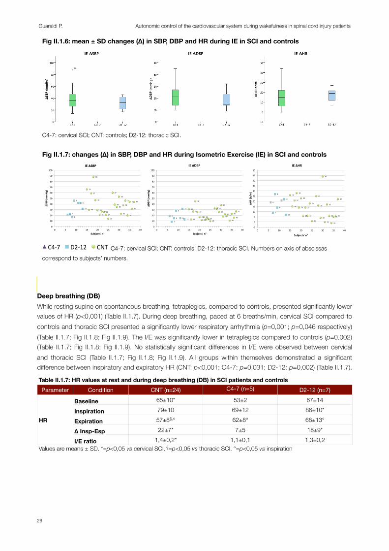
Deep breathing (DB)While resting supine on spontaneous breathing, tetraplegics, compared to controls, presented significantly lower values of HR (p<0,001) (Table II.1.7). During deep breathing, paced at 6 breaths/min, cervical SCI compared to controls and thoracic SCI presented a significantly lower respiratory arrhythmia (p=0,001; p=0,046 respectively) (Table II.1.7; Fig II.1.8; Fig II.1.9). The I/E was significantly lower in tetraplegics compared to controls (p=0,002) (Table II.1.7; Fig II.1.8; Fig II.1.9). No statistically significant differences in I/E were observed between cervical and thoracic SCI (Table II.1.7; Fig II.1.8; Fig II.1.9). All groups within themselves demonstrated a significant difference between inspiratory and expiratory HR (CNT: p<0,001; C4-7: p=0,031; D2-12: p=0,002) (Table II.1.7).
Table II.1.7: HR values at rest and during deep breathing (DB) in SCI patients and controls Table II.1.7: HR values at rest and during deep breathing (DB) in SCI patients and controls Table II.1.7: HR values at rest and during deep breathing (DB) in SCI patients and controls Table II.1.7: HR values at rest and during deep breathing (DB) in SCI patients and controls Table II.1.7: HR values at rest and during deep breathing (DB) in SCI patients and controls Parameter Condition CNT (n=24) C4-7 (n=5) D2-12 (n=7)
HR
Baseline 65±10* 53±2 67±14
HRInspiration 79±10 69±12 86±10*
HR Expiration 57±8§,° 62±8° 68±13°HR
∆ Insp-Esp 22±7* 7±5 18±9*HR
I/E ratio 1,4±0,2* 1,1±0,1 1,3±0,2Values are means ± SD. *=p<0,05 vs cervical SCI. §=p<0,05 vs thoracic SCI. °=p<0,05 vs inspirationValues are means ± SD. *=p<0,05 vs cervical SCI. §=p<0,05 vs thoracic SCI. °=p<0,05 vs inspirationValues are means ± SD. *=p<0,05 vs cervical SCI. §=p<0,05 vs thoracic SCI. °=p<0,05 vs inspirationValues are means ± SD. *=p<0,05 vs cervical SCI. §=p<0,05 vs thoracic SCI. °=p<0,05 vs inspirationValues are means ± SD. *=p<0,05 vs cervical SCI. §=p<0,05 vs thoracic SCI. °=p<0,05 vs inspiration
Fig II.1.6: mean ± SD changes (∆) in SBP, DBP and HR during IE in SCI and controls
18
C4-7: cervical SCI; CNT: controls; D2-12: thoracic SCI.
Fig II.1.7: changes (∆) in SBP, DBP and HR during Isometric Exercise (IE) in SCI and controls
6" 7"
8"
9"11"12"
13"
15"
16"
17"
18"
19"
20"
21"
22"23"24"
25"
27"
28"29"
30"
31"
32"
33"
34"
35"
36"
37"
0"
10"
20"
30"
40"
50"
60"
70"
80"
90"
100"
0" 5" 10" 15" 20" 25" 30" 35" 40"
ΔSBP
%(mmHg
)%
Subjects'%n°%
IE%ΔSBP%%
6"
7"
8"
9"
11"
12"
13"
15"
16"
17"
18"
19"
20"21"
22"23"24"
25"
27"28"
29"
30"
31"32"33"
34"
35"
36"
37"
0"
10"
20"
30"
40"
50"
60"
70"
80"
90"
100"
0" 5" 10" 15" 20" 25" 30" 35" 40"
ΔDBP
%(mmHg
)%
Subjects'%n°%
IE%ΔDBP%
6"
7"
8"
9"
11"12"
13"
15"
16"
17"
18"
19"
20"
21"
22"
23"
24"
25"
27"
28"
29"
30"
31"
32"
33"
34"
35"36"
37"
,5"
0"
5"
10"
15"
20"
25"
30"
35"
40"
45"
50"
0" 5" 10" 15" 20" 25" 30" 35" 40"
ΔHR$(b/m
)$
Subjects'$n°$
IE$ΔHR$
4"
5" 6"
7"8" 9"
11"
12"
13"
14"
15"
16"
17"18"
19"
20"
21"
22"23"
24"
25"
27"
28"
29"
30"
31"
32"
33"
34"
35"
36"
37"
5" 10" 15" 20" 25" 30" 35" 40"Pa#ents')n°)
C4-7" D2-12" CNT" C4-7: cervical SCI; CNT: controls; D2-12: thoracic SCI. Numbers on axis of abscissas correspond to subjects’ numbers.
Guaraldi P. Autonomic control of the cardiovascular system during wakefulness in spinal cord injury patients
28

Cold Face (CF)Baseline SBP, DBP and HR was significantly lower in cervical SCI compared to controls (p=0,005; p=0,049; p=0,007 respectively) (Table II.1.8). Baseline SBP was significantly lower in tetraplegics compared to paraplegics (p=0,024) but no differences were observed in baseline DBP and HR between cervical and thoracic SCI (Table II.1.8).
Cervical SCI compared to thoracic SCI and controls presented a significantly lower SBP, DBP values during the cold face test (SBP: C4-7 vs CNT p=0,001; C4-7 vs D2-12: p=0,003; DBP: C4-7 vs CNT p=0,002; C4-7 vs D2-12 p=0,042) and consequently demonstrated a significantly lower increase in SBP (∆SBP) and DBP (∆DBP) during the manoeuvre (∆SBP: C4-7 vs CNT p=0,005; C4-7 vs D2-12: p=0,004; ∆DBP: C4-7 vs CNT p=0,019; C4-7 vs D2-12 p=0,012) (Table II.1.8; Fig II.1.10; Fig II.1.11). The mean HR was not significantly different in the three groups during the CF, but cervical SCI compared to controls presented a reduced decrease in HR (∆HR) during the cold stimulation of the forehead (p=0,014) (Table II.1.8; Fig II.1.10; Fig II.1.11). No differences were observed in ∆HR between cervical and thoracic SCI (Table II.1.8; Fig II.1.10; Fig II.1.11).
Within subjects analysis demonstrated that both thoracic SCI and controls presented significant increase in BP and decrease in HR while cervical SCI did not (Table II.1.8).
Fig II.1.8: mean ± SD changes (∆) in HR and I/E ratio during DB in SCI and controls
Deep Breathing
1"2" 3"
4"5"
6" 7"
8"
9"
11"
12"13"
14"
15"16"
17"
18"19"20"
21"
22"23"
24"
25"26"
27"
28"
29"
30"
31"
32"33"
34"35"
36"37"
0"
0,2"
0,4"
0,6"
0,8"
1"
1,2"
1,4"
1,6"
1,8"
2"
0" 5" 10" 15" 20" 25" 30" 35" 40"Pa#ents')n°)
DB_I/E)
C4.7" D2.12" CNT"
∆HR I/E
*24
30
1"
2"3"
4"
5"6"
7"
8"
9"
11"
12"
13"
14"
15"16"17"
18"
19"20"
21"
22"
23"
24"
25"26"
27"
28"
29"
30"
31"
32"33"
34"35"
36"37"
0"
10"
20"
30"
40"
50"
60"
70"
80"
90"
100"
0" 5" 10" 15" 20" 25" 30" 35" 40"
HR#(b
/m#
Pa*ents'#n°#
DB_Delta#HR#
C4-7" D2-12" CNT"
* *
*=p<0,05. C4-7: cervical SCI; CNT: controls; D2-12: thoracic SCI; I/E: inspiratory/expiratory ratio.
Fig II.1.9: changes (∆) in HR and I/E ratio during Deep Breathing (DB) in SCI and controls
1"
2"3"
4"
5"6"
7"
8"
9"
11"
12"
13"
14"
15"16"17"
18"
19"20"
21"
22"
23"
24"
25"26"
27"
28"
29"
30"
31"
32"33"
34"35"
36"37"
0"
10"
20"
30"
40"
50"
60"
70"
80"
90"
100"
0" 5" 10" 15" 20" 25" 30" 35" 40"
ΔHR$(b/m
)$
Subjects'$n°$
DB$ΔHR$
1"2" 3"
4"5"
6" 7"
8"
9"
11"
12"
13"
14"
15"
16"
17"
18"19"
20"
21"
22"23"
24"
25"26"
27"
28"
29"
30"
31"
32"33"
34"
35"36"
37"
0"
0,2"
0,4"
0,6"
0,8"
1"
1,2"
1,4"
1,6"
1,8"
2"
0" 5" 10" 15" 20" 25" 30" 35" 40"
Subjects'*n°*
DB*I/E*
4"
5" 6"
7"8" 9"
11"
12"
13"
14"
15"
16"
17"18"
19"
20"
21"
22"23"
24"
25"
27"
28"
29"
30"
31"
32"
33"
34"
35"
36"
37"
5" 10" 15" 20" 25" 30" 35" 40"Pa#ents')n°)
C4-7" D2-12" CNT" C4-7: cervical SCI; CNT: controls; D2-12: thoracic SCI; I/E: inspiratory/expiratory ratio. Numbers on axis of abscissas correspond to subjects’ numbers.
Section II.1
29
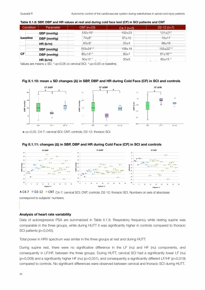
Table II.1.8: SBP, DBP and HR values at rest and during cold face test (CF) in SCI patients and CNTTable II.1.8: SBP, DBP and HR values at rest and during cold face test (CF) in SCI patients and CNTTable II.1.8: SBP, DBP and HR values at rest and during cold face test (CF) in SCI patients and CNTTable II.1.8: SBP, DBP and HR values at rest and during cold face test (CF) in SCI patients and CNTTable II.1.8: SBP, DBP and HR values at rest and during cold face test (CF) in SCI patients and CNTCondition Parameter CNT (n=23) C4-7 (n=5) D2-12 (n=7)
baselineSBP (mmHg) 132±15* 102±23 131±21*
baseline DBP (mmHg) 74±8* 61±10 72±17baseline
HR (b/m) 63±9* 53±4 68±16
CFSBP (mmHg) 153±24*,° 108±18 155±22*,°
CF DBP (mmHg) 85±14*,° 60±7 81±16*,°CF
HR (b/m) 50±12 ° 50±5 60±15 °Values are means ± SD. *=p<0,05 vs cervical SCI. °=p<0,05 vs baseline.Values are means ± SD. *=p<0,05 vs cervical SCI. °=p<0,05 vs baseline.Values are means ± SD. *=p<0,05 vs cervical SCI. °=p<0,05 vs baseline.Values are means ± SD. *=p<0,05 vs cervical SCI. °=p<0,05 vs baseline.Values are means ± SD. *=p<0,05 vs cervical SCI. °=p<0,05 vs baseline.
Analysis of heart rate variabilityData of autoregressive PSA are summarized in Table II.1.9. Respiratory frequency while resting supine was comparable in the three groups, while during HUTT it was significantly higher in controls compared to thoracic SCI patients (p=0,045).
Total power in HRV spectrum was similar in the three groups at rest and during HUTT.
During supine rest, there were no significative difference in the LF (nu) and HF (nu) components, and consequently in LF/HF, between the three groups. During HUTT, cervical SCI had a significantly lower LF (nu) (p=0,009) and a significantly higher HF (nu) (p=0,031), and consequently a significantly different LF/HF (p=0,019) compared to controls. No significant differences were observed between cervical and thoracic SCI during HUTT.
Fig II.1.10: mean ± SD changes (∆) in SBP, DBP and HR during Cold Face (CF) in SCI and controls
Cold Face
1"2"
3"
4"
5"6"
7" 8"
9"11"
12"
13"14"15"16"
17"
18"
19"
20"
21"
22"
23"
24"
25"
27"
28"
29"
30"31"
32"
33"
34"
35"
36"
37"
,20"
,10"
0"
10"
20"
30"
40"
50"
60"
0" 5" 10" 15" 20" 25" 30" 35" 40"
SBP$(m
mHg
)$
Pa+ents'$n°$
CF_Delta$SBP$$
C4,7" D2,12" CNT"
1"
2"3"
4"
5"
6"7"
8"
9"
11"
12"
13"
14"
15"16"
17"
18"
19"20"
21"
22"
23"
24"25"
27"
28"
29"
30"
31"
32"
33"
34"
35"
36"37"
,10"
,5"
0"
5"
10"
15"
20"
25"
30"
0" 5" 10" 15" 20" 25" 30" 35" 40"
DBP$(m
mHg
)$
Pa+ents'$n°$
CF_Delta$DBP$
C4,7" D2,12" CNT"
1" 2"
3"
4"
5" 6"
7"8" 9"
11"
12"
13"
14"
15"
16"
17"18"
19"
20"
21"
22"23"
24"
25"
27"
28"
29"
30"
31"
32"
33"
34"
35"
36"
37"
,40"
,35"
,30"
,25"
,20"
,15"
,10"
,5"
0"
5"
10"
0" 5" 10" 15" 20" 25" 30" 35" 40"
HR#(b
/m#
Pa*ents'#n°#
CF_Delta#HR#
C4,7" D2,12" CNT"
* * * * *
Fig II.4 ∆SBP, ∆DBP and ∆HR during cold face test (CF) in SCI patients and controls
*=p<0,05. C4-7: cervical SCI; CNT: controls; D2-12: thoracic SCI.
Fig II.1.11: changes (∆) in SBP, DBP and HR during Cold Face (CF) in SCI and controls
1"2"
3"
4"
5"6"
7" 8"
9"11"
12"
13"14"15"16"
17"
18"
19"
20"
21"
22"
23"
24"
25"
27"
28"
29"
30"31"
32"
33"
34"
35"
36"
37"
,20"
,10"
0"
10"
20"
30"
40"
50"
60"
0" 5" 10" 15" 20" 25" 30" 35" 40"
ΔSBP
%(mmHg
)%
Subjects'%n°%
CF%ΔSBP%%
1"
2"3"
4"
5"
6" 7"8"
9"
11"
12"
13"
14"
15"16"
17"
18"
19"20"
21"
22"
23"24"25"
27"
28"29"
30"
31"
32"
33"
34"
35"
36"37"
,20"
,10"
0"
10"
20"
30"
40"
50"
60"
0" 5" 10" 15" 20" 25" 30" 35" 40"
ΔDBP
%(mmHg
)%
Subjects'%n°%
CF%ΔDBP%
1" 2"
3"
4"
5" 6"
7"8" 9"
11"
12"
13"
14"
15"
16"
17"18"
19"
20"
21"
22"23"
24"
25"
27"
28"
29"
30"
31"
32"
33"
34"
35"
36"
37"
,40"
,35"
,30"
,25"
,20"
,15"
,10"
,5"
0"
5"
10"
0" 5" 10" 15" 20" 25" 30" 35" 40"
ΔHR$(b/m
)$
Subjects'$n°$
CF$ΔHR$
4"
5" 6"
7"8" 9"
11"
12"
13"
14"
15"
16"
17"18"
19"
20"
21"
22"23"
24"
25"
27"
28"
29"
30"
31"
32"
33"
34"
35"
36"
37"
5" 10" 15" 20" 25" 30" 35" 40"Pa#ents')n°)
C4-7" D2-12" CNT" C4-7: cervical SCI; CNT: controls; D2-12: thoracic SCI. Numbers on axis of abscissas correspond to subjects’ numbers.
Guaraldi P. Autonomic control of the cardiovascular system during wakefulness in spinal cord injury patients
30
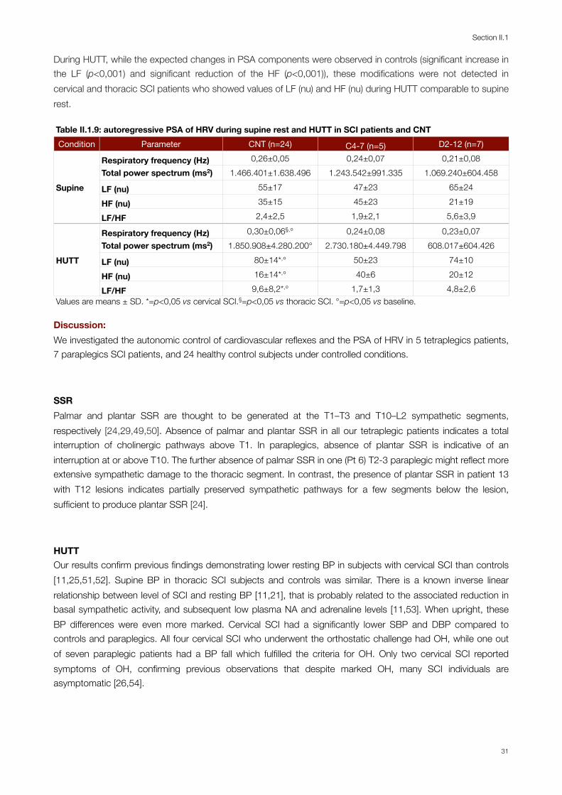
During HUTT, while the expected changes in PSA components were observed in controls (significant increase in the LF (p<0,001) and significant reduction of the HF (p<0,001)), these modifications were not detected in cervical and thoracic SCI patients who showed values of LF (nu) and HF (nu) during HUTT comparable to supine rest.
Table II.1.9: autoregressive PSA of HRV during supine rest and HUTT in SCI patients and CNTTable II.1.9: autoregressive PSA of HRV during supine rest and HUTT in SCI patients and CNTTable II.1.9: autoregressive PSA of HRV during supine rest and HUTT in SCI patients and CNTTable II.1.9: autoregressive PSA of HRV during supine rest and HUTT in SCI patients and CNTTable II.1.9: autoregressive PSA of HRV during supine rest and HUTT in SCI patients and CNTCondition Parameter CNT (n=24) C4-7 (n=5) D2-12 (n=7)
Supine
Respiratory frequency (Hz) 0,26±0,05 0,24±0,07 0,21±0,08
SupineTotal power spectrum (ms2) 1.466.401±1.638.496 1.243.542±991.335 1.069.240±604.458
Supine LF (nu) 55±17 47±23 65±24Supine
HF (nu) 35±15 45±23 21±19Supine
LF/HF 2,4±2,5 1,9±2,1 5,6±3,9
HUTT
Respiratory frequency (Hz) 0,30±0,06§,° 0,24±0,08 0,23±0,07
HUTTTotal power spectrum (ms2) 1.850.908±4.280.200° 2.730.180±4.449.798 608.017±604.426
HUTT LF (nu) 80±14*,° 50±23 74±10HUTT
HF (nu) 16±14*,° 40±6 20±12HUTT
LF/HF 9,6±8,2*,° 1,7±1,3 4,8±2,6Values are means ± SD. *=p<0,05 vs cervical SCI.§=p<0,05 vs thoracic SCI. °=p<0,05 vs baseline.Values are means ± SD. *=p<0,05 vs cervical SCI.§=p<0,05 vs thoracic SCI. °=p<0,05 vs baseline.Values are means ± SD. *=p<0,05 vs cervical SCI.§=p<0,05 vs thoracic SCI. °=p<0,05 vs baseline.Values are means ± SD. *=p<0,05 vs cervical SCI.§=p<0,05 vs thoracic SCI. °=p<0,05 vs baseline.Values are means ± SD. *=p<0,05 vs cervical SCI.§=p<0,05 vs thoracic SCI. °=p<0,05 vs baseline.
Discussion:We investigated the autonomic control of cardiovascular reflexes and the PSA of HRV in 5 tetraplegics patients, 7 paraplegics SCI patients, and 24 healthy control subjects under controlled conditions.
SSRPalmar and plantar SSR are thought to be generated at the T1–T3 and T10–L2 sympathetic segments, respectively [24,29,49,50]. Absence of palmar and plantar SSR in all our tetraplegic patients indicates a total interruption of cholinergic pathways above T1. In paraplegics, absence of plantar SSR is indicative of an interruption at or above T10. The further absence of palmar SSR in one (Pt 6) T2-3 paraplegic might reflect more extensive sympathetic damage to the thoracic segment. In contrast, the presence of plantar SSR in patient 13 with T12 lesions indicates partially preserved sympathetic pathways for a few segments below the lesion, sufficient to produce plantar SSR [24].
HUTTOur results confirm previous findings demonstrating lower resting BP in subjects with cervical SCI than controls [11,25,51,52]. Supine BP in thoracic SCI subjects and controls was similar. There is a known inverse linear relationship between level of SCI and resting BP [11,21], that is probably related to the associated reduction in basal sympathetic activity, and subsequent low plasma NA and adrenaline levels [11,53]. When upright, these BP differences were even more marked. Cervical SCI had a significantly lower SBP and DBP compared to controls and paraplegics. All four cervical SCI who underwent the orthostatic challenge had OH, while one out of seven paraplegic patients had a BP fall which fulfilled the criteria for OH. Only two cervical SCI reported symptoms of OH, confirming previous observations that despite marked OH, many SCI individuals are asymptomatic [26,54].
Section II.1
31

Fig II.1.11: respiratory frequency, LF (nu) and HF (nu) during supine rest and HUTT in SCI and controls
! !
supine HUTT
*
supine HUTT
RespiratoryFrequency
(Hz)
LF(nu)
HF(nu)
! !
*
! !
*
! !!!!
HUTT
*
*
Fig II.5 Respiratory frequency, LF (nu) and HF (nu) while supine and during HUT in SCI patients and controls
1 1
12
32
33
12
12
12
35
32
!!
*12
!
*
supine
12
*=p<0,05. C4-7: cervical SCI; CNT: controls; D2-12: thoracic SCI.
Guaraldi P. Autonomic control of the cardiovascular system during wakefulness in spinal cord injury patients
32
HR responses to orthostatic stress are due to baroreflex-mediated parasympathetic (vagal) withdrawal, usually preserved following SCI [21], and sympathetic activation. Lesions above T6 may disrupt descending sympathetic control of the heart. Resting supine HR in our cervical SCI patients was significantly slower compared to thoracic SCI and controls. Tetraplegics compared to paraplegics and controls did not present a significant increase in HR during the orthostatic challenge and therefore HR in cervical SCI, was significantly slower during HUTT compared to thoracic SCI. The alterations in cervical SCI resting HR have been seen previously [11,55,56], and probably reflect decreased sympathetic chronotropic responses in high lesions [56], and compensation for reduced SV in thoracic SCI [55]. Upright HR responses were similar between cervical SCI and controls, suggesting they are predominantly due to baroreflex-mediated reductions in vagal tone, which is preserved in all subjects. However, the stimulus provided to the baroreceptors would have been greater in cervical SCI subjects, in whom upright blood pressures were lowest. Accordingly, one might expect larger HR increases during orthostatic stress in cervical SCI subjects. The fact that this was not observed in our court may be partly due to increased cardiac baroreflex delay in cervical SCI subjects [1].
Although there were no significative differences between thoracic SCI and controls, within subjects analysis demonstrated that paraplegics, as tetraplegics, did not present a significant increase in HR. Therefore, even if cardiac autonomic control is intact in thoracic SCI, they had a blunted sympathetic response to HUTT [8].
Valsalva manoeuvreVM induced no overshoot in the two cervical SCI patients who were able to perform it adequately, but it was present in most of paraplegics. These findings confirm that the OV is primarily related to persistent cardiac sympathetic activity [10]. Only two thoracic SCI (Patient 6 and 8 with T2-3 and T4-5 lesion respectively) showed no overshoot after the VM. Noteworthy patient 6 had absent SSR in all limbs and therefore a more extensive sympathetic damage.
Isometric exerciseIsometric exercise could be performed only by thoracic SCI, who demonstrated normal pressor responses compared to controls, probably due to an increase in sympathetic activity above the lesion level and an increase in heart rate and cardiac contractility [57]. Although, the rise is SBP was blunted as the splanchnic and lower limb vascular beds could not contribute to the hemodynamic response [12] as demonstrated by the absence of changes of the peripheral resistance of the leg [15], of electrodermal activity [16,17] or skin vasomotor reflexes [14].
Deep BreathingDuring deep breathing cervical SCI compared to controls and thoracic SCI presented a significantly lower respiratory arrhythmia and the I/E was significantly lower in tetraplegics compared to controls. These finding might be due to inability to reduce vagal activity through the pulmonary inflation reflex because of the inability to breathe or an increased vagal activity in relation to hypoxia [58]. Although all groups, including tetraplegics, within themselves demonstrated a significant increase in HR during inspiration compared to expiration.
Section II.1
33

Cold FaceStimulation of the forehead with ice-cold water induced no changes in BP or HR in cervical SCI. Consequently tetraplegic presented significantly lower SBP, DBP values during the manoeuvre compared to thoracic SCI and controls. Instead, no significative differences were observed between thoracic SCI and controls. These results are in agreement with previous findings demonstrating that stimuli above the lesion site have no effect on BP in tetraplegics [11]. Corbett et al. found no change in HR and BP following cutaneous (pinprick and forehead cold) or cerebral (mental arithmetic or noise) stimuli above the lesion [59]. Stimulation above the lesion is not accompanied by changes of the peripheral resistance of the leg or vasoconstriction in the calf [15,59], of electrodermal activity [16,17,24,50], or of skin vasomotor reflexes [14].
Tetraplegics during cold face had no bradycardia compared to paraplegics and controls. This finding might be due to the impossibility to elicit chronotropic responses due to disruption of sympathetic descending control to the heart [56].
Heart rate variability analysisIn our court the LF (nu) component of HRV was present and not significantly different in the three groups during supine rest. During HUTT cervical SCI, compared to thoracic SCI and controls, did not present and increase in LF (nu) and consequently demonstrated a reduced LF (nu) during the orthostatic challenge. Similarly HF (nu) was not significantly different in the three groups while supine, but did not decrease during the postural change and was significantly higher in tetraplegics compared to paraplegics and controls during HUTT. Consequently the LF/HF was similar in the tree groups during supine rest, but was significantly lower in cervical SCI compared to thoracic SCI and controls during HUTT.
Inoue et al. [44] in a study on six completely quadriplegic patients found that LF component was missing in quadriplegics, indicating that sympathetic fibers integrity is required for LF component. Although Claydon and Krassioukov [43] demonstrated that the LF oscillations were not abolished in 14 patients with cervical SCI , but were reduced compared to 17 controls and 12 patients with thoracic cord injury, in both supine and upright positions suggesting that (1) LF after cervical SCI is mediated also by parasympathetic mechanisms, (2) spontaneous rhythmic firing of spinal sympathetic neurons, and (3) an incomplete destruction of descending sympathetic pathways [43,60].
Our results confirm previous findings by Claydon and Krassioukov [43] who showed that HF (nu) tended to be greater in cervical SCI compared to patients with thoracic SCI in the supine and upright position. Although, in our case, HF (nu) was significantly greater in the cervical SCI group compared to controls during HUTT only.
Regarding the LF/HF, previous finding reported conflicting results. Inoue et al. showed that in quadriplegics the LF/HF power ratio was significantly larger as compared with control group. Claydon and Krassioukov [43] instead, demonstrated that the LF/ HF ratio was lower in the cervical SCI group than in control and thoracic SCI groups in both supine and upright positions. In the upright position, LF/HF ratio increased in the control group but did not increase significantly in the cervical SCI or thoracic SCI group. Finally, in study by Grimm et al. [7], LF/HF ratio was found normal. Our study demonstrated that cervical SCI during supine rest had a normal LF/HF ratio compared to controls, suggesting that, despite the interruption of the sympathetic outflow, these patients are able to maintain homeostasis in supine position. Though, during the orthostatic challenge, due to the inability to increase HR via sympathetic chronotropic efferences or increased cardiac baroreflex delay, tetraplegics’ LF/HF was significantly reduced compared to controls.
Guaraldi P. Autonomic control of the cardiovascular system during wakefulness in spinal cord injury patients
34

LimitationsThis study was designed to evaluate cardiovascular control in individuals with SCI under controlled laboratory conditions. However, we must acknowledge that, even if patients were asked to delayed their morning dose until after the experiment and not to assume any medication from the midnight preceding the study, the effect of drugs on cardiovascular parameters could not be completely ruled out. Furthermore, one patient (Pt 12) was on intrathecal baclofen and morphine and could not suspend his treatment. All our subjects were male, therefore we cannot provide any evidence on female SCI patients. Patients and controls were asked to lay down relaxed on the tilt table and not to sleep or talk during the study. However, we did not measure EMG activity, so it is possible that some controls activated the leg muscle pump during the orthostatic challenge and patients may have presented involuntary contractions due to spasticity. However all the recording were visually inspected and artifacts were excluded from analysis.
Conclusions:Non-invasive cardiovascular and sudomotor testing allows to detect the presence and quantify the extension of autonomic dysfunction in SCI patients. Our results confirm previous findings demonstrating that abnormal cardiovascular regulation is correlated with the level of SCI. Cardiovascular autonomic dysfunction, including orthostatic hypotension, was common in tetraplegics, while paraplegics presented near to normal cardiovascular reflexes. The assessment of HRV demonstrated that tetraplegics and paraplegics despite the interruption of the sympathetic outflow, are able to maintain homeostasis in supine position. During HUTT instead, cervical SCI demonstrated an unbalanced sympatho-vagal modulation during HUTT while paraplegics presented near to normal responses.
Section II.1
35

Bibliography:1. Claydon VE, Krassioukov AV (2006) Orthostatic hypotension and autonomic pathways after spinal cord injury. Journal of
neurotrauma 23 (12):1713-1725.
2. Furlan JC, Fehlings MG, Shannon P, Norenberg MD, Krassioukov AV (2003) Descending vasomotor pathways in humans: correlation between axonal preservation and cardiovascular dysfunction after spinal cord injury. Journal of neurotrauma 20 (12):1351-1363.
3. Krassioukov A, Claydon VE (2006) The clinical problems in cardiovascular control following spinal cord injury: an overview. Progress in brain research 152:223-229.
4. Alexander MS, Biering-Sorensen F, Bodner D, Brackett NL, Cardenas D, Charlifue S, Creasey G, Dietz V, Ditunno J, Donovan W, Elliott SL, Estores I, Graves DE, Green B, Gousse A, Jackson AB, Kennelly M, Karlsson AK, Krassioukov A, Krogh K, Linsenmeyer T, Marino R, Mathias CJ, Perkash I, Sheel AW, Schilero G, Schurch B, Sonksen J, Stiens S, Wecht J, Wuermser LA, Wyndaele JJ (2009) International standards to document remaining autonomic function after spinal cord injury. Spinal Cord 47 (1):36-43.
5. Corbett JL (1974) Cardio-vascular responses to prolonged head-up tilting in tetraplegic man. Proceedings of the Australian Association of Neurologists 11:27-33.
6. Houtman S, Oeseburg B, Hughson RL, Hopman MT (2000) Sympathetic nervous system activity and cardiovascular homeostatis during head-up tilt in patients with spinal cord injuries. Clin Auton Res 10 (4):207-212.
7. Grimm DR, De Meersman RE, Almenoff PL, Spungen AM, Bauman WA (1997) Sympathovagal balance of the heart in subjects with spinal cord injury. Am J Physiol 272 (2 Pt 2):H835-842.
8. Wecht JM, Weir JP, Bauman WA (2006) Blunted heart rate response to vagal withdrawal in persons with tetraplegia. Clin Auton Res 16 (6):378-383.
9. Brown R, Macefield VG (2008) Assessing the capacity of the sympathetic nervous system to respond to a cardiovascular challenge in human spinal cord injury. Spinal Cord 46 (10):666-672.
10. Houtman S, Oeseburg B, Hopman MT (1999) Non-invasive assessment of autonomic nervous system integrity in able-bodied and spinal cord-injured individuals. Clin Auton Res 9 (3):115-122.
11. Mathias CJ, Frankel HL (2002) Autonomic disturbances in spinal cord lesions. In: Mathias CJ, Bannister R (eds) Autonomic failure : a textbook of clinical disorders of the autonomic nervous system. 4th edn. Oxford University Press, Oxford ; New York, pp 494–513.
12. Catz A, Bluvshtein V, Pinhas I, Akselrod S, Gelernter I, Nissel T, Vered Y, Bornstein N, Korczyn AD (2008) Cold pressor test in tetraplegia and paraplegia suggests an independent role of the thoracic spinal cord in the hemodynamic responses to cold. Spinal Cord 46 (1):33-38.
13. Karlsson AK, Friberg P, Lonnroth P, Sullivan L, Elam M (1998) Regional sympathetic function in high spinal cord injury during mental stress and autonomic dysreflexia. Brain : a journal of neurology 121 ( Pt 9):1711-1719.
14. Nicotra A, Young TM, Asahina M, Mathias CJ (2005) The effect of different physiological stimuli on skin vasomotor reflexes above and below the lesion in human chronic spinal cord injury. Neurorehabilitation and neural repair 19 (4):325-331.
15. Kooijman M, Rongen GA, Smits P, Hopman MT (2003) Preserved alpha-adrenergic tone in the leg vascular bed of spinal cord-injured individuals. Circulation 108 (19):2361-2367.
16. Brown R, Engel S, Wallin BG, Elam M, Macefield V (2007) Assessing the integrity of sympathetic pathways in spinal cord injury. Auton Neurosci 134 (1-2):61-68.
Guaraldi P. Autonomic control of the cardiovascular system during wakefulness in spinal cord injury patients
36
17. Brown R, Stolzenhein G, Engel S, Macefield VG (2009) Cutaneous vasoconstriction as a measure of incipient autonomic dysreflexia during penile vibratory stimulation in spinal cord injury. Spinal Cord 47 (7):538-544.
18. Erickson RP (1980) Autonomic hyperreflexia: pathophysiology and medical management. Arch Phys Med Rehabil 61 (10):431-440.
19. Guttmann L, Whitteridge D (1947) Effects of bladder distension on autonomic mechanisms after spinal cord injuries. Brain : a journal of neurology 70 (Pt 4):361-404.
20. Soler JM, Previnaire JG, Plante P, Denys P, Chartier-Kastler E (2007) Midodrine improves ejaculation in spinal cord injured men. The Journal of urology 178 (5):2082-2086.
21. Teasell RW, Arnold JM, Krassioukov A, Delaney GA (2000) Cardiovascular consequences of loss of supraspinal control of the sympathetic nervous system after spinal cord injury. Arch Phys Med Rehabil 81 (4):506-516.
22. Karlsson AK (1999) Autonomic dysreflexia. Spinal Cord 37 (6):383-391.
23. Nicotra A, Catley M, Ellaway PH, Mathias CJ (2005) The ability of physiological stimuli to generate the sympathetic skin response in human chronic spinal cord injury. Restorative neurology and neuroscience 23 (5-6):331-339.
24. Cariga P, Catley M, Mathias CJ, Savic G, Frankel HL, Ellaway PH (2002) Organisation of the sympathetic skin response in spinal cord injury. J Neurol Neurosurg Psychiatry 72 (3):356-360.
25. Claydon VE, Elliott SL, Sheel AW, Krassioukov A (2006) Cardiovascular responses to vibrostimulation for sperm retrieval in men with spinal cord injury. J Spinal Cord Med 29 (3):207-216.
26. Claydon VE, Hol AT, Eng JJ, Krassioukov AV (2006) Cardiovascular responses and postexercise hypotension after arm cycling exercise in subjects with spinal cord injury. Arch Phys Med Rehabil 87 (8):1106-1114.
27. Courtois FJ, Gonnaud PM, Charvier KF, Leriche A, Raymond DP (1998) Sympathetic skin responses and psychogenic erections in spinal cord injured men. Spinal Cord 36 (2):125-131.
28. Nagarajarao HS, Kumar BN, Watt JW, Wiredu E, Bhamidimarri K (2006) Bedside assessment of sympathetic skin response after spinal cord injury: a brief report comparing inspiratory gasp and visual stimulus. Spinal Cord 44 (4):217-221.
29. Reitz A, Schmid DM, Curt A, Knapp PA, Schurch B (2002) Sympathetic sudomotor skin activity in human after complete spinal cord injury. Auton Neurosci 102 (1-2):78-84.
30. Garshick E, Kelley A, Cohen SA, Garrison A, Tun CG, Gagnon D, Brown R (2005) A prospective assessment of mortality in chronic spinal cord injury. Spinal Cord 43 (7):408-416.
31. Munakata M, Kameyama J, Nunokawa T, Ito N, Yoshinaga K (2001) Altered Mayer wave and baroreflex profiles in high spinal cord injury. American journal of hypertension 14 (2):141-148.
32. Claydon VE, Steeves JD, Krassioukov A (2006) Orthostatic hypotension following spinal cord injury: understanding clinical pathophysiology. Spinal Cord 44 (6):341-351.
33. Marcus RR, Kalisetti D, Raxwal V, Kiratli BJ, Myers J, Perkash I, Froelicher VF (2002) Early repolarization in patients with spinal cord injury: prevalence and clinical significance. J Spinal Cord Med 25 (1):33-38; discussion 39.
34. Forrest GP (1991) Atrial fibrillation associated with autonomic dysreflexia in patients with tetraplegia. Arch Phys Med Rehabil 72 (8):592-594.
35. Mathias CJ, Bannister R (1999) Investigation of autonomic disorders. In: Mathias CJ, Bannister R (eds) Autonomic failure : a textbook of clinical disorders of the autonomic nervous system. 4th edn. Oxford University Press, Oxford; New York, pp 169-195.
Section II.1
37
36. Ditor DS, Kamath MV, Macdonald MJ, Bugaresti J, McCartney N, Hicks AL (2005) Reproducibility of heart rate variability and blood pressure variability in individuals with spinal cord injury. Clin Auton Res 15 (6):387-393.
37. Akselrod S, Gordon D, Madwed JB, Snidman NC, Shannon DC, Cohen RJ (1985) Hemodynamic regulation: investigation by spectral analysis. Am J Physiol 249 (4 Pt 2):H867-875.
38. Baselli G, Cerutti S, Civardi S, Liberati D, Lombardi F, Malliani A, Pagani M (1986) Spectral and cross-spectral analysis of heart rate and arterial blood pressure variability signals. Computers and biomedical research, an international journal 19 (6):520-534.
39. Parati G, Saul JP, Di Rienzo M, Mancia G (1995) Spectral analysis of blood pressure and heart rate variability in evaluating cardiovascular regulation. A critical appraisal. Hypertension 25 (6):1276-1286.
40. Akselrod S (1988) Spectral analysis of fluctuations in cardiovascular parameters: a quantitative tool for the investigation of autonomic control. Trends in pharmacological sciences 9 (1):6-9.
41. Cevese A, Gulli G, Polati E, Gottin L, Grasso R (2001) Baroreflex and oscillation of heart period at 0.1 Hz studied by alpha-blockade and cross-spectral analysis in healthy humans. J Physiol 531 (Pt 1):235-244.
42. Grasso R, Schena F, Gulli G, Cevese A (1997) Does low-frequency variability of heart period reflect a specific parasympathetic mechanism? J Auton Nerv Syst 63 (1-2):30-38.
43. Claydon VE, Krassioukov AV (2008) Clinical correlates of frequency analyses of cardiovascular control after spinal cord injury. American journal of physiology Heart and circulatory physiology 294 (2):H668-678.
44. Inoue K, Ogata H, Hayano J, Miyake S, Kamada T, Kuno M, Kumashiro M (1995) Assessment of autonomic function in traumatic quadriplegic and paraplegic patients by spectral analysis of heart rate variability. J Auton Nerv Syst 54 (3):225-234.
45. Williams JR (2008) The Declaration of Helsinki and public health. Bulletin of the World Health Organization 86 (8):650-652.
46. Tarvainen MP, Ranta-Aho PO, Karjalainen PA (2002) An advanced detrending method with application to HRV analysis. IEEE transactions on bio-medical engineering 49 (2):172-175.
47. Pagani M, Lombardi F, Guzzetti S, Rimoldi O, Furlan R, Pizzinelli P, Sandrone G, Malfatto G, Dell'Orto S, Piccaluga E, et al. (1986) Power spectral analysis of heart rate and arterial pressure variabilities as a marker of sympatho-vagal interaction in man and conscious dog. Circulation research 59 (2):178-193.
48. Freeman R, Wieling W, Axelrod FB, Benditt DG, Benarroch E, Biaggioni I, Cheshire WP, Chelimsky T, Cortelli P, Gibbons CH, Goldstein DS, Hainsworth R, Hilz MJ, Jacob G, Kaufmann H, Jordan J, Lipsitz LA, Levine BD, Low PA, Mathias C, Raj SR, Robertson D, Sandroni P, Schatz IJ, Schondorf R, Stewart JM, van Dijk JG (2011) Consensus statement on the definition of orthostatic hypotension, neurally mediated syncope and the postural tachycardia syndrome. Autonomic neuroscience : basic & clinical 161 (1-2):46-48.
49. Previnaire JG, Mathias CJ, El Masri W, Soler JM, Leclercq V, Denys P (2010) The isolated sympathetic spinal cord: Cardiovascular and sudomotor assessment in spinal cord injury patients: A literature survey. Annals of physical and rehabilitation medicine 53 (8):520-532.
50. Curt A, Weinhardt C, Dietz V (1996) Significance of sympathetic skin response in the assessment of autonomic failure in patients with spinal cord injury. J Auton Nerv Syst 61 (2):175-180.
51. Mathias CJ (1995) Orthostatic hypotension: causes, mechanisms, and influencing factors. Neurology 45 (4 Suppl 5):S6-11.
52. Sheel AW, Krassioukov AV, Inglis JT, Elliott SL (2005) Autonomic dysreflexia during sperm retrieval in spinal cord injury: influence of lesion level and sildenafil citrate. J Appl Physiol 99 (1):53-58.
Guaraldi P. Autonomic control of the cardiovascular system during wakefulness in spinal cord injury patients
38
53. Maiorov DN, Weaver LC, Krassioukov AV (1997) Relationship between sympathetic activity and arterial pressure in conscious spinal rats. Am J Physiol 272 (2 Pt 2):H625-631.
54. Illman A, Stiller K, Williams M (2000) The prevalence of orthostatic hypotension during physiotherapy treatment in patients with an acute spinal cord injury. Spinal Cord 38 (12):741-747.
55. Jacobs PL, Mahoney ET, Robbins A, Nash M (2002) Hypokinetic circulation in persons with paraplegia. Medicine and science in sports and exercise 34 (9):1401-1407.
56. Mathias CJ (2006) Orthostatic hypotension and paroxysmal hypertension in humans with high spinal cord injury. Progress in brain research 152:231-243.
57. Groothuis JT, Hopman MT (2009) Hemodynamic responses to the cold pressor test in spinal cord-injured individuals; control of the splanchnic vascular bed is the key factor. Spinal Cord 47 (1):95; author reply 96.
58. Mathias C (2012) Autonomic Disturbances in Spinal Cord Injuries. In: Robertson D (ed) Primer on the autonomic nervous system. 3rd edn. Elsevier Academic Press, San Diego, Calif, pp 505-509.
59. Corbett JL, Frankel HL, Harris PJ (1971) Cardiovascular reflex responses to cutaneous and visceral stimuli in spinal man. J Physiol 215 (2):395-409.
60. Koh J, Brown TE, Beightol LA, Ha CY, Eckberg DL (1994) Human autonomic rhythms: vagal cardiac mechanisms in tetraplegic subjects. J Physiol 474 (3):483-495.
Section II.1
39

Guaraldi P. Autonomic control of the cardiovascular system during wakefulness in spinal cord injury patients
40

Section II.2: The circadian rhythm of blood pressure and heart rate in SCI patients
Background:The autonomic nervous system (ANS) is believed to play an integral role in the maintenance of 24-hour cardiovascular hemodynamics, and changes in sympathetic nerve activity (SNA) relate to daytime versus night-time heart rate (HR), and blood pressure (BP) [1]. It has been reported that the post-ganglionic sympathetic nerve activity and blood pressure in normal subjects are both lower during deep non-rapid eye-movement (NREM) sleep than they are while subjects are awake [1]. Arousal stimuli during NREM sleep elicit K complexes, accompanied by bursts of sympathetic nerve activity and a transient increase in blood pressure. While the subject is awake, BP and SNA are elevated under psychologic stress and during exercise but are low during rest [2]. Consequently, the circadian profile of SNA resembles that of the BP [3]. The interruption of the pathway that links the brain and the spinal sympathetic neurons can therefore alter the circadian variation of BP.
Reductions in nocturnal SNA, HR, and BP and relative increases in vagal tone [4] during NREM sleep are thought to be cardioprotective. Absence or diminution of nocturnal reductions in HR, BP, and SNA are associated with increased incidence of stroke [5], congestive heart failure [6], and cardiovascular morbidity [7]. Therefore, 24-h, daytime and nighttime variation in cardiovascular hemodynamics and autonomic function may provide insight into long-term cardiovascular health and/or disease risk.
Cardiovascular disease (CVD) has emerged as a leading cause of mortality in persons with chronic spinal cord injury (SCI), exceeding renal and pulmonary complications [8-10]. Increased morbidity due to CVD, especially coronary heart disease (CHD), is reported in individuals with SCI [9] and the incidence of CVD may occur earlier in life compared to the non-SCI population [10].
Few previous studies assessed the 24-h circadian rhythm of BP and heart rate (HR) in SCI patients [11-17]. Furthermore, all these studies were performed by means of ambulatory blood pressure and/or heart rate recordings and therefore, assessed the day-night differences in cardiovascular parameters but not the proper circadian rhythm. Finally these studies were limited to small groups of patients [13,17], mainly tetraplegics [16-18] and/or were performed in uncontrolled settings [11,12,16,17].
In a recent ambulatory study on 20 tetraplegics, 10 paraplegics T2–T5, 9 paraplegics T7–T12, and 10 controls [11] revealed that daytime systolic blood pressure (SBP) was lower in the tetraplegics group compared to the control and paraplegic groups. Although the mean nighttime SBP was not significant in the four groups, the nocturnal dip in SBP was significantly diminished in the tetraplegics group compared to the other groups. No differences were observed between T2–T5 and T7–T12 paraplegics and controls. Similar results were demonstrated in previous studies by Munakata et al. [15], and Nitsche et al.[16]. In this study [11] daytime and night-time HR were similar in tetraplegics and controls groups, but the nocturnal dip in HR was significantly reduced in tetraplegics compared to controls. Similar HR changes were described by Munakata et al. [15], but inconsistent findings regarding the presence/absence of a HR circadian rhythm in tetraplegics were previously reported [16,17].
Objective: The evidence of increased CVD risk in persons with paraplegia and the paucity of literature focused on 24-h cardiovascular hemodynamic and autonomic function in these individuals have prompted this study. The objectives were: 1) to compare the results of a 24-h continuous recording of BP and HR values in SCI
Section II.2
41
individuals and able-bodied controls under controlled environmental conditions; 2) to examine the impact of the lesion level on the circadian modulation of BP and HR by comparing subjects with a cervical and thoracic lesion. Furthermore, we aimed to assess the interaction between the sleep cycle and cardiovascular parameters in patients with SCI, comparing BP and HR values between sleep phases (NREM sleep, REM sleep) and wake (state-dependent modulation).
Materials and Methods:
SubjectsPatients and controls were the same of Section I (pag 6) and Table I.1 (pag 7).
The investigation conformed with the principles outlined in the Declaration of Helsinki [19]. The protocol was approved by the Institutional Review Board of the University of Bologna. All participants provided informed consent.
Study protocolSystolic and diastolic blood pressure (DBP), heart rate, and wake-sleep cycle were continuously monitored for 24-h from 11:00 am SBP, DBP and HR were monitored beat-to-beat with a Portapres portable recorder (Portapres® Model-2, Finapres Medical Systems, Paasheuvelweg, Amsterdam, Netherland). The sleep-wake cycle was monitored by an ambulatory polygraphic recorder (Albert Grass Heritage®, Colleague TM PSG Model PSG16P-1, Astro-Med, Inc, West Warwick, RI, USA or Neurofax Electroencephalograph EEG-1200, Nihon-Kohden Corp., Tokyo, Japan) recording electroencephalogram (EEG: C3-A2, C4-A1, O2-A1), right and left electro-oculogram, electrocardiogram, electromyogram of the mylohyoideus and left and right anterior tibialis muscles and thoraco-abdominal breathing. During the study subjects were allowed to sleep ad libitum, living in a temperature (24°±1°C) and humidity (40-50%) controlled room, lying in bed except when eating, in a light- dark schedule (dark period: 11:00 pm-7:00 am). The subjects were placed on a 1.800 kcal/day diet divided into three meals (8:00 am, 12:00 am, 6:00 pm) and three snacks (10:00 am, 4:00 pm, 11:00 pm). From midnight preceding the monitoring, subjects were instructed to avoid alcohol and caffeinated beverages and to abstain from smoking.
Sleep parametersThe 24-h sleep-wake cycle starting from 11:00 am on the first day of PSG recording was visually scored in 30 s epochs according to the standardized criteria of Rechtschaffen and Kales [20] as light (stages N1 and N2) non-REM sleep, deep (stage N3) NREM sleep, and REM sleep.
Time-dependent analysisFor each subjects, we analyzed the distribution over the 24-h period of the mean value of SBP, DBP and HR calculated every 15 min.
Guaraldi P. The circadian rhythm of blood pressure and heart rate in SCI patients
42

Day-night pattern of SBP, DBP and HRTo evaluate the nocturnal decline of SBP, DBP and HR, daytime mean values (from 7:00 am-11:00 pm) and night-time mean values (from 11:00 pm-7:00 am) were calculated [21,22]. The difference between night-time and daytime values (∆SBP, ∆DBP, ∆HR) was also calculated.
24-h circadian rhythm of SBP, DBP and HRRhythmicity was analyzed by evaluating the time series for SBP, DBP and HR according to the single cosinor method (Fig II.2.1), using a computerized procedure [23]. The procedure determined whether or not there was a rhythm with a 24-h period (p<0,05) and evaluated the following parameters of the cosinor function with their 95% confidence limits: the mesor (Midline Estimating Statistic of Rhythm: 24-h mean), the amplitude (AMP: one-half the peak to trough distance of the approximated waveform) and the acrophase (ACR: peak time referred to local midnight hour). For each patient (except patient 12) we analyzed the 24-h rhythmicity of a single day, starting at 11:00 am. For all controls subjects and patient 12, we analyzed the 24-h rhythmicity of two consecutive days, the first (day 1) starting at 11:00 am, and the second (day 2) starting at 8:00 am.
Fig II.2.1: rhythmicity analyzed according to the single cosinor method
State-dependent changes in SBP, DBP and HRThe analysis was conducted on the data of the 24-h period of PSG recording. The mean value of each variable in each sleep stage (NREM stage 1, 2, 3 and REM sleep) was calculated. The difference (∆) between the mean value of each variable in any sleep stage and the mean value in wake (W), considered as the reference value, was calculated.
Statistical analysisData are reported as means ± SD. Between subjects data, which were normally distributed, were compared using a repeated measures analysis of variance (ANOVA) with Bonferroni and Dunnett's T3 post hoc tests for multiple comparisons as appropriate. Between subjects analysis from not normally distributed data, were compared using the Kruskal–Wallis test. State-dependent analysis of cardiovascular parameters was performed by mixed factorial ANOVA where the factors were wake-sleep phases (W, NREM stage 1 and 2, SWS, REM) and group (cervical SCI vs thoracic SCI and controls subjects). All statistical analyses were performed with IBM SPSS Statistics version 20.0 and a p<0,05 was considered significant.
Section II.2
43

Results:
Subjects characteristicsSee Section 1 (pag 7).
Time-dependent analysisCervical SCI compared to controls presented significantly lower SBP values during the late morning-early afternoon (11:00 am-1:00 pm), but significantly higher SBP values during the night (1:00 am to 6:30 am) (Fig II.2.2). Similarly, cervical SCI compared to controls had significantly lower DBP values around noon (12:00 pm-2:00 pm), but significantly higher DBP values during the second part of the night (3:00 am to 6:00 am) (Fig II.2.2). Thoracic SCI SBP pattern was similar to controls, with a blunted physiological BP decline during night-time. Thoracic SCI DBP instead, compared to controls, was significantly lower during the late morning-early afternoon (11:00 am-3:00 pm), but similar to controls (with no evident physiological BP decline) during the night. HR pattern was highly variable during the daytime and not statistically different in the three groups. Although, during the night, cervical SCI did not display the physiological decline and HR values were significantly higher in cervical SCI compared to controls from 11:00 pm to 3:00 am (Fig II.2.2). Thoracic SCI HR pattern was half-way from the two other groups (Fig II.2.2).
Fig II.2.2: 24-h pattern of SBP, DBP and HR in SCI patients and controls
90
110
130
150
170
190
210
230
250
08.00 10.00 12.00 14.00 16.00 18.00 20.00 22.00 00.00 02.00 04.00 06.00
45
55
65
75
85
95
08.00 10.00 12.00 14.00 16.00 18.00 20.00 22.00 00.00 02.00 04.00 06.00
SBP(mmHg)
DBP (mmHg)
HR (b/m)
Fig II.2.1 24-h pattern of SBP, DBP and HR in SCI patients and controls
40
50
60
70
80
90
100
110
120
08.00 10.00 12.00 14.00 16.00 18.00 20.00 22.00 00.00 02.00 04.00 06.00 07.45
Guaraldi P. The circadian rhythm of blood pressure and heart rate in SCI patients
44

Day-night pattern of SBP, DBP and HR
The day-night pattern of SBP, DBP and HR is summarized in Table II.2.1. Daytime and night-time SBP, DBP and HR mean values were comparable in SCI and CNT subjects (p>0,05). Although, tetraplegic patients demonstrated a significant night-time increase in SBP (∆SBP), DBP (∆DBP) and HR (∆HR), while controls presented the physiological decline in cardiovascular parameters (∆SBP p=0,001; ∆DBP p=0,001; ∆HR p= 0,019) (Fig II.2.3 and Fig II.2.4). Thoracic SCI presented a modest reduction in SBP during the night which was significantly different from cervical SCI ∆SBP (p=0,033). No statistically significant differences in ∆DBP and ∆HR were observed between paraplegics and tetraplegics.
Table II.2.1 : mean daytime, night-time and day-night changes (∆) in SBP, DBP and HR in SCI and controls Table II.2.1 : mean daytime, night-time and day-night changes (∆) in SBP, DBP and HR in SCI and controls Table II.2.1 : mean daytime, night-time and day-night changes (∆) in SBP, DBP and HR in SCI and controls Table II.2.1 : mean daytime, night-time and day-night changes (∆) in SBP, DBP and HR in SCI and controls Table II.2.1 : mean daytime, night-time and day-night changes (∆) in SBP, DBP and HR in SCI and controls Condition Parameter CNT (n=14) C4-7 (n=5) D2-12 (n=8)
DaytimeSBP (mmHg) 127±10 127±18 125±15
Daytime DBP (mmHg) 75±5 71±10 67±8Daytime
HR (b/m) 66±13 62±6 67±13
Night-timeSBP (mmHg) 114±11 138±35 120±16
Night-time DBP (mmHg) 67±5 77±18 65±9Night-time
HR (b/m) 57±11 63±3 62±11
∆ (night-time-daytime)SBP (mmHg) -13±5* 11±20 -5±10*
∆ (night-time-daytime) DBP (mmHg) -7±3* 5±10 -3±5∆ (night-time-daytime)
HR (b/m) -9±7* 1±5 -5±5Values are means ± SD. *=p<0,05 vs. cervical SCI. Values are means ± SD. *=p<0,05 vs. cervical SCI. Values are means ± SD. *=p<0,05 vs. cervical SCI. Values are means ± SD. *=p<0,05 vs. cervical SCI. Values are means ± SD. *=p<0,05 vs. cervical SCI.
Fig II.2.3: mean day-night changes (∆) in SBP, DBP and HR in SCI patients and controls
*
**
*∆SBP ∆DBP ∆HR
Fig II.2.2 Daytime-night-time Delta (∆) in SBP, DBP and HR in SCI patients and controls
17
6
*=p<0,05. C4-7: cervical SCI; CNT: controls; D2-12: thoracic SCI; SCI: spinal cord injury.
Fig II.2.4: day-night changes (∆) in SBP, DBP and HR in SCI patients and controls
1"
2"
3"
4"
5"
6"
7"8"
9"
11"
12"
12,5"
13"
14"
14,5"
15"15,5"16"16,5"
17"17,5"
18"18,5"19"
19,5"20"
20,5"
-30"
-20"
-10"
0"
10"
20"
30"
40"
50"
0" 5" 10" 15" 20" 25"
SBP$(m
mHg
)$
Subjects'$n°$
∆SBP$$
1" 2"
3"
4"
5"
6"
7"
8"
9"
11"
12"
12,5"
13"14"
14,5"
15"15,5"
16"
16,5"
17"17,5"
18"18,5"19"
19,5"
20"
20,5"
-15"
-10"
-5"
0"
5"
10"
15"
20"
25"
0" 5" 10" 15" 20" 25"
DBP$(m
mHg
)$
Subjects'$n°$
∆DBP$$
1"
2" 3"
4"
5"
6"
7"8"
9"11"
12"12,5"
13"
14"14,5"
15"15,5"
16"16,5"
17"17,5"18"
18,5"19"19,5"
20"20,5"
-30"
-20"
-10"
0"
10"
20"
30"
40"
50"
0" 5" 10" 15" 20" 25"
HR#(b
/m)#
Subjects'#n°#
∆HR##
4"
5" 6"
7"8" 9"
11"
12"
13"
14"
15"
16"
17"18"
19"
20"
21"
22"23"
24"
25"
27"
28"
29"
30"
31"
32"
33"
34"
35"
36"
37"
5" 10" 15" 20" 25" 30" 35" 40"Pa#ents')n°)
C4-7" D2-12" CNT" C4-7: cervical SCI; CNT: controls; D2-12: thoracic SCI. Numbers on axis of abscissas correspond to subjects’ numbers.
Section II.2
45
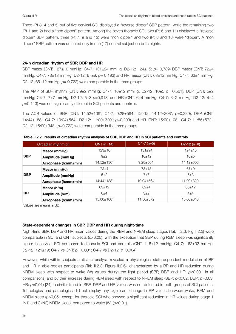
Three (Pt 3, 4 and 5) out of five cervical SCI displayed a “reverse dipper” SBP pattern, while the remaining two (Pt 1 and 2) had a “non dipper” pattern. Among the seven thoracic SCI, two (Pt 6 and 11) displayed a “reverse dipper” SBP pattern, three (Pt 7, 9 and 12) were “non dipper” and two (Pt 8 and 13) were “dipper”. A “non dipper” SBP pattern was detected only in one (17) control subject on both nights.
24-h circadian rhythm of SBP, DBP and HRSBP mesor (CNT: 127±10 mmHg; C4-7: 131±24 mmHg; D2-12: 124±15; p= 0,789) DBP mesor (CNT: 72±4 mmHg; C4-7: 73±13 mmHg; D2-12: 67±9; p= 0,193) and HR mesor (CNT: 63±12 mmHg; C4-7: 62±4 mmHg; D2-12: 65±12 mmHg, p= 0,722) were comparable in the three groups.
The AMP of SBP rhythm (CNT: 9±2 mmHg; C4-7: 16±12 mmHg; D2-12: 10±5 p= 0,561), DBP (CNT: 5±2 mmHg; C4-7: 7±7 mmHg; D2-12: 5±3 p=0,918) and HR (CNT: 6±4 mmHg; C4-7: 3±2 mmHg; D2-12: 4±4 p=0,113) was not significantly different in SCI patients and controls.
The ACR values of SBP (CNT: 14:52±136’; C4-7: 9:28±564’; D2-12: 14:12±308’; p=0,389), DBP (CNT: 14:44±188’; C4-7: 10:04±564’; D2-12: 11:00±320’; p=0,209) and HR (CNT: 15:00±108’; C4-7: 11:56±572’; D2-12: 15:00±348’; p=0,722) were comparable in the three groups.
Table II.2.2 : results of circadian rhythm analysis of SBP, DBP and HR in SCI patients and controls Table II.2.2 : results of circadian rhythm analysis of SBP, DBP and HR in SCI patients and controls Table II.2.2 : results of circadian rhythm analysis of SBP, DBP and HR in SCI patients and controls Table II.2.2 : results of circadian rhythm analysis of SBP, DBP and HR in SCI patients and controls Table II.2.2 : results of circadian rhythm analysis of SBP, DBP and HR in SCI patients and controls
Circadian rhythm ofCircadian rhythm of CNT (n=14) C4-7 (n=5) D2-12 (n=8)
SBPMesor (mmHg) 123±10 131±24 124±15
SBP Amplitude (mmHg) 9±2 16±12 10±5SBP
Acrophase (h:mm±min) 14:52±136’ 9:28±564’ 14:12±308’
DBPMesor (mmHg) 72±4 73±13 67±9
DBP Amplitude (mmHg) 5±2 7±7 5±3DBP
Acrophase (h:mm±min) 14:44±188’ 10:04±564’ 11:00±320’
HRMesor (b/m) 63±12 62±4 65±12
HR Amplitude (b/m) 6±4 3±2 4±4HR
Acrophase (h:mm±min) 15:00±108’ 11:56±572’ 15:00±348’Values are means ± SD.Values are means ± SD.Values are means ± SD.Values are means ± SD.Values are means ± SD.
State-dependent changes in SBP, DBP and HR during night-timeNight-time SBP, DBP and HR mean values during the REM and NREM sleep stages (Tab II.2.3; Fig II.2.5) were comparable in SCI and CNT subjects (p>0,05), with the exception that SBP during REM sleep was significantly higher in cervical SCI compared to thoracic SCI and controls (CNT: 116±12 mmHg; C4-7: 162±32 mmHg; D2-12: 121±19; C4-7 vs CNT: p= 0,001; C4-7 vs D2-12: p=0,004).
However, while within subjects statistical analysis revealed a physiological state-dependent modulation of BP and HR in able-bodies participants (Tab II.2.3; Figure II.2.6), characterized by a BP and HR reduction during NREM sleep with respect to wake (W) values during the light period (SBP, DBP and HR: p<0,001 in all comparisons) and by their increase during REM sleep with respect to NREM sleep (SBP: p<0,02, DBP: p=0,03, HR: p=0,01) [24], a similar trend in SBP, DBP and HR values was not detected in both groups of SCI patients. Tetraplegics and paraplegics did not display any significant change in BP values between wake, REM and NREM sleep (p>0,05), except for thoracic SCI who showed a significant reduction in HR values during stage 1 (N1) and 2 (N2) NREM sleep compared to wake (W) (p<0,01).
Guaraldi P. The circadian rhythm of blood pressure and heart rate in SCI patients
46

Table II.2.3 : state dependent changes in SBP, DBP and HR in SCI patients and controlsTable II.2.3 : state dependent changes in SBP, DBP and HR in SCI patients and controlsTable II.2.3 : state dependent changes in SBP, DBP and HR in SCI patients and controlsTable II.2.3 : state dependent changes in SBP, DBP and HR in SCI patients and controlsTable II.2.3 : state dependent changes in SBP, DBP and HR in SCI patients and controlsParameterParameter CNT (n=14) C4-7 (n=5) D2-12 (n=8)
SBP (mmHg)
W 127±10 132±22 127±14SBP (mmHg) N1 116±12° 119±21 125±16SBP (mmHg)
N2 112±10° 129±29 121±16
SBP (mmHg)
N3 111±8° 121±34 111±10
SBP (mmHg)
REM 116±12*,° 162±32 121±19*DBP (mmHg)
W 75±4 74±12 69±9DBP (mmHg) N1 68±6° 67±10 66±7DBP (mmHg)
N2 66±5° 72±15 65±8
DBP (mmHg)
N3 65±4° 67±19 62±8
DBP (mmHg)
REM 68±6° 77±23 65±10HR (b/m) W 66±13 63±6 68±12HR (b/m)
N1 56±11° 60±4 62±11°HR (b/m)
N2 55±11° 61±3 60±11°
HR (b/m)
N3 54±10° 58±2 61±11
HR (b/m)
REM 56±11° 65±4 62±12Values are means ± SD. *=p<0,05 vs. cervical SCI. °=p<0,05 vs within group wake (W) valuesValues are means ± SD. *=p<0,05 vs. cervical SCI. °=p<0,05 vs within group wake (W) valuesValues are means ± SD. *=p<0,05 vs. cervical SCI. °=p<0,05 vs within group wake (W) valuesValues are means ± SD. *=p<0,05 vs. cervical SCI. °=p<0,05 vs within group wake (W) valuesValues are means ± SD. *=p<0,05 vs. cervical SCI. °=p<0,05 vs within group wake (W) values
Fig II.2.5: state dependent changes in SBP, DBP and HR in SCI and controls (between groups)Fig II.2.3 State dependent changes in SBP, DBP and HR in SCI patients and controls (between groups)
SBP DBP HR
* *
*=p<0,01 vs. cervical SCI *=p<0,01 vs. cervical SCI.
C4-7: cervical SCI; CNT: controls; D2-12: thoracic SCI; SCI: spinal cord injury.
Discussion: To our knowledge this is the first study which assessed the 24-h circadian rhythm in individuals with tetraplegia and paraplegia with continuos recording of cardiovascular parameters under controlled environmental conditions. Furthermore, this is the first study which evaluated the state-dependent modulation of BP and HR in SCI patients and healthy controls.
Section II.2
47
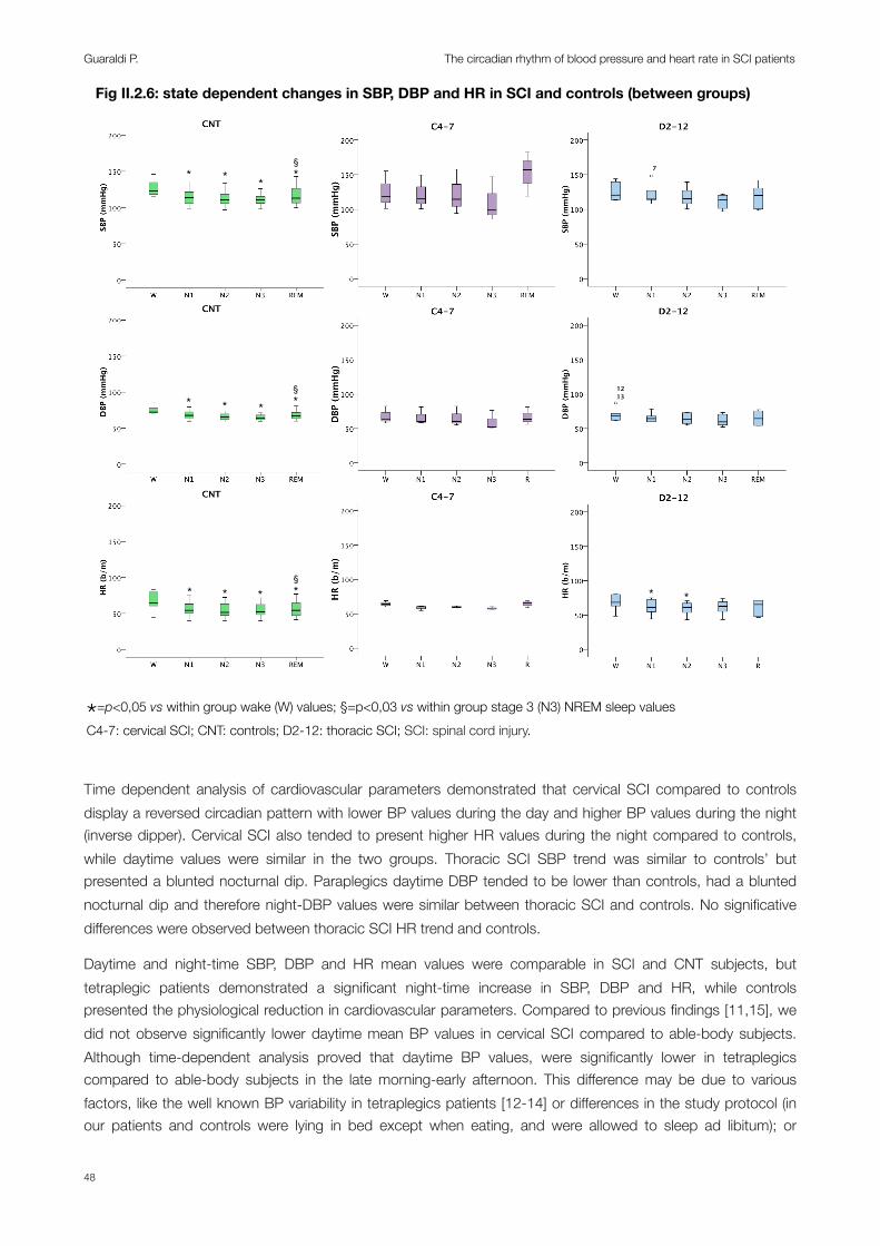
Fig II.2.6: state dependent changes in SBP, DBP and HR in SCI and controls (between groups)Fig II.2.4 State dependent changes in SBP, DBP and HR in SCI patients and controls (within groups)
* * *
* * *§*
* * * * *
*=p<0,05 vs. within group wake (W) values
1213
7§*
§*
*=p<0,05 vs within group wake (W) values; §=p<0,03 vs within group stage 3 (N3) NREM sleep values C4-7: cervical SCI; CNT: controls; D2-12: thoracic SCI; SCI: spinal cord injury.
Time dependent analysis of cardiovascular parameters demonstrated that cervical SCI compared to controls display a reversed circadian pattern with lower BP values during the day and higher BP values during the night (inverse dipper). Cervical SCI also tended to present higher HR values during the night compared to controls, while daytime values were similar in the two groups. Thoracic SCI SBP trend was similar to controls’ but presented a blunted nocturnal dip. Paraplegics daytime DBP tended to be lower than controls, had a blunted nocturnal dip and therefore night-DBP values were similar between thoracic SCI and controls. No significative differences were observed between thoracic SCI HR trend and controls.
Daytime and night-time SBP, DBP and HR mean values were comparable in SCI and CNT subjects, but tetraplegic patients demonstrated a significant night-time increase in SBP, DBP and HR, while controls presented the physiological reduction in cardiovascular parameters. Compared to previous findings [11,15], we did not observe significantly lower daytime mean BP values in cervical SCI compared to able-body subjects. Although time-dependent analysis proved that daytime BP values, were significantly lower in tetraplegics compared to able-body subjects in the late morning-early afternoon. This difference may be due to various factors, like the well known BP variability in tetraplegics patients [12-14] or differences in the study protocol (in our patients and controls were lying in bed except when eating, and were allowed to sleep ad libitum); or
Guaraldi P. The circadian rhythm of blood pressure and heart rate in SCI patients
48
concomitant medications. Therefore, despite this discrepancy, we confirmed the previous findings demonstrating reversed day-night BP changes in cervical SCI and a blunted nocturnal BP dip in paraplegics.
Regarding HR day-night variations, previous studies provided conflicting results. Nitsche et al. reported that complete and incomplete tetraplegic patients preserved the HR day-night physiological modifications [16], while according to Munakata et al. and Rosado Rivera et al. the day-night differences in HR were significantly reduced in cervical SCI compared to thoracic SCI and controls. In a previous study on 8 tetraplegic subjects Christ reported that only 5 patients demonstrated the HR day-night physiological modifications, while 3 didn’t [17]. In our study daytime and night-time mean HR were similar in the three groups, but the nocturnal decline in HR was reduced in tetraplegics compared to controls. Furthermore, time dependent analysis revealed that cervical SCI tended to present higher HR values between 11:00 pm and 3:00 am compared to controls.
The state dependent analysis of SBP, DBP and HR changes during the night-time confirmed that tetraplegics did not present the physiological state-dependent modulation of BP and HR, as observed in controls, characterized by BP and HR reduction during NREM sleep with respect to wake values and by their increase during REM sleep with respect to NREM sleep [24]. Furthermore this analysis revealed that even paraplegics did not present the state-dependent modulation of BP and HR during sleep, despite their near to normal daytime and night-time cardiovascular changes.
Previous studies attributed the reported BP changes to the autonomic dysfunction resulting from the SCI, which determined an absent increase in sympathetic activity during daytime hours in tetraplegics, and to the preservation of physiological nocturnal dipping of BP values in controls [15,16]. This hypothesis was strengthened by the finding of Casiglia et al., who demonstrated that in able-bodied subjects leg resistance was significantly higher during waking hours (when the sympathetic system is more activated) than during sleep, while in subjects with spinal cord injury no difference was detected between day-time and night-time [14]. Similarly, the differences in 24-h HR between tetraplegics and controls were attributed to the disruption of sympathetic chronotropic efferents to the heart in high SCI lesions and to the physiological lowering of HR values during sleep in controls [25]. Although, as demonstrated in Section II.1, the autonomic control of the cardiovascular system is severely affected in cervical SCI but near to normal in thoracic SCI. Therefore, the observed alteration in 24-h BP an HR profile, at least in thoracic SCI, cannot be explained solely by the changes in SNA due to the SCI. Other pathogenetic mechanism may be involved. It is tempting to speculate that the reduction in slow wave sleep we observed in both cervical and thoracic SCI (see Section I) could play a role in altering the 24-h BP and HR profile in these patients, similarly to what was previously described in obstructive sleep apnea (OSA) [26,27] or other disorders causing sleep fragmentation [28]. Whether these finding are the direct consequence of SCI or secondary to the sleep related breathing disorders frequently associated with this condition is still to be determined.
Previous studies demonstrated an increased risk of CVD and mortality in SCI patients [10,29,30]. The increased risk for CVD is not limited to tetraplegic patients, but there is evidence suggesting that individuals with paraplegia are at increased risk for CVD [31], silent ischemia [32], and CHD [33], and the relative risk of CHD can also be 70% greater in individuals with chronic paraplegia compared to individuals with chronic tetraplegia [9]. Furthermore, a recent series of publications found that a third of the Swedish paraplegic population is at greater risk for CVD disease than age-matched non-SCI controls [34-36]. Therefore is tempting to speculate that a reverse/absent or blunted BP nocturnal dip and a failure of the state-dependent modulation of cardiovascular parameters may predispose individuals with tetraplegia and paraplegia to an increased risk of CVD and mortality.
Finally, between subjects state dependent analysis showed that tetraplegics, paraplegics and controls presented comparable SBP, DBP and HR mean values during the REM and NREM sleep stages, with the
Section II.2
49
exception that SBP during REM sleep was significantly higher in cervical SCI compared to thoracic SCI and controls. REM sleep in humans is characterized by a great variability of BP and HR together with breathing irregularities, depending directly on central changes in the regulation of the autonomic outflow that activated peripherally controlled variables. BP rises are sudden, irregular and often wide. During REM, muscle sympathetic nerve activity increases above the levels recorded during wakefulness and this increase is more pronounced than the changes in HR and BP [1,37]. Three out of five cervical patients had a confirmed diagnosis of OSA and one patient reported sleep related breathing disorders. Therefore, we could speculate that the increase in SBP observed during REM sleep in cervical SCI could be related to the breathing irregularities which are prominent during this sleep stage and to the associated mechanical factors and muscle spasms.
Limitations:Although the study was performed under controlled environmental conditions a possible limitation of the study is that episodes of autonomic hyperreflexia related to spasm, bladder distention or other stimuli may have occurred during the recording, especially in cervical SCI. Furthermore for ethical reason we could not discontinue patients medications and the possible effect of drugs should be taken in account. Finally, because the study population was comprised of men with complete spinal cord injuries, applicability of the findings to females or a cohort of individuals with incomplete SCI cannot be ascertained.
Conclusions:Overall, our results confirm previous ambulatory findings which demonstrate that day-night BP changes are abolished/reversed in cervical SCI and blunted in thoracic SCI. Consistent with some previous findings we did not observe any HR day-night variations in the three groups but tetraplegic patients presented higher HR values during the night compared to controls. Furthermore, thanks to state dependent analysis, we demonstrated that both cervical and thoracic SCI did not display the physiological modulation of BP and HR between wake, NREM and REM sleep. Whether these findings are the direct consequence of the autonomic dysfunction due to the SCI or secondary to the sleep alterations observed in these patients (see Section I) is still to be determined. Although, due to the possible relation with increased CVD and mortality cardiovascular autonomic scrutiny should be an integral part of SCI examination and the assessment of the 24-h circadian rhythm of SBP, DBP and HR should be brought in clinical practice. Finally, studies should be designed to investigate the short-term and long-term effects of various interventions (e.g. exercise training, pharmacological interventions) on normalization of 24-h cardiovascular and autonomic function in individuals with SCI.
Guaraldi P. The circadian rhythm of blood pressure and heart rate in SCI patients
50

Bibliography:1. Somers VK, Dyken ME, Mark AL, Abboud FM (1993) Sympathetic-nerve activity during sleep in normal subjects. The New
England journal of medicine 328 (5):303-307.
2. Lundin S, Jern S, Hansson L (1986) Testing neural control of the circulation in patients: hemodynamic responses to exercise and stressful stimuli. In: Zanchetti A, Tarazi RC (eds) Handbook of hypertension. vol 8. Pathophysiology of hypertension : regulatory mechanisms. Elsevier ; New York, NY : Sole distributor for the USA and Canada, Elsevier Science Pub. Co., Amsterdam ; New York, pp xxi, 703 p.
3. Linsell CR, Lightman SL, Mullen PE, Brown MJ, Causon RC (1985) Circadian rhythms of epinephrine and norepinephrine in man. J Clin Endocrinol Metab 60 (6):1210-1215.
4. Elsenbruch S, Harnish MJ, Orr WC (1999) Heart rate variability during waking and sleep in healthy males and females. Sleep 22 (8):1067-1071.
5. O'Brien E, Sheridan J, O'Malley K (1988) Dippers and non-dippers. Lancet 2 (8607):397.
6. Ingelsson E, Bjorklund-Bodegard K, Lind L, Arnlov J, Sundstrom J (2006) Diurnal blood pressure pattern and risk of congestive heart failure. JAMA : the journal of the American Medical Association 295 (24):2859-2866.
7. Verdecchia P, Schillaci G, Gatteschi C, Zampi I, Battistelli M, Bartoccini C, Porcellati C (1993) Blunted nocturnal fall in blood pressure in hypertensive women with future cardiovascular morbid events. Circulation 88 (3):986-992.
8. Samsa GP, Patrick CH, Feussner JR (1993) Long-term survival of veterans with traumatic spinal cord injury. Arch Neurol 50 (9):909-914.
9. Groah SL, Weitzenkamp D, Sett P, Soni B, Savic G (2001) The relationship between neurological level of injury and symptomatic cardiovascular disease risk in the aging spinal injured. Spinal Cord 39 (6):310-317.
10. Myers J, Lee M, Kiratli J (2007) Cardiovascular disease in spinal cord injury: an overview of prevalence, risk, evaluation, and management. Am J Phys Med Rehabil 86 (2):142-152.
11. Rosado-Rivera D, Radulovic M, Handrakis JP, Cirnigliaro CM, Jensen AM, Kirshblum S, Bauman WA, Wecht JM (2011) Comparison of 24-hour cardiovascular and autonomic function in paraplegia, tetraplegia, and control groups: implications for cardiovascular risk. J Spinal Cord Med 34 (4):395-403.
12. Frisbie JH (2007) Unstable baseline blood pressure in chronic tetraplegia. Spinal Cord 45 (1):92-95.
13. Tolbert G, Tuck ML (2004) Ambulatory blood pressure monitoring in persons with chronic spinal cord injury. J Spinal Cord Med 27 (5):476-480.
14. Casiglia E, Pizziol A, Piacentini F, Biasin R, Onesto C, Tikhonoff V, Prati R, Palatini P, Pessina AC (1999) 24-hour leg and forearm haemodynamics in transected spinal cord subjects. Cardiovasc Res 41 (1):312-316.
15. Munakata M, Kameyama J, Kanazawa M, Nunokawa T, Moriai N, Yoshinaga K (1997) Circadian blood pressure rhythm in patients with higher and lower spinal cord injury: simultaneous evaluation of autonomic nervous activity and physical activity. J Hypertens 15 (12 Pt 2):1745-1749.
16. Nitsche B, Perschak H, Curt A, Dietz V (1996) Loss of circadian blood pressure variability in complete tetraplegia. J Hum Hypertens 10 (5):311-317.
17. Christ JE (1979) An analysis of circadian rhythmicity of heart rate in tetraplegic human subjects. Paraplegia 17 (2):251-258.
18. Krum H, Louis WJ, Brown DJ, Jackman GP, Howes LG (1991) Diurnal blood pressure variation in quadriplegic chronic spinal cord injury patients. Clin Sci (Lond) 80 (3):271-276.
Section II.2
51

19. Williams JR (2008) The Declaration of Helsinki and public health. Bulletin of the World Health Organization 86 (8):650-652.
20. Rechtschaffen A, Kales A (1968) A manual of standardized terminology, techniques and scoring system for sleep stages of human subjects. . National Institutes of Health publication,, vol no 204. U. S. National Institute of Neurological Diseases and Blindness, Neurological Information Network, Bethesda, Md.,.
21. O'Brien E, Asmar R, Beilin L, Imai Y, Mallion JM, Mancia G, Mengden T, Myers M, Padfield P, Palatini P, Parati G, Pickering T, Redon J, Staessen J, Stergiou G, Verdecchia P (2003) European Society of Hypertension recommendations for conventional, ambulatory and home blood pressure measurement. J Hypertens 21 (5):821-848.
22. Fagard RH, Staessen JA, Thijs L (1997) Optimal definition of daytime and night-time blood pressure. Blood pressure monitoring 2 (6):315-321.
23. Mojon A, Fernandez JR, Hermida RC (1992) Chronolab: an interactive software package for chronobiologic time series analysis written for the Macintosh computer. Chronobiology international 9 (6):403-412.
24. Mancia G (1993) Autonomic modulation of the cardiovascular system during sleep. The New England journal of medicine 328 (5):347-349.
25. Mathias CJ (2006) Orthostatic hypotension and paroxysmal hypertension in humans with high spinal cord injury. Progress in brain research 152:231-243.
26. Noda A, Okada T, Hayashi H, Yasuma F, Yokota M (1993) 24-hour ambulatory blood pressure variability in obstructive sleep apnea syndrome. Chest 103 (5):1343-1347.
27. Wolf J, Hering D, Narkiewicz K (2010) Non-dipping pattern of hypertension and obstructive sleep apnea syndrome. Hypertension research : official journal of the Japanese Society of Hypertension 33 (9):867-871.
28. Carrington MJ, Trinder J (2008) Blood pressure and heart rate during continuous experimental sleep fragmentation in healthy adults. Sleep 31 (12):1701-1712.
29. Krassioukov A, Claydon VE (2006) The clinical problems in cardiovascular control following spinal cord injury: an overview. Progress in brain research 152:223-229.
30. Garshick E, Kelley A, Cohen SA, Garrison A, Tun CG, Gagnon D, Brown R (2005) A prospective assessment of mortality in chronic spinal cord injury. Spinal Cord 43 (7):408-416.
31. Nash MS, Mendez AJ (2007) A guideline-driven assessment of need for cardiovascular disease risk intervention in persons with chronic paraplegia. Arch Phys Med Rehabil 88 (6):751-757.
32. Bauman WA, Raza M, Spungen AM, Machac J (1994) Cardiac stress testing with thallium-201 imaging reveals silent ischemia in individuals with paraplegia. Arch Phys Med Rehabil 75 (9):946-950.
33. Bauman WA, Spungen AM, Raza M, Rothstein J, Zhang RL, Zhong YG, Tsuruta M, Shahidi R, Pierson RN, Jr., Wang J, et al. (1992) Coronary artery disease: metabolic risk factors and latent disease in individuals with paraplegia. The Mount Sinai journal of medicine, New York 59 (2):163-168.
34. Wahman K, Nash MS, Lewis JE, Seiger A, Levi R (2011) Cardiovascular disease risk and the need for prevention after paraplegia determined by conventional multifactorial risk models: the Stockholm spinal cord injury study. Journal of rehabilitation medicine : official journal of the UEMS European Board of Physical and Rehabilitation Medicine 43 (3):237-242.
35. Wahman K, Nash MS, Lewis JE, Seiger A, Levi R (2010) Increased cardiovascular disease risk in Swedish persons with paraplegia: The Stockholm spinal cord injury study. Journal of rehabilitation medicine : official journal of the UEMS European Board of Physical and Rehabilitation Medicine 42 (5):489-492.
Guaraldi P. The circadian rhythm of blood pressure and heart rate in SCI patients
52
36. Wahman K, Nash MS, Westgren N, Lewis JE, Seiger A, Levi R (2010) Cardiovascular disease risk factors in persons with paraplegia: the Stockholm spinal cord injury study. Journal of rehabilitation medicine : official journal of the UEMS European Board of Physical and Rehabilitation Medicine 42 (3):272-278.
37. Liguori R, Donadio V, Foschini E, Di Stasi V, Plazzi G, Lugaresi E, Montagna P (2000) Sleep stage-related changes in sympathetic sudomotor and vasomotor skin responses in man. Clin Neurophysiol 111 (3):434-439.
Section II.2
53

Guaraldi P. The circadian rhythm of blood pressure and heart rate in SCI patients
54
Section III
BODY CORE TEMPERATURE REGULATION IN SPINAL CORD INJURY PATIENTS
‘‘Something’s wrong. I’m hot. No, wait, now I’m cold. But why am I sweating? I never sweat. My legs won’t stop jumping. Now my hands are in tight fists and my torso is tight as well. Why does my scalp itch? Oh no, the headache is starting. I’m getting dysreflexic.”
Kim Anderson

Section III: BODY CORE TEMPERATURE REGULATION IN SPINAL CORD INJURY PATIENTS
Background:The circadian rhythm of body core temperature (BcT) in diurnally active humans shows a nadir in the morning between 4:00 and 6:00 am and a peak 1–4 h before habitual bedtime [1]. The main temperature regulator in the hypothalamus depends on afferent feedback to adequately regulate BcT [2,3], and relies on the peripheral nervous system to produce heat via muscle activity [4] or to elicit heat loss through vasodilatation and sweat gland activity [5]. If the afferent and/or efferent arms of the sympathetic nerve system are lost, regulation of BcT may be impaired [6,7].
Dependent on the site and extent of the lesion, individuals with a spinal cord injury (SCI) have an abnormal cardiovascular regulation and sudomotor dysfunction below the level of the lesion [8] resulting in an impaired afferent input to, and efferent signals from the central thermoregulatory center [9,10]. Consequently SCI individuals depend on the region above the lesion for thermoregulation [11,12] and may present a thermal dysfunction [6,11,13]. Indeed, hypothermia may occur in patients with high SCI as shivering is diminished and they may be unable to vasoconstrict the cutaneous circulation compared with subjects with a lower lesion [6,11,13]. The reverse may occur causing hyperthermia because of the inability to sweat and to reflexly vasodilate in the periphery, so as to lose heat [14].
Interestingly, thermoregulatory and hypnic mechanisms are thought to be linked [15]. Humans usually initiate sleep on the downward slope of the circadian body temperature rhythm [16], when the rate of change of core temperature and peripheral heat loss are maximal and when endogenous levels of melatonin begin to rise and cortisol decline [17,18]. Body heat loss before lights off, via selective vasodilatation of distal skin regions, promotes the ability to initiate and maintain sleep [19]. Therefore it could be hypothesized that the impaired thermoregulatory function in SCI patient, causing an altered circadian variation of BcT may affect the sleep-wake cycle. Indeed, previous studies have identified a high prevalence of sleep disorders in SCI [20]. The elevated 15–40% prevalence of sleep disorders in SCI individuals [21-24] is higher than the ~4% prevalence typically reported in middle-aged men [25]. Furthermore, nocturnal release of melatonin, a hormone that contributes to the normal sleep pattern [26], contributes to the 24-h variation of BcT. The neural pathway for the endogenous production of melatonin passes through the cervical spinal cord [26,27] and, therefore, may be altered or even absent in SCI individuals with a cervical lesion [28,29].
However, so far only a single recent study examined, with formal chronobiological analyses, the hypothesis that an altered circadian variation of BcT is present in SCI (Fig III.1) [30]. The authors measured continuously and simultaneously intestinal BcT (telemetry system) and physical activity (ambulatory activity monitor) levels in 8 tetraplegics, 7 paraplegics, and 8 able-bodied controls during a 24-h period of “normal” living. In tetraplegics, the mean dominant period length was 6–8 h shorter (p=0,02), whereas the mean BcT during the night-time was significantly higher (p=0,005). Tetraplegics showed a 5-h phase-advanced circadian trough time (p=0,04), and more variable relationships between physical activity and BcT (p=0,03). Taken together these data indicate that tetraplegics demonstrate a pronounced disturbance of the circadian variation of BcT, whereas the variation of BcT in paraplegics was comparable to able-bodied controls.
Section III
55
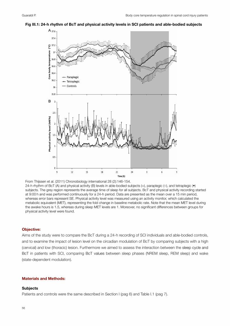
Objective: Aims of the study were to compare the BcT during a 24-h recording of SCI individuals and able-bodied controls, and to examine the impact of lesion level on the circadian modulation of BcT by comparing subjects with a high (cervical) and low (thoracic) lesion. Furthermore we aimed to assess the interaction between the sleep cycle and BcT in patients with SCI, comparing BcT values between sleep phases (NREM sleep, REM sleep) and wake (state-dependent modulation).
Materials and Methods:
SubjectsPatients and controls were the same described in Section I (pag 6) and Table I.1 (pag 7).
Fig III.1: 24-h rhythm of BcT and physical activity levels in SCI patients and able-bodied subjects
impact of time-of-day was found for Tcore reactivity( p = .01; Figure 3). Although no interaction betweentime-of-day and groupwas found ( p = .56), the variabilityin the Tcore–physical activity relationship was signifi-cantly higher for tetraplegics (one-factor general linearmodel [GLM] on within-subjects SD over time-of-day:p = .03). In tetraplegics, the relation of Tcore to physicalactivity changed markedly 4–10 h after sleep. This isconsistent with the early afternoon increase in Tcore intetraplegics, despite little change in physical activity(Figure 2B).
DISCUSSION
The main purpose of this study was to examine the circa-dian variation of the Tcore in able-bodied controls and
SCI individuals with a “high” (cervical) or “low” (thor-acic) lesion. Tcore across the 24-h period in able-bodied controls demonstrated typical 24-h variation,i.e., an early evening peak in Tcore followed by a rapiddecline and subsequent plateau phase in Tcore duringsleep (Lack et al., 2008). However, dependent on lesionlevel, SCI subjects demonstrated a marked difference inthe circadian variation of Tcore. Tetraplegics showed ashorter time period during which the biphasic variationin Tcore was completed and an earlier nocturnalphasing of the Tcore trough value. These differencescannot be fully explained by physical activity levels,since physical activity was consistent over the 24-hperiod in these individuals, thus making relationshipsbetween Tcore and physical activity rather weak. Eventhough paraplegic subjects have a significantly reduced
FIGURE 2. 24-h rhythm of Tcore (A) and physical activity (B) levels in able-bodied subjects ( ), paraplegic (!), and tetraplegic (•) sub-jects. The grey region represents the average time of sleep for all subjects. Tcore and physical activity recording started at 09:00 h and wasperformed continuously for a 24-h period. Data are presented as the mean over a 15-min period, whereas error bars represent SE. Physicalactivity level was measured using an activity monitor, which calculated the metabolic equivalent (MET), representing the fold change inbaseline metabolic rate. Note that the mean MET level during the awake hours is!1.5, whereas during sleep MET levels are!1. Moreover,no significant differences between groups for physical activity level were found.
D. H. J. Thijssen et al.
Chronobiology International
Chr
onob
iol I
nt D
ownl
oade
d fr
om in
form
ahea
lthca
re.c
om b
y D
S D
iffus
ioni
Sci
entif
iche
- U
nica
l on
11/0
7/11
For p
erso
nal u
se o
nly.
From Thijssen et al. (2011) Chronobiology international 28 (2):146-154.24-h rhythm of BcT (A) and physical activity (B) levels in able-bodied subjects (−), paraplegic (○), and tetraplegic (•) subjects. The grey region represents the average time of sleep for all subjects. BcT and physical activity recording started at 9:00 h and was performed continuously for a 24-h period. Data are presented as the mean over a 15 min period, whereas error bars represent SE. Physical activity level was measured using an activity monitor, which calculated the metabolic equivalent (MET), representing the fold change in baseline metabolic rate. Note that the mean MET level during the awake hours is 1.5, whereas during sleep MET levels are 1. Moreover, no significant differences between groups for physical activity level were found.
Guaraldi P. Body core temperature regulation in spinal cord injury patients
56
The investigation conformed with the principles outlined in the Declaration of Helsinki [31]. The protocol was approved by the Institutional Review Board of the University of Bologna. All participants provided written informed consent.
Study protocolBcT and wake-sleep cycle were continuously monitored for 24-h from 11:00 am. Rectal temperature was monitored every 2 minutes by a Mini-loggerTM portable device. The sleep-wake cycle was monitored by an ambulatory polygraphic recorder (Albert Grass Heritage®, Colleague TM PSG Model PSG16P-1, Astro-Med, Inc, West Warwick, RI, USA or Neurofax Electroencephalograph EEG-1200, Nihon-Kohden Corp., Tokyo, Japan) recording electroencephalogram (EEG: C3-A2, C4-A1, O2-A1), right and left electrooculogram, electrocardiogram, and electromyogram of the mylohyoideus, left and right anterior tibialis muscles and thoraco-abdominal breathing. During the study subjects were allowed to sleep ad libitum, living in a temperature (24 ± 1 °C) and humidity (40-50%) controlled room, lying in bed except when eating, in a light-dark schedule (dark period: 11:00 pm-7:00 am). The subjects were placed on a 1.800 kcal/day diet, divided into three meals (8 am, 12:00 am, 6:00 pm) and three snacks (10:00 am, 4:00 pm, 11:00 pm). From midnight preceding the monitoring, subjects were instructed to avoid alcohol and caffeinated beverages and to abstain from smoking.
Sleep parametersThe 24-h sleep-wake cycle starting from 11:00 am on the first day of polysomnographic (PSG) recording was visually scored in 30 s epochs according to the standardized criteria of Rechtschaffen and Kales [32] as light (stages N1 and N2) non-REM sleep (NREM), deep (stages N3) NREM sleep, and REM sleep.
Time-dependent analysisWe compared the mean value of BcT, calculated every 15 min in each participant, in search for between groups significative differences.
Day-night pattern of BcTTo evaluate the day-night pattern of BcT daytime mean values (from 7:00 am to 11:00 pm) and night-time mean values (from 11:00 pm to 7:00 am) were calculated [33,34]. The nocturnal decline of BcT was determined by the difference between night-time and daytime values (∆BcT).
24-h circadian rhythm of BcTRhythmicity was analyzed by evaluating the time series for BcT according to the single cosinor method, using a computerized procedure (Fig III.2) [35]. The procedure determined whether or not there was a rhythm with a 24-h period (p<0,05) and evaluated the following parameters of the cosinor function with their 95% confidence limits: the midline estimating statistic of rhythm (mesor: 24-h mean), the amplitude (AMP: one-half the peak to trough distance of the approximated waveform) and the acrophase (ACR: peak time referred to local midnight hour). For each patient (except patient 12) we analyzed the 24-h rhythmicity of a single day, starting at 11:00 am. For all controls subjects and patient 12, we analyzed the 24-h rhythmicity of two consecutive days, the first (day 1) starting at 11:00 am, and the second (day 2) starting at 8:00 am.
Section III
57
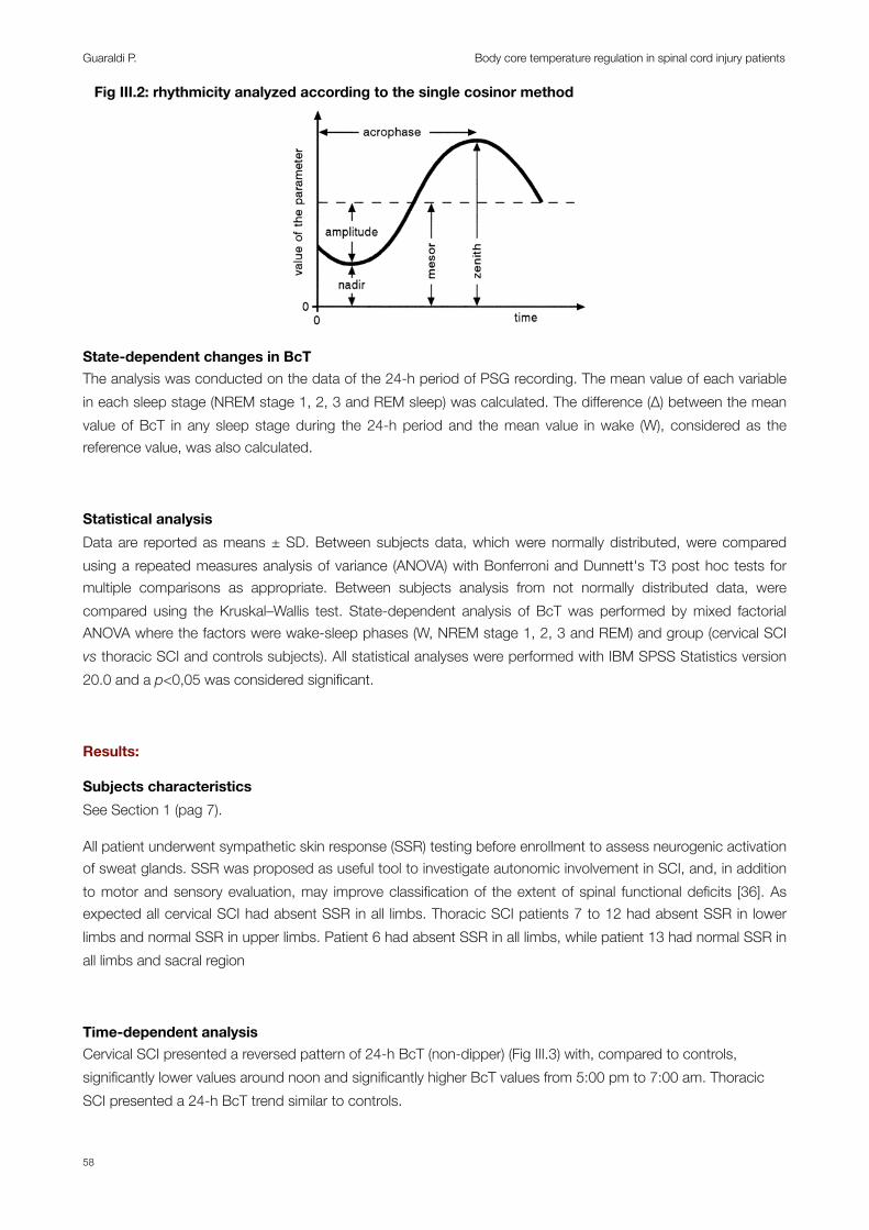
State-dependent changes in BcTThe analysis was conducted on the data of the 24-h period of PSG recording. The mean value of each variable in each sleep stage (NREM stage 1, 2, 3 and REM sleep) was calculated. The difference (∆) between the mean value of BcT in any sleep stage during the 24-h period and the mean value in wake (W), considered as the reference value, was also calculated.
Statistical analysisData are reported as means ± SD. Between subjects data, which were normally distributed, were compared using a repeated measures analysis of variance (ANOVA) with Bonferroni and Dunnett's T3 post hoc tests for multiple comparisons as appropriate. Between subjects analysis from not normally distributed data, were compared using the Kruskal–Wallis test. State-dependent analysis of BcT was performed by mixed factorial ANOVA where the factors were wake-sleep phases (W, NREM stage 1, 2, 3 and REM) and group (cervical SCI vs thoracic SCI and controls subjects). All statistical analyses were performed with IBM SPSS Statistics version 20.0 and a p<0,05 was considered significant.
Results:
Subjects characteristicsSee Section 1 (pag 7).
All patient underwent sympathetic skin response (SSR) testing before enrollment to assess neurogenic activation of sweat glands. SSR was proposed as useful tool to investigate autonomic involvement in SCI, and, in addition to motor and sensory evaluation, may improve classification of the extent of spinal functional deficits [36]. As expected all cervical SCI had absent SSR in all limbs. Thoracic SCI patients 7 to 12 had absent SSR in lower limbs and normal SSR in upper limbs. Patient 6 had absent SSR in all limbs, while patient 13 had normal SSR in all limbs and sacral region
Time-dependent analysisCervical SCI presented a reversed pattern of 24-h BcT (non-dipper) (Fig III.3) with, compared to controls, significantly lower values around noon and significantly higher BcT values from 5:00 pm to 7:00 am. Thoracic SCI presented a 24-h BcT trend similar to controls.
Fig III.2: rhythmicity analyzed according to the single cosinor method
Guaraldi P. Body core temperature regulation in spinal cord injury patients
58

Fig III.3: 24-h pattern of BcT in SCI patients and controlsFig III.1 24-h pattern of BcT in SCI patients and controls
BcT(°C)
36
37
38
39
08.00 10.00 12.00 14.00 16.00 18.00 20.00 22.00 00.00 02.00 04.00 06.00
BcT: body core temperature; C4-7: cervical SCI; CNT: controls; D2-12: thoracic SCI; SCI: spinal cord injury.
Day-night pattern of BcTThe 24-h pattern of BcT is shown in Fig III.3 and summarized in Table III.1, Fig III.4 and Fig III.5. Daytime and night-time BcT mean values were significantly higher in cervical SCI compared to thoracic SCI and CNT subjects (p=0,009 and p=0,004 respectively for daytime BcT; p=0,000 and p=0,000 respectively for night-time BcT). The night-time fall in BcT (∆BcT) (Fig III.4, Fig III.5) was significantly reduced in tetraplegic patients compared to controls and paraplegics (p=0,001 and p=0,048 respectively).
Table III.1: daytime BcT, night-time BcT and day-night changes (∆) in BcT in SCI and controlsTable III.1: daytime BcT, night-time BcT and day-night changes (∆) in BcT in SCI and controlsTable III.1: daytime BcT, night-time BcT and day-night changes (∆) in BcT in SCI and controlsTable III.1: daytime BcT, night-time BcT and day-night changes (∆) in BcT in SCI and controlsTable III.1: daytime BcT, night-time BcT and day-night changes (∆) in BcT in SCI and controls
Circadian rhythm ofCircadian rhythm of CNT (n=14) C4-7 (n=6) D2-12 (n=8)
BcT (°C)Daytime 37,07±0,16* 37,56±0,55 36,98±0,20*
BcT (°C) Night-time 36,63±0,20* 37,76±0,59 36,76±0,33*BcT (°C)
∆ -0,44±0,23* 0,2±0,46 -0,22±0,27*Values are means ± SD. *=p<0,05 vs cervical SCI. Values are means ± SD. *=p<0,05 vs cervical SCI. Values are means ± SD. *=p<0,05 vs cervical SCI. Values are means ± SD. *=p<0,05 vs cervical SCI. Values are means ± SD. *=p<0,05 vs cervical SCI.
Fig III.4: mean daytime, night-time and day-night changes (∆) in BcT in SCI patients and controls
* *
daytime BcT (°C) night-time BcT (°C) ∆BcT (°C)
Fig III.2 Daytime, night-time and Delta (∆) day-night BcT in SCI patients and controls
* ** *
*=p<0,05. BcT: body core temperature; C4-7: cervical SCI; CNT: controls; D2-12: thoracic SCI; ∆: day-night changes.
Section III
59
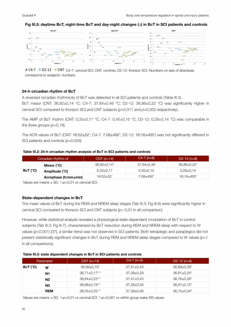
Fig III.5: daytime BcT, night-time BcT and day-night changes (∆) in BcT in SCI patients and controls
1"
2"
2,5"
3"
4"
5"
6"
7"
8" 9"
11"
12"
12,5"
13"
14"14,5"
15"15,5"
16"16,5"
17"
17,5"
18,5"
19"19,5"
20"20,5"
36,5"
37"
37,5"
38"
38,5"
39"
0" 5" 10" 15" 20" 25"
BcT$(C°)$
Subjects'$n°$
day$BcT$$
1"
2"2,5"
3"
4"
5"
6"
7"
8"
9"
11"
12"
12,5"
13" 14"
14,5"15"
15,5"
16"16,5"
17"17,5"
18,5"19"
19,5"20"20,5"
36"
36,5"
37"
37,5"
38"
38,5"
39"
0" 5" 10" 15" 20" 25"
BcT$(C°)$
Subjects'$n°$
night$BcT$$
1"
2"
2,5"
3"
4"
5"
6"
7"
8"
9"
11" 12"
12,5"
13"
14"
14,5"
15"15,5"
16"16,5"
17"17,5"
18,5"19"
19,5"20"20,5"
-1,2"
-1"
-0,8"
-0,6"
-0,4"
-0,2"
0"
0,2"
0,4"
0,6"
0,8"
0" 5" 10" 15" 20" 25"
∆"BC
T"(C°)"
Subjects'"n°"
∆BCT""
4"
5" 6"
7"8" 9"
11"
12"
13"
14"
15"
16"
17"18"
19"
20"
21"
22"23"
24"
25"
27"
28"
29"
30"
31"
32"
33"
34"
35"
36"
37"
5" 10" 15" 20" 25" 30" 35" 40"Pa#ents')n°)
C4-7" D2-12" CNT" C4-7: cervical SCI; CNT: controls; D2-12: thoracic SCI. Numbers on axis of abscissas correspond to subjects’ numbers.
24-h circadian rhythm of BcTA reversed circadian rhythmicity of BcT was detected in all SCI patients and controls (Table III.2).BcT mesor (CNT: 36,92±0,14 °C; C4-7: 37,64±0,49 °C; D2-12: 36,86±0,22 °C) was significantly higher in cervical SCI compared to thoracic SCI and CNT subjects (p=0,011 and p=0,002 respectively).
The AMP of BcT rhythm (CNT: 0,33±0,17 °C; C4-7: 0,45±0,15 °C; D2-12: 0,29±0,14 °C) was comparable in the three groups (p=0,19).
The ACR values of BcT (CNT: 16:52±52’; C4-7: 7:08±492’; D2-12: 16:16±400’) was not significantly different in SCI patients and controls (p=0,059)
Table III.2: 24-h circadian rhythm analysis of BcT in SCI patients and controls Table III.2: 24-h circadian rhythm analysis of BcT in SCI patients and controls Table III.2: 24-h circadian rhythm analysis of BcT in SCI patients and controls Table III.2: 24-h circadian rhythm analysis of BcT in SCI patients and controls Table III.2: 24-h circadian rhythm analysis of BcT in SCI patients and controls
Circadian rhythm ofCircadian rhythm of CNT (n=14) C4-7 (n=6) D2-12 (n=8)
BcT (°C)Mesor (°C) 36,92±0,14* 37,64±0,49 36,86±0,22*
BcT (°C) Amplitude (°C) 0,33±0,17 0,45±0,15 0,29±0,14BcT (°C)
Acrophase (h:mm±min) 16:52±52’ 7:08±492’ 16:16±400’Values are means ± SD. *=p<0,01 vs cervical SCI. Values are means ± SD. *=p<0,01 vs cervical SCI. Values are means ± SD. *=p<0,01 vs cervical SCI. Values are means ± SD. *=p<0,01 vs cervical SCI. Values are means ± SD. *=p<0,01 vs cervical SCI.
State-dependent changes in BcTThe mean values of BcT during the REM and NREM sleep stages (Tab III.3; Fig III.6) were significantly higher in cervical SCI compared to thoracic SCI and CNT subjects (p< 0,01 in all comparison).
However, while statistical analysis revealed a physiological state-dependent modulation of BcT in control subjects (Tab III.3; Fig III.7), characterized by BcT reduction during REM and NREM sleep with respect to W values (p<0,001) [37], a similar trend was not observed in SCI patients. Both tetraplegic and paraplegics did not present statistically significant changes in BcT during REM and NREM sleep stages compared to W values (p=1 in all comparisons).
Table III.3: state dependent changes in BcT in SCI patients and controlsTable III.3: state dependent changes in BcT in SCI patients and controlsTable III.3: state dependent changes in BcT in SCI patients and controlsTable III.3: state dependent changes in BcT in SCI patients and controlsTable III.3: state dependent changes in BcT in SCI patients and controls
ParameterParameter CNT (n=14) C4-7 (n=5) D2-12 (n=8)BcT (°C) W 36,99±0,15* 37,51±0,43 36,89±0,28*BcT (°C)
N1 36,71±0,17*,° 37,38±0,29 36,81±0,24*BcT (°C)
N2 36,64±0,23*,° 37,47±0,43 36,76±0,28*
BcT (°C)
N3 36,68±0,19*,° 37,28±0,34 36,67±0,12*
BcT (°C)
REM 36,55±0,25*,° 37,39±0,46 36,75±0,34*
Values are means ± SD. *=p<0,01 vs cervical SCI. °=p<0,001 vs within group wake (W) values Values are means ± SD. *=p<0,01 vs cervical SCI. °=p<0,001 vs within group wake (W) values Values are means ± SD. *=p<0,01 vs cervical SCI. °=p<0,001 vs within group wake (W) values Values are means ± SD. *=p<0,01 vs cervical SCI. °=p<0,001 vs within group wake (W) values Values are means ± SD. *=p<0,01 vs cervical SCI. °=p<0,001 vs within group wake (W) values
Guaraldi P. Body core temperature regulation in spinal cord injury patients
60
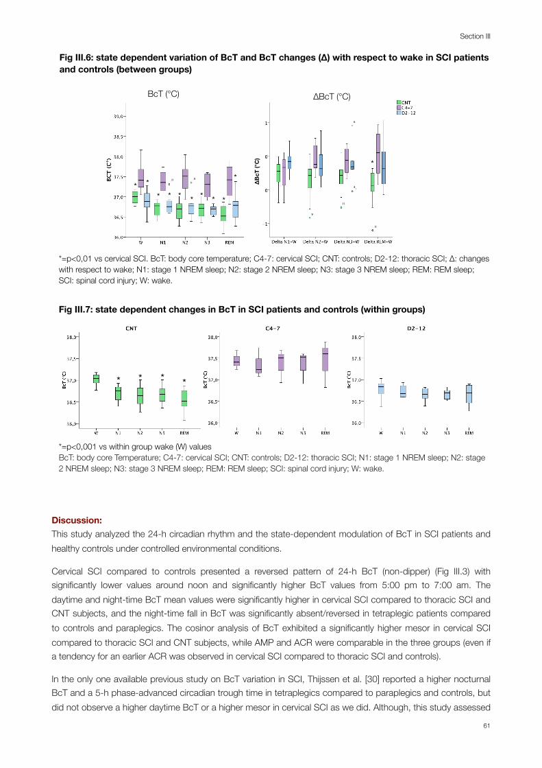
Fig III.6: state dependent variation of BcT and BcT changes (∆) with respect to wake in SCI patients and controls (between groups)Fig III.3 State dependent BcT and changes (∆) with respect to wake in SCI patients and controls (between groups)
∆BcT (°C)
*
BcT (°C)
* ** * * * * * *
*1111
9
11
11
6
4
∆BcT
(°C
)*=p<0,01 vs cervical SCI. BcT: body core temperature; C4-7: cervical SCI; CNT: controls; D2-12: thoracic SCI; ∆: changes with respect to wake; N1: stage 1 NREM sleep; N2: stage 2 NREM sleep; N3: stage 3 NREM sleep; REM: REM sleep; SCI: spinal cord injury; W: wake.
Fig III.7: state dependent changes in BcT in SCI patients and controls (within groups)Fig III.4 State dependent changes in BcT in SCI patients and controls (within groups)
*=p<0,05 vs. within group wake (W) values
* * * *
*=p<0,001 vs within group wake (W) valuesBcT: body core Temperature; C4-7: cervical SCI; CNT: controls; D2-12: thoracic SCI; N1: stage 1 NREM sleep; N2: stage 2 NREM sleep; N3: stage 3 NREM sleep; REM: REM sleep; SCI: spinal cord injury; W: wake.
Discussion: This study analyzed the 24-h circadian rhythm and the state-dependent modulation of BcT in SCI patients and healthy controls under controlled environmental conditions.
Cervical SCI compared to controls presented a reversed pattern of 24-h BcT (non-dipper) (Fig III.3) with significantly lower values around noon and significantly higher BcT values from 5:00 pm to 7:00 am. The daytime and night-time BcT mean values were significantly higher in cervical SCI compared to thoracic SCI and CNT subjects, and the night-time fall in BcT was significantly absent/reversed in tetraplegic patients compared to controls and paraplegics. The cosinor analysis of BcT exhibited a significantly higher mesor in cervical SCI compared to thoracic SCI and CNT subjects, while AMP and ACR were comparable in the three groups (even if a tendency for an earlier ACR was observed in cervical SCI compared to thoracic SCI and controls).
In the only one available previous study on BcT variation in SCI, Thijssen et al. [30] reported a higher nocturnal BcT and a 5-h phase-advanced circadian trough time in tetraplegics compared to paraplegics and controls, but did not observe a higher daytime BcT or a higher mesor in cervical SCI as we did. Although, this study assessed
Section III
61

patients during their standard routine activities at their home, and, even if they were required to remain indoors, constant stable environmental condition could not be guaranteed. The current study, instead, was performed under controlled ambient conditions, in order to exclude the well documented influence exerted on BcT rhythm by external factors such as light-dark cycle, food intake, sleep, room temperature, humidity, and body position (the so called photic and non-photic entrainment (zeitgebers) of the human circadian system) [38,39]. The presence/absence of these confounders may explain the discrepancies between their findings and our results. Similarly to Thijssen et al. we did not find any important differences in the paraplegic 24-h variation in BcT compared to controls. Therefore, even if thoracic SCI have a reduced surface area available for thermoregulation, this may be enough to maintain BcT in the physiological range, or different mechanisms, other than skin thermoregulation, may be involved in 24-h regulation of BcT. Based on previous observations demonstrating that the neural pathway for the endogenous production of melatonin passes through the cervical spinal cord [27,29] and that tetraplegics show no nocturnal release of melatonin [28,29] Thijssen et al. hypothesized that these changes in endocrine function may contribute to the observed differences in BcT, especially since tetraplegics demonstrated the most pronounced change in the 24-h variation in BcT. However, no previous study examined contextually melatonin release and BcT in SCI in order to establish a relationship between these two variables. Furthermore, our data demonstrate a 24-h (daytime and night-time) elevation of BcT in cervical SCI compared to thoracic SCI and CNT subjects which may be difficult to relate to the pulsatile secretion of melatonin.
Important new insights were provided by the state-dependent analysis of BcT in our study. This analysis revealed, as expected, that BcT was significantly higher in cervical SCI during wake, NREM and REM sleep stages compared to controls and thoracic SCI. Furthermore, it demonstrated that both tetraplegics and paraplegics did not display the physiological modulation of BcT between wake, NREM and REM sleep during the 24-h. Indeed, BcT was not reduced during REM and NREM sleep with respect to wakefulness in cervical and thoracic SCI patients.
In Section I we discussed the differences occurring in the sleep structure between SCI patients and controls, demonstrating that both cervical and thoracic SCI presented a higher prevalence of light sleep and a reduction in slow wave sleep (SWS) (Section I, Fig I.1, Fig I.2, Table I.3). As demonstrated by previous studies, the sleep disturbances observed in patients with SCI cannot be explained solely by disruptions in breathing during sleep [21,40,41]. Indeed, even if sleep related breathing disorders (SRBD) may exert an important role in determining the higher number of arousals observed in our cervical SCI group (Section I, Fig I.3, Fig I.4, Table I.3) and their increased proportion of light sleep, they could not explain the alteration in sleep architecture observed in thoracic SCI, since paraplegics had a apnea-hypopnea index (AHI) comparable to controls (Section I, Table I.3).
Therefore, other factors may play a role in determining the predominance of light sleep observed in tetraplegics and paraplegics, including their inability to regulate thermal function. Although, the reverse could be also possible, being the reduced SWS to interfere with body temperature changes. Further studies will be necessary to assess the relationship between the sleep disorders and the circadian variation of BcT observed in SCI patients.
Conclusions:
Overall, our results confirm previous findings which demonstrate that tetraplegics present a pronounced disturbance of the circadian variation of BcT. Furthermore, thanks to state dependent analysis, we also demonstrated that both cervical and thoracic SCI did not display the physiological modulation of BcT between
Guaraldi P. Body core temperature regulation in spinal cord injury patients
62

wake, NREM and REM sleep. The pathogenetic mechanism and the possible consequence of such alterations are yet to be determined.
Section III
63

Bibliography:1. Waterhouse J, Drust B, Weinert D, Edwards B, Gregson W, Atkinson G, Kao S, Aizawa S, Reilly T (2005) The circadian
rhythm of core temperature: origin and some implications for exercise performance. Chronobiology international 22 (2):207-225.
2. Berner NJ, Heller HC (1998) Does the preoptic anterior hypothalamus receive thermoafferent information? Am J Physiol 274 (1 Pt 2):R9-18.
3. Satinoff E (1978) Neural organization and evolution of thermal regulation in mammals. Science 201 (4350):16-22.
4. Morton JP, MacLaren DP, Cable NT, Bongers T, Griffiths RD, Campbell IT, Evans L, Kayani A, McArdle A, Drust B (2006) Time course and differential responses of the major heat shock protein families in human skeletal muscle following acute nondamaging treadmill exercise. J Appl Physiol 101 (1):176-182.
5. Johnson JM, Kellogg DL, Jr. (2010) Thermoregulatory and thermal control in the human cutaneous circulation. Front Biosci (Schol Ed) 2:825-853.
6. Boot CR, Binkhorst RA, Hopman MT (2006) Body temperature responses in spinal cord injured individuals during exercise in the cold and heat. International journal of sports medicine 27 (8):599-604.
7. Price MJ, Campbell IG (1999) Thermoregulatory responses of spinal cord injured and able-bodied athletes to prolonged upper body exercise and recovery. Spinal Cord 37 (11):772-779.
8. Mathias CJ, Frankel HL (2002) Autonomic disturbances in spinal cord lesions. In: Mathias CJ, Bannister R (eds) Autonomic failure : a textbook of clinical disorders of the autonomic nervous system. 4th edn. Oxford University Press, Oxford ; New York, pp 494–513.
9. Freund PR, Brengelmann GL, Rowell LB, Halar E (1984) Attenuated skin blood flow response to hyperthermia in paraplegic men. J Appl Physiol 56 (4):1104-1109.
10. Theisen D, Vanlandewijck Y, Sturbois X, Francaux M (2001) Cutaneous vascular response and thermoregulation in individuals with paraplegia during sustained arm-cranking exercise. International journal of sports medicine 22 (2):97-102.
11. Guttmann L, Silver J, Wyndham CH (1958) Thermoregulation in spinal man. J Physiol 142 (3):406-419.
12. Sawka MN, Latzka WA, Pandolf KB (1989) Temperature regulation during upper body exercise: able-bodied and spinal cord injured. Medicine and science in sports and exercise 21 (5 Suppl):S132-140.
13. Khan S, Plummer M, Martinez-Arizala A, Banovac K (2007) Hypothermia in patients with chronic spinal cord injury. J Spinal Cord Med 30 (1):27-30.
14. Price MJ (2006) Thermoregulation during exercise in individuals with spinal cord injuries. Sports Med 36 (10):863-879.
15. Atkinson G, Davenne D (2007) Relationships between sleep, physical activity and human health. Physiology & behavior 90 (2-3):229-235.
16. Czeisler CA, Zimmerman JC, Ronda JM, Moore-Ede MC, Weitzman ED (1980) Timing of REM sleep is coupled to the circadian rhythm of body temperature in man. Sleep 2 (3):329-346.
17. Campbell SS, Broughton RJ (1994) Rapid decline in body temperature before sleep: fluffing the physiological pillow? Chronobiology international 11 (2):126-131.
18. Zulley J, Wever R, Aschoff J (1981) The dependence of onset and duration of sleep on th circadian rhythm of rectal temperature. Pflugers Archiv : European journal of physiology 391 (4):314-318.
Guaraldi P. Body core temperature regulation in spinal cord injury patients
64

19. Raymann RJ, Swaab DF, Van Someren EJ (2007) Skin temperature and sleep-onset latency: changes with age and insomnia. Physiology & behavior 90 (2-3):257-266.
20. Scheer FA, Zeitzer JM, Ayas NT, Brown R, Czeisler CA, Shea SA (2006) Reduced sleep efficiency in cervical spinal cord injury; association with abolished night time melatonin secretion. Spinal Cord 44 (2):78-81.
21. Biering-Sorensen F, Biering-Sorensen M (2001) Sleep disturbances in the spinal cord injured: an epidemiological questionnaire investigation, including a normal population. Spinal Cord 39 (10):505-513.
22. Burns SP, Little JW, Hussey JD, Lyman P, Lakshminarayanan S (2000) Sleep apnea syndrome in chronic spinal cord injury: associated factors and treatment. Arch Phys Med Rehabil 81 (10):1334-1339.
23. Hyyppa MT, Kronholm E (1989) Quality of sleep and chronic illnesses. Journal of clinical epidemiology 42 (7):633-638.
24. Jensen MP, Hirsh AT, Molton IR, Bamer AM (2009) Sleep problems in individuals with spinal cord injury: frequency and age effects. Rehabil Psychol 54 (3):323-331.
25. Young T, Palta M, Dempsey J, Skatrud J, Weber S, Badr S (1993) The occurrence of sleep-disordered breathing among middle-aged adults. The New England journal of medicine 328 (17):1230-1235.
26. Pandi-Perumal SR, Trakht I, Spence DW, Srinivasan V, Dagan Y, Cardinali DP (2008) The roles of melatonin and light in the pathophysiology and treatment of circadian rhythm sleep disorders. Nature clinical practice Neurology 4 (8):436-447.
27. Moore RY (1996) Neural control of the pineal gland. Behavioural brain research 73 (1-2):125-130.
28. Li Y, Jiang DH, Wang ML, Jiao DR, Pang SF (1989) Rhythms of serum melatonin in patients with spinal lesions at the cervical, thoracic or lumbar region. Clinical endocrinology 30 (1):47-56.
29. Zeitzer JM, Ayas NT, Shea SA, Brown R, Czeisler CA (2000) Absence of detectable melatonin and preservation of cortisol and thyrotropin rhythms in tetraplegia. J Clin Endocrinol Metab 85 (6):2189-2196.
30. Thijssen DH, Eijsvogels TM, Hesse M, Ballak DB, Atkinson G, Hopman MT (2011) The effects of thoracic and cervical spinal cord lesions on the circadian rhythm of core body temperature. Chronobiology international 28 (2):146-154.
31. Williams JR (2008) The Declaration of Helsinki and public health. Bulletin of the World Health Organization 86 (8):650-652.
32. Rechtschaffen A, Kales A (1968) A manual of standardized terminology, techniques and scoring system for sleep stages of human subjects. . National Institutes of Health publication,, vol no 204. U. S. National Institute of Neurological Diseases and Blindness, Neurological Information Network, Bethesda, MD.
33. O'Brien E, Asmar R, Beilin L, Imai Y, Mallion JM, Mancia G, Mengden T, Myers M, Padfield P, Palatini P, Parati G, Pickering T, Redon J, Staessen J, Stergiou G, Verdecchia P (2003) European Society of Hypertension recommendations for conventional, ambulatory and home blood pressure measurement. J Hypertens 21 (5):821-848.
34. Fagard RH, Staessen JA, Thijs L (1997) Optimal definition of daytime and night-time blood pressure. Blood pressure monitoring 2 (6):315-321.
35. Mojon A, Fernandez JR, Hermida RC (1992) Chronolab: an interactive software package for chronobiologic time series analysis written for the Macintosh computer. Chronobiology international 9 (6):403-412.
36. Nicotra A, Catley M, Ellaway PH, Mathias CJ (2005) The ability of physiological stimuli to generate the sympathetic skin response in human chronic spinal cord injury. Restorative neurology and neuroscience 23 (5-6):331-339.
Section III
65

37. Mancia G (1993) Autonomic modulation of the cardiovascular system during sleep. The New England journal of medicine 328 (5):347-349.
38. Mayer G, Hellmann F, Leonhard E, Meier-Ewert K (1997) Circadian temperature and activity rhythms in unmedicated narcoleptic patients. Pharmacology, biochemistry, and behavior 58 (2):395-402.
39. Mistlberger RE, Skene DJ (2005) Nonphotic entrainment in humans? Journal of biological rhythms 20 (4):339-352.
40. McEvoy RD, Mykytyn I, Sajkov D, Flavell H, Marshall R, Antic R, Thornton AT (1995) Sleep apnoea in patients with quadriplegia. Thorax 50 (6):613-619.
41. Adey WR, Bors E, Porter RW (1968) EEG sleep patterns after high cervical lesions in man. Arch Neurol 19 (4):377-383.
Guaraldi P. Body core temperature regulation in spinal cord injury patients
66

CONCLUSIONS


CONCLUSIONS
Overall the major findings of the current study are:
1. Chronic cervical and thoracic SCI have a compromised sleep, characterized by an augmented proportion N2 NREM sleep and reduced SWS compared to controls. No statistical differences in any sleep parameter were found between cervical and thoracic SCI patients. While the sleep disturbances in cervical SCI could be related to SRBD, higher AHI, and possibly to an alteration in melatonin secretion, the pathogenesis of the sleep disorder in thoracic SCI is yet to be determined.
2. Abnormal cardiovascular regulation is correlated with the level of SCI. Cardiovascular autonomic dysfunction, including orthostatic hypotension, was common in tetraplegics, while paraplegics presented near to normal cardiovascular reflexes.
3. The assessment of HRV demonstrated that tetraplegics and paraplegics despite the interruption of the sympathetic outflow, are able to maintain homeostasis in supine position. During HUTT instead, cervical SCI demonstrated an unbalanced sympatho-vagal modulation while paraplegics presented near to normal responses.
4. Day-night BP changes are abolished/reversed in cervical SCI and blunted in thoracic SCI. We did not observe any HR day-night variations in the three groups but tetraplegic patients presented higher HR values during the night compared to controls.
5. State dependent analysis of BP and HR changes demonstrated that both cervical and thoracic SCI did not display the physiological modulation of BP and HR between wake, NREM and REM sleep. Whether these finding are the direct consequence of the autonomic dysfunction due to the SCI or secondary to the sleep alterations observed in these patients is still to be determined.
6. Tetraplegics present a pronounced disturbance of the circadian variation of BcT.
7. State dependent analysis of variation of BcT, revealed that both cervical and thoracic SCI did not display the physiological modulation of BcT between wake, NREM and REM sleep. The pathogenetic mechanism and the possible consequence of such alterations are yet to be determined.
Further studies should be designed to investigate the effects of various interventions (e.g. treating OSA, reducing PLMS, ameliorating the thermoregulation, etc...) on the association between sleep alteration, autonomic dysfunction and thermoregulatory failure we documented, in order to detect the presence and the direction of a causal relationship between these parameters.
67

68

BIBLIOGRAPHY
- Adey WR, Bors E, Porter RW (1968) EEG sleep patterns after high cervical lesions in man. Arch Neurol 19 (4):377-383.
- Akselrod S (1988) Spectral analysis of fluctuations in cardiovascular parameters: a quantitative tool for the investigation of autonomic control. Trends in pharmacological sciences 9 (1):6-9.
- Akselrod S, Gordon D, Madwed JB, Snidman NC, Shannon DC, Cohen RJ (1985) Hemodynamic regulation: investigation by spectral analysis. Am J Physiol 249 (4 Pt 2):H867-875.
- Alexander MS, Biering-Sorensen F, Bodner D, Brackett NL, Cardenas D, Charlifue S, Creasey G, Dietz V, Ditunno J, Donovan W, Elliott SL, Estores I, Graves DE, Green B, Gousse A, Jackson AB, Kennelly M, Karlsson AK, Krassioukov A, Krogh K, Linsenmeyer T, Marino R, Mathias CJ, Perkash I, Sheel AW, Schilero G, Schurch B, Sonksen J, Stiens S, Wecht J, Wuermser LA, Wyndaele JJ (2009) International standards to document remaining autonomic function after spinal cord injury. Spinal Cord 47 (1):36-43.
- Atkinson G, Davenne D (2007) Relationships between sleep, physical activity and human health. Physiology & behavior 90 (2-3):229-235.
- Ayas NT, Epstein LJ, Lieberman SL, Tun CG, Larkin EK, Brown R, Garshick E (2001) Predictors of loud snoring in persons with spinal cord injury. J Spinal Cord Med 24 (1):30-34.
- Baselli G, Cerutti S, Civardi S, Liberati D, Lombardi F, Malliani A, Pagani M (1986) Spectral and cross-spectral analysis of heart rate and arterial blood pressure variability signals. Computers and biomedical research, an international journal 19 (6):520-534.
- Bauman WA, Raza M, Spungen AM, Machac J (1994) Cardiac stress testing with thallium-201 imaging reveals silent ischemia in individuals with paraplegia. Arch Phys Med Rehabil 75 (9):946-950.
- Bauman WA, Spungen AM, Raza M, Rothstein J, Zhang RL, Zhong YG, Tsuruta M, Shahidi R, Pierson RN, Jr., Wang J, et al. (1992) Coronary artery disease: metabolic risk factors and latent disease in individuals with paraplegia. The Mount Sinai journal of medicine, New York 59 (2):163-168.
- Bensmail D, Marquer A, Roche N, Godard AL, Lofaso F, Quera-Salva MA (2012) Pilot study assessing the impact of intrathecal baclofen administration mode on sleep-related respiratory parameters. Arch Phys Med Rehabil 93 (1):96-99.
- Bensmail D, Quera Salva MA, Roche N, Benyahia S, Bohic M, Denys P, Bussel B, Lofaso F (2006) Effect of intrathecal baclofen on sleep and respiratory function in patients with spasticity. Neurology 67 (8):1432-1436.
- Berner NJ, Heller HC (1998) Does the preoptic anterior hypothalamus receive thermoafferent information? Am J Physiol 274 (1 Pt 2):R9-18.
- Biering-Sorensen F, Biering-Sorensen M (2001) Sleep disturbances in the spinal cord injured: an epidemiological questionnaire investigation, including a normal population. Spinal Cord 39 (10):505-513.
- Biering-Sorensen F, Jennum P, Laub M (2009) Sleep disordered breathing following spinal cord injury. Respir Physiol Neurobiol 169 (2):165-170.
Bibliography
69

- Bonekat HW, Andersen G, Squires J (1990) Obstructive disordered breathing during sleep in patients with spinal cord injury. Paraplegia 28 (6):392-398.
- Boot CR, Binkhorst RA, Hopman MT (2006) Body temperature responses in spinal cord injured individuals during exercise in the cold and heat. International journal of sports medicine 27 (8):599-604.
- Brown LK, Heffner JE, Obbens EA (2000) Transverse myelitis associated with restless legs syndrome and periodic movements of sleep responsive to an oral dopaminergic agent but not to intrathecal baclofen. Sleep 23 (5):591-594.
- Brown R, Engel S, Wallin BG, Elam M, Macefield V (2007) Assessing the integrity of sympathetic pathways in spinal cord injury. Auton Neurosci 134 (1-2):61-68.
- Brown R, Macefield VG (2008) Assessing the capacity of the sympathetic nervous system to respond to a cardiovascular challenge in human spinal cord injury. Spinal Cord 46 (10):666-672.
- Brown R, Stolzenhein G, Engel S, Macefield VG (2009) Cutaneous vasoconstriction as a measure of incipient autonomic dysreflexia during penile vibratory stimulation in spinal cord injury. Spinal Cord 47 (7):538-544.
- Burns SP, Little JW, Hussey JD, Lyman P, Lakshminarayanan S (2000) Sleep apnea syndrome in chronic spinal cord injury: associated factors and treatment. Arch Phys Med Rehabil 81 (10):1334-1339.
- Cahan C, Gothe B, Decker MJ, Arnold JL, Strohl KP (1993) Arterial oxygen saturation over time and sleep studies in quadriplegic patients. Paraplegia 31 (3):172-179.
- Campbell SS, Broughton RJ (1994) Rapid decline in body temperature before sleep: fluffing the physiological pillow? Chronobiology international 11 (2):126-131.
- Cariga P, Catley M, Mathias CJ, Savic G, Frankel HL, Ellaway PH (2002) Organisation of the sympathetic skin response in spinal cord injury. J Neurol Neurosurg Psychiatry 72 (3):356-360.
- Carrington MJ, Trinder J (2008) Blood pressure and heart rate during continuous experimental sleep fragmentation in healthy adults. Sleep 31 (12):1701-1712.
- Casiglia E, Pizziol A, Piacentini F, Biasin R, Onesto C, Tikhonoff V, Prati R, Palatini P, Pessina AC (1999) 24-hour leg and forearm haemodynamics in transected spinal cord subjects. Cardiovasc Res 41 (1):312-316.
- Catz A, Bluvshtein V, Pinhas I, Akselrod S, Gelernter I, Nissel T, Vered Y, Bornstein N, Korczyn AD (2008) Cold pressor test in tetraplegia and paraplegia suggests an independent role of the thoracic spinal cord in the hemodynamic responses to cold. Spinal Cord 46 (1):33-38.
- Cevese A, Gulli G, Polati E, Gottin L, Grasso R (2001) Baroreflex and oscillation of heart period at 0.1 Hz studied by alpha-blockade and cross-spectral analysis in healthy humans. J Physiol 531 (Pt 1):235-244.
- Christ JE (1979) An analysis of circadian rhythmicity of heart rate in tetraplegic human subjects. Paraplegia 17 (2):251-258.
- Claydon VE, Elliott SL, Sheel AW, Krassioukov A (2006) Cardiovascular responses to vibrostimulation for sperm retrieval in men with spinal cord injury. J Spinal Cord Med 29 (3):207-216.
- Claydon VE, Hol AT, Eng JJ, Krassioukov AV (2006) Cardiovascular responses and postexercise hypotension after arm cycling exercise in subjects with spinal cord injury. Arch Phys Med Rehabil 87 (8):1106-1114.
Guaraldi P. Bibliography
70
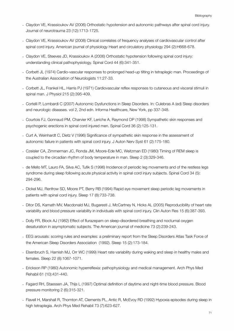
- Claydon VE, Krassioukov AV (2006) Orthostatic hypotension and autonomic pathways after spinal cord injury. Journal of neurotrauma 23 (12):1713-1725.
- Claydon VE, Krassioukov AV (2008) Clinical correlates of frequency analyses of cardiovascular control after spinal cord injury. American journal of physiology Heart and circulatory physiology 294 (2):H668-678.
- Claydon VE, Steeves JD, Krassioukov A (2006) Orthostatic hypotension following spinal cord injury: understanding clinical pathophysiology. Spinal Cord 44 (6):341-351.
- Corbett JL (1974) Cardio-vascular responses to prolonged head-up tilting in tetraplegic man. Proceedings of the Australian Association of Neurologists 11:27-33.
- Corbett JL, Frankel HL, Harris PJ (1971) Cardiovascular reflex responses to cutaneous and visceral stimuli in spinal man. J Physiol 215 (2):395-409.
- Cortelli P, Lombardi C (2007) Autonomic Dysfunctions in Sleep Disorders. In: Culebras A (ed) Sleep disorders and neurologic diseases. vol 2, 2nd edn. Informa Healthcare, New York, pp 337-348.
- Courtois FJ, Gonnaud PM, Charvier KF, Leriche A, Raymond DP (1998) Sympathetic skin responses and psychogenic erections in spinal cord injured men. Spinal Cord 36 (2):125-131.
- Curt A, Weinhardt C, Dietz V (1996) Significance of sympathetic skin response in the assessment of autonomic failure in patients with spinal cord injury. J Auton Nerv Syst 61 (2):175-180.
- Czeisler CA, Zimmerman JC, Ronda JM, Moore-Ede MC, Weitzman ED (1980) Timing of REM sleep is coupled to the circadian rhythm of body temperature in man. Sleep 2 (3):329-346.
- de Mello MT, Lauro FA, Silva AC, Tufik S (1996) Incidence of periodic leg movements and of the restless legs syndrome during sleep following acute physical activity in spinal cord injury subjects. Spinal Cord 34 (5):294-296.
- Dickel MJ, Renfrow SD, Moore PT, Berry RB (1994) Rapid eye movement sleep periodic leg movements in patients with spinal cord injury. Sleep 17 (8):733-738.
- Ditor DS, Kamath MV, Macdonald MJ, Bugaresti J, McCartney N, Hicks AL (2005) Reproducibility of heart rate variability and blood pressure variability in individuals with spinal cord injury. Clin Auton Res 15 (6):387-393.
- Dolly FR, Block AJ (1982) Effect of flurazepam on sleep-disordered breathing and nocturnal oxygen desaturation in asymptomatic subjects. The American journal of medicine 73 (2):239-243.
- EEG arousals: scoring rules and examples: a preliminary report from the Sleep Disorders Atlas Task Force of the American Sleep Disorders Association (1992). Sleep 15 (2):173-184.
- Elsenbruch S, Harnish MJ, Orr WC (1999) Heart rate variability during waking and sleep in healthy males and females. Sleep 22 (8):1067-1071.
- Erickson RP (1980) Autonomic hyperreflexia: pathophysiology and medical management. Arch Phys Med Rehabil 61 (10):431-440.
- Fagard RH, Staessen JA, Thijs L (1997) Optimal definition of daytime and night-time blood pressure. Blood pressure monitoring 2 (6):315-321.
- Flavell H, Marshall R, Thornton AT, Clements PL, Antic R, McEvoy RD (1992) Hypoxia episodes during sleep in high tetraplegia. Arch Phys Med Rehabil 73 (7):623-627.
Bibliography
71
- Forrest GP (1991) Atrial fibrillation associated with autonomic dysreflexia in patients with tetraplegia. Arch Phys Med Rehabil 72 (8):592-594.
- Freeman R, Wieling W, Axelrod FB, Benditt DG, Benarroch E, Biaggioni I, Cheshire WP, Chelimsky T, Cortelli P, Gibbons CH, Goldstein DS, Hainsworth R, Hilz MJ, Jacob G, Kaufmann H, Jordan J, Lipsitz LA, Levine BD, Low PA, Mathias C, Raj SR, Robertson D, Sandroni P, Schatz IJ, Schondorf R, Stewart JM, van Dijk JG (2011) Consensus statement on the definition of orthostatic hypotension, neurally mediated syncope and the postural tachycardia syndrome. Autonomic neuroscience : basic & clinical 161 (1-2):46-48.
- Freund PR, Brengelmann GL, Rowell LB, Halar E (1984) Attenuated skin blood flow response to hyperthermia in paraplegic men. J Appl Physiol 56 (4):1104-1109.
- Frisbie JH (2007) Unstable baseline blood pressure in chronic tetraplegia. Spinal Cord 45 (1):92-95.
- Furlan JC, Fehlings MG, Shannon P, Norenberg MD, Krassioukov AV (2003) Descending vasomotor pathways in humans: correlation between axonal preservation and cardiovascular dysfunction after spinal cord injury. Journal of neurotrauma 20 (12):1351-1363.
- Garshick E, Kelley A, Cohen SA, Garrison A, Tun CG, Gagnon D, Brown R (2005) A prospective assessment of mortality in chronic spinal cord injury. Spinal Cord 43 (7):408-416.
- Grasso R, Schena F, Gulli G, Cevese A (1997) Does low-frequency variability of heart period reflect a specific parasympathetic mechanism? J Auton Nerv Syst 63 (1-2):30-38.
- Grimm DR, De Meersman RE, Almenoff PL, Spungen AM, Bauman WA (1997) Sympathovagal balance of the heart in subjects with spinal cord injury. Am J Physiol 272 (2 Pt 2):H835-842.
- Groah SL, Weitzenkamp D, Sett P, Soni B, Savic G (2001) The relationship between neurological level of injury and symptomatic cardiovascular disease risk in the aging spinal injured. Spinal Cord 39 (6):310-317.
- Groothuis JT, Hopman MT (2009) Hemodynamic responses to the cold pressor test in spinal cord-injured individuals; control of the splanchnic vascular bed is the key factor. Spinal Cord 47 (1):95; author reply 96.
- Guttmann L, Silver J, Wyndham CH (1958) Thermoregulation in spinal man. J Physiol 142 (3):406-419.
- Guttmann L, Whitteridge D (1947) Effects of bladder distension on autonomic mechanisms after spinal cord injuries. Brain : a journal of neurology 70 (Pt 4):361-404.
- Houtman S, Oeseburg B, Hopman MT (1999) Non-invasive assessment of autonomic nervous system integrity in able-bodied and spinal cord-injured individuals. Clin Auton Res 9 (3):115-122.
- Houtman S, Oeseburg B, Hughson RL, Hopman MT (2000) Sympathetic nervous system activity and cardiovascular homeostatis during head-up tilt in patients with spinal cord injuries. Clin Auton Res 10 (4):207-212.
- Hyyppa MT, Kronholm E (1989) Quality of sleep and chronic illnesses. Journal of clinical epidemiology 42 (7):633-638.
- Iber C, Ancoli-Israel S, Chesson A, Quan S (eds) (2007) The AASM manual for the scoring of sleep and associated events: rules terminology and technical specifications. 1st edn. American Academy of Sleep Medicine, Westchester, IL
Guaraldi P. Bibliography
72
- Illman A, Stiller K, Williams M (2000) The prevalence of orthostatic hypotension during physiotherapy treatment in patients with an acute spinal cord injury. Spinal Cord 38 (12):741-747.
- Ingelsson E, Bjorklund-Bodegard K, Lind L, Arnlov J, Sundstrom J (2006) Diurnal blood pressure pattern and risk of congestive heart failure. JAMA : the journal of the American Medical Association 295 (24):2859-2866.
- Inoue K, Ogata H, Hayano J, Miyake S, Kamada T, Kuno M, Kumashiro M (1995) Assessment of autonomic function in traumatic quadriplegic and paraplegic patients by spectral analysis of heart rate variability. J Auton Nerv Syst 54 (3):225-234.
- Jacobs PL, Mahoney ET, Robbins A, Nash M (2002) Hypokinetic circulation in persons with paraplegia. Medicine and science in sports and exercise 34 (9):1401-1407.
- Jensen MP, Hirsh AT, Molton IR, Bamer AM (2009) Sleep problems in individuals with spinal cord injury: frequency and age effects. Rehabil Psychol 54 (3):323-331.
- Johnson JM, Kellogg DL, Jr. (2010) Thermoregulatory and thermal control in the human cutaneous circulation. Front Biosci (Schol Ed) 2:825-853.
- Karlsson AK (1999) Autonomic dysreflexia. Spinal Cord 37 (6):383-391.
- Karlsson AK, Friberg P, Lonnroth P, Sullivan L, Elam M (1998) Regional sympathetic function in high spinal cord injury during mental stress and autonomic dysreflexia. Brain : a journal of neurology 121 ( Pt 9):1711-1719.
- Khan S, Plummer M, Martinez-Arizala A, Banovac K (2007) Hypothermia in patients with chronic spinal cord injury. J Spinal Cord Med 30 (1):27-30.
- Klefbeck B, Sternhag M, Weinberg J, Levi R, Hultling C, Borg J (1998) Obstructive sleep apneas in relation to severity of cervical spinal cord injury. Spinal Cord 36 (9):621-628.
- Koh J, Brown TE, Beightol LA, Ha CY, Eckberg DL (1994) Human autonomic rhythms: vagal cardiac mechanisms in tetraplegic subjects. J Physiol 474 (3):483-495.
- Kooijman M, Rongen GA, Smits P, Hopman MT (2003) Preserved alpha-adrenergic tone in the leg vascular bed of spinal cord-injured individuals. Circulation 108 (19):2361-2367.
- Krassioukov A, Claydon VE (2006) The clinical problems in cardiovascular control following spinal cord injury: an overview. Progress in brain research 152:223-229.
- Krum H, Louis WJ, Brown DJ, Jackman GP, Howes LG (1991) Diurnal blood pressure variation in quadriplegic chronic spinal cord injury patients. Clin Sci (Lond) 80 (3):271-276.
- Lee MS, Choi YC, Lee SH, Lee SB (1996) Sleep-related periodic leg movements associated with spinal cord lesions. Mov Disord 11 (6):719-722.
- Li Y, Jiang DH, Wang ML, Jiao DR, Pang SF (1989) Rhythms of serum melatonin in patients with spinal lesions at the cervical, thoracic or lumbar region. Clinical endocrinology 30 (1):47-56.
- Liguori R, Donadio V, Foschini E, Di Stasi V, Plazzi G, Lugaresi E, Montagna P (2000) Sleep stage-related changes in sympathetic sudomotor and vasomotor skin responses in man. Clin Neurophysiol 111 (3):434-439.
Bibliography
73
- Linsell CR, Lightman SL, Mullen PE, Brown MJ, Causon RC (1985) Circadian rhythms of epinephrine and norepinephrine in man. J Clin Endocrinol Metab 60 (6):1210-1215.
- Lundin S, Jern S, Hansson L (1986) Testing neural control of the circulation in patients: hemodynamic responses to exercise and stressful stimuli. In: Zanchetti A, Tarazi RC (eds) Handbook of hypertension. vol 8. Pathophysiology of hypertension : regulatory mechanisms. Elsevier ; New York, NY : Sole distributor for the USA and Canada, Elsevier Science Pub. Co., Amsterdam ; New York, pp xxi, 703 p.
- Maiorov DN, Weaver LC, Krassioukov AV (1997) Relationship between sympathetic activity and arterial pressure in conscious spinal rats. Am J Physiol 272 (2 Pt 2):H625-631.
- Mancia G (1993) Autonomic modulation of the cardiovascular system during sleep. The New England journal of medicine 328 (5):347-349.
- Marcus RR, Kalisetti D, Raxwal V, Kiratli BJ, Myers J, Perkash I, Froelicher VF (2002) Early repolarization in patients with spinal cord injury: prevalence and clinical significance. J Spinal Cord Med 25 (1):33-38; discussion 39.
- Marino RJ, Barros T, Biering-Sorensen F, Burns SP, Donovan WH, Graves DE, Haak M, Hudson LM, Priebe MM (2003) International standards for neurological classification of spinal cord injury. J Spinal Cord Med 26 Suppl 1:S50-56.
- Mathias C (2012) Autonomic Disturbances in Spinal Cord Injuries. In: Robertson D (ed) Primer on the autonomic nervous system. 3rd edn. Elsevier Academic Press, San Diego, Calif, pp 505-509.
- Mathias CJ (1995) Orthostatic hypotension: causes, mechanisms, and influencing factors. Neurology 45 (4 Suppl 5):S6-11.
- Mathias CJ (2006) Orthostatic hypotension and paroxysmal hypertension in humans with high spinal cord injury. Progress in brain research 152:231-243.
- Mathias CJ, Bannister R (1999) Investigation of autonomic disorders. In: Mathias CJ, Bannister R (eds) Autonomic failure : a textbook of clinical disorders of the autonomic nervous system. 4th edn. Oxford University Press, Oxford; New York, pp 169-195.
- Mathias CJ, Frankel HL (2002) Autonomic disturbances in spinal cord lesions. In: Mathias CJ, Bannister R (eds) Autonomic failure : a textbook of clinical disorders of the autonomic nervous system. 4th edn. Oxford University Press, Oxford ; New York, pp 494–513.
- Mayer G, Hellmann F, Leonhard E, Meier-Ewert K (1997) Circadian temperature and activity rhythms in unmedicated narcoleptic patients. Pharmacology, biochemistry, and behavior 58 (2):395-402.
- McEvoy RD, Mykytyn I, Sajkov D, Flavell H, Marshall R, Antic R, Thornton AT (1995) Sleep apnoea in patients with quadriplegia. Thorax 50 (6):613-619.
- Mistlberger RE, Skene DJ (2005) Nonphotic entrainment in humans? Journal of biological rhythms 20 (4):339-352.
- Mojon A, Fernandez JR, Hermida RC (1992) Chronolab: an interactive software package for chronobiologic time series analysis written for the Macintosh computer. Chronobiology international 9 (6):403-412.
- Moore RY (1996) Neural control of the pineal gland. Behavioural brain research 73 (1-2):125-130.
Guaraldi P. Bibliography
74

- Morton JP, MacLaren DP, Cable NT, Bongers T, Griffiths RD, Campbell IT, Evans L, Kayani A, McArdle A, Drust B (2006) Time course and differential responses of the major heat shock protein families in human skeletal muscle following acute nondamaging treadmill exercise. J Appl Physiol 101 (1):176-182.
- Munakata M, Kameyama J, Kanazawa M, Nunokawa T, Moriai N, Yoshinaga K (1997) Circadian blood pressure rhythm in patients with higher and lower spinal cord injury: simultaneous evaluation of autonomic nervous activity and physical activity. J Hypertens 15 (12 Pt 2):1745-1749.
- Munakata M, Kameyama J, Nunokawa T, Ito N, Yoshinaga K (2001) Altered Mayer wave and baroreflex profiles in high spinal cord injury. American journal of hypertension 14 (2):141-148.
- Myers J, Lee M, Kiratli J (2007) Cardiovascular disease in spinal cord injury: an overview of prevalence, risk, evaluation, and management. Am J Phys Med Rehabil 86 (2):142-152.
- Nagarajarao HS, Kumar BN, Watt JW, Wiredu E, Bhamidimarri K (2006) Bedside assessment of sympathetic skin response after spinal cord injury: a brief report comparing inspiratory gasp and visual stimulus. Spinal Cord 44 (4):217-221.
- Nash MS, Mendez AJ (2007) A guideline-driven assessment of need for cardiovascular disease risk intervention in persons with chronic paraplegia. Arch Phys Med Rehabil 88 (6):751-757.
- Nicotra A, Catley M, Ellaway PH, Mathias CJ (2005) The ability of physiological stimuli to generate the sympathetic skin response in human chronic spinal cord injury. Restorative neurology and neuroscience 23 (5-6):331-339.
- Nicotra A, Young TM, Asahina M, Mathias CJ (2005) The effect of different physiological stimuli on skin vasomotor reflexes above and below the lesion in human chronic spinal cord injury. Neurorehabilitation and neural repair 19 (4):325-331.
- Nitsche B, Perschak H, Curt A, Dietz V (1996) Loss of circadian blood pressure variability in complete tetraplegia. J Hum Hypertens 10 (5):311-317.
- Noda A, Okada T, Hayashi H, Yasuma F, Yokota M (1993) 24-hour ambulatory blood pressure variability in obstructive sleep apnea syndrome. Chest 103 (5):1343-1347.
- Norrbrink Budh C, Hultling C, Lundeberg T (2005) Quality of sleep in individuals with spinal cord injury: a comparison between patients with and without pain. Spinal Cord 43 (2):85-95.
- O'Brien E, Asmar R, Beilin L, Imai Y, Mallion JM, Mancia G, Mengden T, Myers M, Padfield P, Palatini P, Parati G, Pickering T, Redon J, Staessen J, Stergiou G, Verdecchia P (2003) European Society of Hypertension recommendations for conventional, ambulatory and home blood pressure measurement. J Hypertens 21 (5):821-848.
- O'Brien E, Sheridan J, O'Malley K (1988) Dippers and non-dippers. Lancet 2 (8607):397.
- Pagani M, Lombardi F, Guzzetti S, Rimoldi O, Furlan R, Pizzinelli P, Sandrone G, Malfatto G, Dell'Orto S, Piccaluga E, et al. (1986) Power spectral analysis of heart rate and arterial pressure variabilities as a marker of sympatho-vagal interaction in man and conscious dog. Circulation research 59 (2):178-193.
- Pandi-Perumal SR, Trakht I, Spence DW, Srinivasan V, Dagan Y, Cardinali DP (2008) The roles of melatonin and light in the pathophysiology and treatment of circadian rhythm sleep disorders. Nature clinical practice Neurology 4 (8):436-447.
Bibliography
75

- Parati G, Saul JP, Di Rienzo M, Mancia G (1995) Spectral analysis of blood pressure and heart rate variability in evaluating cardiovascular regulation. A critical appraisal. Hypertension 25 (6):1276-1286.
- Paulus W, Schomburg ED (2006) Dopamine and the spinal cord in restless legs syndrome: does spinal cord physiology reveal a basis for augmentation? Sleep medicine reviews 10 (3):185-196.
- Previnaire JG, Mathias CJ, El Masri W, Soler JM, Leclercq V, Denys P (2010) The isolated sympathetic spinal cord: Cardiovascular and sudomotor assessment in spinal cord injury patients: A literature survey. Annals of physical and rehabilitation medicine 53 (8):520-532.
- Price MJ (2006) Thermoregulation during exercise in individuals with spinal cord injuries. Sports Med 36 (10):863-879.
- Price MJ, Campbell IG (1999) Thermoregulatory responses of spinal cord injured and able-bodied athletes to prolonged upper body exercise and recovery. Spinal Cord 37 (11):772-779.
- Raymann RJ, Swaab DF, Van Someren EJ (2007) Skin temperature and sleep-onset latency: changes with age and insomnia. Physiology & behavior 90 (2-3):257-266.
- Rechtschaffen A, Kales A (1968) A manual of standardized terminology, techniques and scoring system for sleep stages of human subjects. . National Institutes of Health publication,, vol no 204. U. S. National Institute of Neurological Diseases and Blindness, Neurological Information Network, Bethesda, MD.
- Reitz A, Schmid DM, Curt A, Knapp PA, Schurch B (2002) Sympathetic sudomotor skin activity in human after complete spinal cord injury. Auton Neurosci 102 (1-2):78-84.
- Rosado-Rivera D, Radulovic M, Handrakis JP, Cirnigliaro CM, Jensen AM, Kirshblum S, Bauman WA, Wecht JM (2011) Comparison of 24-hour cardiovascular and autonomic function in paraplegia, tetraplegia, and control groups: implications for cardiovascular risk. J Spinal Cord Med 34 (4):395-403.
- Samsa GP, Patrick CH, Feussner JR (1993) Long-term survival of veterans with traumatic spinal cord injury. Arch Neurol 50 (9):909-914.
- Satinoff E (1978) Neural organization and evolution of thermal regulation in mammals. Science 201 (4350):16-22.
- Sawka MN, Latzka WA, Pandolf KB (1989) Temperature regulation during upper body exercise: able-bodied and spinal cord injured. Medicine and science in sports and exercise 21 (5 Suppl):S132-140.
- Scheer FA, Zeitzer JM, Ayas NT, Brown R, Czeisler CA, Shea SA (2006) Reduced sleep efficiency in cervical spinal cord injury; association with abolished night time melatonin secretion. Spinal Cord 44 (2):78-81.
- Sheel AW, Krassioukov AV, Inglis JT, Elliott SL (2005) Autonomic dysreflexia during sperm retrieval in spinal cord injury: influence of lesion level and sildenafil citrate. J Appl Physiol 99 (1):53-58.
- Short DJ, Stradling JR, Williams SJ (1992) Prevalence of sleep apnoea in patients over 40 years of age with spinal cord lesions. J Neurol Neurosurg Psychiatry 55 (11):1032-1036.
- Sleep-related breathing disorders in adults: recommendations for syndrome definition and measurement techniques in clinical research. The Report of an American Academy of Sleep Medicine Task Force (1999). Sleep 22 (5):667-689.
Guaraldi P. Bibliography
76
- Smith HR (2008) The basic neurology of sleep. In: Smith HR, Comella C, Högl B (eds) Sleep medicine. Cambridge University Press, Cambridge ; New York, pp 1-8.
- Soler JM, Previnaire JG, Plante P, Denys P, Chartier-Kastler E (2007) Midodrine improves ejaculation in spinal cord injured men. The Journal of urology 178 (5):2082-2086.
- Somers VK, Dyken ME, Mark AL, Abboud FM (1993) Sympathetic-nerve activity during sleep in normal subjects. The New England journal of medicine 328 (5):303-307.
- Star AM, Osterman AL (1988) Sleep apnea syndrome after spinal cord injury. Report of a case and literature review. Spine (Phila Pa 1976) 13 (1):116-117.
- Stockhammer E, Tobon A, Michel F, Eser P, Scheuler W, Bauer W, Baumberger M, Muller W, Kakebeeke TH, Knecht H, Zach GA (2002) Characteristics of sleep apnea syndrome in tetraplegic patients. Spinal Cord 40 (6):286-294.
- Taasan VC, Block AJ, Boysen PG, Wynne JW (1981) Alcohol increases sleep apnea and oxygen desaturation in asymptomatic men. The American journal of medicine 71 (2):240-245.
- Tarvainen MP, Ranta-Aho PO, Karjalainen PA (2002) An advanced detrending method with application to HRV analysis. IEEE transactions on bio-medical engineering 49 (2):172-175.
- Teasell RW, Arnold JM, Krassioukov A, Delaney GA (2000) Cardiovascular consequences of loss of supraspinal control of the sympathetic nervous system after spinal cord injury. Arch Phys Med Rehabil 81 (4):506-516.
- Telles SC, Alves RC, Chadi G (2011) Periodic limb movements during sleep and restless legs syndrome in patients with ASIA A spinal cord injury. J Neurol Sci 303 (1-2):119-123.
- Theisen D, Vanlandewijck Y, Sturbois X, Francaux M (2001) Cutaneous vascular response and thermoregulation in individuals with paraplegia during sustained arm-cranking exercise. International journal of sports medicine 22 (2):97-102.
- Thijssen DH, Eijsvogels TM, Hesse M, Ballak DB, Atkinson G, Hopman MT (2011) The effects of thoracic and cervical spinal cord lesions on the circadian rhythm of core body temperature. Chronobiology international 28 (2):146-154.
- Tolbert G, Tuck ML (2004) Ambulatory blood pressure monitoring in persons with chronic spinal cord injury. J Spinal Cord Med 27 (5):476-480.
- Tran K, Hukins C, Geraghty T, Eckert B, Fraser L (2009) Sleep-disordered breathing in spinal cord-injured patients: a short-term longitudinal study. Respirology 15 (2):272-276.
- Verdecchia P, Schillaci G, Gatteschi C, Zampi I, Battistelli M, Bartoccini C, Porcellati C (1993) Blunted nocturnal fall in blood pressure in hypertensive women with future cardiovascular morbid events. Circulation 88 (3):986-992.
- Vetrugno R, Provini F, Plazzi G, Cortelli P, Montagna P (2005) Propriospinal myoclonus: a motor phenomenon found in restless legs syndrome different from periodic limb movements during sleep. Mov Disord 20 (10):1323-1329.
Bibliography
77

- Wahman K, Nash MS, Lewis JE, Seiger A, Levi R (2010) Increased cardiovascular disease risk in Swedish persons with paraplegia: The Stockholm spinal cord injury study. Journal of rehabilitation medicine : official journal of the UEMS European Board of Physical and Rehabilitation Medicine 42 (5):489-492.
- Wahman K, Nash MS, Lewis JE, Seiger A, Levi R (2011) Cardiovascular disease risk and the need for prevention after paraplegia determined by conventional multifactorial risk models: the Stockholm spinal cord injury study. Journal of rehabilitation medicine : official journal of the UEMS European Board of Physical and Rehabilitation Medicine 43 (3):237-242.
- Wahman K, Nash MS, Westgren N, Lewis JE, Seiger A, Levi R (2010) Cardiovascular disease risk factors in persons with paraplegia: the Stockholm spinal cord injury study. Journal of rehabilitation medicine : official journal of the UEMS European Board of Physical and Rehabilitation Medicine 42 (3):272-278.
- Waterhouse J, Drust B, Weinert D, Edwards B, Gregson W, Atkinson G, Kao S, Aizawa S, Reilly T (2005) The circadian rhythm of core temperature: origin and some implications for exercise performance. Chronobiology international 22 (2):207-225.
- Wecht JM, Weir JP, Bauman WA (2006) Blunted heart rate response to vagal withdrawal in persons with tetraplegia. Clin Auton Res 16 (6):378-383.
- Williams JR (2008) The Declaration of Helsinki and public health. Bulletin of the World Health Organization 86 (8):650-652.
- Wolf J, Hering D, Narkiewicz K (2010) Non-dipping pattern of hypertension and obstructive sleep apnea syndrome. Hypertension research : official journal of the Japanese Society of Hypertension 33 (9):867-871.
- Yokota T, Hirose K, Tanabe H, Tsukagoshi H (1991) Sleep-related periodic leg movements (nocturnal myoclonus) due to spinal cord lesion. J Neurol Sci 104 (1):13-18.
- Young T, Palta M, Dempsey J, Skatrud J, Weber S, Badr S (1993) The occurrence of sleep-disordered breathing among middle-aged adults. The New England journal of medicine 328 (17):1230-1235.
- Zeitzer JM, Ayas NT, Shea SA, Brown R, Czeisler CA (2000) Absence of detectable melatonin and preservation of cortisol and thyrotropin rhythms in tetraplegia. J Clin Endocrinol Metab 85 (6):2189-2196.
- Zulley J, Wever R, Aschoff J (1981) The dependence of onset and duration of sleep on th circadian rhythm of rectal temperature. Pflugers Archiv : European journal of physiology 391 (4):314-318.
Guaraldi P. Bibliography
78

APPENDIX


Age distribution in SCI patients and controls
1"
2"
3"
4"
5"
6"
7"
8"
9"
11"
12"12,5"
13"14"
15"
16" 17"
18"
19"
20"14,5"
15,5"
16,5" 17,5"
18,5"
19,5"
20,5"
0"
10"
20"
30"
40"
50"
60"
70"
0" 5" 10" 15" 20" 25"
years&
Subjects'&n°&
Age&
4"
5" 6"
7"8" 9"
11"
12"
13"
14"
15"
16"
17"18"
19"
20"
21"
22"23"
24"
25"
27"
28"
29"
30"
31"
32"
33"
34"
35"
36"
37"
5" 10" 15" 20" 25" 30" 35" 40"Pa#ents')n°)
C4-7" D2-12" CNT" C4-7: cervical SCI; CNT: controls; D2-12: thoracic SCI; SCI: spinal cord injury. Numbers on axis of abscissas correspond to subjects’ numbers.
BMI distribution in SCI patients and controls
1"
2"
3" 4"5"
6"
7"
8"
9" 11"
12"12,5"13"
14"
15"
16"
17" 18"
19"
20"
14,5"
15,5"
16,5"
17,5" 18,5"
19,5"
20,5"
0"
5"
10"
15"
20"
25"
30"
35"
0" 5" 10" 15" 20" 25"
Subjects'*n°*
BMI*
4"
5" 6"
7"8" 9"
11"
12"
13"
14"
15"
16"
17"18"
19"
20"
21"
22"23"
24"
25"
27"
28"
29"
30"
31"
32"
33"
34"
35"
36"
37"
5" 10" 15" 20" 25" 30" 35" 40"Pa#ents')n°)
C4-7" D2-12" CNT" C4-7: cervical SCI; CNT: controls; D2-12: thoracic SCI; SCI: spinal cord injury. Numbers on axis of abscissas correspond to subjects’ numbers.
APPENDIX: Section I
79

Total Sleep Time (TST) and Sleep Efficiency in SCI patients and controls
C4-7: cervical SCI; CNT: controls; D2-12: thoracic SCI; SCI: spinal cord injury.
APPENDIX: Section I
80
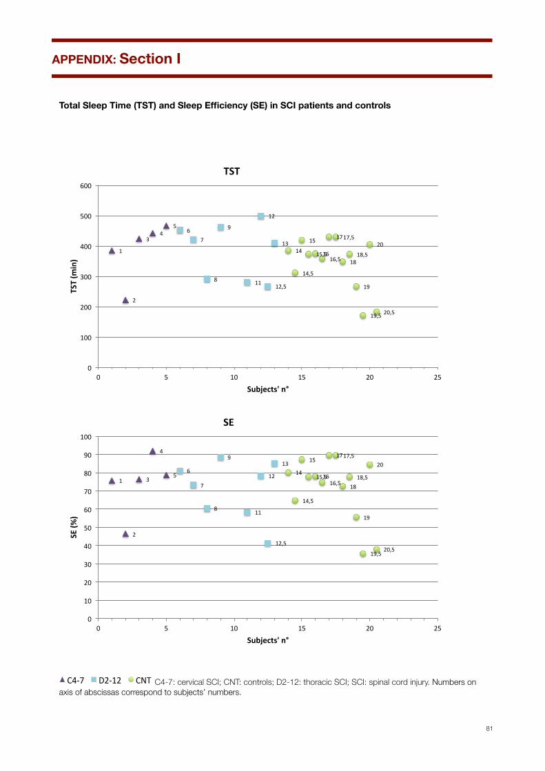
Total Sleep Time (TST) and Sleep Efficiency (SE) in SCI patients and controls
1"
2"
3"4"
5"6"
7"
8"
9"
11"
12"
12,5"
13"14"
15"
16"
17"
18"
19"
20"
14,5"
15,5"16,5"
17,5"
18,5"
19,5" 20,5"
0"
100"
200"
300"
400"
500"
600"
0" 5" 10" 15" 20" 25"
TST#(m
in)#
Subjects'#n°#
TST#
1"
2"
3"
4"
5"6"
7"
8"
9"
11"
12"
12,5"
13"14"
15"
16"
17"
18"
19"
20"
14,5"
15,5"16,5"
17,5"
18,5"
19,5"20,5"
0"
10"
20"
30"
40"
50"
60"
70"
80"
90"
100"
0" 5" 10" 15" 20" 25"
SE#(%
)#
Subjects'#n°#
SE#
4"
5" 6"
7"8" 9"
11"
12"
13"
14"
15"
16"
17"18"
19"
20"
21"
22"23"
24"
25"
27"
28"
29"
30"
31"
32"
33"
34"
35"
36"
37"
5" 10" 15" 20" 25" 30" 35" 40"Pa#ents')n°)
C4-7" D2-12" CNT" C4-7: cervical SCI; CNT: controls; D2-12: thoracic SCI; SCI: spinal cord injury. Numbers on axis of abscissas correspond to subjects’ numbers.
APPENDIX: Section I
81
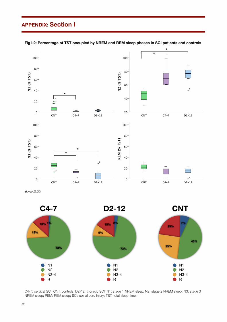
Fig I.2: Percentage of TST occupied by NREM and REM sleep phases in SCI patients and controls
Fig I.1 Percentage of sleep phases in SCI patients and controls
*
**
**
19
27
27
*=p<0,05
Percentages of sleep stages
13%
13%
73%
1%
C4-7
N1N2N3-4R
15%
9%
73%
2%
D2-12
N1N2N3-4R
23%
25%45%
7%
CNT
N1N2N3-4R
C4-7: cervical SCI; CNT: controls; D2-12: thoracic SCI; N1: stage 1 NREM sleep; N2: stage 2 NREM sleep; N3: stage 3 NREM sleep; REM: REM sleep; SCI: spinal cord injury; TST: total sleep time.
APPENDIX: Section I
82
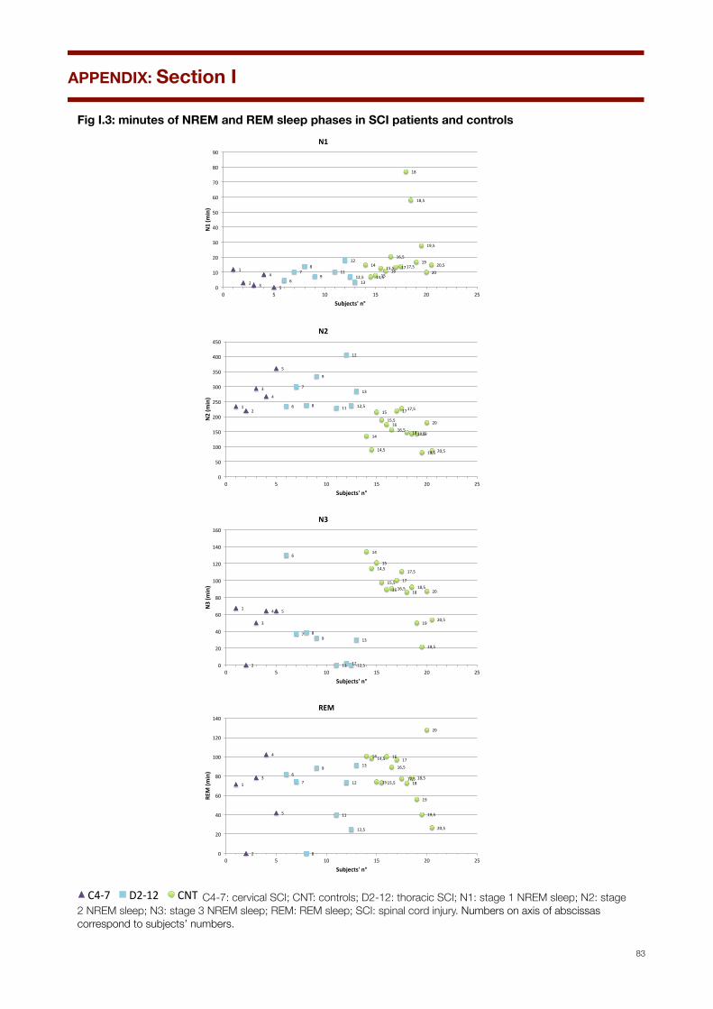
Fig I.3: minutes of NREM and REM sleep phases in SCI patients and controls
1"
2" 3"
4"
5"6"
7"8"
9"11"
12"
12,5"13"
14"
15"16"
17"
18"
19"
20"14,5"
15,5"
16,5"
17,5"
18,5"
19,5"
20,5"
0"
10"
20"
30"
40"
50"
60"
70"
80"
90"
0" 5" 10" 15" 20" 25"
N1#(m
in)#
Subjects'#n°#
N1#
1"2"
3"
4"
5"
6"
7"
8"
9"
11"
12"
12,5"
13"
14"
15"
16"
17"
18" 19"
20"
14,5"
15,5"
16,5"
17,5"
18,5"
19,5" 20,5"
0"
50"
100"
150"
200"
250"
300"
350"
400"
450"
0" 5" 10" 15" 20" 25"
N2#(m
in)#
Subjects'#n°#
N2#
1"
2"
3"
4" 5"
6"
7" 8"9"
11" 12"12,5"
13"
14"
15"
16"
17"
18"
19"
20"
14,5"
15,5"16,5"
17,5"
18,5"
19,5"
20,5"
0"
20"
40"
60"
80"
100"
120"
140"
160"
0" 5" 10" 15" 20" 25"
N3#(m
in)#
Subjects'#n°#
N3#
1"
2"
3"
4"
5"
6"
7"
8"
9"
11"
12"
12,5"
13"
14"
15"
16"17"
18"
19"
20"
14,5"
15,5"
16,5"
17,5" 18,5"
19,5"
20,5"
0"
20"
40"
60"
80"
100"
120"
140"
0" 5" 10" 15" 20" 25"
REM$(m
in)$
Subjects'$n°$
REM$
4"
5" 6"
7"8" 9"
11"
12"
13"
14"
15"
16"
17"18"
19"
20"
21"
22"23"
24"
25"
27"
28"
29"
30"
31"
32"
33"
34"
35"
36"
37"
5" 10" 15" 20" 25" 30" 35" 40"Pa#ents')n°)
C4-7" D2-12" CNT" C4-7: cervical SCI; CNT: controls; D2-12: thoracic SCI; N1: stage 1 NREM sleep; N2: stage 2 NREM sleep; N3: stage 3 NREM sleep; REM: REM sleep; SCI: spinal cord injury. Numbers on axis of abscissas correspond to subjects’ numbers.
APPENDIX: Section I
83

Fig I.4: arousals, periodic limb movement and phase shifts in SCI patients and controls
Arousals
Periodic Limb Movement of Sleep
Fase shifts and sleep fragmentation
Fig I.2 Arousals, periodic limb movement and sleep fragmentation in SCI patients and controls
*
18
25
18
*=p<0,05C4-7: cervical SCI; CNT: controls; D2-12: thoracic SCI; SCI: spinal cord injury, PLMS: periodic limb movement during sleep.
APPENDIX: Section I
84

Fig I.5: arousals, periodic limb movement and phase shifts in SCI patients and controls
1"
2"
3"
4"
5"
6"
7" 8"
9" 11"
12"
12,5"
13" 14"
15" 16" 17"18"
19" 20"14,5"
15,5"
16,5"
17,5"
18,5"19,5"
20,5"
0"
10"
20"
30"
40"
50"
60"
70"
80"
0" 5" 10" 15" 20" 25"
Pa#ents')n°)
Arousal)Index)
C4.7" D2.12" CNT"
1" 2"
3"
4" 5" 6" 7" 8" 9"
11"
12"
12,5"
13" 14" 15" 16"
17"
18"19"
20"14,5" 15,5"16,5"
17,5"18,5"
19,5"20,5"0"
20"
40"
60"
80"
100"
120"
140"
160"
180"
0" 5" 10" 15" 20" 25"
Pa#ents')n°)
PLMS)Index)
C4.7" D2.12" CNT"
ArousalIndex
PLMSIndex
Phase Shift
Index
1"
2"
3"
4" 5"
6"
7"
8"
9"
11"
12"
12,5"
13"14"
15"
16"17"
18"19"
20"14,5" 15,5"
16,5" 17,5"18,5"
19,5"
20,5"
0"
10"
20"
30"
40"
50"
60"
70"
0" 5" 10" 15" 20" 25"
Subjects'*n°*
Phase*Shi1*Index*
C4.7" D2.12" CNT"
4"
5" 6"
7"8" 9"
11"
12"
13"
14"
15"
16"
17"18"
19"
20"
21"
22"23"
24"
25"
27"
28"
29"
30"
31"
32"
33"
34"
35"
36"
37"
5" 10" 15" 20" 25" 30" 35" 40"Pa#ents')n°)
C4-7" D2-12" CNT" C4-7: cervical SCI; CNT: controls; D2-12: thoracic SCI; PLMS: periodic limb movement during sleep. Numbers on axis of abscissas correspond to subjects’ numbers.
APPENDIX: Section I
85

APPENDIX: Section II.1
86
Supine SBP, DBP and HR values in SCI patients and controls
1"2"
3"
4"
5"
6"
7"
8"
9" 11"
12"
13"
14"15"
16" 17" 18" 19" 20"
21" 22"
23"
24"
25"26"
27"
28"
29" 30" 31"32"
33"
34"
35"
36" 37"
0"
20"
40"
60"
80"
100"
120"
140"
160"
180"
0" 5" 10" 15" 20" 25" 30" 35" 40"
SBP$(m
mHg
)$
Subjects'$n°$
baseline$SBP$$
1" 2"
3"4"
5"
6"7"
8" 9"11"
12"
13"
14"
15"
16"17"
18" 19"
20"
21"22"
23"
24"
25"
26"
27" 28"
29"
30"
31"
32"33"
34"
35" 36"
37"
0"
10"
20"
30"
40"
50"
60"
70"
80"
90"
0" 5" 10" 15" 20" 25" 30" 35" 40"
DBP$(m
mHg
)$
Subjects'$n°$
baseline$DBP$
1" 2"
3"4"
5"
6"
7"8"
9"
11"
12"
13"
14"15"
16"
17"
18"
19"
20" 21"
22"
23"
24"
25"26"
27"
28"
29"
30"
31"
32"
33"
34"
35"36"
37"
0"
10"
20"
30"
40"
50"
60"
70"
80"
90"
0" 5" 10" 15" 20" 25" 30" 35" 40"
HR#(b
/m)#
Subjects'#n°#
baseline#HR##
4"
5" 6"
7"8" 9"
11"
12"
13"
14"
15"
16"
17"18"
19"
20"
21"
22"23"
24"
25"
27"
28"
29"
30"
31"
32"
33"
34"
35"
36"
37"
5" 10" 15" 20" 25" 30" 35" 40"Pa#ents')n°)
C4-7" D2-12" CNT" C4-7: cervical SCI; CNT: controls; D2-12: thoracic SCI. Numbers on axis of abscissas correspond to subjects’ numbers.
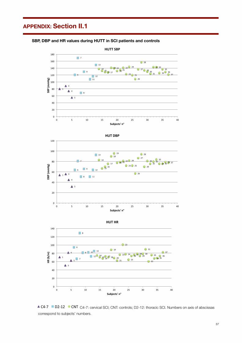
SBP, DBP and HR values during HUTT in SCI patients and controls
1"3"
4"
5"
6"
7"
8"
9"
11"12"
13"
14"15"
16" 17"
18" 19"20" 21"
22"
23"
24"
25"
26"
27"
28"
29"30"
31"
32" 33"
34"
35"36"
37"
0"
20"
40"
60"
80"
100"
120"
140"
160"
180"
0" 5" 10" 15" 20" 25" 30" 35" 40"
SBP$(m
mHg
)$
Subjects'$n°$
HUTT$SBP$
1" 3"
4"
5"
6"
7"
8"
9"
11"
12"
13"
14"
15"16"
17"
18"19"
20" 21"22"
23"
24"
25"
26"
27"
28"
29"
30"31"
32"33"
34" 35"36" 37"
0"
20"
40"
60"
80"
100"
120"
0" 5" 10" 15" 20" 25" 30" 35" 40"
DBP$(m
mHg
)$
Subjects'$n°$
HUT$DBP$
1"
3"
4"
5"
6"
7"
8"
9" 11"
12"
13"
14"15"16"
17"18"
19"
20"21"
22"
23"
24"
25"
26"27"28"
29"30"
31"
32"
33"34"35"
36"
37"
0"
20"
40"
60"
80"
100"
120"
140"
0" 5" 10" 15" 20" 25" 30" 35" 40"
HR#(b
/m)#
Subjects'#n°#
HUT#HR#
4"
5" 6"
7"8" 9"
11"
12"
13"
14"
15"
16"
17"18"
19"
20"
21"
22"23"
24"
25"
27"
28"
29"
30"
31"
32"
33"
34"
35"
36"
37"
5" 10" 15" 20" 25" 30" 35" 40"Pa#ents')n°)
C4-7" D2-12" CNT" C4-7: cervical SCI; CNT: controls; D2-12: thoracic SCI. Numbers on axis of abscissas correspond to subjects’ numbers.
APPENDIX: Section II.1
87
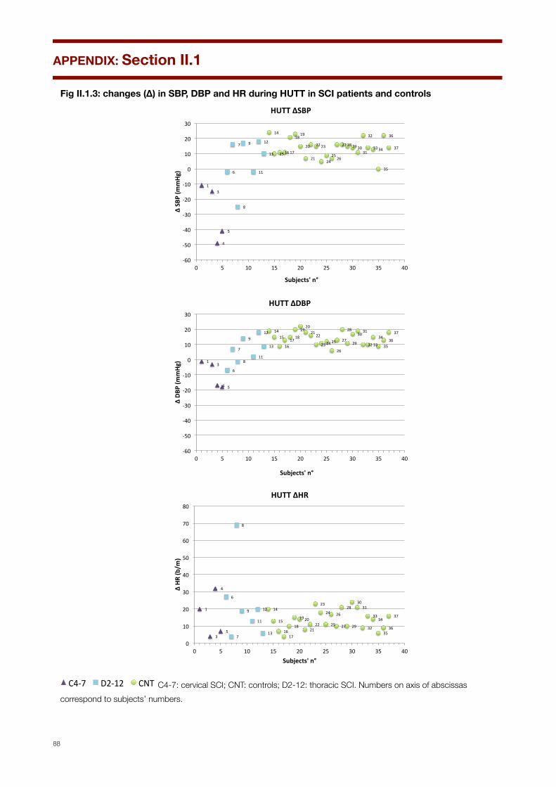
Fig II.1.3: changes (∆) in SBP, DBP and HR during HUTT in SCI patients and controls
1"3"
4"
5"
6"
7"
8"
9"
11"
12"
13"
14"
15"16"17"
18"19"
20"
21"
22"23"
24"25"
26"
27"28"29"30"31"
32"
33"34"
35"
36"
37"
,60"
,50"
,40"
,30"
,20"
,10"
0"
10"
20"
30"
0" 5" 10" 15" 20" 25" 30" 35" 40"
Δ"SB
P"(m
mHg
)"
Subjects'"n°"
HUTT"ΔSBP""
1"3"
4" 5"
6"
7"
8"
9"
11"
12"
13"
14"15"
16"17"
18"
19"20"
21"22"
23"24"25"
26"
27"
28"
29"
30"31"
32"33"
34"
35"36"
37"
,60"
,50"
,40"
,30"
,20"
,10"
0"
10"
20"
30"
0" 5" 10" 15" 20" 25" 30" 35" 40"
Δ"DB
P"(m
mHg
)"
Subjects'"n°"
HUTT"ΔDBP"
1"
3"
4"
5"
6"
7"
8"
9"
11"
12"
13"
14"
15"
16"17"
18"
19"20"
21"22"
23"
24"
25"
26"
27"
28"
29"
30"31"
32"
33"34"
35"36"
37"
0"
10"
20"
30"
40"
50"
60"
70"
80"
0" 5" 10" 15" 20" 25" 30" 35" 40"
Δ"HR
"(b/m
)"
Subjects'"n°"
HUTT"ΔHR"
4"
5" 6"
7"8" 9"
11"
12"
13"
14"
15"
16"
17"18"
19"
20"
21"
22"23"
24"
25"
27"
28"
29"
30"
31"
32"
33"
34"
35"
36"
37"
5" 10" 15" 20" 25" 30" 35" 40"Pa#ents')n°)
C4-7" D2-12" CNT" C4-7: cervical SCI; CNT: controls; D2-12: thoracic SCI. Numbers on axis of abscissas correspond to subjects’ numbers.
APPENDIX: Section II.1
88

Fig II.1.4: SBP, DBP and HR means±SD during supine rest and HUTT in SCI patients and controls
!
*
!
* *
!
*supine HUTT
!
*
!
* **
!
*
* **
supine HUTT
supine HUTT
SBP(mmHg)
DBP(mmHg)
HR(bpm)
CNT C4-7 D2-12 CNT C4-7 D2-12
CNT C4-7 D2-12 CNT C4-7 D2-12
CNT C4-7 D2-12 CNT C4-7 D2-12
200
100
150
50
0
100
60
80
40
20
0
150
90
120
60
0
28
19
26
23
*
30
*=p<0,05. C4-7: cervical SCI; CNT: controls; D2-12: thoracic SCI; SCI: spinal cord injury.
APPENDIX: Section II.1
89

Fig II.1.5: overshoot (OV) and valsalva ratio (VR) in SCI patients and controls
1"
5"
7"
9"
11"
12"13"
14"
15"
16"
17"
18"
19"
20"
21"
22"
23"
24"
25"
26"
27"
28"
29"
30"
31"
32"
33"
34"
35"
36"
37"
,10"
0"
10"
20"
30"
40"
50"
60"
70"
80"
90"
0" 5" 10" 15" 20" 25" 30" 35" 40"
OV#(m
mHg
)#
Subjects'#n°#
Overshoot#
1"
5"6"
7"
8"
9"
11"
12"
13"
14"
15"16"
17"
18"19"
20"
21"22"
23"
24"
25"26"
27"
28"
29"
30"
31"32"
33"34"
35"36"
37"
0"
0,5"
1"
1,5"
2"
2,5"
3"
0" 5" 10" 15" 20" 25" 30" 35" 40"
Subjects'*n°*
VR*
4"
5" 6"
7"8" 9"
11"
12"
13"
14"
15"
16"
17"18"
19"
20"
21"
22"23"
24"
25"
27"
28"
29"
30"
31"
32"
33"
34"
35"
36"
37"
5" 10" 15" 20" 25" 30" 35" 40"Pa#ents')n°)
C4-7" D2-12" CNT" C4-7: cervical SCI; CNT: controls; D2-12: thoracic SCI. Numbers on axis of abscissas correspond to subjects’ numbers.
APPENDIX: Section II.1
90

Fig II.1.6: mean ± SD changes (∆) in SBP, DBP and HR during Isometric Exercise (IE) in SCI and controls
C4-7: cervical SCI; CNT: controls; D2-12: thoracic SCI; SCI: spinal cord injury.
APPENDIX: Section II.1
91

Fig II.1.7: changes (∆) in SBP, DBP and HR during Isometric Exercise (IE) in SCI and controls
6" 7"
8"
9"11"12"
13"
15"
16"
17"
18"
19"
20"
21"
22"23"24"
25"
27"
28"29"
30"
31"
32"
33"
34"
35"
36"
37"
0"
10"
20"
30"
40"
50"
60"
70"
80"
90"
100"
0" 5" 10" 15" 20" 25" 30" 35" 40"
ΔSBP
%(mmHg
)%
Subjects'%n°%
IE%ΔSBP%%
6"
7"
8"
9"
11"
12"
13"
15"
16"
17"
18"
19"
20"21"
22"23"24"
25"
27"28"
29"
30"
31"32"33"
34"
35"
36"
37"
0"
10"
20"
30"
40"
50"
60"
70"
80"
90"
100"
0" 5" 10" 15" 20" 25" 30" 35" 40"
ΔDBP
%(mmHg
)%
Subjects'%n°%
IE%ΔDBP%
6"
7"
8"
9"
11"12"
13"
15"
16"
17"
18"
19"
20"
21"
22"
23"
24"
25"
27"
28"
29"
30"
31"
32"
33"
34"
35"36"
37"
,5"
0"
5"
10"
15"
20"
25"
30"
35"
40"
45"
50"
0" 5" 10" 15" 20" 25" 30" 35" 40"
ΔHR$(b/m
)$
Subjects'$n°$
IE$ΔHR$
4"
5" 6"
7"8" 9"
11"
12"
13"
14"
15"
16"
17"18"
19"
20"
21"
22"23"
24"
25"
27"
28"
29"
30"
31"
32"
33"
34"
35"
36"
37"
5" 10" 15" 20" 25" 30" 35" 40"Pa#ents')n°)
C4-7" D2-12" CNT" C4-7: cervical SCI; CNT: controls; D2-12: thoracic SCI; SCI: spinal cord injury. Numbers on axis of abscissas correspond to subjects’ numbers.
APPENDIX: Section II.1
92
Fig II.1.8: mean ± SD changes (∆) in HR and I/E during Deep Breathing (DB) in SCI and controls
Deep Breathing
1"2" 3"
4"5"
6" 7"
8"
9"
11"
12"13"
14"
15"16"
17"
18"19"20"
21"
22"23"
24"
25"26"
27"
28"
29"
30"
31"
32"33"
34"35"
36"37"
0"
0,2"
0,4"
0,6"
0,8"
1"
1,2"
1,4"
1,6"
1,8"
2"
0" 5" 10" 15" 20" 25" 30" 35" 40"Pa#ents')n°)
DB_I/E)
C4.7" D2.12" CNT"
∆HR I/E
*24
30
1"
2"3"
4"
5"6"
7"
8"
9"
11"
12"
13"
14"
15"16"17"
18"
19"20"
21"
22"
23"
24"
25"26"
27"
28"
29"
30"
31"
32"33"
34"35"
36"37"
0"
10"
20"
30"
40"
50"
60"
70"
80"
90"
100"
0" 5" 10" 15" 20" 25" 30" 35" 40"
HR#(b
/m#
Pa*ents'#n°#
DB_Delta#HR#
C4-7" D2-12" CNT"
* *
*=p<0,05. C4-7: cervical SCI; CNT: controls; D2-12: thoracic SCI; I/E: inspiratory/expiratory ratio. SCI: spinal cord injury.
APPENDIX: Section II.1
93
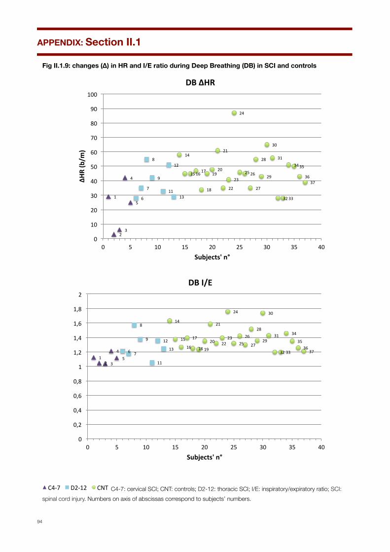
Fig II.1.9: changes (∆) in HR and I/E ratio during Deep Breathing (DB) in SCI and controls
1"
2"3"
4"
5"6"
7"
8"
9"
11"
12"
13"
14"
15"16"17"
18"
19"20"
21"
22"
23"
24"
25"26"
27"
28"
29"
30"
31"
32"33"
34"35"
36"37"
0"
10"
20"
30"
40"
50"
60"
70"
80"
90"
100"
0" 5" 10" 15" 20" 25" 30" 35" 40"
ΔHR$(b/m
)$
Subjects'$n°$
DB$ΔHR$
1"2" 3"
4"5"
6" 7"
8"
9"
11"
12"
13"
14"
15"
16"
17"
18"19"
20"
21"
22"23"
24"
25"26"
27"
28"
29"
30"
31"
32"33"
34"
35"36"
37"
0"
0,2"
0,4"
0,6"
0,8"
1"
1,2"
1,4"
1,6"
1,8"
2"
0" 5" 10" 15" 20" 25" 30" 35" 40"
Subjects'*n°*
DB*I/E*
4"
5" 6"
7"8" 9"
11"
12"
13"
14"
15"
16"
17"18"
19"
20"
21"
22"23"
24"
25"
27"
28"
29"
30"
31"
32"
33"
34"
35"
36"
37"
5" 10" 15" 20" 25" 30" 35" 40"Pa#ents')n°)
C4-7" D2-12" CNT" C4-7: cervical SCI; CNT: controls; D2-12: thoracic SCI; I/E: inspiratory/expiratory ratio; SCI: spinal cord injury. Numbers on axis of abscissas correspond to subjects’ numbers.
APPENDIX: Section II.1
94
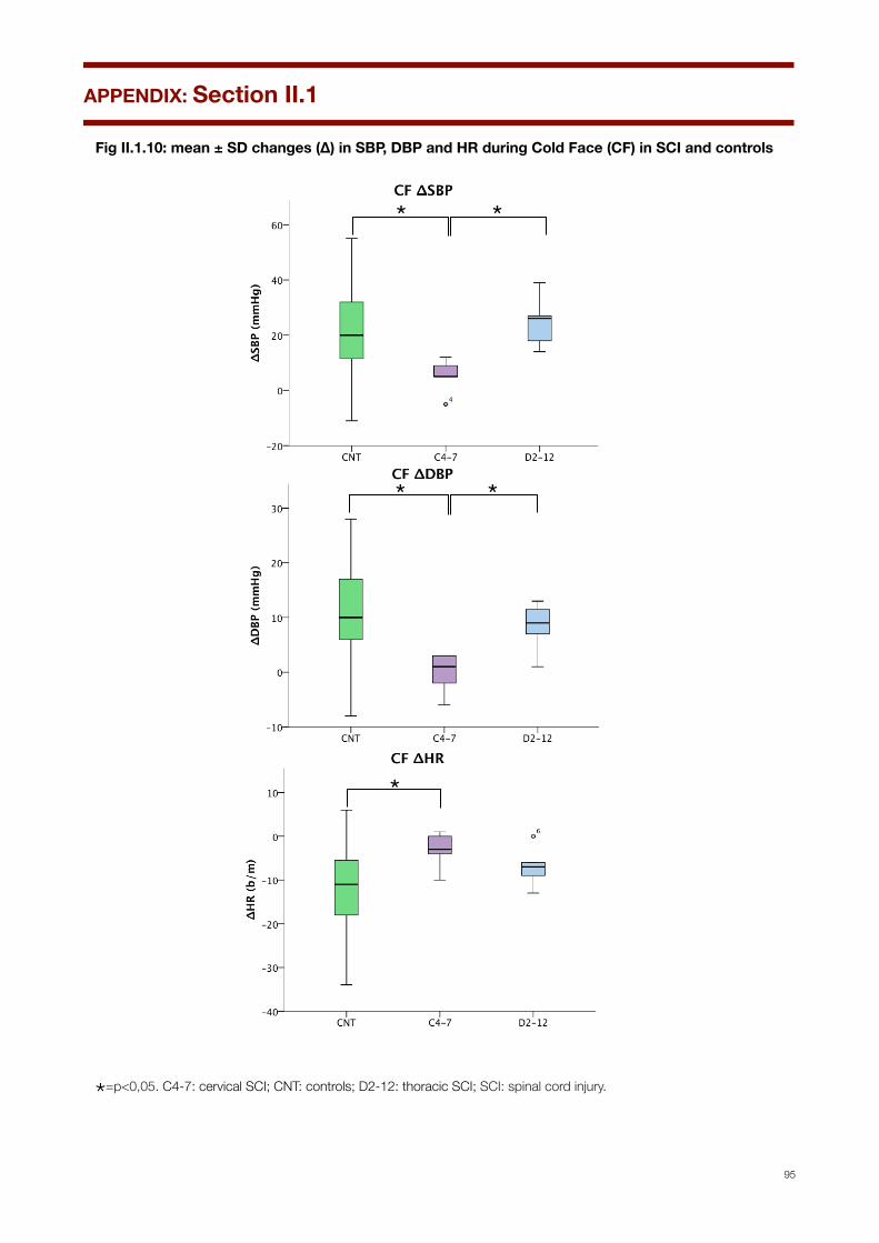
Fig II.1.10: mean ± SD changes (∆) in SBP, DBP and HR during Cold Face (CF) in SCI and controls
Cold Face
1"2"
3"
4"
5"6"
7" 8"
9"11"
12"
13"14"15"16"
17"
18"
19"
20"
21"
22"
23"
24"
25"
27"
28"
29"
30"31"
32"
33"
34"
35"
36"
37"
,20"
,10"
0"
10"
20"
30"
40"
50"
60"
0" 5" 10" 15" 20" 25" 30" 35" 40"
SBP$(m
mHg
)$
Pa+ents'$n°$
CF_Delta$SBP$$
C4,7" D2,12" CNT"
1"
2"3"
4"
5"
6"7"
8"
9"
11"
12"
13"
14"
15"16"
17"
18"
19"20"
21"
22"
23"
24"25"
27"
28"
29"
30"
31"
32"
33"
34"
35"
36"37"
,10"
,5"
0"
5"
10"
15"
20"
25"
30"
0" 5" 10" 15" 20" 25" 30" 35" 40"
DBP$(m
mHg
)$
Pa+ents'$n°$
CF_Delta$DBP$
C4,7" D2,12" CNT"
1" 2"
3"
4"
5" 6"
7"8" 9"
11"
12"
13"
14"
15"
16"
17"18"
19"
20"
21"
22"23"
24"
25"
27"
28"
29"
30"
31"
32"
33"
34"
35"
36"
37"
,40"
,35"
,30"
,25"
,20"
,15"
,10"
,5"
0"
5"
10"
0" 5" 10" 15" 20" 25" 30" 35" 40"
HR#(b
/m#
Pa*ents'#n°#
CF_Delta#HR#
C4,7" D2,12" CNT"
* * * * *
Fig II.4 ∆SBP, ∆DBP and ∆HR during cold face test (CF) in SCI patients and controls
*=p<0,05. C4-7: cervical SCI; CNT: controls; D2-12: thoracic SCI; SCI: spinal cord injury.
APPENDIX: Section II.1
95

Fig II.1.11: changes (∆) in SBP, DBP and HR during Cold Face (CF) in SCI and controls
1"2"
3"
4"
5"6"
7" 8"
9"11"
12"
13"14"15"16"
17"
18"
19"
20"
21"
22"
23"
24"
25"
27"
28"
29"
30"31"
32"
33"
34"
35"
36"
37"
,20"
,10"
0"
10"
20"
30"
40"
50"
60"
0" 5" 10" 15" 20" 25" 30" 35" 40"
ΔSBP
%(mmHg
)%
Subjects'%n°%
CF%ΔSBP%%
1"
2"3"
4"
5"
6" 7"8"
9"
11"
12"
13"
14"
15"16"
17"
18"
19"20"
21"
22"
23"24"25"
27"
28"29"
30"
31"
32"
33"
34"
35"
36"37"
,20"
,10"
0"
10"
20"
30"
40"
50"
60"
0" 5" 10" 15" 20" 25" 30" 35" 40"
ΔDBP
%(mmHg
)%
Subjects'%n°%
CF%ΔDBP%
1" 2"
3"
4"
5" 6"
7"8" 9"
11"
12"
13"
14"
15"
16"
17"18"
19"
20"
21"
22"23"
24"
25"
27"
28"
29"
30"
31"
32"
33"
34"
35"
36"
37"
,40"
,35"
,30"
,25"
,20"
,15"
,10"
,5"
0"
5"
10"
0" 5" 10" 15" 20" 25" 30" 35" 40"
ΔHR$(b/m
)$
Subjects'$n°$
CF$ΔHR$
4"
5" 6"
7"8" 9"
11"
12"
13"
14"
15"
16"
17"18"
19"
20"
21"
22"23"
24"
25"
27"
28"
29"
30"
31"
32"
33"
34"
35"
36"
37"
5" 10" 15" 20" 25" 30" 35" 40"Pa#ents')n°)
C4-7" D2-12" CNT" C4-7: cervical SCI; CNT: controls; D2-12: thoracic SCI; SCI: spinal cord injury. Numbers on axis of abscissas correspond to subjects’ numbers.
APPENDIX: Section II.1
96
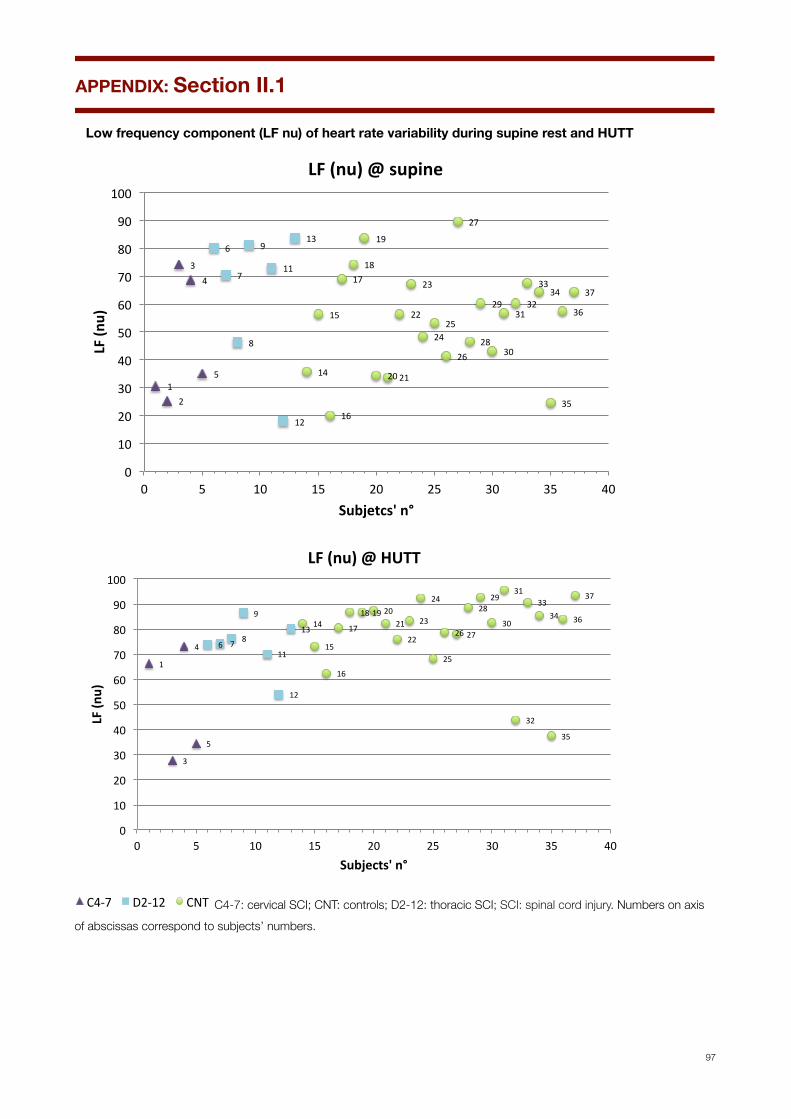
Low frequency component (LF nu) of heart rate variability during supine rest and HUTT
1"2"
3"4"
5"
6"
7"
8"
9"
11"
12"
13"
14"
15"
16"
17"18"
19"
20"21"
22"
23"
24"25"
26"
27"
28"
29"
30"
31"32"
33"34"
35"
36"
37"
0"
10"
20"
30"
40"
50"
60"
70"
80"
90"
100"
0" 5" 10" 15" 20" 25" 30" 35" 40"
LF#(n
u)#
Subjetcs'#n°#
LF#(nu)#@#supine#
1"
3"
4"
5"
6" 7" 8"
9"
11"
12"
13"14"
15"
16"
17"
18"19"20"21"
22"
23"
24"
25"
26"27"
28"29"
30"
31"
32"
33"34"
35"
36"
37"
0"
10"
20"
30"
40"
50"
60"
70"
80"
90"
100"
0" 5" 10" 15" 20" 25" 30" 35" 40"
LF#(n
u)#
Subjects'#n°#
LF#(nu)#@#HUTT#
4"
5" 6"
7"8" 9"
11"
12"
13"
14"
15"
16"
17"18"
19"
20"
21"
22"23"
24"
25"
27"
28"
29"
30"
31"
32"
33"
34"
35"
36"
37"
5" 10" 15" 20" 25" 30" 35" 40"Pa#ents')n°)
C4-7" D2-12" CNT" C4-7: cervical SCI; CNT: controls; D2-12: thoracic SCI; SCI: spinal cord injury. Numbers on axis of abscissas correspond to subjects’ numbers.
APPENDIX: Section II.1
97
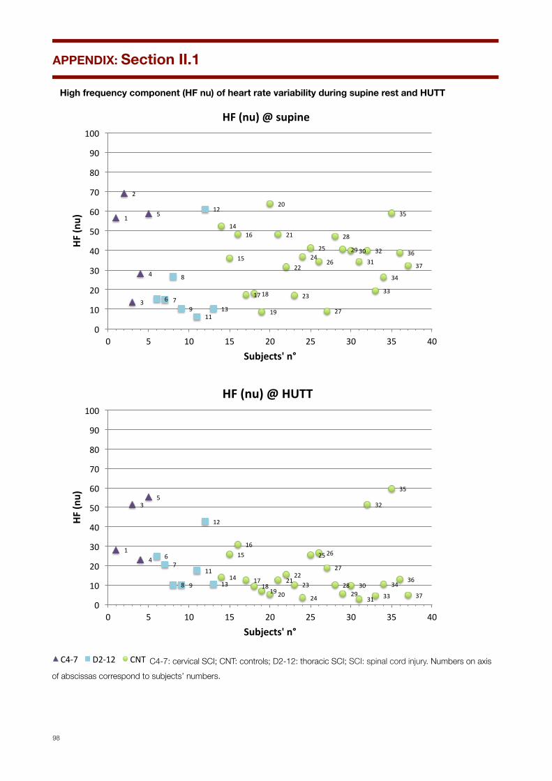
High frequency component (HF nu) of heart rate variability during supine rest and HUTT
1"
2"
3"
4"
5"
6" 7"
8"
9"11"
12"
13"
14"
15"
16"
17"18"
19"
20"
21"
22"
23"
24"25"
26"
27"
28"
29"30"31"
32"
33"
34"
35"
36"
37"
0"
10"
20"
30"
40"
50"
60"
70"
80"
90"
100"
0" 5" 10" 15" 20" 25" 30" 35" 40"
HF#(n
u)#
Subjects'#n°#
HF#(nu)#@#supine#
1"
3"
4"
5"
6"7"
8" 9"
11"
12"
13"14"
15"16"
17"18"
19"20"
21"22"
23"
24"
25"26"
27"
28"29"
30"
31"
32"
33"
34"
35"
36"
37"0"
10"
20"
30"
40"
50"
60"
70"
80"
90"
100"
0" 5" 10" 15" 20" 25" 30" 35" 40"
HF#(n
u)#
Subjects'#n°#
HF#(nu)#@#HUTT##
4"
5" 6"
7"8" 9"
11"
12"
13"
14"
15"
16"
17"18"
19"
20"
21"
22"23"
24"
25"
27"
28"
29"
30"
31"
32"
33"
34"
35"
36"
37"
5" 10" 15" 20" 25" 30" 35" 40"Pa#ents')n°)
C4-7" D2-12" CNT" C4-7: cervical SCI; CNT: controls; D2-12: thoracic SCI; SCI: spinal cord injury. Numbers on axis of abscissas correspond to subjects’ numbers.
APPENDIX: Section II.1
98

LF/HF ratio during supine rest and HUTT
1" 2"
3"
4"5"
6" 7"
8"
9"
11"
12"
13"
14"15"
16"
17"18"
19"
20"21"22"
23"
24"25"26"
27"
28"29"30"31"32"33"
34"
35"36"37"
0"
5"
10"
15"
20"
25"
30"
35"
0" 5" 10" 15" 20" 25" 30" 35" 40"
Subjects'*n°*
LF/HF*(nu)*@*supine*
1"3"
4"
5"
6" 7"
8"
9"
11"
12"
13"14"
15"16"
17"
18"
19"
20"
21"22"
23"
24"
25"26"27"
28"
29"
30"
31"
32"
33"
34"
35"
36"
37"
0"
5"
10"
15"
20"
25"
30"
35"
0" 5" 10" 15" 20" 25" 30" 35" 40"
Subjects'*n°*
LF/HF*(nu)*@*HUTT*
4"
5" 6"
7"8" 9"
11"
12"
13"
14"
15"
16"
17"18"
19"
20"
21"
22"23"
24"
25"
27"
28"
29"
30"
31"
32"
33"
34"
35"
36"
37"
5" 10" 15" 20" 25" 30" 35" 40"Pa#ents')n°)
C4-7" D2-12" CNT" C4-7: cervical SCI; CNT: controls; D2-12: thoracic SCI; SCI: spinal cord injury. Numbers on axis of abscissas correspond to subjects’ numbers.
APPENDIX: Section II.1
99
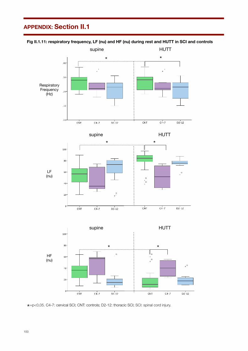
Fig II.1.11: respiratory frequency, LF (nu) and HF (nu) during rest and HUTT in SCI and controls
! !
supine HUTT
*
supine HUTT
RespiratoryFrequency
(Hz)
LF(nu)
HF(nu)
! !
*
! !
*
! !!!!
HUTT
*
*
Fig II.5 Respiratory frequency, LF (nu) and HF (nu) while supine and during HUT in SCI patients and controls
1 1
12
32
33
12
12
12
35
32
!!
*12
!
*
supine
12
*=p<0,05. C4-7: cervical SCI; CNT: controls; D2-12: thoracic SCI; SCI: spinal cord injury.
APPENDIX: Section II.1
100
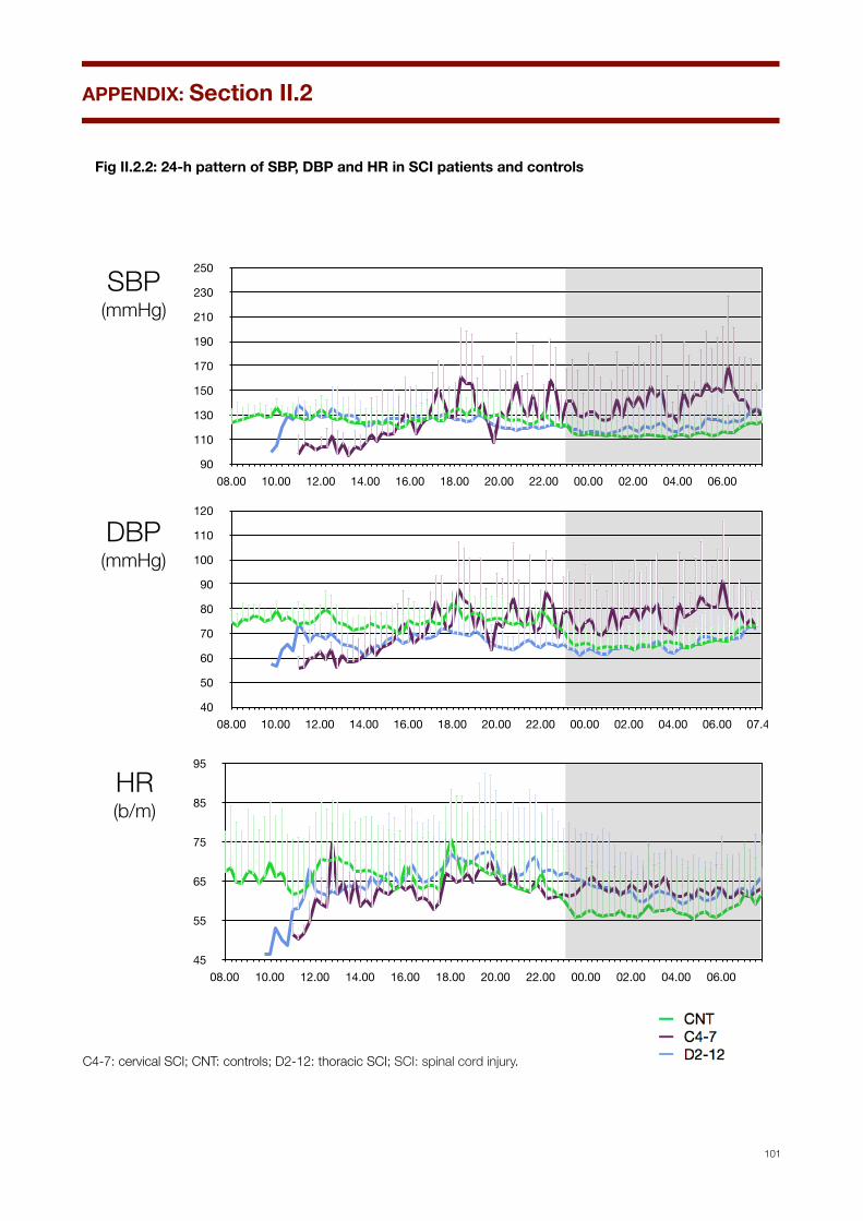
Fig II.2.2: 24-h pattern of SBP, DBP and HR in SCI patients and controls
90
110
130
150
170
190
210
230
250
08.00 10.00 12.00 14.00 16.00 18.00 20.00 22.00 00.00 02.00 04.00 06.00
45
55
65
75
85
95
08.00 10.00 12.00 14.00 16.00 18.00 20.00 22.00 00.00 02.00 04.00 06.00
SBP(mmHg)
DBP (mmHg)
HR (b/m)
Fig II.2.1 24-h pattern of SBP, DBP and HR in SCI patients and controls
40
50
60
70
80
90
100
110
120
08.00 10.00 12.00 14.00 16.00 18.00 20.00 22.00 00.00 02.00 04.00 06.00 07.45
C4-7: cervical SCI; CNT: controls; D2-12: thoracic SCI; SCI: spinal cord injury.! ! !
APPENDIX: Section II.2
101

24-h pattern of SBP, DBP and HR in each SCI patient and control
0
31
63
94
125
156
188
219
250
8:00 10:00 12:00 14:00 16:00 18:00 20:00 22:00 0:00 2:00 4:00 6:00
SBP(mmHg)
DBP (mmHg)
HR (b/m)
0
15
30
45
60
75
90
105
120
8:00 10:00 12:00 14:00 16:00 18:00 20:00 22:00 0:00 2:00 4:00 6:00
30
41
53
64
75
86
98
109
120
8:00 10:00 12:00 14:00 16:00 18:00 20:00 22:00 0:00 2:00 4:00 6:00
C4-7: cervical SCI; CNT: controls; D2-12: thoracic SCI; SCI: spinal cord injury.! ! !
APPENDIX: Section II.2
102
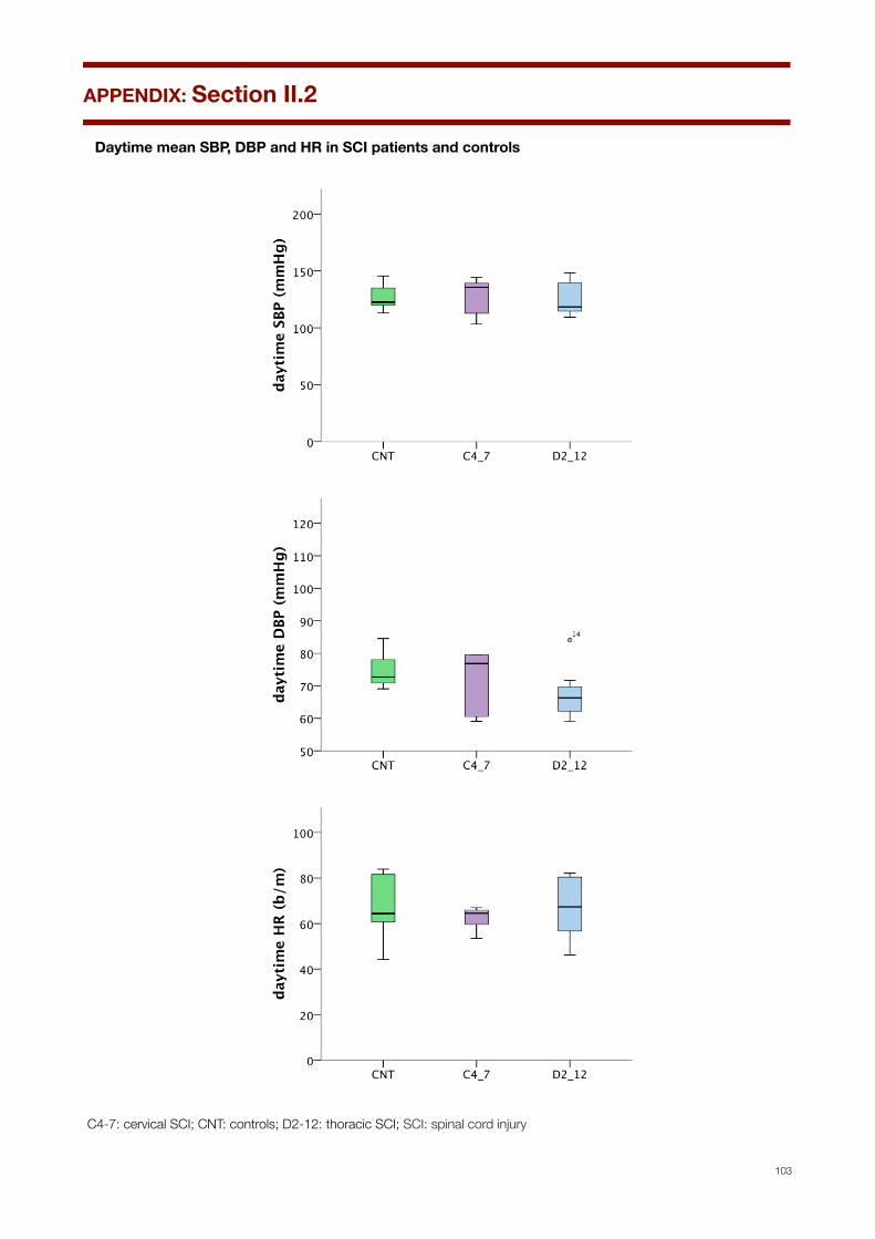
Daytime mean SBP, DBP and HR in SCI patients and controls
C4-7: cervical SCI; CNT: controls; D2-12: thoracic SCI; SCI: spinal cord injury
APPENDIX: Section II.2
103

Daytime SBP, DBP and HR in SCI patients and controls
1"
2"3"
4"
5"
6"
7"
8" 9"
11"
12"
12,5"
13"
14"14,5"15"15,5"
16"16,5"
17"
17,5"18"18,5"
19"19,5"20"20,5"
0"
20"
40"
60"
80"
100"
120"
140"
160"
0" 5" 10" 15" 20" 25"
SBP$(m
mHg
)$
Subjects'$n°$
day7me$SBP$$
1"
2"3"
4"
5"
6" 7"
8"9"
11"
12"12,5"
13"
14"14,5"15"15,5"
16"16,5"
17"
17,5"18"
18,5"
19"19,5"20"20,5"
0"
10"
20"
30"
40"
50"
60"
70"
80"
90"
0" 5" 10" 15" 20" 25"
DBP$(m
mHg
)$
Subjects'$n°$
day8me$DBP$$
1" 2"
3"
4"
5"
6"
7"
8"9"
11"
12"
12,5"
13"14"14,5"
15"
15,5"16"16,5"
17"17,5"
18"18,5"
19"19,5"
20"20,5"
0"
10"
20"
30"
40"
50"
60"
70"
80"
90"
0" 5" 10" 15" 20" 25"
HR#(b
/m)#
Subjects'#n°#
day6me#HR##
4"
5" 6"
7"8" 9"
11"
12"
13"
14"
15"
16"
17"18"
19"
20"
21"
22"23"
24"
25"
27"
28"
29"
30"
31"
32"
33"
34"
35"
36"
37"
5" 10" 15" 20" 25" 30" 35" 40"Pa#ents')n°)
C4-7" D2-12" CNT" C4-7: cervical SCI; CNT: controls; D2-12: thoracic SCI. Numbers on axis of abscissas correspond to subjects’ numbers.
APPENDIX: Section II.2
104

Night-time mean SBP, DBP and HR in SCI patients and controls
C4-7: cervical SCI; CNT: controls; D2-12: thoracic SCI; SCI: spinal cord injury.
APPENDIX: Section II.2
105
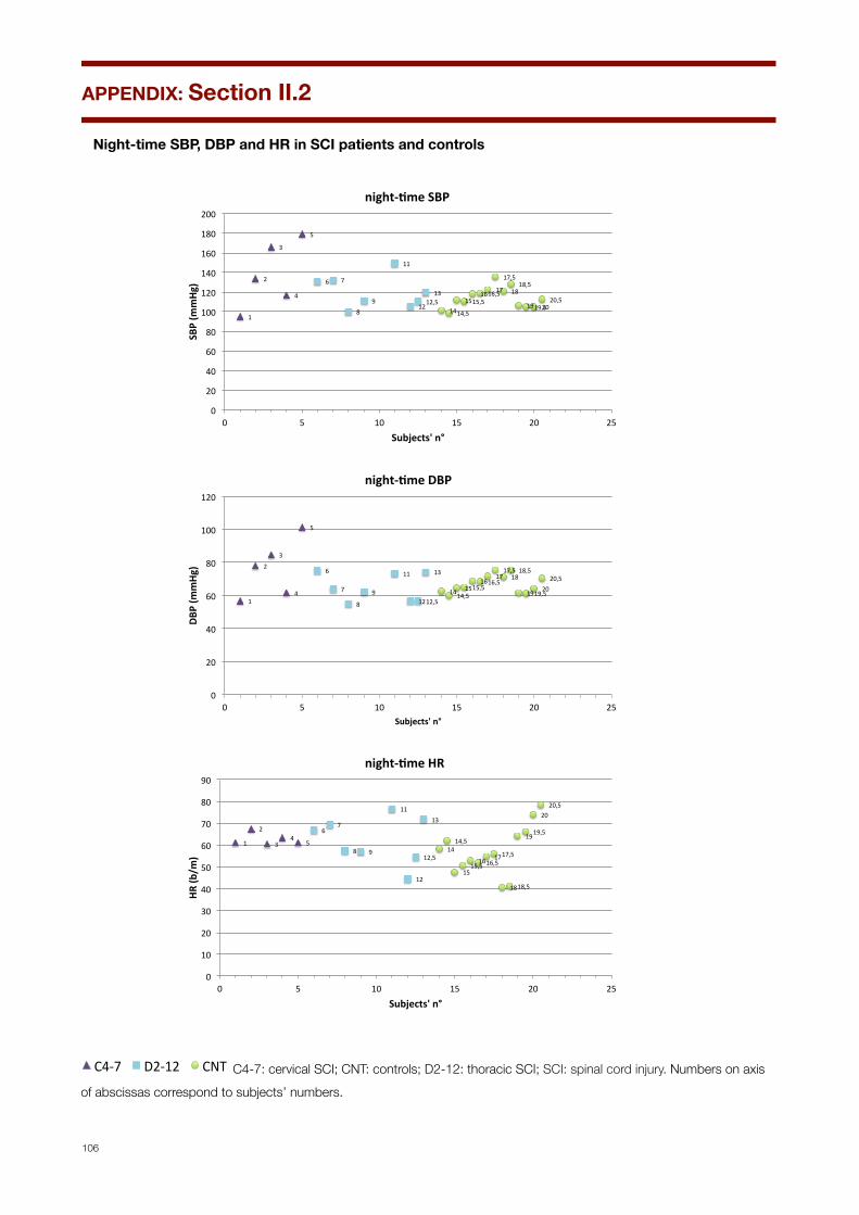
Night-time SBP, DBP and HR in SCI patients and controls
1"
2"
3"
4"
5"
6" 7"
8"9"
11"
12"12,5"
13"
14"14,5"
15"15,5"16"16,5"
17"
17,5"
18"18,5"
19"19,5"20"20,5"
0"
20"
40"
60"
80"
100"
120"
140"
160"
180"
200"
0" 5" 10" 15" 20" 25"
SBP$(m
mHg
)$
Subjects'$n°$
night67me$SBP$$
1"
2"
3"
4"
5"
6"
7"
8"
9"
11"
12"12,5"
13"
14"14,5"15"15,5"
16"16,5"17"
17,5"18"
18,5"
19"19,5"20"
20,5"
0"
20"
40"
60"
80"
100"
120"
0" 5" 10" 15" 20" 25"
DBP$(m
mHg
)$
Subjects'$n°$
night78me$DBP$$
1"
2"
3"4"
5"
6"7"
8" 9"
11"
12"
12,5"
13"
14"14,5"
15"15,5"
16"16,5"17"17,5"
18"18,5"
19"19,5"
20"20,5"
0"
10"
20"
30"
40"
50"
60"
70"
80"
90"
0" 5" 10" 15" 20" 25"
HR#(b
/m)#
Subjects'#n°#
night67me#HR##
4"
5" 6"
7"8" 9"
11"
12"
13"
14"
15"
16"
17"18"
19"
20"
21"
22"23"
24"
25"
27"
28"
29"
30"
31"
32"
33"
34"
35"
36"
37"
5" 10" 15" 20" 25" 30" 35" 40"Pa#ents')n°)
C4-7" D2-12" CNT" C4-7: cervical SCI; CNT: controls; D2-12: thoracic SCI; SCI: spinal cord injury. Numbers on axis of abscissas correspond to subjects’ numbers.
APPENDIX: Section II.2
106

Fig II.2.3: mean day-night changes (∆) in SBP, DBP and HR in SCI patients and controls
*
**
*∆SBP ∆DBP ∆HR
Fig II.2.2 Daytime-night-time Delta (∆) in SBP, DBP and HR in SCI patients and controls
17
6
*
**
*∆SBP ∆DBP ∆HR
Fig II.2.2 Daytime-night-time Delta (∆) in SBP, DBP and HR in SCI patients and controls
17
6
*
**
*∆SBP ∆DBP ∆HR
Fig II.2.2 Daytime-night-time Delta (∆) in SBP, DBP and HR in SCI patients and controls
17
6
*=p<0,05. C4-7: cervical SCI; CNT: controls; D2-12: thoracic SCI; SCI: spinal cord injury
APPENDIX: Section II.2
107
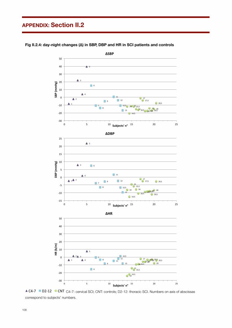
Fig II.2.4: day-night changes (∆) in SBP, DBP and HR in SCI patients and controls
1"
2"
3"
4"
5"
6"
7"8"
9"
11"
12"
12,5"
13"
14"
14,5"
15"15,5"16"16,5"
17"17,5"
18"18,5"19"
19,5"20"
20,5"
-30"
-20"
-10"
0"
10"
20"
30"
40"
50"
0" 5" 10" 15" 20" 25"
SBP$(m
mHg
)$
Subjects'$n°$
∆SBP$$
1" 2"
3"
4"
5"
6"
7"
8"
9"
11"
12"
12,5"
13"14"
14,5"
15"15,5"
16"
16,5"
17"17,5"
18"18,5"19"
19,5"
20"
20,5"
-15"
-10"
-5"
0"
5"
10"
15"
20"
25"
0" 5" 10" 15" 20" 25"
DBP$(m
mHg
)$
Subjects'$n°$
∆DBP$$
1"
2" 3"
4"
5"
6"
7"8"
9"11"
12"12,5"
13"
14"14,5"
15"15,5"
16"16,5"
17"17,5"18"
18,5"19"19,5"
20"20,5"
-30"
-20"
-10"
0"
10"
20"
30"
40"
50"
0" 5" 10" 15" 20" 25"
HR#(b
/m)#
Subjects'#n°#
∆HR##
4"
5" 6"
7"8" 9"
11"
12"
13"
14"
15"
16"
17"18"
19"
20"
21"
22"23"
24"
25"
27"
28"
29"
30"
31"
32"
33"
34"
35"
36"
37"
5" 10" 15" 20" 25" 30" 35" 40"Pa#ents')n°)
C4-7" D2-12" CNT" C4-7: cervical SCI; CNT: controls; D2-12: thoracic SCI. Numbers on axis of abscissas correspond to subjects’ numbers.
APPENDIX: Section II.2
108

Fig II.2.5: state dependent changes in SBP, DBP and HR in SCI and controls (between groups)
Fig II.2.3 State dependent changes in SBP, DBP and HR in SCI patients and controls (between groups)
SBP DBP HR
* *
*=p<0,01 vs. cervical SCI
Fig II.2.3 State dependent changes in SBP, DBP and HR in SCI patients and controls (between groups)
SBP DBP HR
* *
*=p<0,01 vs. cervical SCI
Fig II.2.3 State dependent changes in SBP, DBP and HR in SCI patients and controls (between groups)
SBP DBP HR
* *
*=p<0,01 vs. cervical SCI *=p<0,01 vs. cervical SCI.
C4-7: cervical SCI; CNT: controls; D2-12: thoracic SCI; SCI: spinal cord injury.
APPENDIX: Section II.2
109

Fig II.2.6: state dependent changes in SBP, DBP and HR in SCI and controls (between groups)Fig II.2.4 State dependent changes in SBP, DBP and HR in SCI patients and controls (within groups)
* * *
* * *§*
* * * * *
*=p<0,05 vs. within group wake (W) values
1213
7§*
§*
*=p<0,05 vs within group wake (W) values; §=p<0,03 vs within group stage 3 (N3) NREM sleep values C4-7: cervical SCI; CNT: controls; D2-12: thoracic SCI; SCI: spinal cord injury.
APPENDIX: Section II.2
110

Fig III.3: 24-h pattern of BcT in SCI patients and controlsFig III.1 24-h pattern of BcT in SCI patients and controls
BcT(°C)
36
37
38
39
08.00 10.00 12.00 14.00 16.00 18.00 20.00 22.00 00.00 02.00 04.00 06.00
BcT: body core temperature; C4-7: cervical SCI; CNT: controls; D2-12: thoracic SCI; SCI: spinal cord injury.
24-h pattern of BcT in each SCI patient and control
Fig III.1 24-h pattern of BcT in SCI patients and controls
BcT(°C)
36
37
38
39
08.00 10.00 12.00 14.00 16.00 18.00 20.00 22.00 00.00 02.00 04.00 06.00
36,0
36,5
37,0
37,5
38,0
38,5
39,0
8:00 10:00 12:00 14:00 16:00 18:00 20:00 22:00 0:00 2:00 4:00 6:00
BcT(°C)
BcT: body core temperature; C4-7: cervical SCI; CNT: controls; D2-12: thoracic SCI; SCI: spinal cord injury.
APPENDIX: Section III
111
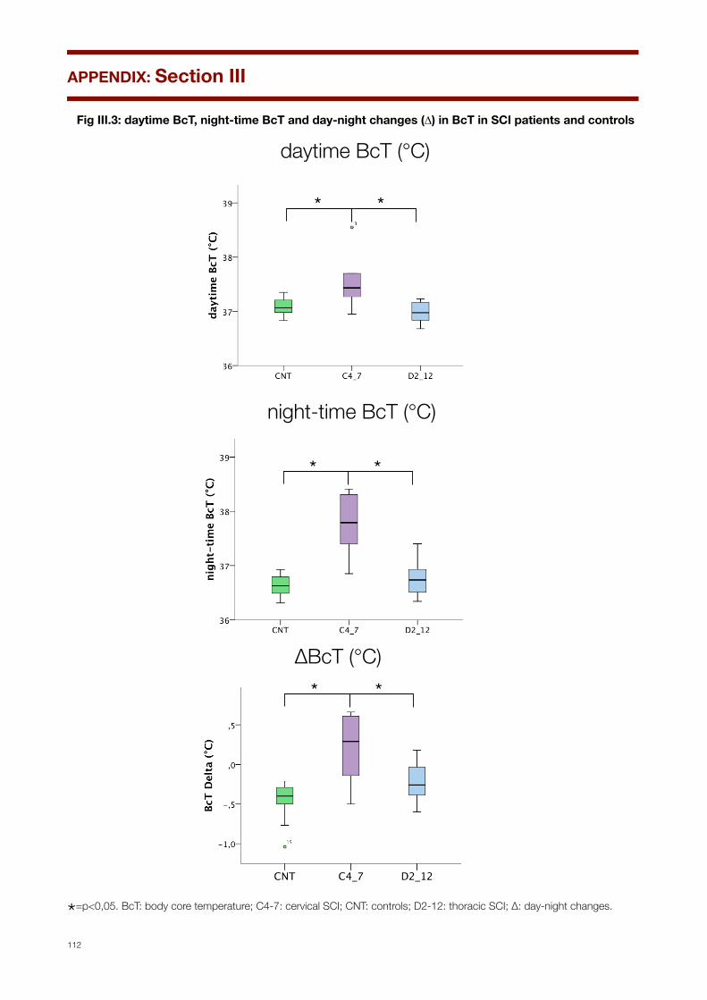
Fig III.3: daytime BcT, night-time BcT and day-night changes (∆) in BcT in SCI patients and controls
* *
daytime BcT (°C)
Fig III.2 Daytime, night-time and Delta (∆) day-night BcT in SCI patients and controls
night-time BcT (°C)
* *
∆BcT (°C)* *
* *
daytime BcT (°C)
Fig III.2 Daytime, night-time and Delta (∆) day-night BcT in SCI patients and controls
night-time BcT (°C)
* *
∆BcT (°C)* *
* *
daytime BcT (°C)
Fig III.2 Daytime, night-time and Delta (∆) day-night BcT in SCI patients and controls
night-time BcT (°C)
* *
∆BcT (°C)* *
*=p<0,05. BcT: body core temperature; C4-7: cervical SCI; CNT: controls; D2-12: thoracic SCI; ∆: day-night changes.
APPENDIX: Section III
112
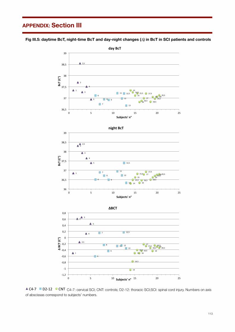
Fig III.5: daytime BcT, night-time BcT and day-night changes (∆) in BcT in SCI patients and controls
1"
2"
2,5"
3"
4"
5"
6"
7"
8" 9"
11"
12"
12,5"
13"
14"14,5"
15"15,5"
16"16,5"
17"
17,5"
18,5"
19"19,5"
20"20,5"
36,5"
37"
37,5"
38"
38,5"
39"
0" 5" 10" 15" 20" 25"
BcT$(C°)$
Subjects'$n°$
day$BcT$$
1"
2"2,5"
3"
4"
5"
6"
7"
8"
9"
11"
12"
12,5"
13" 14"
14,5"15"
15,5"
16"16,5"
17"17,5"
18,5"19"
19,5"20"20,5"
36"
36,5"
37"
37,5"
38"
38,5"
39"
0" 5" 10" 15" 20" 25"
BcT$(C°)$
Subjects'$n°$
night$BcT$$
1"
2"
2,5"
3"
4"
5"
6"
7"
8"
9"
11" 12"
12,5"
13"
14"
14,5"
15"15,5"
16"16,5"
17"17,5"
18,5"19"
19,5"20"20,5"
-1,2"
-1"
-0,8"
-0,6"
-0,4"
-0,2"
0"
0,2"
0,4"
0,6"
0,8"
0" 5" 10" 15" 20" 25"
∆"BC
T"(C°)"
Subjects'"n°"
∆BCT""
4"
5" 6"
7"8" 9"
11"
12"
13"
14"
15"
16"
17"18"
19"
20"
21"
22"23"
24"
25"
27"
28"
29"
30"
31"
32"
33"
34"
35"
36"
37"
5" 10" 15" 20" 25" 30" 35" 40"Pa#ents')n°)
C4-7" D2-12" CNT" C4-7: cervical SCI; CNT: controls; D2-12: thoracic SCI;SCI: spinal cord injury. Numbers on axis of abscissas correspond to subjects’ numbers.
APPENDIX: Section III
113
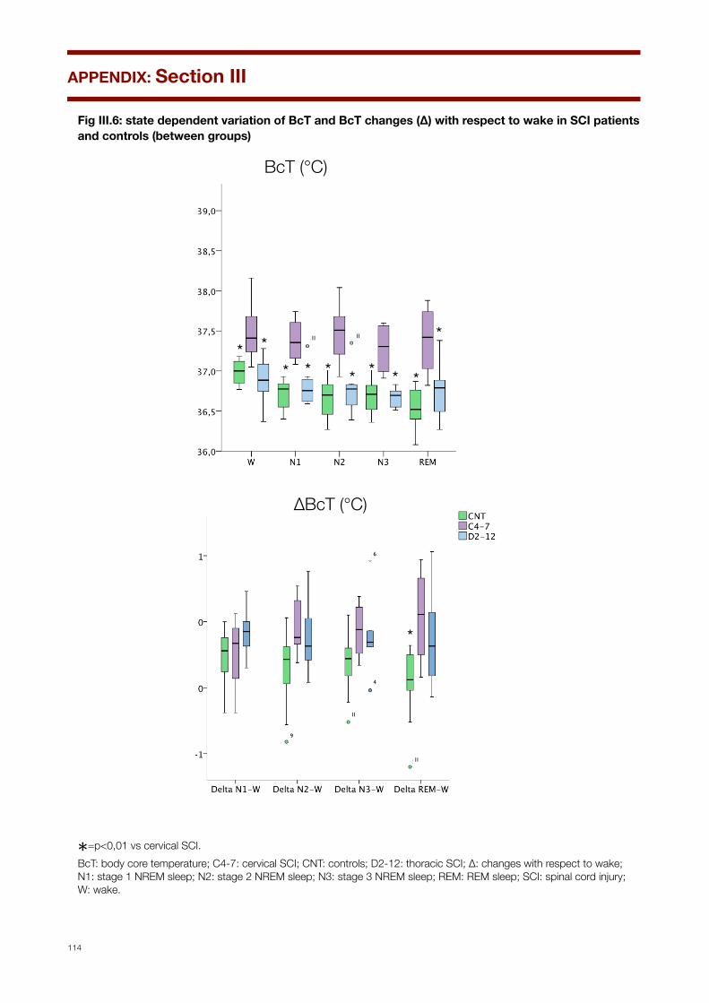
Fig III.6: state dependent variation of BcT and BcT changes (∆) with respect to wake in SCI patients and controls (between groups)
Fig III.3 State dependent BcT and changes (∆) with respect to wake in SCI patients and controls (between groups)
∆BcT (°C)
*
BcT (°C)
* ** * * * * * *
*1111
9
11
11
6
4
∆BcT
(°C
)
Fig III.3 State dependent BcT and changes (∆) with respect to wake in SCI patients and controls (between groups)
∆BcT (°C)
*
BcT (°C)
* ** * * * * * *
*1111
9
11
11
6
4
∆BcT
(°C
)
*=p<0,01 vs cervical SCI.BcT: body core temperature; C4-7: cervical SCI; CNT: controls; D2-12: thoracic SCI; ∆: changes with respect to wake; N1: stage 1 NREM sleep; N2: stage 2 NREM sleep; N3: stage 3 NREM sleep; REM: REM sleep; SCI: spinal cord injury; W: wake.
APPENDIX: Section III
114
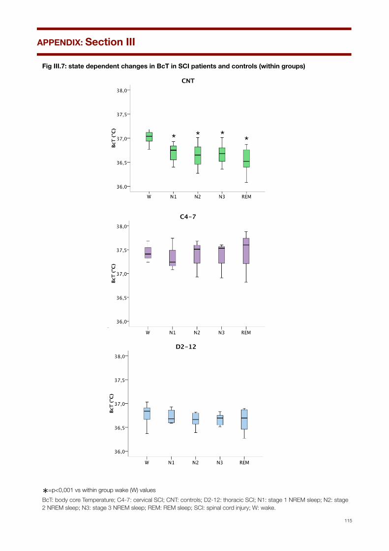
Fig III.7: state dependent changes in BcT in SCI patients and controls (within groups)Fig III.4 State dependent changes in BcT in SCI patients and controls (within groups)
*=p<0,05 vs. within group wake (W) values
* * * *
Fig III.4 State dependent changes in BcT in SCI patients and controls (within groups)
*=p<0,05 vs. within group wake (W) values
* * * *
Fig III.4 State dependent changes in BcT in SCI patients and controls (within groups)
*=p<0,05 vs. within group wake (W) values
* * * *
*=p<0,001 vs within group wake (W) valuesBcT: body core Temperature; C4-7: cervical SCI; CNT: controls; D2-12: thoracic SCI; N1: stage 1 NREM sleep; N2: stage 2 NREM sleep; N3: stage 3 NREM sleep; REM: REM sleep; SCI: spinal cord injury; W: wake.
APPENDIX: Section III
115

APPENDIX: Section III
116
ACKNOWLEDGEMENTS
Questa tesi di dottorato è il frutto del lavoro e della collaborazione di diverse persone. Ognuno nel suo singolo ha contribuito in maniera significativa e ha portato idee e contribuiti senza le quali questo risultato non sarebbe stato possibile.
Anzitutto, è il frutto delle brillanti idee e della grande capacità e competenza del Prof Pietro Cortelli; sono fiero e onorato di aver potuto seguire la sua Scuola in tutti questi anni. Questo volume vuole essere il mio modo di ringraziarlo e di restituirgli, almeno in parte, i grandi insegnamenti che da lui ho ricevuto.
È frutto della collaborazione con l’Unità Spinale dell’Azienda Ospedaliera Carreggi Firenze, col Dott Giuseppe De Scisciolo, Dott. Sergio Aito, Dott Riccardo Caramelli e Dott.ssa Vincenza Schiavone che hanno accettato di partecipare allo studio e inviato i pazienti.
È frutto della supervisione della Dott.ssa Federica Provini, a cui devo tutto quello che ho imparato sulla medicina del sonno e che ringrazio per la sua semplice positività e costante incoraggiamento.
È frutto dei consigli e del lavoro “pionieristico” delle mie colleghe Dott.ssa Daniela Grimaldi e Dott.ssa Giovanna Calandra, che con le loro pubblicazioni precedenti mi hanno aperto una strada e semplificato la vita, oltre ad aver fornito i preziosi dati sui controlli utilizzati per lo studio. Ringrazio in particolare Giovanna per aver tacitamente sopportato il mio disordine e per la complicità derivata dall’aver passato tante ore insieme lavorando nella nostra “mansarda”, ma anche mangiando cioccolata e ogni schifezza possibile proveniente dalle macchinette...
È soprattutto il frutto della perizia di un personale tecnico insostituibile come Giorgio Barletta, in grado di chiarire ogni dubbio oltre che di fornire tracciati di una qualità ineccepibile, e della silenziosa presenza di Annagrazia Cecere, che, oltre ad aver fornito un’inestimabile aiuto nella lettura dei tracciati si è anche presa cura di mia figlia Agata mente io scrivevo la tesi!
È il frutto del sostegno della mia famiglia, e in particolare dei miei genitori Paola e Giampaolo, a cui devo semplicemente tutto.
È il frutto della pazienza di Achira e Agata, che hanno sopportato senza lamentarsi questo mio lavoro senza orari e senza fine settimana. Grazie, spero di restituirvi presto il tempo che vi è stato sottratto, perché solo a voi spetta, ed è con voi che io lo voglio passare.


![Neonatal Thermoregulation - University of · PDF fileNeonatal Thermoregulation Julia Petty. ... A care study. Journal of Neonatal Nursing. ... 5 Thermoregulation [Compatibility Mode]](https://img.pdfslide.net/doc/110x75/5aafe83f7f8b9a6b308de3c0/neonatal-thermoregulation-university-of-thermoregulation-julia-petty-a-care.jpg)