Embed Size (px)
Citation preview
Annals of Academy of Romanian Scientists Series: Medical Sciences
Online ISSN 2285-4150 Volume 4, Number 2/2013 73
Spinal disorders in children and teenagers: clinical data, imagistics and therapeutic principles
Gh. BURNEI1,2,∗∗∗∗, Stefan GAVRILIU1, 2, Costel VLAD2,
Cezar TOMESCU2, Raluca Alexandra GHIłĂ2, Ileana GEORGESCU2,
Cezara Alexandra DUGHILĂ3, Mădălina MACADON2
Abstract
Spinal pathology in children and teenagers may be evaluated by the presence of back pain and by the various surgical procedures for different spinal disorders. In our statistics about 40% of the patients presented with back pain and 24% of them, after thorough investigations, had vertebral column lesions. 16% had no obvious bony lesions. MRI with contrast and discography are highlightening intervertebral disk lesions of different degrees. Out of all patients with discal problems only 2% presented discal hernia.
Idiopathic scoliosis represent the most frequent spinal deformity and a very small number of patients accuse back pain, either due to arthritis lesions of the joint processes or to discal issues.
The second most frequent spinal issue is represented by Scheuemann's kyphosis. This disease is usually encountered in teenagers and it requires a surgical procedure, especially if the kyphosis is greater than 75 degrees and it is associated with pain when sitting and during school activities.
Congenital scoliosis has an incidence of 1/4.000 births in Romania. In Romania there are approximately 300 cases with congenital scoliosis, 80 cases being children and teenagers. The first surgical procedure with somatic instrumentation in Romania has been done in “Maria Sklodowska Curie” hospital by Prof. Dr. Gh. Burnei in 2000 with hemivertebra resection and somatic instrumentation with total correction of the axial deviation.
Nowadays in Romania there should be a serious effort to be done in order to detect early spinal disorders to avoid the onset of severe, rigid deformities which may only be treated by extended surgical procedures with high neurological risks and high costs. The data in this paper are meant to guide the medical persons in an early detection of spinal issues. The school
1 University of Medicine and Pharmacy “Carol Davila”, Bucharest, Romania 2 Clinical Emergency Hospital for Children “Maria Sklodowska Curie”, ”Alexandru Pesamosca” Clinics, Bucharest,
Romania 3 Clinical Emergency Hospital for Children “Sf. Maria”, Iaşi, Romania
* Address for correspondence: Prof. Dr. Gh. Burnei, University of Medicine and Pharmacy “Carol Davila”, Bucharest,
Romania, Clinical Emergency Hospital for Children “Maria Sklodowska Curie”, 20, C-tin Brâncoveanu, email:
Gh. BURNEI∗, Stefan GAVRILIU, Costel VLAD, Cezar TOMESCU, Raluca Alexandra GHIłĂ, Ileana GEORGESCU, Cezara Alexandra DUGHILĂ, Mădălina MACADON
74
screening program has to be adapted to our conditions in order that all scoliosis with 40 degrees and kyphosis with 50 degrees to be monitored by centers where spinal procedures are performed.
Keywords: spinal deformities, scoliosis, Scheuermann kyphosis, lordosis, treatment principles
Rezumat
Patologia rahisului la copil şi adolescent poate fi evaluată atât sub aspectul durerilor rahidiene, cât şi din punct de vedere al intervenŃiilor operatorii efectuate pentru diverse afecŃiuni. În cazuistica noastră 40% din pacienŃi se prezintă cu dureri rahidiene şi în urma investigaŃiilor efectuate, 24% dintre aceştia prezintă leziuni osteoarticulare ale coloanei vertebrale. La 16% nu se decelează leziuni osoase. IRM cu substanŃă de contrast şi discografia pun în evidenŃă leziuni ale discurilor intervertebrale de diverse grade. Din totalul pacienŃilor cu discopatii doar 2 % au prezentat hernii de disc.
Scolioza idiopatică reprezintă cea mai frecventă diformitate a coloanei vertebrale şi un număr foarte redus de cazuri acuză durere rahidiană, fie datorită unor leziuni artrozice ale proceselor articulare, fie a discopatiilor.
Pe locul 2 ca frecvenŃă se situează cifoza juvenilă Scheuermann. Această afecŃiune este depistată de obicei la vârsta adolescentului mare şi impune o atitudine terapeutică chirurgicală, mai ales când cifoza care depăŃeste 75 de grade se insoteste de durere in pozitia sezind, a activitatii scolare si de studiu.
Scoliozele congenitale au o incidenŃă în România de 1 la 4.000 de naşteri. În România sunt diagnosticaŃi actualmente circa 300 de pacienŃi cu scolioză congenitală, dintre care 80 sunt copii şi adolescenŃi. Prima intervenŃie chirurgicală cu instrumentare somatică din România a fost efectuată la spitalul “Maria Sklodowska Curie” de Prof. Dr. Gh. Burnei în anul 2000 la un copil la care s-a practicat rezecŃia hemivertebrei şi instrumentare somatică cu corecŃia totală a deviaŃiei axiale.
Actualmente în România trebuie depus un efort susŃinut pentru depistarea precoce a afecŃiunilor coloanei vertebrale pentru a evita evoluŃia spre diformitîŃi severe, rigide, care nu pot fi corectate decât prin intervenŃii de mare amploare cu risc neurologic major şi costuri enorme. Datele consemnate în această lucrare au ca obiectiv orientarea cadrelor medicale angajate în depistarea precoce a acestor afecŃiuni. Programul de screening şcolar trebuie adaptat sistemului nostru de organizare astfel încât toŃi pacienŃii cu scolioze cu unghi Cobb de 40 de grade şi cifoze juvenile ce depăşesc 50 de grade să fie luaŃi în evidenŃă de centrele unde se efectuează intervenŃii chirurgicale.
Cuvinte-cheie: diformităŃi spinale, scolioză, cifoză Scheuermann, lordoză, principii de tratament

Spinal disorders in children and teenagers: clinical data, imagistics and therapeutic principles
75
1. THE SPINAL COLUMN
1.1. Anatomical data
Spinal column is the most important segment of the locomotor apparatus, of
which are linked all the other segments that make up the torso (thorax and pelvis),
respectively upper and lower limbs. Spinal column gives the body symmetry,
direction of movement and protects our communication systems providing
stability, due to the superposition of many bone pieces from the original
cartilaginous tube.
Due to spinal curvatures, projection of the centers of gravity of various
segments aren’t found on the projection line of the general body center of gravity.
Therefore, the action of gravity cause, from one vertebra to another, rotational
stresses which tend to accentuate curves and which must be neutralized, because
otherwise the column would collapse. Forces that oppose rotational stresses are
ligaments. Projection center of the thoracic spinal segment passes anterior. It
would collapse if there would not intervene the vertebral common posterior
ligament, interspinous ligaments and ligamentum flavum. The situation is
reversed at the lumbar and cervical spine: projection of the center of gravity goes
posteriorly from the column, and the forces that oppose collapse are represented
by the resistance of the vertebral common anterior ligament. Vertebral ligaments
were therefore designed to alleviate a great deal of stresses.
Other items that are designed to absorb the forces of demand are
intervertebral discs. They don’t stay in tension, like ligaments, but under pressure.
Between these two categories of anatomical elements, ligaments on one side and
intervertebral discs on the other, subject to contrary forces, are established a
certain state of balance, named intrinsic equilibrium. In orthostatism and during
rest the spinal column has a vertical direction and a slightly sinuous form,
especially in the sagittal plane. In physics it is known that an elastic column, with
curves, offers a higher resistance to vertical pressures, than a perfectly straight
column. Curves absorb vertical shocks and favors maintaining column balance on
the pelvis, thus easing spinal muscle belt efforts.
This attitude and this form are maintained thanks to muscle tonicity,
ligaments and discs elasticity and due to anatomical jointing of the 34 vertebrae
composing the spinal column; vertebrae adapts to each other by different joint
surfaces.
1.2. Biomechanics
Column movements, regardless of their magnitude, are complex movements,
involving several segments of the spine. They are made by combining slight
displacement of the vertebral bodies, which occur at the level of the intervertebral
Gh. BURNEI∗, Stefan GAVRILIU, Costel VLAD, Cezar TOMESCU, Raluca Alexandra GHIłĂ, Ileana GEORGESCU, Cezara Alexandra DUGHILĂ, Mădălina MACADON
76
discs, and at the other joints. These movements are limited by the resistance of
ligaments, by the intervertebral joints shape and by the degree of compressibility
of the fibro-cartilaginous tissue that makes up the disc.
Small intervertebral movements are only possible due to the presence of the
nucleus pulposus. Vertebral movements run on nucleus pulposus as a shaft,
nucleus pulposus playing the role of a real mechanical ball. This means such as a
ball all movements are possible; though they will be limited and dependent on the
various conformations and positions of the articular apophyses, by the spinal
ligaments and by the spinal muscular „brace”.
By the pressure of the liquid located between its components, nucleus
pulposus has the ability to be flexible. Due to this property spinal movements are
possible and excessive pressure effects or shocks suffered by the spinal column
are removed. In a forced flexion attitude, occurs approximation of the vertebral
bodies by their anterior side, by partially compressing anterior half disc and by
slightly pushing nucleus pulposus posterior. In extension, things happen vice
versa. Movements are made possible through the integral role intervertebral disc
play, forming a unitary organ. The researches performed to date have shown that
if the nucleus pulposus should be considered „the bearing” on who are running the
movements of the spine, the fibrous ring remains the most important element of
the intervertebral disc that resists compression and decompression forces.
As is known, spine shows complex movements resulting from cumulative
micro-movements of all intervertebral joints: flexion-extension, lateral bending,
rotation, and - as a result thereof – circumduction.
Ventral flexion of the trunk on the legs is achieved through participation not
only by the spine, but also by the hips. Sacrum being fixed, the rest of the spine
can fully execute a movement of flexion, but not all the segments equally
participate. Largest amplitude in flexion is performed in the cervical and lumbar
region. Anterior concave arch that is formed by column in its entirety is not an
arc of circle, but a curved line, composed of three segments, namely: one with
smaller radius, which is formed by the cervical spine, one with larger radius,
which represents the thoracic spineand finally, one with a small radius of lumbar
region.
In maximum flexion the transverse line that extends the axis plane intersects
vertical line at an angle of 140-160 degrees. In moderate flexion, anterior portion
of the intervertebral disc is compressed, while the posterior common vertebral
ligament, ligamentum flavum, interspinous ligaments, supraspinous ligament and
the back muscles are all under tension.
In orthostatic position, in extension, things happen exactly the opposite. In
the lumbar region, extension reaches up to 30 degrees, in the thoracic one up to 55
degrees and in the cervical region up to 60 degrees. In extension movement, the
posterior portions of the intervertebral discs are compressed, while the anterior
Spinal disorders in children and teenagers: clinical data, imagistics and therapeutic principles
77
common vertebral ligament is under tension. The lateral tilt movement is
approximately 16.6 degrees, with a maximum of amplitude in the thoracic
segment. When there is some degree of twisting of the spine, the trunk is leaning
further lateral. The rotation movement is maximum in the cervical region, where it
reach 75 degrees. Thoracic spine rotates slightly and only if it tilts laterally. In the
lumbar segment, and in predilect mannerthoraco-lumbar, rotation movement
execute when the column is in extension. When the column is flexed, the rotation
movement in the lumbar segment is impossible, because the vertebrae condyles
are placed vertically in the joints and stop the movement, the same case applies
inflexion when the lateral tilt of the lumbar segment can’t be done. The flexion-
extension movement is around a transverse axis passing through the upper part of
the atlas glenoidal cavity (1).
Morphofunctional complexity of the column has as consequence varied
pathology, with symptomatology as complex, with pathophysiological
mechanisms neurosomatic, neurovegetative, vascular, osteoarticular, ligamentary,
in the pathologic processes of traumatic, inflammatory, postural, tumoral order.
To complete this unitary look must be considered visceral relationships and
particular individual reaction. Also, it must be revealed the fact that some of the
chronic spinal disorders is installed gradually over years and sometimes decades,
with a long evolution, most often asymptomatic, suggesting the idea of a
functional adaptation, a compensating form of balance, common to other organs.
This explains why many of the bearers of important radiological alterations shows
no sign of disease or why noisy clinical manifestations are accompanied by
minimal or absent radiological alterations, so that the spine semiology and clinical
examination shows the great importance. The rich somatic and autonomic
innervation of the spine justifies largely, the echo on the nervous system in spinal
disorders. Indeed, some of these patients may occur at clinical examination as
mentally ill, secondary to a spinal disease, by disrupting the dynamics of the
central nervous system after ascending excitation. These cortical components
secondary or primitive and the visceral neuroreflexe manifestations, complicates
clinical examination therefore is required a unitary vision, analytical, with careful
investigation of all apparatus and systems.
1.3. Statics
There are five general types of postures recognized: normal back, round, flat,
flat-concave and flat-round.
Normal back is the type of posture in which spinal curvatures in the anterior-
posterior sense shows a normal arching, and the pelvis has a normal tilt. Round
back is very common, thoracic convexity descends, including the lumbar
vertebrae and the lumbar concavity shrinks in size and depth; the pelvis is slightly

Gh. BURNEI∗, Stefan GAVRILIU, Costel VLAD, Cezar TOMESCU, Raluca Alexandra GHIłĂ, Ileana GEORGESCU, Cezara Alexandra DUGHILĂ, Mădălina MACADON
78
tilted ventral and caudal. Flat back is more rare than the round back. Thoracic
kyphosis and lumbar lordosis disappear, but still maintain pelvic tilt. Shoulder
blades appear embossed posterior. Flat concave back (lordotic) is less common.
Lumbar concavity is more emphasized, but simultaneously it accentuates thoracic
convexity (2).
2. DIAGNOSIS
The inclination of vertebral column in the sagittal plane, the frontal plane or
horizontally, and often in several planes at once, causes kyphosis, lordosis, or
pathological scoliosis.
2.1. Symptomatology
The dominant symptom of spinal conditions is pain, a purely subjective
element, whose detailed analysis may reveal some shades so particular that directs
the examiner attention to a more pronounced distress of a certain morphological
structure.
It is more important to conceptualize the back pain as a potential neurological
symptom and such the analysis of the entire neurological system relevant. It may
occur a number of neurological symptoms such as: veiled view, double vision,
syncope, headache, hearing loss, motor non-coordination, fine or gross. Also can
install arm pain, fatigue, sensitivity problems, followed by paresthesia and
heaviness of hands. Paresthesias may be accused at the chest and at the back (3).
Then it’s analyzed the existing pain in joints, interdependence between pain and
menstrual periods, and weather effects.
2.2. Clinical examination
At the physical examination of the patient are followed: cervical lordosis,
thoracic kyphosis, lumbar lordosis. At skin inspection, it finds any changes that
may occur in such cases: macular hirsutism, subcutaneous lesions, localized
hyperpigmentation, multiple lipomas, brown spots, dermal sinuses. Are palpable
bony prominences of the cervical, thoracic and lumbar-sacral regions and the
adjacent soft tissues. The physician is interested in whether the patient has rib
hump (gibus) and if scoliosis is clinically evident. Overall flexibility of the spine
in the thoracic-lumbar segment is measured by placing the patient in flexion, in
extension, laterally tilted and axial rotated left and right. With the patients eated
reflexes are checked at the kneecap level. The flexibility of all joints is verified,
also the secondary sexual characteristics are inspected (hair growth and
development of the scrotum).
Spinal disorders in children and teenagers: clinical data, imagistics and therapeutic principles
79
Also, in the case of back pain can be useful complete blood tests, and
urinalysis, in the case in which the patient is suspected juvenile arthritis,
investigations are corresponding to. Therewith can be frequently established a
strong link between endocrine disorders and some local deformations of the spine.
„Scoliotic disease” begins with predilection between 10 and 12 years,
through a fairly typical clinical picture. Child grows in height in a very short time,
although it feeds inadequate (4). These children are pale and CBC highlights
hypochromic anemia. Their musculature is less tonic and therefore they are easily
fatigued. Sometimes these children experience night sweats and complain of pain
between the shoulder blades, or vague, without being able to specify the place, but
with some irradiation to the abdomen. Many of the patients had obvious endocrine
disorders and the majority is observed with more or less obvious hypertrophy
thyroid body. Basal metabolism is altered. Some patients, few in number, are
apathetic, they are quickly fatigued and start a school activity with lower value,
others, on the contrary, are more irritable, with moist skin, with occasionally
insomnia. Girls are often seen with menstrual disorders in this time (5).
2.3. Radiological exploration
From the moment it was concluded that it is necessary a radiological
evaluation of patients with back pain, the question arises which is the optimal
method of investigation. Radiological tests are those that provide more precise
data for diagnosis, x-rays are usually done in two incidents: anteroposterior and
lateral. Antero-posterior incidence highlights rotation of the pedicles,
interpedicular distance and pedicleserosion. Lateral incidence provides
information about the structure and alignment of the vertebral bodies, about
apophysis, flat ends and also about bone density. In addition are shown sagittal
curvature of the lumbar spine, height of the spaces between the vertebrae and the
presence of bone calcification. Some specialists request also the oblique
radiograph plan because in 20% of cases, it highlights spondylosis.
In some cases, to assess scoliosis or kyphosis, radiographs are indicated
seated laterally and seated anteroposterior.
2.4. Imaging explorations
CT-scan of the bones is a method of investigation of bones useful in back
pain. Perhaps the most common use of this method is the detection of spondylosis
in athletes under 20 years old presenting bone changes that occur after intensive
stress, performed over a long period of time. The biggest advantage of the method
of investigation by computed tomography is the reconstruction details sagittal and
axial plane. Computed tomography can be used to identify intracanal fragments
Gh. BURNEI∗, Stefan GAVRILIU, Costel VLAD, Cezar TOMESCU, Raluca Alexandra GHIłĂ, Ileana GEORGESCU, Cezara Alexandra DUGHILĂ, Mădălina MACADON
80
that may occur after the rachis fractures and spinal canal stenosis in congenital
scoliosis.
A non-invasive technique that may allow the discovery of asymptomatic
pathologies, before the onset of neurological consequences, is magnetic
resonance. On the basis of investigations carried out on a group of patients, with
or without neurological symptoms, subject to the investigation by this method,
anatomical factors of the condition was defined, giving more information than the
CT or myelography. The method has proved useful in assessing the etiology of
scoliosis in children, to identify lesions that change the spinal alignment and can
precipitate a neurological catastrophe. It also helps in the analysis of acute and
chronic infections of the skeletal muscles and differentiation of soft tissue by bone
infections. It is the recommended evaluation method of assessing spinal infections
and was found superior to myelography or computed tomography in following
conditions: epidural abscess without meningitis, myelitis, infection, osteomyelitis
and interdiscal space infection (6). It is also useful in differentiating tumors from
infections.
3. SCOLIOSIS
Scoliosis is a three-dimensional deformation of the spine. 80% of scoliosis
cases are of unknown etiology (7). Several hypotheses have been advanced
involving genetic, skeletal, toxic, chemical, mechanical or biomechanical and
neurohormonal factors.
It is a permanent lateral deviation of the spine, which can be volunteerly
reduced by patient, but can not reproduced it in reverse. The name of scoliosis is
based on the convexity location and the number of curves that is achieved: single
curvature, which may be total or with short range and multiple bends, double or
triple.
3.1 Classification
Scoliosis with a single curvature and small radiuses are usually congenital
defects or injury. Scoliosis with total unique curvature are commonly polio or
rickets. Scoliosis can have multiple etiopathogenesis:
• Idiopathic: Juvenile (90 %)
Infantile
• Congenital: Numerical abnormalities
Morphological abnormalities
Segmentation faults

Spinal disorders in children and teenagers: clinical data, imagistics and therapeutic principles
81
Complex abnormalities
Cleidocranian dysostosis
Aplasia or ribs synostosis
• Sistemic diseases: Morquio disease
Dischondroplasia
Ehlers-Danlos syndrome
Arthrogryposis
• Known cause Traumatic:
- fracture-dislocations
- herniated disc
Infectious:
- spondylitis
- thoracic-pulmonary disease
Deficiency:
- rickets
Neurological:
- polio
- tabes
- syringomyelia
- neurofibromatosis
Postural:
- visual and hearing disorders
- vicious professional positions
Static (compensatory) - secondary to other diseases:
- torticollis
- elevated scapula
- pelvic limb length discrepancies
- pelvic, hip or shoulder deformities.
Scoliosis can be compensated or uncompensated. However, scoliosis can be
divided into unfixed forms, intermediate and fixed. Thereof, scoliosis can be
classified into:
- Grade I: reductible scoliosis
- Grade II: fixed scoliosis, which can be corrected
- Grade III: fixed osseous scoliosis
In case of analyzed scolioses several morphological characteristics were
observed:
- Deformation of the vertebral bodies
- Asymmetry of the pedicles
Gh. BURNEI∗, Stefan GAVRILIU, Costel VLAD, Cezar TOMESCU, Raluca Alexandra GHIłĂ, Ileana GEORGESCU, Cezara Alexandra DUGHILĂ, Mădălina MACADON
82
- Facet joint asymmetry and bad orientation
- Deformation of the spinal processes
- Asymmetry of the transverse processes
- Deformation of the spinal canal.
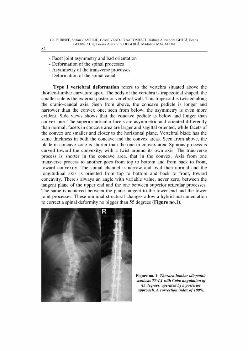
Type I vertebral deformation refers to the vertebra situated above the
thoraco-lumbar curvature apex. The body of the vertebra is trapezoidal shaped, the
smaller side is the external posterior vertebral wall. This trapezoid is twisted along
the cranio-caudal axis. Seen from above, the concave pedicle is longer and
narrower than the convex one; seen from below, the asymmetry is even more
evident. Side views shows that the concave pedicle is below and longer than
convex one. The superior articular facets are asymmetric and oriented differently
than normal; facets in concave area are larger and sagittal oriented, while facets of
the convex are smaller and closer to the horizontal plane. Vertebral blade has the
same thickness in both the concave and the convex areas. Seen from above, the
blade in concave zone is shorter than the one in convex area. Spinous process is
curved toward the convexity, with a twist around its own axis. The transverse
process is shorter in the concave area, that in the convex. Axis from one
transverse process to another goes from top to bottom and from back to front,
toward convexity. The spinal channel is narrow and oval than normal and the
longitudinal axis is oriented from top to bottom and back to front, toward
concavity. There's always an angle with variable value, never zero, between the
tangent plane of the upper end and the one between superior articular processes.
The same is achieved between the plane tangent to the lower end and the lower
joint processes. These minimal structural changes allow a hybrid instrumentation
to correct a spinal deformity no bigger than 55 degrees (Figure no.1).
Figure no. 1: Thoraco-lumbar idiopathic
scoliosis T5-L1 with Cobb angulation of
45 degrees, operated by a posterior
approach. A correction index of 100%.
Spinal disorders in children and teenagers: clinical data, imagistics and therapeutic principles
83
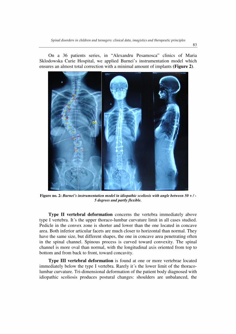
On a 36 patients series, in “Alexandru Pesamosca” clinics of Maria
Sklodowska Curie Hospital, we applied Burnei’s instrumentation model which
ensures an almost total correction with a minimal amount of implants (Figure 2).
Figure no. 2: Burnei’s instrumentation model in idiopathic scoliosis with angle between 50 + / -
5 degrees and partly flexible.
Type II vertebral deformation concerns the vertebra immediately above
type I vertebra. It´s the upper thoraco-lumbar curvature limit in all cases studied.
Pedicle in the convex zone is shorter and lower than the one located in concave
area. Both inferior articular facets are much closer to horizontal than normal. They
have the same size, but different shapes, the one in concave area penetrating often
in the spinal channel. Spinous process is curved toward convexity. The spinal
channel is more oval than normal, with the longitudinal axis oriented from top to
bottom and from back to front, toward concavity.
Type III vertebral deformation is found at one or more vertebrae located
immediately below the type I vertebra. Rarely it´s the lower limit of the thoraco-
lumbar curvature. Tri-dimensional deformation of the patient body diagnosed with
idiopathic scoliosis produces postural changes: shoulders are unbalanced, the
Gh. BURNEI∗, Stefan GAVRILIU, Costel VLAD, Cezar TOMESCU, Raluca Alexandra GHIłĂ, Ileana GEORGESCU, Cezara Alexandra DUGHILĂ, Mădălina MACADON
84
pelvis is oblique and there is a major gibosity. Usually these patients present
obvious respiratory failure and the Cobb angle excedes 80-90 degrees. Surgical
procedures on a rigid spinal column do not allow total correction by somatic
instrumentation and usually there rises the need of a double instrumentation on the
concave side with lateral traction by means of a rod. The partial correction is
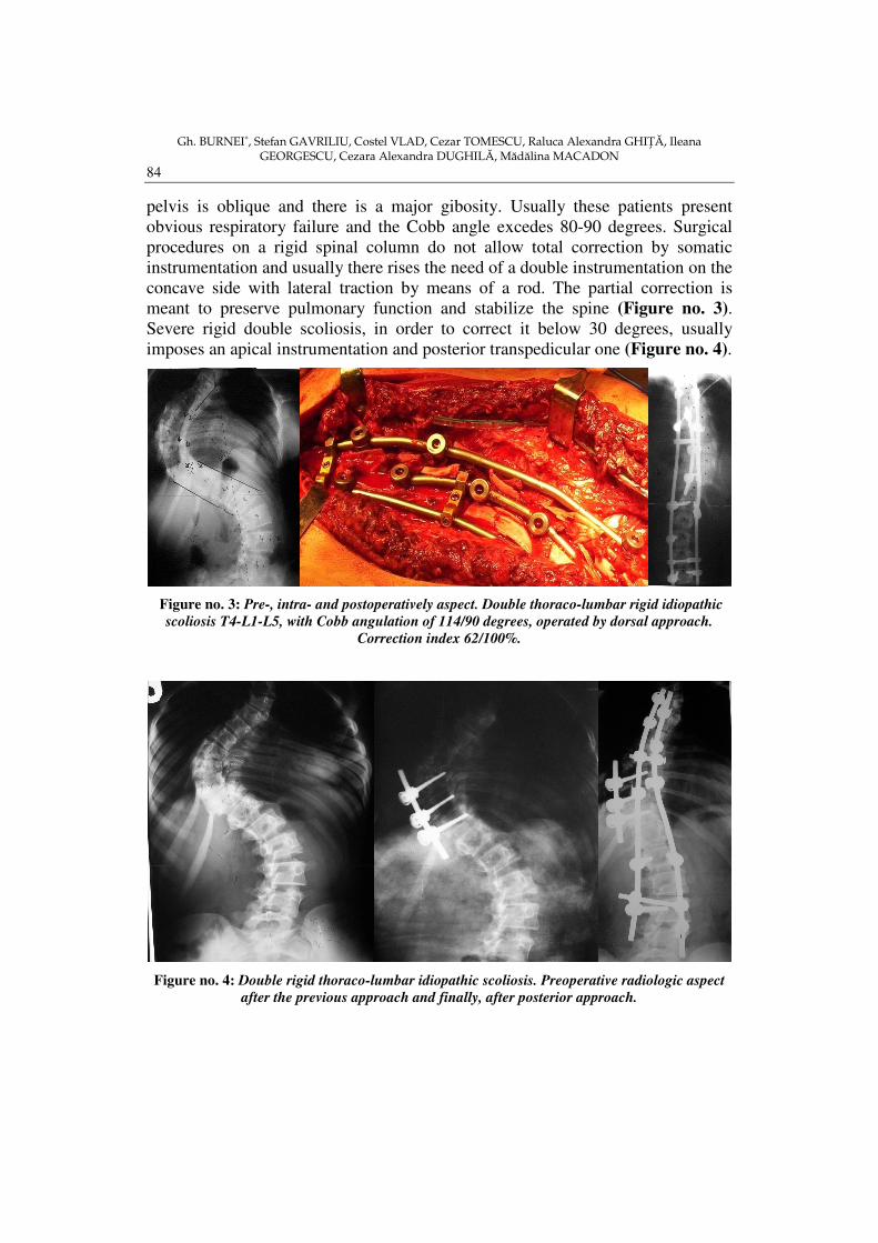
meant to preserve pulmonary function and stabilize the spine (Figure no. 3).
Severe rigid double scoliosis, in order to correct it below 30 degrees, usually
imposes an apical instrumentation and posterior transpedicular one (Figure no. 4).
Figure no. 3: Pre-, intra- and postoperatively aspect. Double thoraco-lumbar rigid idiopathic
scoliosis T4-L1-L5, with Cobb angulation of 114/90 degrees, operated by dorsal approach.
Correction index 62/100%.
Figure no. 4: Double rigid thoraco-lumbar idiopathic scoliosis. Preoperative radiologic aspect
after the previous approach and finally, after posterior approach.

Spinal disorders in children and teenagers: clinical data, imagistics and therapeutic principles
85
Idiopathic scoliosis in teenagers with a 75 to 90 degrees, partially flexible, a
Cobb angle correction about 30%, detected by the X-rays bending probe may be
fully corrected by „all body” vertebral instrumentation (Figure 5).
Figure no. 5: Total transpedicular instrumentation T7-S1 with almost complete correction of
the spinal deformity.
Clinical researches conducted on a group of children and adolescents with
idiopathic scoliosis were made by studying thoracic and lumbar region
radiographs of the spine. Deformation is still appreciated by measurements of X-
rays and less accurate measurements of the surface. Clinical measurements of
spinal radiographs can define the lateral deflection and rotation of the spine.
Lateral deflection is usually measured and defined by the Cobb angle. Nash-Moe
method, which measures the pedicles movement, is widely used. Pedriolle
measures the vertebrae rotation through his own method using a machine called
¨torsion-meter¨. Bunnell invented a "projector " which measures the rotation of
the vertebrae by comparing the position of the spinous process with the vertebral
body. Axis around which produce axial rotation is not the same at all levels of the
spine, so three-dimensional measurement, which is used more frequently, is
considered by the authors to be the most accurate. In thoracic area the rotation
axis passes through the body of the vertebra and in lumbar area passes through the
arch. Explanation for this phenomenon lies in the changing of inclination of the
T11 and T12 joint forces.
Gibbus in thoracic scoliosis is measured at the curvature apex using torsion
meter and for more accuracy, measurement is correlated with Cobb angle and with

Gh. BURNEI∗, Stefan GAVRILIU, Costel VLAD, Cezar TOMESCU, Raluca Alexandra GHIłĂ, Ileana GEORGESCU, Cezara Alexandra DUGHILĂ, Mădălina MACADON
86
apical vertebral rotation (8). The measurements are performed with the patient
seated in three standard positions: sitting, bent forward, bent forward and tilted.
Gibbus measurement results in each of these three positions are compared
with serial measurements at several levels, with pedicles and spinous processes
rotation, made on anteroposterior radiographs of the spine.
Types of curvature have been classified as: simple thoracic curve, simple
lumbar and double curvature (9).
3.2 Congenital scoliosis: spinal anomalies
They are caused by abnormal development of the vertebrae. Typically, they
are rare, less common than idiopathic scoliosis.
Congenital anomalies of the spine can be:
Simple, benign - not induce spinal deformities;
Complex - causing severe spinal deformities.
Some of these severe birth defects are present at birth, others develop in
childhood and adolescence, becoming progressively severe, with abnormal growth
of the spine, they can be associated with many other malformations. A "C" shaped
bend of column in a newborn caused by pelvic obliquity and inadequate
intrauterine position should not be diagnosed as congenital scoliosis.
Congenital abnormalities of the spine can be caused by a segmentation
defects or formation defects, but they are often the result of both pathogenic
factors.
3.2.1 Segmentation defects have as a result a column like a non-segmented
rod segmentation defects that result in a column of a non-segmented rod. Two or
more vertebrae may be affected, involving vertebral bodies, the rear elements or
even combinations thereof. Unilateral unsegmented column is the most exposed
for deformation. When segmentation is flawed lateral and on one side, the
resulting deformation is a severe progressive scoliosis, anterior non-segmentation
produce kyphosis, bilateral posterior non-segmentation produce lordosis, and the
unilateral one produces lordose-scoliosis. Symmetric circumferential
segmentation defect creates a "block" which causes vertebral angular or rotational
deformation of the spine, but vertical growth of the spine is affected.
3.2.2 Formation defects can be partial or complete. A partial unilateral
Formation failure produces a sharpening of the vertebra vertebra and confer a
trapezoidal shape. A a slight pedicular trace can be seen on the radiograph.
Hemivertebra, cause of failure of complete unilateral formation may be non-
segmented, or segmented semisegmentată from adjacent vertebrae. Hemivertebra
can be balanced or unbalanced. A segmented hemivertebra is completely separate

Spinal disorders in children and teenagers: clinical data, imagistics and therapeutic principles
87
from adjacent vertebrae. In this case, as a result of vertebral asymmetrical growth
scoliosis is developed. A hemivertebra semi-segmented is separated from one of
the adjacent vertebrae (upper or lower) by a disk or by a vertebral growth
membrane, normal, but united with the other adjacent vertebrae. Hemivertebral
resection in optimal timing, 1 to 4 years of age, allows total correction by somatic
instrumentation (Figure 6).
Figure no. 6: Congenital scoliosis with hemivertebra through formation defect - L2-L3
supernumerary hemivertebra, operated by a double approach, dorsal and ventral.
A non-segmented hemivertebra is connected to both adjacent vertebrae
(upper and lower) without disks or growth membranes. In the absence of
asymmetric growth, a non-segmented hemivertebra does not cause progressive
deformation of the spine. If there are two or more hemivertebrae on the same side
of the column, the column degree of mismatch is higher and produces severe
spinal deformities. Scoliosis can be balanced, if both hemivertebrae are placed
symmetrically, but the degree of curvature remains progressive. Hemivertebrae
placed on both sides of the spine, in a 5 and above normal vertebrae row induce
double congenital scoliosis requiring a double approach with somatic
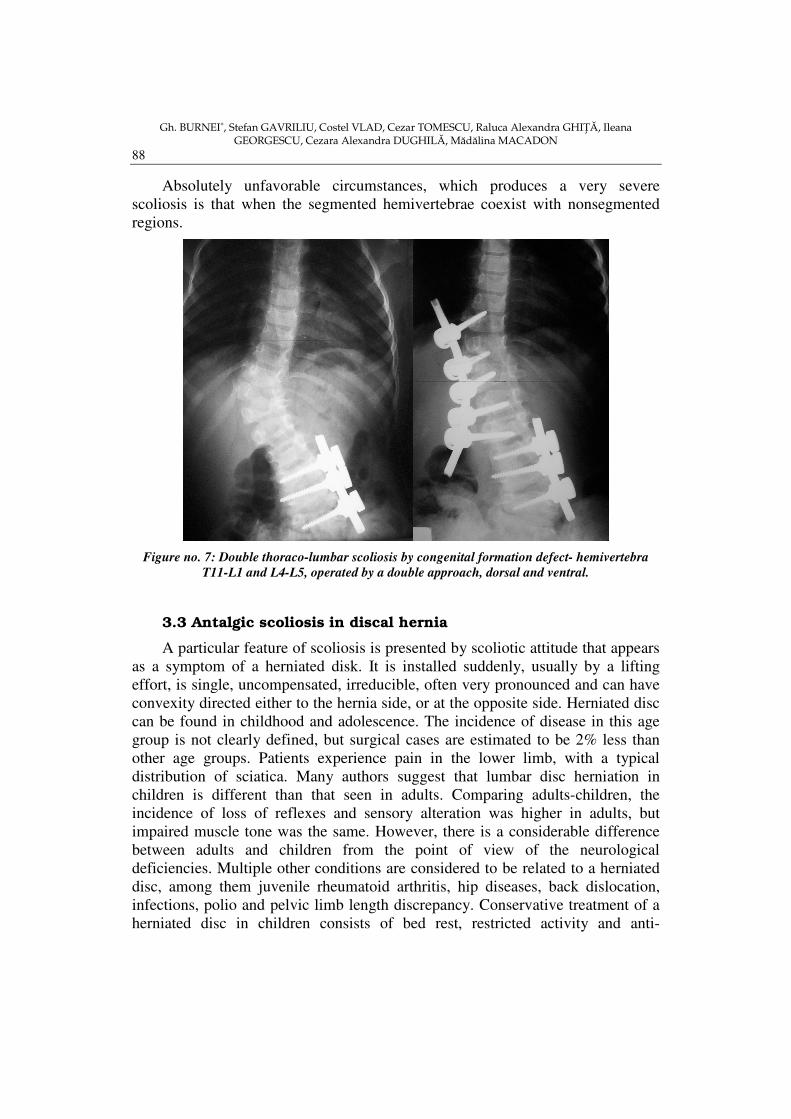
instrumentation (Figure 7, next page).
Kyphoses will occur when the anterior vertebral body is aplastic or
hypoplastic due to defects in formation and posterior elements are normally
developed. One or more vertebrae may present defects. Resulting kyphosis is
more angular than round.
Congenital absence of sacrum and lumbar zone is an extreme form of
formation defect of the whole caudal spine.
Gh. BURNEI∗, Stefan GAVRILIU, Costel VLAD, Cezar TOMESCU, Raluca Alexandra GHIłĂ, Ileana GEORGESCU, Cezara Alexandra DUGHILĂ, Mădălina MACADON
88
Absolutely unfavorable circumstances, which produces a very severe
scoliosis is that when the segmented hemivertebrae coexist with nonsegmented
regions.
Figure no. 7: Double thoraco-lumbar scoliosis by congenital formation defect- hemivertebra
T11-L1 and L4-L5, operated by a double approach, dorsal and ventral.
3.3 Antalgic scoliosis in discal hernia
A particular feature of scoliosis is presented by scoliotic attitude that appears
as a symptom of a herniated disk. It is installed suddenly, usually by a lifting
effort, is single, uncompensated, irreducible, often very pronounced and can have
convexity directed either to the hernia side, or at the opposite side. Herniated disc
can be found in childhood and adolescence. The incidence of disease in this age
group is not clearly defined, but surgical cases are estimated to be 2% less than
other age groups. Patients experience pain in the lower limb, with a typical
distribution of sciatica. Many authors suggest that lumbar disc herniation in
children is different than that seen in adults. Comparing adults-children, the
incidence of loss of reflexes and sensory alteration was higher in adults, but
impaired muscle tone was the same. However, there is a considerable difference
between adults and children from the point of view of the neurological
deficiencies. Multiple other conditions are considered to be related to a herniated
disc, among them juvenile rheumatoid arthritis, hip diseases, back dislocation,
infections, polio and pelvic limb length discrepancy. Conservative treatment of a
herniated disc in children consists of bed rest, restricted activity and anti-
Spinal disorders in children and teenagers: clinical data, imagistics and therapeutic principles
89
inflammatory medication. Preoperative evaluation includes diagnostic tests to
confirm and locate the disease. For pathogenic diagnosis some experts propose
lesions visualization by conducting an MRI test, others proposing
electromyography or tomography.
Antalgic scoliosis is the result of nerve root compression of the intervertebral
disc, which is often inflamed. Therefore, when the hernia has purely "invaded"
nerve root, as is usually happens with the L5-S1 disc, for releasing the root and
for distance increasing tilt on the affected side is needed to cause "direct
scoliosis". When the hernia is outside the root, as usually happens with the L4-L5
disc, for release is needed a lateral tilt on the healthy side, which departs root from
hernia and causes a "cross scoliosis".
We note that the terms direct and cross scoliosis have a somewhat
inappropriate use as scoliosis is termed, classically, based on the convexity
orientation. If we were following this terminology, scoliosis with the convexity on
the hernia side, would be direct and with the convexity on the opposite side of the
hernia, would be the cross one. So we must take into account the nomenclature
used to avoid confusion. This fact determined Forestier to suggest replacement of
the cross scoliosis term with crossed vertebral inflection, on the opposite side of
the hernia and direct scoliosis term with direct vertebral inflection, on the hernia
side.
Disc sciatic pain is accompanied by the presence of several points where pain
is obtained by applying pressure on affected nerves, namely: gluteal point,
femoral points, fibular point, Achilles point and medial plantar point.
Pain obtained by pressing the iliac artery is common to 80% of cases, at
patients suffering from herniated disc. The presence of this sign has a similar
meaning to painful points and is due to perivascular sympathetic
hyperexcitability.
3.4 Postural scoliosis
Improper posture may be associated with medium scoliosis, characterized by
a long thoraco-lumbar curvature. The rotation of the vertebrae towards curve
convexity isn’t revealed; gibbus is not visible atribs level or unilateral
proeminence of the paraspinal muscles on the convex curvature area. Scoliosis is
very flexible, but disappears when the patient is asked to stand upright or in lateral
flexion on the convex curvature side.
Postural scoliosis does not progress and does not become structural. It is of
little clinical importance and usually no treatment is indicated, physical exercise is
sufficient.

Gh. BURNEI∗, Stefan GAVRILIU, Costel VLAD, Cezar TOMESCU, Raluca Alexandra GHIłĂ, Ileana GEORGESCU, Cezara Alexandra DUGHILĂ, Mădălina MACADON
90
3.5 Static scoliosis due to lower limb length discrepancy
In this kind of non-structural scoliosis occurs only a long thoraco-lumbar
curvature, located usually from the cervical-thoracic junction to the sacrum.
Curvature convexity is towards the lowered semi-pelvis, respectively toward the
shorter limb. A slight rotation of the vertebrae appears and because the column is
ventral arcuate, minimal rotation is to the concave side of the curvature, as
opposed to the structural scoliosis, which the rotation istoward the convexity of
curvature.
Spinal curvature is present in the sitting position. Column loops evenly on
both sides in lateral flexion of the trunk. The patient can voluntarily correct the
lateral deviation of the spine. Discrepancy correction between the lengths of the
lower limbswith orthopedic boots, straightens the pelvis, removing scoliosis. On
radiography, the column does not show structural changes at the vertebrae level
(10).
4. KYPHOSIS
It is the most frequent deviation of the vertebral column, which is caused by
increased curvature of the thoracic and appears in a large variety of disease:
• Congenital and hereditary Brachispondylitis
Microspondylitis
Achondroplasty
Osteopsathyrosis
Putti platyspondylia
• Traumatic Fracture and fracture-dislocation
Disk hernia
Drug-induced kyphosis
• Infectious Pott's disease
Typhoid / paratyphoid spondylitis
Melitococcal spondylitis
Vertebral osteomyelitis
Mycotical spondylitis
• Rheumatic Ankylopoetic spondylitis
Deforming chronic rheumatism
• Tumoral Primitive
Secondary

Spinal disorders in children and teenagers: clinical data, imagistics and therapeutic principles
91
• Endocrine Osteoporosis in Cushing's Syndrome
Postmenopausal osteoporosis
Hyperthyroid osteoporosis
• Deficiency Ricketts
Osteomalacia
Painful osteoporosis of digestive diseases
• Dystrophic Senile or presenile kyphosis
Scheuermann vertebral epiphysitis
• Neuropsychiatric Hystero-traumatic Kyphosis
• Posture Professional
Flat foot
Clinically, there are two main types of kyphosis: kyphosis with small radius
of curvature (short arch) and large radius of curvature kyphosis (long arch).
Kyphosis with short arch appears in diseases that, destroying one or more
vertebral bodies, lead to a compaction thereof, as happens in vertebral fractures,
Pott disease, spondylitis, vertebral osteomyelitis, in primary postmenopausal
osteoporosis etc. Long arch kyphosis occurs in conditions that interests backbone
on a longer distance, as in presenile and senile primary osteoporosis, ankylosing
spondylitis, vertebral epiphysis etc.
Pathological kyphosis can be installed either by emphasizing physiological
kyphosis (when the condition interests thoracic region), or by deleting a
physiological lordosis (when the disease involves cervical or lumbar region).
Kyphosis is essentially the attitude or deformity of the spine that takes the most
convenient position for improving pain, knowing that anterior vertebral ligament
and the front of the spine are most richly innervated.
A special category of kyphosis is composed by the compressionof the
vertebral bodies in osteoporosis or generalized osteopathy. Their differential
diagnosis is possible only on the basis of radiological examination and laboratory
tests. At teenagers’ osteoporosis, alkaline phosphatase increase only in the case of
recent fractures.
Kyphoses severity can be measured and objectified using various
kyphometres.

Gh. BURNEI∗, Stefan GAVRILIU, Costel VLAD, Cezar TOMESCU, Raluca Alexandra GHIłĂ, Ileana GEORGESCU, Cezara Alexandra DUGHILĂ, Mădălina MACADON
92
4.1 Juvenile kyphosis - Scheuermann's disease
Kyphosis is installed at puberty and it manifests by reducing the anterior
volume of one or more vertebrae, deformation that produces certain radiographic
changes. This characteristic change of the vertebral bodies, with the anterior
height reduction was first described by Scheurmann in 1920.
Radiologically, Scheuermann's disease is defined as a kyphosis which
includes at least three adjacent vertebrae, with a decrease of the anterior height of
5° or more. The diagnosis is placed after 11-12 years because typical vertebrae
edge changes and kyphoses are not obvious before 10 years. Statistically, it was
observed that the condition has a higher incidence in girls than in boys.
The cause of this condition is still unknown, despite the many theories
advanced in the literature. Vertebrae changes was explained as a result of aseptic
necrosis of the apophyseal cartilage ring, that disturbs the growth in height of the
anterior vertebral body. Also the mechanical and static forces acting on the
spinehave an etiologicimportance. Ultrastructural and histo-chemical studies
revealed changes in the growth of the cartilage plateau and vertebral matrix,
observing a high percentage of collagen-protoglicanic. Therefore, enchondral
ossificationand longitudinal growth of the vertebrae are reduced. It may not
appears ring apophyses necrosis or abnormal intervertebral discs .
In advanced cases, anterior longitudinal ligament is contracted and
thickened, acting as a spring along kyphosis. Smaller anterior vertebral body has
variable values .
Typically, patients have a faulty posture and accuse pain in kyphosis region.
Physical appearance depends on the location of the apex of kyphosis. In almost
three-quarters of cases, the disease is purely thoracic, thoraco-lumbar to a quarter
of patients and lumbar location in rare. If the disease is located in the chest area,
there is an emphasis on normal kyphosis, an increased lumbar lordosis and a
protruding abdomen. The shoulders are held back and the center of gravity falls
behind the sacrum, with exaggerated pelvic tilt. As a rule, cervical lordosis
becomes increased. Exaggerated cervical and lumbar lordosis are acompensatory
phenomena.
Scheuermann´s juvenile kyphosis diagnosis is possible only after
radiological investigation. Vertebrae becomes cuneiform in the center of kyphotic
area and decreases cranial and caudal. This vertebral deformation is due to
delayed longitudinal growth of the anterior vertebra part. Measuring vertebra
compression is analyzed by profile radiography, by drawing lines through the
levels of the two planes and by determining the angle between these two lines
with a goniometer. The boundary between normal and abnormal vertebral
configuration is 5 degrees. Kyphotic angle is the angle between the upper plateau
of the cranial kyphotic vertebra and lower plateau of thecaudal vertebra. The
intervertebral discs are normal in the early stages of the disease, maintaining

Spinal disorders in children and teenagers: clinical data, imagistics and therapeutic principles
93
height between trapezoidal vertebrae; with time it narrows, especially in the center
of the kyphotic area. Antero-posterior diameter of trapezoidal vertebra can be
increased.
In the early stages, posture defects can be corrected active and passive,
gradually, over a period of 6-9 months, after which the kyphosis is final.
A clinical and radiological analysis of condition established following steps
in pathological evolution:
- Functional phase - clinically characterized by improper posture, at the age
of 9-10 years. There may be observed exaggeration of thoracic kyphosis but does
not register pain or other clinical symptoms. Scheuermann´s disease is difficult to
diagnose at this stage.
- Typical changes phase, at the age of 12-18 years, in which the kyphosis
and possibly scoliosis installs. Patients complains of back pain and fatigue.
Radiologically, this phase is characterized by reducing the anterior height of the
vertebrae and presence of irregularities at the anterion edge of the plateau.
- Late phase - seen in adults. Appear narrowing of the intervertebral discs
and deformation of the vertebrae edges. Spinal pain are permanent and muscles
weakness in the kyphosis region.
After lengthy analysis of several Scheuermann´s juvenile kyphosis cases
were drawn several conclusions, namely:
• Spinal pain and fatigue are less common after growth, having no effect on
the working capacity of the patient;
• Approximately half of the mature patients had spinal pain and almost a
quarter suffer from lumbar discs degeneration;
• When the kyphotic area is low or very long, interesting the medular
channel and the second lumbar vertebra, lumbar disc degeneration induces great
pain;
• Radiological, there is a slow kyphosis progression, due to the slow
formation of the trapezoidal vertebrae and narrowing of the vertebrae discs.
Treatment
Scheuermann´s juvenile kyphosis treatment objectives are directed toward
pain relief, correction of the kyphosis degree, prevention of the kyphotic growth,
improving physical appearance. The choice of treatment is taking into account the
following factors:
- Age of the patient;
- Phase of the condition;
- The structural degree and rigidity of kyphosis;
- The posterior convexity location;
- Presence of pain;
- Possible association with scoliosis;
Gh. BURNEI∗, Stefan GAVRILIU, Costel VLAD, Cezar TOMESCU, Raluca Alexandra GHIłĂ, Ileana GEORGESCU, Cezara Alexandra DUGHILĂ, Mădălina MACADON
94
- Psycho-social factors.
Not all patients with this disease require treatment. If a patient is skeletally
mature, with an asymmetric acceptable kyphosis, it can be put underobservation
without treatment. Overall physical exercises aim to improve patient outfit, but it
must be convinced that only exercises doesn’t have correction effect on the
vertebrae shape or reduction of the fixed kyphoses degree (12).
Surgical correction of Scheuermann´s disease is rarely indicated. It’s applied
only on mature patients with local chronic pain, kyphotic curves with 60°-70° or
more and trapezoidal vertebrae angle greater than 10°. For the permanent
correction are needed both anterior and posteriorspinal fusion. Posterior
instrumentation provides a good correction.
Complications
A severe complication but rare with Scheuermann´s disease is compression
of the spinal cord; may be due to a herniated disk injury and direct mechanical
compression of the spinal cord or spinal channel narrowing in the kyphotic area.
It can also be seen atypical forms of the disease which is manifested by
changes in the vertebrae body, or by narrowing the anterior vertebral body, but
without any other vertebral modifications.
4.2. Congenital kyphosis
Congenital kyphosis may be caused by formation defects or by segmentation
defects of the vertebral bodies.
Type I (formation defect) is characterized by partially or total aplasia of the
vertebral bodies. May be affected one, two or even three vertebral bodies .
A - agenesis of T1 vertebral body
B - L1 vertebral body agenesis and microspondylitis of the adjacent
vertebra (T12), at both vertebrae being able to notice the presence of
pedicles.
C - microspondylitis L1 vertebra
D - microspondylitis of two adjacent vertebrae (T11 and T12).
Type II (segmentation defect) affects the anterior side of two or more
adjacent vertebrae.
E - type II congenital kyphosis due to segmentation fault at the front of
the three adjacent vertebrae
F - the absence of supero-posterior corner of cuneiform vertebra
G - cuneiform vertebrae (side view)
H - cuneiform vertebra (anteroposterior view).

Spinal disorders in children and teenagers: clinical data, imagistics and therapeutic principles
95
Kyphosis correction is performed by a posterior approach in case of a
hemisegmented or non-segmented cuneiform vertebra or a double approach,
anterior and posterior, if the vertebra is segmented (Figure no. 8). The most
affected area by kyphosis is between T10 and L2, however, can be observed
anywhere between T4 and L5. Kyphotic deformation is usually observed in
neonates and becomes more pronounced when children start to walk. With the
increase of the column, kyphosis is emphasized. The deformity may be that severe
in teenagers or young adults that it induces respiratory failure and imposes
restrictions not only in physical activities, but daily one, too (Figure no. 9).
Figure no. 8: Pre-and postoperative radiological aspects in case of a thoracic T6 cuneiform
vertebra kyphosis.
Figure no. 9: Extreme kyphoscoliosis with thoracic insufficiency syndrome due to late
diagnosis. We intervened to stabilize the deformity in order to prevent onset of spinal stenosis by
compression and emphasizing respiratory failure syndrome, heart failure and premature exitus.

Gh. BURNEI∗, Stefan GAVRILIU, Costel VLAD, Cezar TOMESCU, Raluca Alexandra GHIłĂ, Ileana GEORGESCU, Cezara Alexandra DUGHILĂ, Mădălina MACADON
96
In children it is not painful and does not appear muscle spasms, but in adults,
pain is a symptom of degenerative arthritic changes. Typically, the height of the
patient is less than average.
Paraplegia caused by spinal cord compression occurs frequently in
congenital kyphoses Sequential neurological examinations of the whole column
must be made, myelography examination or magnetic resonance visualization
method.
Deformation can not be corrected and stopped from evolution by
conservative methods. Non-operative measures are ineffective. The surgical
procedure is dependent on the type of kyphosis, the severity of the deformation,
on the age of the patient. Anterior unsegmented column is treated by posterior
spondylodesis, if deflection angle is medium or moderate and has no particular
clinical significance. Posterior spondylodesis extends from the first vertebrae,
located proximal to the affected area, to the first vertebra located distal to this
zone. Moreover, posterior spinal fusion prevents increased kyphosis. Optimal age
for posterior fusion is between 1 and 2 years.
When the distortion is severe column fusions are required both anterior and
posterior (11). If the posterior fusion does not ensure the stability of the kyphotic
column, pseud-arthrosis appears and the kyphosis is progressing.
5. LORDOSIS
Is the third significant deviation of the spine and is characterized by
emphasizing the dorsal flexion of the spine. Headquarted in lumbar and cervical
region can stretch to thoracic region and sometimes can interest the spine
completely. In many cases it is about lordosis attitudeobvious in orthostatism that
isdeleted in supine position. They are the result of static tilt adaptations or
dynamic asynergy.
By static tilt we mean compensation at the joints level and lumbar discs, for
that normal tilting of the pelvis does not cause spinal deformation, which thus
recovers at its base. We encounter this at the women who wear high heels or in
case of bilateral congenital dislocation.
Lordosis by dynamic disequilibrium correspond to a compromise between
the extensor muscles of the trunk and flexors, being the result of a abdominal wall
muscle atonia or abdominal distension by: tumors, enteroptosis or pregnancy.
In some cases we can findbone fixed lordosis (lumbarization,
spondylolisthesis). In these cases, although lordosis is very pronounced,gluteal
regions doesn’t stand out (as in hip congenital dislocation), but remain deleted,
and thorax seems to be clogged in the pelvis, the soft tissues above coxal bones
showing more or less pronouncedditches.

Spinal disorders in children and teenagers: clinical data, imagistics and therapeutic principles
97
Lordosis depth is estimated using a lead bob, measuring the distance that
separates the plumb bob from the top of the farthest spinous process. Normal
pelvis is tilted by 12° related to axis of the pelvic limbs, tending to vertical and
hyperlordosis. If the deformity is reducible seated and by bending forward, it is
noticed how lumbar muscles relaxes and the medial hole is filled. If it isn’t
reducible, the flexion is made form the torso and hips, while lumbar region tilts as
a block, keeping the concavity.
Progressing in age hyperlordosis leads to suffering of the spinous apophyses,
inducing wear injuries that cause pain when touching.
The causes of lordosis can include: carrying heavy loads on head, wearing
high heels, traumatickyphosis decompensation, S1 lumbarization,
spondylolisthesis, etc. In some conditions we may find the physiological cervical
or lumbar lordosis deletion. This symptom can be a sign of disc suffering, of
fracture-dislocation or spondylitis. Erasing lordosis doesn’t have a specific
character but in relation with pacient’s normal posture.
References
1. Burnei G, Gavriliu S, Georgescu I. - Patologia rahisului la copii şi adolescenŃi (I) – coloana vertebrală, date anatomice, biomecanică, statică, diagnostic, anomalii congenitale ale coloanei vertebrale –– ViaŃa Medicală, nr. 23 (805), anul XVII, 20 iunie 2005, pag. 7;
2. Burnei G, Gavriliu S, Georgescu I. - Patologia rahisului la copii şi adolescenŃi (II) – cifoza congenitală, cifoza juvenilă Scheuermann, tratament, complicaŃii - ViaŃa Medicală, nr. 24 (806), anul XVII, 17 iunie 2005, pag.7;
3. Altaf F, Gibson A, Dannawi Z, Noordeen H. - Adolescent idiopathic scoliosis. BMJ 2013;346:f2508;
4. Hasler CC. - A brief overview of 100 years of history of surgical treatment for adolescent idiopathic scoliosis. J Child Orthop. 2013 Feb; 7(1):57-62. Epub 2012 Dec 5;
5. Burton MS. Diagnosis and treatment of adolescent idiopathic scoliosis. Pediatr Ann. 2013 Nov;42(11):224-8. doi: 10.3928/00904481- 20131022-09;
6. Burnei G, Gavriliu S, Georgescu I. - Patologia rahisului la copii şi adolescenŃi (III) – scolioza antalgică în hernia de disc, scolioze antalgice, scolioze statice, datorate inegalităŃii membrelor inferioare, lordoza - ViaŃa Medicală, nr. 25 (807), anul XVII, 24 iunie 2005, pag.7;
7. Konieczny MR, Senyurt H, Krauspe R. - Epidemiology of adolescent idiopathic scoliosis. J Child Orthop. 2013 Feb;7(1):3-9. Epub 2012 Dec 11;
Gh. BURNEI∗, Stefan GAVRILIU, Costel VLAD, Cezar TOMESCU, Raluca Alexandra GHIłĂ, Ileana GEORGESCU, Cezara Alexandra DUGHILĂ, Mădălina MACADON
98
8. Schlenzka D, Yrjönen T. - Bracing in adolescent idiopathic scoliosis. J Child Orthop. 2013 Feb;7(1):51-55. Epub 2012 Nov 30;
9. Hawes MC, O'Brien JP. - The transformation of spinal curvature into spinal deformity: pathological processes and implications for treatment. Scoliosis 2006, 1:3 doi:10.1186/1748-7161-1-3;
10. Burnei G, Vlad C, Dan D, Gavriliu S, Georgescu I. - The Results Of A Spinal Instrumentation Model In 17 Patients With Idiopatic Scoliosis, – World IV Interdisciplinary Congress on Spine Care, July 29th-August 1st, 2007, Istanbul, Turkey;
11. Deniz Olgun Z, Yazici M. - Posterior instrumentation and fusion. J Child Orthop. 2013 Feb;7(1):69-76. Epub 2012 Dec 25;
12. Chan A, Lou E, Hill D. - Review of current technologies and methods supplementing brace treatment in adolescent idiopathic scoliosis. J Child Orthop. 2013 Oct;7(4):309-16. doi: 10.1007/s11832-013-0500-0. Epub 2013 May 28.