Embed Size (px)
Citation preview
Vol. 6: 11-16, 1989 I DISEASES OF AQUATIC ORGANISMS Dis. aquat. Org.
Published February 27
Spiral swimming behavior due to cranial and vertebral lesions associated with Cytophaga psychrophila infections in salmonid fishes
Michael L. Kent1,*, J. M. ~ r o f f ~ , J. K. ~ o r r i s o n ~ , W. T. yasutake4, R. A. Holts
' Battelle Marine Science Laboratory, 439 West Sequim Bay Road. Sequim, Washington 98382. USA Department of Medicine, School of Veterinary Medicine, University of California, Davis, California 95616, USA
Olympia Fish Health Center, 2625 Parkmount Ln., Bldg. A., Olympia, Washington 98502, USA U.S. Fish and Wildlife Service, Seattle National Fishery Research Center, Bldg. 204 Naval Station, Seattle, Washington 98115.
USA Oregon Department of Fish and Wildlife, Department of Microbiology, Oregon State University. Corvallis, Oregon 97331.
USA
ABSTRACT: Cytophaga psychrophila (Order Cytophagales) infections of the cranium and anterior vertebrae in salmonid fishes were associated with ataxia, spiral swimming along the long axis of the fish, and death. The syndrome was observed in 2 to 10% of underyearling coho salmon Oncorhynchus kisutch, rainbow trout Salrno gairdned, and steelhead trout S. gairdneri at several private, state, and federal hatcheries in Washington and Oregon, USA. Affected f.ish did not recover and ultimately died. Histoloqcal examination of affected fish consistently revealed subacute to chronic periostitis, osteitis, meningitis, and ganglioneuritis. Inflammation and periosteal proliferation of the anterior vertebrae at the junction of the vertebral column with the cranium with extension into the cranial case was a consistent feature of the disease. The adjacent nervous tissue, particularly the medulla, was often compressed by the proliferative lesion and this may have caused the observed ataxia. Though bacteria were seldom observed in these lesions, C. psychrophila was isolated in culture from the cranial cavity of all affected fish that were tested. Epizootiological observations suggested that the bacterium is the etiologic agent of the disease because the spiral swimming behavior and lesions were only observed in populations which had recovered from acute C. psychrophila infections, known as 'cold-water disease'
INTRODUCTION
Cytophaga psychrophila (Order Cytophagales), the causative agent of cold-water disease, is a wide-spread pathogen of cultured salmonid fishes in North America (Pacha & Ordal 1970, Schachte 1983). Epizootics typi- cally occur at between 4 and 15 "C in underyearling fish (Conrad & DeCew 1967), but yearling coho Oncorhyn- chus kisutch and chinook salmon 0. tshawytscha may also be affected (Holt 1972, Wood 1974). The bacteria often infected the external surfaces, resulting in ulcera- tion of the dermis and necrosis of the underlying mus- cle (Wolke 1975). The disease may also progress to septicemia with necrosis observed in internal organs (Wolke 19751, and bacteria can be isolated from these
' Present address: Department of Fisheries and Oceans, Pacific Biological Station. Nanaimo, B. C., Canada V9R 5K6
infected tissues (Wood 1974). Though Wood & Yasutake (1956) reported Little inflammatory response in the visceral organs, Borg (1960) reported a mild mononuclear infiltration associated with the disease.
A chronic form of the infection resulting in lordosis and scoliosis was observed in juvenile coho salmon (Conrad & DeCew 1967, Holt 1972), and Wood (1974) surmised that these lesions were caused by bacterial destruction of muscle bundles adjacent to vertebrae which resulted in unequal tension on the spinal col- umn. Spiral swimming behavior has been observed in fish from hatcheries where cold-water disease is en- zootic (Yasutake 1965), and we have observed this syndrome at several hatcheries in Oregon and Washington, USA. Presented here are histological, bac- teriological, and epizootiological data that indicate that this ataxic swimming behavior was due to chronic inflammation in the cranial cavity and anterior verte- brae associated with C. psycl~rophila infections.
3 Inter-Research/Printed in F. R. Germany
Dis. aquat. Org.
MATERIALS AND METHODS
The primary subjects of this investigation were juve- nile coho salmon Oncorhynchus kisutch reared at a freshwater hatchery in Washington State, USA. These fish were exam~nedfrom 29 May to 11 June 1987. In addi- tion, occurrences of this disease in government hatch- eries in Oregon and Washington are reported (Table 1) to indicate the geographical distribution of the syndrome.
Histopathology. The anterior third of the body and visceral organs of 27 juvenjle coho salmon exhibiting spiral swimming behavior along the long axis were fixed in Davidson's solution (Humason 1979). Tissues were decalcified in a formic acid-citric acid solution and then processed using standard histological tech- niques (Luna 1968). Sections were stained with Harns' haernatoxylin and eosin, Brown and Brenn Gram stain, Warthin-Starry silver stain. Periodic acid-Schiffs (PAS) stain, Heidenhain's stain for osteoid, or the Giemsa stain (Luna 1968). Ten clinically normal fish from a raceway containing effected fish were also examined.
Bacteriology. Tissues from the posterior of the cra- nial space of 11 affected and 6 clinically normal fish were cultured for bacteria by anesthetizing the fish with MS-222, disinfecting the surface of the head with 70 % ethanol, and aseptically opening the cranial case. Inocula were then obtained from the posterior region of the cranium with calcium alginate swabs. Inocula were
Table 1 Occurrence of spiral swimming behavior in salmonid fishes associated with cerebral and anterior vertebral lesions in Washington (WA) and Oregon. (OR), USA, with year when
condition was first recognized
Species Location Year
Oncorhynchus Willard federal 1963 kisutch hatchery, WA 0. kisutch Quilcene federal L963
hatchery, WA 0. kisutch Nehalem state 1980
hatchery, OR 0, kisutch Sapdy state 1981
hatchery, OR 0. kisutch Little White Salmon 1986
federal hatchey. WA 0. kisutch Makah federal 1986
hatchery, WA 0. kisutch Humptulips state 1987
hatchery, WA 0, kisutch Willapa state 1987
hatchery. WA 0. kisutch A private hatchery. WA 1987
(present study) Salmo gairdnen Q d c e n e federal 1984
hatchery, WA S. galrdneri Chambers Creek state 1987
hatchery, WA
streaked on charcoal agar (Daly & Stevenson 1985) or Tryptic Soy Agar (Difco) and cultured at 15OC and at room temperature.
Bacterial isolates were tested for their serological relatedness to Cytophaga psychrophila and Flexibacter columnaris using slide agglutination tests with the appropriate antisera, diluted to 1 : 50. The antisera to C. psychrophila and F. colunlnaris were obtained from the fish disease laboratory, Department of Microbiology, Oregon State University, Corvallis, Oregon, USA.
RESULTS
Clinical signs and mortality
Coho salmon reared at a private hatchery in Washing- ton showing spiral swirr-ming behav~or along the !ccg axis and loss of equilibrium first exhibited mortality when they weighed ca 5 g. As the disease progressed the fish often remained motionless on their sides at the water surface until agitated, when they would resume erratic sw~mming. Affected fish did not recover and typically died 3 to 5 d after the clinical signs were first observed. Some affected fish displayed dark pigmenta- tion on either the right or left longitudinal half of the body and others occasionally exhibited dorsal swelling posterior to the skull. Fish were initially maintained at 15°C. The temperature was lowered to 10°C ca 2 mo after onset of the disease and this apparently did not affect the course of the disease. In 1987, ca 2 % of the underyearling coho at this hatchery exhibited these clinical signs, and all affected fish eventually died. The condition persisted in this population for several months and fish were submitted for necropsy at 14 g (avg. wt).
Histopathology
Histological examination of affected fish revealed subacute and chronic periostitis, osteitis, meningitis, and ganglioneuritis (Figs. 1 to 4) . Inflammation and periosteal proliferation of th.e vertebrae at the junction of the vertebral column and the caudal limit of the cranium were observed in all fish. The lesions often extended anteriorly into the caudal aspect of the cranium resulting in subacute meningitis. The adjacent neural trssue, particularly the medulla, was often com- pressed and had secondary necrosis and hemorrhage (Figs. 1 and 2). The posterior cranial nerves and anterior - most spinal nerves were commonly affected (Fig. 3). Inflammation often extended into the connective tissue stroma peripheral to the auditory canal and into the muscle bundles adjacent to the antenor vertebrae. The mixed inflammatory infiltrate consisted of mononuclear
Kent et a1 Cytophaga cranial infections in salrnonlds I.
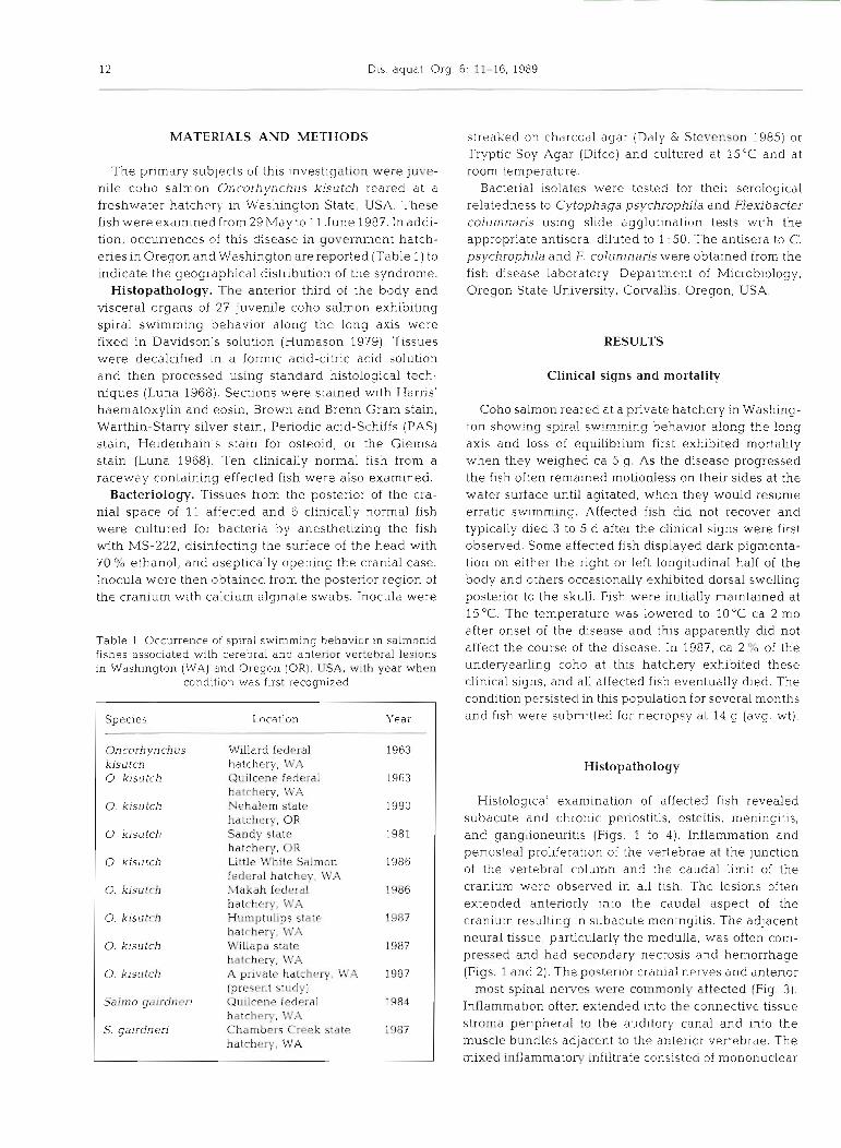
Fig 1 Oncorhynchus kisutch. Posterior aspect of the cranlal rav~ty w t h penosteal prohferatlon and ~nflarnrnation (P) of the anterior vertebrae (A) with extension into the cranial cavltya9The junction of the medulla (M) and the splnaI cord IS apparently compressed w~th areas of hemorrhage (arrows]. The lesion encircles the medulla as indfcated by focl at the dorsal and ventral
aspects (F) H & E Bar -. 250 m
inflammatory cells and polymorphonuclear leukocytes. Heidenhain's stain for osteoid revealed foci of osteoid formation within areas of periosteal prohferatlon.
In one specimen, there was inflammation of the con- nective tissue stroma of the ventral pharyngeal area (Figs. 4 and 5). The lesion was centered around bony elements of the gill arches undergoing endochondral ossification with destruction of the bony elements.
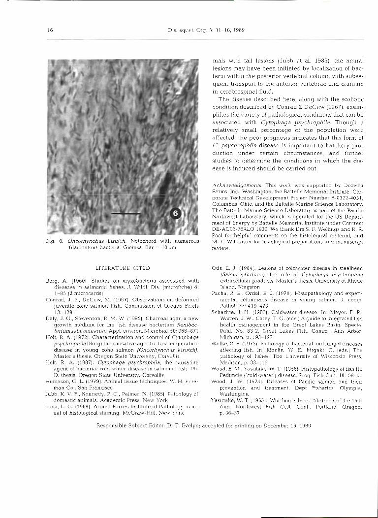
Special stains of the coho salmon tlssues froin this study group did not reveal bacterial organisms associ- ated wlth the lesions. However, Giemsa-stained sec- tions from a coho salmon exhibiting identical lesions from the Willipa state fish hatchery in Washington (Table 1) revealed numerous filamentous bacteria in a lesion involving the notochord (Fig. 6).
lnltlal lsolations varied dramatically between flsh whereas only 3 or 4 colonles were isolated from some fish, other plates revealed numerous Cytophaga col- onles into the tertiary streak. Consistent with C psy- chrophila, the colonies were yellow and the bacterla were Gram-negatlve filamentous rods that dld not grow at 25°C. Furthermore, a representative isolate cross-reacted serologically wlth C. psychroph~la anti- serum at 1 50 on a slide agglutlnat~on test
Epizootiology
Coho salmon, steelhead trout Salmo galrdnen and ralnbow trout S. gairdneri at several government hatcheries In Washington and Oregon exhibited slmilar clinical and histological changes, and these locations
Bacteriology are listed in Table l The table was compiled from observations made by personnel of the Unlted States
Cytophaga psychrophila was isolated from the cra- Fish and Wildlife Service (J Mornson, W Yasutake) nial samples of all affected fish examined on both and the Oregon Department of Fisheries (R Holt) media. The density of colonies on the plates in the Usually 1 to 2 % of the fish at these hatcheries were
14 Dis. aquat. Org. 6: 11-16, 1989
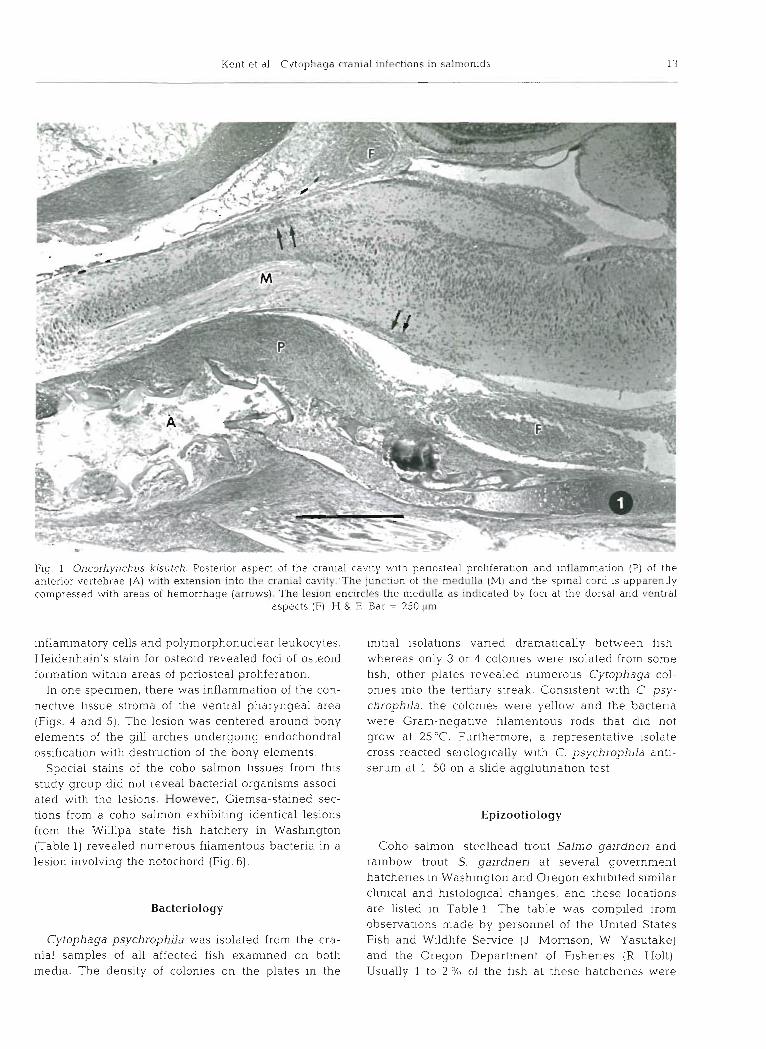
Figs. 2 and 3. Oncorhynchus kisutch. m High magnification of the lesion adjacent to the medulla. There is periosteal proliferation and inflammation (P) with extension through the meninges (M) to the adjacent nervous tissue (NI. H & E. Bar = 25 pm. Flg. Periosteal proliferation and inflammation (P) surrounding a neural ganglion (G) in the posterior cranium. H. & E. Bar
= 100 pm
affected, but at the Sandy and Nehalem hatcheries in sponded well with clinical signs. Furthermore, involve- Oregon ca 1 0 % of the fish died with this syndr0m.e In ment of cranial ganglia associated with the aud~tory 1980 and 1981. canal may have been contributing factors.
Bacteriological and epizootiological evidence sup- ports the hypothesis that Cytophaga psychrophila is
DISCUSSION the causative agent of the disease. To our knowledge, C. psychrophila is enzootic at all of the hatcheries
The cranial and vertebral lesions were most likely where we have observed the cran~al lesions. Moreover, the cause of the ataxia obsewed in affected salmon. the disease reported here usually occurred in fish The subacute to chronic proliferative lesions involving wh.ich had recently recovered from the typical form of the cranium and anterior vertebrae, dnd associated the infection, referred to as cold-water disease. Bac- lesions in the adjacent neural tissue, Lvere observed in terial cultures from the cranium of affected fish consi- all the fish examined that exhibited the spiral swlm- stently revealed the organ.ism, whereas cultures from ming behavior. The hind brain is important in motor fish without the lesions from the same water system control and, therefore, the histological lesions corre- revealed no bacteria. Some cultures contained as few

Kent et al.: Cytophaga cranial infections in salmonids 15
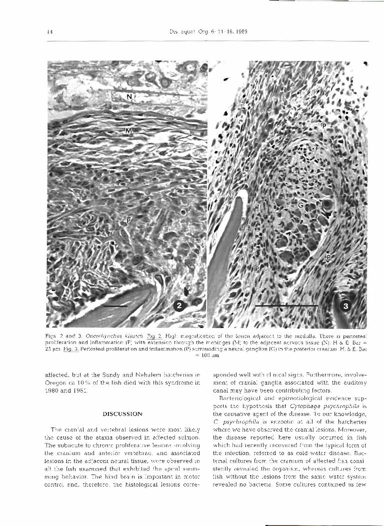
Figs. 4 and 5. Oncorhjrnchus kisutch. m Periosteal proliferation (P), inflammation, and degenerative osteoid (0) in the third gill arch. Note extension of the inflammation involving the center of the bone undergoing endochondral ossification. H & E. Bar =
250 btm. Fig High magnification in an area of periosteal proliferation showing osteoblasts surrounding osteoid (0) H. & E. Bar =
25 pm
as 3 bacterial colonies, which would account for the inability to detect the organisms in most tissue sections.
Similar clinical signs have not been reported in experimentally induced infections (Otis 1984). In one attempt, Holt (1987) was not able to produce the cranial lesions or spiral swimming behavior in fish inoculated subcutaneously with an isolate obtained from the brain of a fish exhibiting this behavior, but typical 'cold- water' disease and mortality was induced in 88 % of the exposed fish. This may indicate that specific conditions are required to cause this chronic infection or that only a small percentage of fish infected with Cytophaga psychrophila develop these lesions. C. psychrophila may be transmitted with eggs (Borg 1960, Schachte 1983) and fish can be infected at essentially any age (Holt 1972). The time of initial exposure may be critical for ultimate development of chronic forms of the infec- tion. Hatcheries usually treat fish affected with cold- water disease with oxytetracycline. Possibly the lesions reported in the present study, and presumably the concurrent bacterial infection, are initiated at sites that
accumulate relatively small amounts of the antibiotic. Initial observations indicate that further treatment with antibiotics, including oxytetracycline, is not efficacious in treating this chronic form of the bacterial infection and that affected fish ultimately die.
The periosteum associated with the central nervous system appeared to be the primary site of infection. Infectious agents and inflammatory processes may take 4 routes to the nervous system; by invasion of peripheral nerves, by direct extension from adjacent structures, by a hematogenous route or by direct implantation from a penetrating injury (Jubb et al. 1985). All but the latter are possible methods by which the cranial infections were established; ganglia were often involved, exten- sion of the inflammation from adjacent tissues was observed in most of the lesions, and Cytophaga psy- chrophila has been detected in the vascular system in previous studies (Wood & Yasutake 1956).
Fish with acute Cytophaga psychrophila infections (cold-water disease) often have caudal lesions that extend to the vertebrae. As with certain domestic mam-
16 Dis. aquat. Org. 6: 11-16, 1989
Fig. 6. Oncorhynchus kisutch. Notochord with numerous filamentous bacteria. Giemsa. Bar = 10 pm
LITERATURE CITED
Borg, A. (1960). Studies on myxobacteria associated with diseases in salmonid fishes. J. Wildl. Dis. (microfiche) 8: 1-85 (2 microcards)
Conrad, J . F . , DeCew, M. (1967). 0bservat.ions on deformed juvenile coho salmon Fish. Commission of Oregon Briefs 13: 129
Daly, .I. G., Stevenson, R. M. W (1985). Charcoal agar, a new growth medium for the fish disease bacterium Renibac- terium salmoninarum. Appl. environ. Microbiol. 50: 868-87 1
Holt, R. A. (1972). Characterization and control of Cytophaga psychrophila (Borg) the causative agent of low temperature disease in young coho salmon (Oncorl~ynchus kisutch). Master's thesis, Oregon State University, Corvallis
Holt, R. A. (1987). Cytophaga psychrophila, the c~iusative agent of bacteria1 cold-water disease in salmon~d fish. Ph D. thesis. Oregon State University, Corvallis
Humason, G. L. (1979), Animal tissue techniques. W. H. Ft.ee- man Co., San Francisco
Jubb, K. V. F., Kennedy, P. C . , Palmer, N. (1985). Pathology of domestic animals. Academic Press. New York
Luna, L. G. (1968). Armed Forces Institute of Patholog? man- ual of histological staining. McGraw-Hill, Ne1.1. Y'firk
mals with tail lesions (Jubb et al. 1985), the neural lesions may have been initiated by localization of bac- teria within the posterior vertebral collimn with subse- quent transport to the anterior vertebrae and cranium in cerebrospinal fluid.
The disease described here, along with the scoliotic condition described by Conrad & DeCew (1967), exem- plifies the variety of pathological cond~tions that can be associated with Cytophaga psychrophila. Though a relatively small percentage of the population were affected, the poor prognosis indicates that this form of C. psychrophila disease is important to hatchery pro- duction under certain circumstances, and further studies to determine the conditions in which the dis- ease is induced should be carried out.
Acknowledgements. This work was supported by Domsea Farms. Inc.. Washington, the Bat tc l l~ Memorial Institute, Cor- porate Technical Development Project Number 3-0322-4051, Columbus, Ohio, and the Battelle Manne Science Laboratory. The Battelle Marine Science Laboratory is part of the Pacific Northwest Laboratory, which is operated for the US Depart- ment of Energy by Battelle Memorial Institute under Contract DE-AC06-76RLO 1830. We thank Drs S. F. Wellings and R. R . Pool for helpful comments on the histological material, and M. T Wilkinson for histological preparations and manuscript review.
Otis, E. J. (1984). Lesions of coldwater hsease in steelhead (Salmo gairdnen): the role of Cytophaga psychrophila extracellular products. Master's thesis, University of Rhode Island, Kingston .
Pacha, R . E , Ordal, E J. (1970). Histopathology and experi- mental columnans disease in young sal.mon. J . cornp. Pathol. 77. 419-423
Schachte, J. H. (1983). Coldwater disease. In: Meyer, F. P., Warren, J W., Carey, T. G. (eds.) A guide to integrated f~sh . health management in the Great Lakes Basm. Spec~al Publ. No. 83-2, Great Lakes Fish. Comm.. Ann Arbor. Michigan, p. 193-197
Wolke. R. E. (1975). Pathology of bacterial and fungal diseases affecting fish. In: Ribclin. W E., Mlgaki, G. (eds.) The pathology of fishes. The University of Wisconsin Press, Madison, p. 33-1 1.6
Wood, E. M , Yasutake, W. T (1956). Histopathology of fish 111. Peduncle ('cold-water') disease. Prog. Fish Cult. 18: 58-61
Wood, J. W. (1974). Diseases of Pacific salmon and their prevention and t.reatrnent. Dept Fisheries, Olympia, Washington
Yasutake, W. T (1965). 'Whirling' sllvcrs Abstracts of thc 16th Ann. Northwest Fish Cult. Conf., Portland, Oregon, p. 3 6 3 7
Responsible Subject Editor: Dr T. Evelyn; accepted for printing on December 16, 1988


![Scanned by CamScannercms.dm.uba.ar/.../1ercuat2019/algebra_I/2019-06-07.pdf · SOC SOL. . ths SOC . e 2) IAS . COHO coho S) C coho SC es tm u] 7 . šsn (e) 6S Y Co . qos C5GJCO es](https://img.pdfslide.net/doc/110x75/611efeea79eaf129dc196a1a/scanned-by-soc-sol-ths-soc-e-2-ias-coho-coho-s-c-coho-sc-es-tm-u-7-sn.jpg)