Embed Size (px)
Citation preview

ORIGINAL ARTICLE Open Access
SPL36 Encodes a Receptor-like ProteinKinase that Regulates Programmed CellDeath and Defense Responses in RiceR. A. O. Yuchun1*†, J. I. A. O. Ran1†, W. A. N. G. Sheng1†, W. U. Xianmei2, Y. E. Hanfei1, P. A. N. Chenyang1,L. I. Sanfeng2, Xin Dedong1, Z. H. O. U. Weiyong3, D. A. I. Gaoxing3, H. U. Juan1, R. E. N. Deyong2* andW. A. N. G. Yuexing2*
Abstract
Lesion mimic mutants spontaneously produce disease spots in the absence of biotic or abiotic stresses. Analyzinglesion mimic mutants’ sheds light on the mechanisms underlying programmed cell death and defense-relatedresponses in plants. Here, we isolated and characterized the rice (Oryza sativa) spotted leaf 36 (spl36) mutant, whichwas identified from an ethyl methanesulfonate-mutagenized japonica cultivar Yundao population. spl36 displayedspontaneous cell death and enhanced resistance to rice bacterial pathogens. Gene expression analysis suggestedthat spl36 functions in the disease response by upregulating the expression of defense-related genes. Physiologicaland biochemical experiments indicated that more cell death occurred in spl36 than the wild type and that plantgrowth and development were affected in this mutant. We isolated SPL36 by map-based cloning. A single basesubstitution was detected in spl36, which results in a cysteine-to-arginine substitution in SPL36. SPL36 is predicted toencode a receptor-like protein kinase containing leucine-rich domains that may be involved in stress responses inrice. spl36 was more sensitive to salt stress than the wild type, suggesting that SPL36 also negatively regulates thesalt-stress response. These findings suggest that SPL36 regulates the disease resistance response in rice by affectingthe expression of defense- and stress-related genes.
Keywords: Defense response, Receptor-like protein kinase, Lesion mimic mutant, Rice, Salt resistance, SPL36
BackgroundThe lesion mimic phenotype is characterized by thespontaneous production of disease spots of various sizesand shapes on the leaves and leaf sheaths (and evenstalks and seeds) in the absence of abiotic or bioticstress. Lesion mimics are the result of apoptosis causedby the hypersensitive response (HR) (Petrov et al., 2015).
Lesion mimic mutants in rice can be divided into the ini-tial (local) type and the spreading type based on pheno-type and whether a dominant or recessive mutation ispresent. The first lesion mimic mutant in plants was re-ported in maize by the American scientist R. A. Emersonin the 1920s (Lu et al., 2012). Sekiguchi Lesion (sl), the firstlesion mimic mutant identified in rice, was discovered bythe Japanese scientist Sekiguchi in the mid-1960s as a nat-urally occurring mutant (Liu et al., 2004).The mechanism underlying the generation of lesion
mimics is complex and regulated by multiple factors, in-cluding both internal and external factors. Internal fac-tors include the altered expression of disease resistance-related genes, uncontrolled programmed cell death(PCD), metabolic disorders, defense signaling molecules,
© The Author(s). 2021 Open Access This article is licensed under a Creative Commons Attribution 4.0 International License,which permits use, sharing, adaptation, distribution and reproduction in any medium or format, as long as you giveappropriate credit to the original author(s) and the source, provide a link to the Creative Commons licence, and indicate ifchanges were made. The images or other third party material in this article are included in the article's Creative Commonslicence, unless indicated otherwise in a credit line to the material. If material is not included in the article's Creative Commonslicence and your intended use is not permitted by statutory regulation or exceeds the permitted use, you will need to obtainpermission directly from the copyright holder. To view a copy of this licence, visit http://creativecommons.org/licenses/by/4.0/.
* Correspondence: [email protected]; [email protected];[email protected]†R. A. O. Yuchun, J. I. A. O. Ran and W. A. N. G. Sheng contributed equally tothis work.1Zhejiang Provincial Key Laboratory of Biotechnology on Specialty EconomicPlants, Zhejiang Normal University, Jinhua 321004, China2State Key Laboratory of Rice Biology, China National Rice Research Institute,Hangzhou 310006, ChinaFull list of author information is available at the end of the article
Yuchun et al. Rice (2021) 14:34 https://doi.org/10.1186/s12284-021-00475-y
and the loss of protease function; external factors in-clude temperature and light. For example, SPL7, the firstlesion mimic mutant gene successfully cloned in rice,encodes the heat shock protein HSFA4, a transcriptionfactor that plays a negative role in the apoptosis pathway(Yamanouchi et al., 2002). SPL7 is highly similar tomaize HSFb, tomato HSF8, and Arabidopsis HSF21 andHSF1, all of which regulate apoptosis in plants; mutantsof these genes show lesion mimic characteristics. Thephenotype of spl18 mutant of rice is associated with theinsertion of a T-DNA activation tag that enhances theexpression of genes around the insertion site (Moriet al., 2007). OsATL, encoding an acyltransferase homo-log that induces allergic reactions to tobacco, is located~ 500 bp downstream of the inserted T-DNA activationtag. This gene is expressed at low levels in wild-type ricebut at high levels in spl18, resulting in the occurrence oflesion mimics due to the abnormal expression of diseaseresistance genes.Rice plants with mutations in NLS1, encoding a CC-
NB-LRR protein, accumulate large amounts of H2O2
and salicylic acid and show abnormal expression ofresistance-related genes, leading to the appearance of le-sion mimics in leaf sheaths (Tang et al., 2011). The pro-tein that is altered in the lesion mimic mutant spl11contains U-box and ARM (armadillo) repeat domainsand undergoes ubiquitination and protein-protein inter-actions when expressed in yeast and mammalian systems(Zeng et al., 2002). The similarity of this protein to otherplant U-box-ARM proteins is mainly limited to the U-box and ARM repeat regions. A single base substitutionwas detected in the mutant spl11 gene, resulting in thepremature termination of translation of the encodedproteins. The E3 ubiquitin ligase activity of this proteinis dependent on the presence of an intact U-box domain,indicating that ubiquitination plays a role in plant celldeath and defense and suggesting that spontaneouslyformed lesion mimics are associated with uncontrolledPCD (Zeng et al., 2004). OsSSI2 encodes fatty acid de-hydrogenase (FAD), which also plays a negative role inthe defense response in rice. The loss of function ofFAD results in lesion mimics and delayed leaf growth(Jiang et al., 2009). Mutations in a gene encoding uridinediphosphate-N-acetylglucosamine pyrophosphorylase(UAP1), which functions during glucose metabolism,can also lead to the appearance of lesion mimics in riceleaves (Jung et al., 2005).Most lesion mimic mutants in rice show enhanced dis-
ease resistance to some extent. Among the more than 80mutants that have been identified, 11 mutants (includingspl1, spl9, spl10, cdr1, and cdr3) show enhanced blast re-sistance (Liu et al., 2004; Yoshimura et al., 1997; Takaha-shi et al., 1999); 12 mutants (including spl21, spl24,lmes1, hm197, and hm83) show enhanced bacterial
blight resistance (Wu et al., 2008); 19 mutants (includingspl14, bl3, and Lmr) show enhanced blast resistance andbacterial blight resistance (Mizobuchi et al., 2002); andone, lmm1, shows both enhanced blast resistance andsheath blight resistance. By contrast, the disease resist-ance of spl2, spl3, spl4, spl6, spl7, and ncr1 is unchangedor even reduced compared to the wild type (Kang et al.,2007; Campbell and Ronald, 2005).Plant receptor-like protein kinases (RLKs) occupy im-
portant metabolic positions and are abundant in plants;rice has approximately 1130 RLK genes (Nguyen et al.,2015). Plant RLKs are composed of intracellular, extra-cellular, and transmembrane regions (Ye et al., 2017).Most RLKs contain an extracellular receptor domain(ECLB), a transmembrane domain (TM), and a proteinkinase contact response domain (PKC) (Walker, 1994;Zhang, 1998).The leucine-rich repeat (LRR) receptor-like protein ki-
nases are a subtype of RLKs that are involved in plantstress responses and defense-related processes, includingdisease responses. The Cf gene family of tomato leafmold encodes proteins with LRR structures; differencesin the amino acid sequences of the LRR motifs of differ-ent proteins in the same family are responsible for thespecificity of ligand binding (Thomas et al., 1998). Theresistance gene FLS2 in Arabidopsis has a similar struc-ture to the sequence encoding the extracellular domainof the tomato Cf gene family (Gómez-Gómez et al.,2001). Upon binding to ligands (avirulence gene prod-ucts of rice bacterial blight pathogens), the extracellularLRR structure of rice Xa21 induces intracellular kinasephosphorylation and produces a series of cellular re-sponses that protect the plant from pathogens (Songet al., 1995; Park and Ronald, 2012). These findings indi-cate that the LRR structure plays an important role inidentifying the basic structures of pathogens andmicroorganisms.Here, to further explore the signal transduction
pathways of LRR-type receptor kinases in response tostress signals in rice, we isolated and characterizedthe novel lesion mimic mutant spotted leaf 36 (spl36).This mutant shows spots at the tillering stage and en-hanced resistance to bacterial blight. We cloned theSPL36 gene by map-based cloning and demonstratedthat it encodes a receptor-like protein kinase receptorthat is expressed in all tissues and at all developmen-tal stages after the tillering stage and is localized tothe plasma membrane. A high frequency of cell death,changes in chloroplast structure, and activation ofdefense-related responses were observed in the spl36mutant. We demonstrate that the loss of function ofSPL36 is responsible for the cell death, prematuresenescence, and activation of the defense response inthis mutant.
Yuchun et al. Rice (2021) 14:34 Page 2 of 14

Materials and MethodsPlant Materials and Growth ConditionsThe spotted leaf mutant spl36 was isolated from amethanesulfonate (EMS)-induced mutant library of Yun-dao rice (wild type, WT). The mutant was hybridizedwith TN1 as the male parent, and the F1 offspring andF2 population were grown in the rice experimental fieldof Zhejiang Normal University, Jinhua City, ZhejiangProvince, China during the summer of 2018 and 2019.The F2 populations of both spl36/ZF802 and spl36/TN1were used for genetic analysis, and the F2 recessive indi-viduals of spl36/TN1 were used for gene fine map-ping.The agronomic traits of wild-type and spl36 plantswere statistically analyzed, including plant height, tillernumber, grain number per panicle, seed setting rate, and1000-grain weight. The results were analyzed based onthe average of 10 replicates.
Measuring Photosynthetic Parameters and ChlorophyllContentFrom 9:30 a.m.to 11:00 a.m. on sunny days, 10 individualplants with relatively uniform growth were harvested.The photosynthetic parameters of wild type and mutantplants were measured with an LI-6400XT portablephotosynthesis tester. Three to five representative flagleaves were treated and measured, and each leaf wasmeasured in triplicate (the mean value was taken as onereplicate). During the measurement, red and blue lightsources were used, the light intensity was constant at1200 μmol/m2, the temperature was 30 °C, the CO2 con-centration was the concentration in the air, and the hu-midity level was the relative humidity in the atmosphere.For chlorophyll measurements, five wild type and mu-tant plants with relatively uniform growth vigor were se-lected. The leaves were weighed, and 0.05 g of leaf tissuewas cut into pieces and soaked in 25mL 1:1 ethanol:acetone solution; three replicates were subjected to thedarkening reaction for 24 h, followed by shaking. Theabsorbance values at 663 nm, 645 nm, and 470 nm weremeasured with a spectrophotometer, and the photosyn-thetic pigment content was calculated and statisticallyanalyzed by Student’s t-test.
Preparation of Rice ProtoplastsTo generate rice protoplasts, 60 rice seedlings were cul-tured for 12–15 days. The leaves were removed from theplants with sterile scissors, leaving only the stalk. Thematerial was cut into 0.5-mm strips and placed into asterilized 50 mL triangular flask. After adding 30mL 0.6mol/L mannitol, the sample was incubated for 15 minwith shaking in the dark at 28 °C and 50 r/min. Aftershaking, the mannitol solution was poured off, and thesample was transferred to a clean sterile triangular flask.Enzymatic hydrolysis was performed by adding 20mL
enzyme solution (1.5% fibrinase R-10, 0.75% segregationenzyme R-10, 0.6 mol/L mannitol, 10 mmol/L MES, 10mmol/L CaCl2, 0.1% BSA, pH 5.7) and incubating at28 °C with shaking at 60 ~ 80 R /min for 4 ~ 5 h. Follow-ing enzymatic hydrolysis, the same volume of W5 solu-tion (154 mmol/L NaCl, 125 mmol/L CaCl2, 5 mmol/LKCl, 2 mmol/L MES, pH 5.7) was added to the sample.Pre-cooled. After 15 s of severe shock, the sample waspassed through a nylon filter. The protoplasts werecleaned with W5 solution, centrifuged at 1000 r/min for5 min, and the supernatant discarded; this step was re-peated 3 to 5 times. Following the addition of MMg so-lution, the protoplasts were collected and examined bymicroscopy and incubated on ice for 40 min.
Histochemical AnalysisThe content and concentration of malondialdehyde(MDA) and the enzymatic activity of superoxide dismut-ase of peroxidase (POD) were analyzed following themanufacturer’s instructions (Nanjing Jiancheng Bio-engineering Institute, Nanjing, China). The MDA andH2O2 contents and SOD and POD activity were mea-sured when the tillering stage was first visible in spl36.Apoptosis was detected by TUNEL assay. FAA fixativewas prepared before sampling and placed into a 2 mLcentrifuge tube (Liang and Zhou., 2018). When the mu-tant lesion phenotype was apparent in spl36 leaves, leaftissues showing this phenotype were harvested, alongwith wild-type leaves at the corresponding position, cutinto clumps, and placed in the 2/3 position of FAA fixa-tive for fixation. The leaf tissue was vacuum infiltrateduntil it sank to the bottom. The tube was sealed withParafilm and stored in a refrigerator at 4 °C. A TUNELapoptosis detection kit (Roche, Cat No.1684817) wasused to measure apoptosis in the samples (Inada et al.,1998).
Linkage Analysis and Mapping of spl36SSR primers that are evenly distributed over the 12 ricechromosomes (from our laboratory) were used to screenthe mutants and TN1 for polymorphisms (Supplemen-tary Table 2). Twenty-one individual F2 lesion mimicspl36/TN1 plants were used for linkage analysis to pre-liminarily confirm the chromosomal location of the tar-get gene. A new InDel marker with relatively goodpolymorphism was developed in the mapping interval,and the target gene was precisely mapped using a singleplant showing the mutant phenotype in the F2 segregatingpopulation of spl36/TN1. Genomic DNA was extractedfrom the samples using the hexadecyltrimethylammoniumbromide (CTAB) method (Wu et al., 2008). The PCR mix-ture included 1 μL DNA template, 1 μL 10 × PCR buffer,0.5 μL each of forward and reverse primers (10 μmol/L),1 μL dNTPs, 0.2 μL rTaq, and H2O to a final volume of
Yuchun et al. Rice (2021) 14:34 Page 3 of 14

10 μL. The PCR amplification program was as follows:pre-denaturation at 94 °C for 4min; denaturation at 94 °Cfor 30 s, annealing at 55–60 °C for 30 s (depending on theprimers), extension at 72 °C for 30 s, 40 cycles; and a finalextension at 72 °C for 10min. The PCR products wereseparated by electrophoresis on a 4% agarose gel, photo-graphed, and the data stored in a gel imager and read. Theprimers used for mapping are shown in SupplementaryTable 3.
Vector ConstructionFor functional complementation of the spl36 mutants,the complete genomic DNA fragment (including thepromoter) of wild-type SPL36 was amplified by PCRwith primers spl36-CPT-F/ spl36-CPT-R and used toconstruct the vector for transformation by insertion intoempty binary vector pCAMBIA1300 via Clontech In-Fusion PCR (TaKaRa). The full-length SPL36 open read-ing frame (ORF) was amplified with the primer pairspl36-GFP-F/spl36-GFP-R, and the coding sequence ofSPL36 was inserted into binary vector pHQSN contain-ing the 35S promoter (p35S::SPL36) for subcellularlocalization analysis. The SPL36 promoter was intro-duced into the expression vector pCAMBIA1305.1, andthe expression of SPL36 in rice tissues was revealedusing the GUS reporter gene. GFP fluorescence was ob-served by confocal laser-scanning microscopy (LeicaTCS SP5, Leica, Germany). The primers used for vectorconstruction are shown in Supplementary Table 4.
Inoculation TestXanthomonas oryzae pv. Oryzae, Xoo (the causal agentof bacterial blight) was inoculated onto the flag leaves ofwild-type Yundao and the spl36 mutant at the tilleringstage using the leaf clipping method (The selected leavesshould be fresh and free of disease spots and senes-cence). Specifically, healthy, fully unfolded rice leaveswere selected, and the tip of each leaf (~ 1 cm) was cutwith scissors that were dipped into Xanthomonas oryzaepv. Oryzae, Xoo solution before making each cut. Thephenotypes of the inoculated leaves were observed at 5and 10 days after inoculation and the lesion length mea-sured and photographed.
Quantitative Reverse-Transcription PCR AnalysisLeaf, root, stem, leaf sheath, panicle, and grain sampleswere collected from wild type and mutant plants at eachstage of development. RNA was extracted from the sam-ples using an RNAprep Pure Plant Kit (Cat No. DP441,Tiangen Biotech, Beijing, China) and amplified using aReverTra-Plus-reverse transcription kit (Cat No.FSQ-301, Toyobo, Japan) and backup for post-reverse tran-scription. Reverse-transcription PCR (qRT-PCR) wasused to detect the expression of defense-related genes
and the expression of SPL36 in tissues at each stage,with the OsActin gene used as an internal reference(GenBank accession number: NM001058705). The reac-tion mixtures contained 2 μL cDNA template, 10 μL 2 ×SYBR qPCR mix, 0.8 μL each of forward and reverseprimers, and ddH2O to a final volume of 20 μL. The re-action program was 95 °C for 30 s; 95 °C for 5 s, 55 °C for10 s; and 72 °C for 5 s for 40 cycles. Each reaction wasperformed in triplicate, and the relative expression levelsof premature senescence-associated genes were calcu-lated using the 2 -ΔΔCt method. RT-PCR was performedusing a quantitative fluorescence gene amplification in-strument (qTOWER3G; Jena, Germany). Data were ana-lyzed using SPSS 19.0 software and Excel. Student’s t-test was used to analyze the significance of differences.The primers used for qRT-PCR are shown in Supple-mentary Table 5.
Salt Stress AssayPlate test: Seeds were collected from wild-type Yundaoand spl36 plants, washed, spread on 200 mM NaCl MSmedium, and cultured at 28 °C in the light; MS mediumwithout NaCl was used as a control. The assay was per-formed in triplicate, and the germination rates of theseeds were determined at each stage. After nine days ofculture, root lengths were measured and photographed.Salt stress assay at the seedling stage: Wild-type and
mutant seedlings were grown hydroponically for ap-proximately two weeks, and seedlings with roughly thesame level of growth were selected for the assay. Wild-type and mutant seedlings were transferred to standardnutrient solution with or without 150 mM NaCl and cul-tured for four days. The salt-stressed seedlings weretransferred to standard nutrient solution and allowed torecover for three days, followed by the detection of plantsurvival rates, fresh weight, conductivity, and prolinecontent.
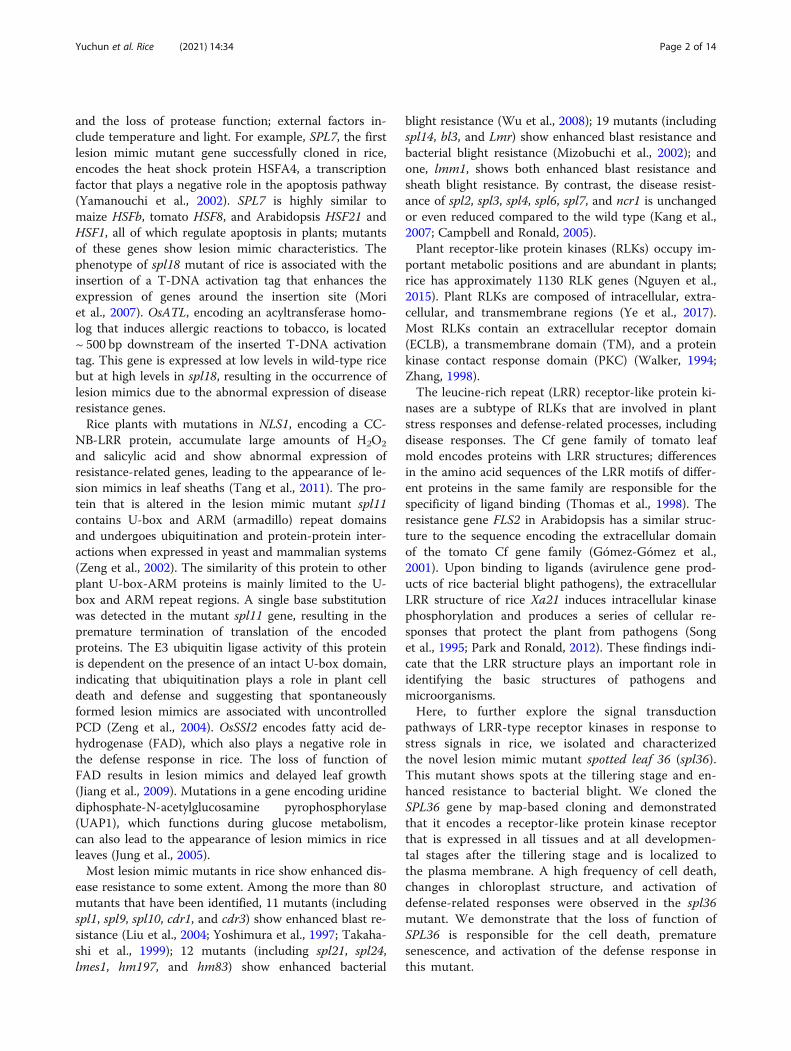
ResultsPhenotype of the Lesion Mimic Mutant spl36Under normal growing conditions in the summer, spl36leaves were not significantly different from those of thewild type before the tillering stage. At the tillering stage,the lesion mimic phenotype appeared in the leaf apex(Fig. 1a). From the tillering stage to the heading stage,these necrotic spots became more severe and graduallyspread throughout the leaf (Fig. 1b). To investigatewhether lesion formation is induced by light in spl36, asin most lesion mimics, we covered spl36 leaves with 2–3 cm aluminum foil at the tillering stage and used un-covered mutant leaves as controls. After seven days, nospread of lesion mimics had occurred in the coveredareas of leaves, whereas lesion mimics appeared on un-covered control leaves (Fig. 1c). These results indicate
Yuchun et al. Rice (2021) 14:34 Page 4 of 14

that the lesion mimic phenotype in spl36 is induced bylight. In addition, the major agronomic traits, includingplant height, grain number per panicle, and 1000-grainweight, were significantly reduced in spl36 vs. wild-typeplants (Fig. 1d–i).
SPL36 Regulates Plant Growth and DevelopmentDue to the negative agronomic changes in spl36, we rea-soned that the growth and development of the mutantwere affected after the appearance of the lesion mimics
because of reduced photosynthesis (Han et al., 2015).We observed chloroplast ultrastructure by transmissionelectron microscopy and found that spl36 chloroplastswere atrophied and smaller than those in the WT andhad disorganized lamellae (Fig. 2a–d). The contents ofboth chlorophyll a and chlorophyll b at the tilleringstage were significantly reduced in spl36 compared tothe wild type (Fig. 2e). In addition, the net photosyn-thetic rates were significantly reduced in spl36 vs. thewild type (Fig. 2g).
Fig. 1 Phenotypes of the spl36 mutant. a Lesions appear at the tillering stage. Bar = 6 cm. b Lesions first appear at the tip of the leaf (WT andspl36 at the tillering stage). c Effect of light on lesion formation under natural conditions; spl36 before shading (1,2). spl36 shaded for 7 days (3,4).d-i Statistical analysis of important agronomic traits in WT and spl36 at the maturity stage. Values are means ±SD (n = 10); ** indicates significanceat P≤ 0.01 by Student’s t test
Yuchun et al. Rice (2021) 14:34 Page 5 of 14

To further explore the effects of the mutation onchloroplast development and chlorophyll biosynthesis,we examined the expression of chloroplast development-and pigment synthesis-related genes in wild-type andmutant plants at the mature stage by qRT-PCR. The ex-pression levels of NYC1, OsClpP5, OsSIG1, OsPORA,and OsCAO1 were significantly reduced in the mutantvs. the wild type (Fig. 2f). These results suggest thatSPL36 influences plant growth and development via itseffects on chloroplast structure.
SPL36 Regulates ROS Accumulation and Cell Death in RiceThe TUNEL (terminal deoxynucleotidyl transferasedUTP nick end labeling) assay is used to detect DNAfragmentation, a marker of programmed cell death (Kimet al., 2009). The TUNEL signal in the nuclei of mutantspl36 cells was intense and randomly distributed,whereas only a weak TUNEL signal was detected in thewild type (Fig. 3a-d). In addition, the accumulation of re-active oxygen species (ROS) at high concentrations leadsto an oxidative burst, which causes cell damage and eventriggers programmed cell death (Kim and Coulombe,2010). H2O2 contents and peroxidase (POD) activity aredirectly related to the accumulation of ROS. Superoxidedismutase (SOD) plays an important role in scavengingO2− in plants. We therefore measured H2O2 content,
POD activity, and SOD activity in the plants and foundthat a large amount of H2O2 accumulated in spl36 (Fig.3e), while POD and SOD activities were significantly re-duced in this mutant vs. the wild type (Fig. 3g-h). Thereduced activity of these enzymes negatively affects theremoval of peroxide and negative oxygen ions, resultingin the accumulation of ROS. In addition, membranelipid peroxidation occurs when plant organs age or suf-fer damage under stress. Malondialdehyde (MDA) is thefinal decomposition product of membrane lipid peroxi-dation, and therefore MDA content can reflect the de-gree of damage in stressed plants. The MDA contentwas significantly higher in spl36 than in the wild type(Fig. 3f). These results indicate that the lesion mimics inspl36 mutants are caused by ROS accumulation and irre-versible membrane damage.
SPL36 Regulates Defense Responses in RiceMost rice lesion mimic mutants show enhanced resist-ance to pathogens. To investigate whether spl36 plantsshowed increased resistance to rice pathogens, we per-formed an inoculation assay on wild-type Yundao andspl36 plants at the tillering stage and used the leaf clip-ping method to inoculate the plants with rice bacterialblight strain HM73. We detected changes in the inocula-tion site and length of the lesion mimics in the mutant
Fig. 2 Chloroplast development and net photosynthetic rate in wild-type and mutant plants. a-d Chloroplast ultrastructure in wild-typeand mutant plants, a, c: leaf cells at 6000X; b, d: leaf cells at 40,000X; N: nucleus; Thy: chloroplast; Og: osmium granules; Bar = 1 μm. e Chlorophyllcontent in the leaves of wild-type and mutant plants at the tillering stage. f Relative expression of chloroplast development and pigmentsynthesis-related genes. g Net photosynthetic rate in wild-type and mutant plants at the tillering stage
Yuchun et al. Rice (2021) 14:34 Page 6 of 14

at 5 and 10 days after inoculation, respectively. At fivedays after inoculation, the leaf apex of the wild typeshowed obvious necrotic spots, whereas the mutant didnot show obvious disease spots. At 10 days after inocula-tion, wild-type disease spots were significantly longerthan those of the mutant (Fig. 4a-e). These results indi-cate that resistance to Xanthomonas oryzae pv. Oryzae,Xoo is significantly enhanced after the emergence of dis-ease spots in spl36.To explore the mechanism underlying the enhanced
resistance of spl36 to bacterial pathogens, we examinedthe expression of defense-related genes in the wild typeand mutants at the tillering stage by qRT-PCR. The ex-pression levels of defense genes MAPK12, WRKY53,BIMK2, AOS2, ASP90, LYP6, PR2, PR1a, and PR1b weresignificantly elevated in the mutants (Fig. 4f). Thus, theloss of function of the SPL36-encoded protein triggers adefense response in rice, leading to the enhanced resist-ance of spl36 to pathogens.
Genetic Analysis and Map-Based Cloning of SPL36We hybridized spl36 as the female parent with ZF802 ofjaponica cultivar TN1. The F1 plants did not show lesionmimics. The segregation ratio of the normal phenotypeto lesion mimic phenotype in the F2 population was es-sentially in compliance with a 3:1 ratio, indicating thatthe spl36 phenotype is caused by a mutation in a singlerecessive nuclear gene (Supplementary Table 1). Weused a selection of polymorphic markers from 238
insertion and deletion tags to map the mutant gene in21 F2 individuals with a lesion mimic phenotype andmapped the mutation site to a location between B12–5and B12–6 on chromosome 12 (Fig. 5a). The SPL36 lo-cation was further refined to a region between JHL-3and JHL-7 by genotyping 148 mutant F2 individualsfrom the same cross and adding four additional poly-morphic tags (Fig. 5b). Using 554 additional F2 mutantindividuals and four newly developed polymorphic tags,we ultimately mapped SPL36 to a 60 kb region betweenmarkers InDel1 and InDel2 (Fig. 5c). Analysis usinghttps://rice.plantbiology.msu.edu/ predicted that this re-gion contains 11 open reading frames (ORFs) encodingseven expressed proteins and four functional proteins(Fig. 5d). Sequencing and alignment revealed that inspl36, gene LOC_Os12g08180 was mutated (Fig. 5e): nu-cleotide T at position 1462 in the coding region of thisgene was replaced by C (Fig. 5f), resulting in the changeof the encoded amino acid from cysteine to arginine(Fig. 5g). Therefore, LOC_Os12g08180 is the candidategene for SPL36.
Functional Complementation of the spl36 Mutant withLOC_Os12g08180To determine whether the single base substitution inLOC_Os12g08180 is indeed associated with the spl36phenotype, we constructed the vector pGSPL36, whichcontained genomic DNA fragments including the pro-moter of the SPL36 gene in wild-type Yundao, and
Fig. 3 Physiological and biochemical analysis of wild-type and mutant plants. a- d TUNEL assay of DNA fragmentation in mesophyll cells.Bar = 100 μm. e-f H2O2 and MDA contents of leaves in spl36 and WT plants at the heading stage. g- h POD and SOD activities in the leaves spl36and WT plants at the heading stage. POD: peroxidase; SOD: superoxide dismutase; MDA: malondialdehyde; WT: wild type
Yuchun et al. Rice (2021) 14:34 Page 7 of 14

Fig. 4 SPL36 regulates defense responses in rice. a-d Phenotypes of wild-type and spl36 leaves at 5 days and 10 days after inoculation withthe bacterial blight pathogen HM73. e Statistical analysis of the length of bacterial leaf blight lesions. f Relative expression of defense-relatedgenes in uninoculated plants
Fig. 5 Genetic and physical maps of the SPL36 gene. a The SPL36 gene was localized to chromosome 12 between InDel markers B12–5 andB12–6. b The SPL36 gene was delimited to the JHL-3 to JHL-7 interval on chromosome 12 using 148 F2 mutant individuals; marker names andnumber of recombinants (Rec.) are shown. C Fine genetic mapping of the SPL36 gene based on 554 mutant F2 individuals. d 11 putative ORFslocated in the ~ 60-kb region of this locus. e Gene structure of LOC_Os12g08180. f Sequence analysis of the T-to-C mutation site in wild type andspl36. g The mutation results in a change from cysteine to arginine in the encoded protein
Yuchun et al. Rice (2021) 14:34 Page 8 of 14

introduced it into spl36 by Agrobacterium tumefaciens-mediated transformation. The corresponding empty vec-tor pEmV was used as a control. Of the 60 T0 plants, 54were positive transformants. All of these transformantshad the same phenotype as the wild type (Fig. 6a), whileplants transformed with the control vector showed thesame lesion mimic phenotype as spl36 (Fig. 6b). Theseresults demonstrate that LOC_Os12g08180 is SPL36 andthat the single base substitution in spl36 leads to the ap-pearance of the lesion mimic phenotype of the mutant.
Expression Analysis of SPL36We performed reverse-transcription quantitative PCR(qRT-PCR) to analyze the expression of SPL36 in variousorgans. SPL36 was expressed in all organs, with higherexpression levels in leaves, leaf sheaths, and roots andlower expression levels in stems and panicles. SPL36 wasexpressed at significantly higher levels in all organs ofspl36 compared to wild-type Yundao (Fig. 7a). Toanalyze the spatiotemporal expression pattern of SPL36more precisely, we constructed the vector pSPL36::GUSby fusing the GUS gene with the promoter of SPL36from the wild type. We then performed Agrobacteriumtumefaciens-mediated transformation to obtain trans-genic plants. We stained various organs of thetransgene-positive plants and observed GUS signal invarious tissues (Fig. 7b-f). Our results showed thatSPL36 was expressed in tested organs and tissues includ-ing the root, leaf, leaf sheath, stem and panicle. Notably,SPL36 transcript level was more abundant in the leafrelative to other tissues or organs. Relative strong GUSsignals were observed in the leaf, whereas weak signalswere also found in the root, culm, leaf sheath, and pan-icle. These findings were consistent with the RT-qPCRresults.
Subcellular Localization of SPL36To determine the subcellular location of SPL36, wefused the full-length coding sequence of SPL36 to the N-
terminus of green fluorescent protein (GFP). When tran-siently expressed in rice protoplasts, the GFP signal ap-peared on the plasma membrane and cytoplasm and thesignal of 35S::GFP appeared on membrane, cytoplasm,and nucleus (Fig. 8a–d). To verify this observation, wetransformed Nicotiana benthamiana leaves with a plas-mid containing the SPL36-GFP fusion vector. SPL36-GFP protein was detected on the membrane and cyto-plasm and 35S::GFP protein was detected on the mem-brane, cytoplasm, and nucleus (Fig. 8e–h). These resultsindicate that SPL36 localizes to the membrane andcytoplasm.
SPL36 Is Involved in Salt Stress Responses in RiceAfter verifying that the single base substitution of LOC_Os12g08180 was responsible for the lesion mimic pheno-type of spl36, we determined that this gene encodes areceptor-like protein kinase. Plant receptor-like proteinkinases play regulatory roles in plant growth and devel-opment and disease resistance (Afzal et al., 2008; LiLiyun et al., 2008), and most of them function in stressresponses. To investigate whether SPL36 is involved instress response-related pathways, we performed saltstress assays in which we placed wild-type and mutantseeds in flat dishes for the germination assay and grewseedlings hydroponically in the absence and presence ofNaCl for the seedling assay.In the absence of salt treatment, no significant differ-
ence in the germination rates of spl36 vs. wild-type seedswas detected over a one-week period. Under salt treat-ment, the germination rates of both mutant and wild-type seeds decreased significantly, while the germinationrate of the wild type was also significantly lower thanthat of the mutant. After 9 days of culture in the germin-ation assay, we measured the lengths of the root por-tions of salt-treated and control seedlings. Under controlconditions, there was no significant difference in stemlength between wild-type and mutant seedlings. By con-trast, under NaCl treatment, the stems were significantly
Fig. 6 Functional complementation of the spl36 mutant with LOC_Os12g08180. a The phenotype of the spl36 mutant transformed with thegenomic sequence of SPL36 (pGSPL36) was completely recovered to that of the wild type. The insets show enlarged views of leaf sections withlesion spots. Bar = 8 cm. b Transgenic plants were verified by the presence of the hygromycin selectable marker gene. pEmV: empty vector
Yuchun et al. Rice (2021) 14:34 Page 9 of 14

Fig. 7 Expression analysis of SPL36. a Expression of SPL36 in various organs of wild type and spl36 plants analyzed by quantitative RT-PCR. b-fHistochemical signals from the SPL36 promoter-GUS reporter gene. GUS signals were detected in the root b, stem c, leaf d, leaf sheath e, andpanicle f
Fig. 8 Subcellular localization of SPL36. a-c Transient assay in rice protoplasts. h-j N. benthamiana leaf assay. d-f and k-m 35S::GFP controlsused in the assays. SPL36-GFP: SPL36 GFP fusion protein. Bar = 20 μm
Yuchun et al. Rice (2021) 14:34 Page 10 of 14

shorter in mutant vs. wild-type seedlings (SupplementaryFig. 1). We also grew wild-type and mutant seedlingshydroponically for four weeks, cultured them in high-salt medium for four days, returned them to normalconditions for recovery, and examined them three dayslater. No significant changes were detected in the wildtype following four days of treatment, with the pheno-type recovering after the return to normal conditions. Bycontrast, while spl36 showed significant leaf bendingafter salt treatment, and the plants did not recover oreven died following their return to normal conditions.Our analysis of fresh weight, conductivity, and final sur-vival of plants before and after treatment and under con-trol conditions revealed that spl36 was more sensitive tosalt treatment than the wild type (Fig. 9). In summary,SPL36 is involved in salt-stress responses in rice.
DiscussionLesion mimic mutants are extremely valuable for study-ing PCD and defense-related responses in plant cells. Inthe present study, we selected the lesion mimic mutantspl36 from a mutant library generated by mutagenizingwild-type Yundao rice using EMS. There were no obvi-ous phenotypic differences between this mutant and thewild type at the seedling stage. However, reddish-brown
disease spots appeared in the mutant at the leaf apex atthe tillering stage and gradually spread throughout theleaf. We observed the chloroplast ultrastructures of boththe wild type and mutant at this stage and measuredtheir photosynthetic rates. The appearance of lesionmimics led to significant changes in chloroplast struc-ture in the mutant. Since chloroplasts are the sites ofphotosynthesis (Wu et al., 2018), the appearance of le-sion mimics affected both the growth and developmentof the plants. Altered chloroplast structure is also a dir-ect cause of the decline in multiple agronomic traits inplants (Ishikawa et al., 2001).Using map-based cloning, we mapped the SPL36 gene
within a 60 Kb interval on chromosome 12. Based on in-formation in the rice genome database (https://rice.plantbiology.msu.edu/), this interval contains 11 ORFs,including genes for seven expressed proteins and fourfunctional proteins. We amplified the genomic se-quences in this region in the mutant and wild type byPCR. Sequence alignment and sequencing analysis re-vealed that nucleotide T at position 1462 in the codingregion of the gene LOC_Os12g08180 was replaced withC, resulting in a change in the encoded amino acid fromcysteine to arginine. Using a functional complementa-tion assay, we determined that this gene is SPL36.
Fig. 9 Analysis of salt-stress responses in wild type and spl36 plants at the seedling stage. a Phenotypes wild-type and mutant seedlings beforeand after 150 mM NaCl treatment. b Conductivity of wild-type and mutant seedlings before and after 150 mM NaCl treatment. c Fresh weights ofwild-type and mutant seedlings before and after 150 mM NaCl treatment. d Survival rates of wild-type and mutant seedlings after 150 mMNaCl treatment
Yuchun et al. Rice (2021) 14:34 Page 11 of 14

Structural analysis of the protein encoded by this geneshowed that this protein is a receptor-like protein kinasereceptor containing multiple leucine-repeat domains(Supplementary Fig. 2).Leucine-rich receptor-like kinases (LRR-RLKs) are
closely related to stress and defense responses in plants.The PRK1 gene was initially isolated from Arabidopsisin 1997 by Hong et al. (Hong et al., 1997), who demon-strated that the LRR domain of PRK1 functions inprotein-protein interactions, as well as the perception ofstress signals from the environment. In 2014, Yang et al.(Yang et al., 2014) identified new LRR-RLKs in wild soy-bean and demonstrated that GsLRPK improves droughtresistance when exogenously expressed in Arabidopsis.OsGIRL1 is upregulated in response to abiotic stressesincluding salt, osmotic stress, and heat, and the stress-related hormones salicylic acid and abscisic acid but isdownregulated in response to jasmonic acid; OsGIRL1 islocated in the plasma membrane. The biological func-tion of OsGIRL1 was explored by exogenously expressingthis gene in Arabidopsis plants and examining plant re-sponses to irradiation, salt pressure, osmotic pressure,and thermal stress (Park et al., 2014). The LRR type re-ceptor protein kinase gene OsRLK1 is induced by lowtemperature and salt stress in rice (Lee et al., 2004).CALRR1 in pepper is induced not only by exposure toanthrax pathogens but also under abiotic stress condi-tions such as high salt, abscisic acid, and wounding (Junget al., 2004). Here, using two different salt stress proto-cols, we demonstrated that spl36 was more sensitivethan the wild type to salt treatment. Although these re-sults can be explained by the finding that a missensemutation in the coding region of LOC_Os12g08180 ledto the loss of protein function, the specific mechanismremains to be elucidated and will be explored in thefuture.LRR-RLKs are primarily associated with abiotic stress
responses in plants, while their relationships with PCDand disease resistance have not been reported. We veri-fied the higher frequency of cell death in spl36 by per-forming a TUNEL assay and measured H2O2 and MDAlevels and POD and SOD activities in the wild type andmutant. spl36 accumulated more ROS than the wildtype, which led to an oxidative burst and ultimatelyPCD. Since lesion mimics arise spontaneously in spl36,our findings indicate that SPL36 negatively regulatesPCD in rice.Most previously reported lesion mimic mutants show
some enhancement of disease resistance. To investigatewhether SPL36 is involved in disease resistance in rice,we used the leaf clipping method to inoculate wild-typeand mutant plants with the bacterial blight pathogenHM73 and found that spl36 had significant resistance tothis pathogen. However, it remains to be determined
whether spl36 has broad-spectrum resistance. We alsoanalyzed the expression of several defense-related genesin the wild type and mutant. The defense genesMAPK12, WRKY53, BIMK2, AOS2, LYP6, PR1a, andPR1b were significantly upregulated in the mutant vs.the wild type. OsWRKY53 encodes a transcriptional acti-vator that plays an important role in the excitation-induced defense signal transduction pathway in rice(Chujo et al., 2007; Tian et al., 2017). OsAOS2 expres-sion in leaves is significantly induced by rice blast. Theexpression of OsAOS2 driven by the PBZ1 promoter ac-tivated the expression of other pathogenesis-relatedgenes, thereby increasing resistance to rice blast (Meiet al., 2006). OsBIMK2 plays an important role in ricedisease resistance responses (Song et al., 2006). LYP6 en-codes a protein containing cytolytic enzyme motifs thatfunctions as a pattern recognition receptor for bacterialpeptidoglycan and fungal chitin and plays a dual role inthe recognition of peptidoglycan and chitin during in-nate immunity in rice (Liu et al., 2012). OsPR1a andOsPR1b are pathogenesis-related genes (Agrawal et al.,2000). Based on our results, we propose that SPL36 reg-ulates the disease resistance response in rice by upregu-lating the expression of defense genes, but the specificmechanism requires further investigation.
ConclusionWe cloned a novel spotted leaf gene (SPL36) encoding areceptor-like protein kinase that contains repeated leu-cine domains and may be involved in stress responses inrice. This is the first report of the involvement of areceptor-like protein kinase in disease resistance-relatedpathways in rice. We demonstrated that the loss of func-tion of SPL36 results in enhanced resistance to patho-gens and increased salt sensitivity. We are currentlyconducting an in-depth study to determine whether thespl36 mutant has broad-spectrum resistance to patho-gens and to uncover the role of SPL36 in the salt stressresponse in rice.
AbbreviationsEMS: Ethyl methyl sulfonate; GFP: Green fluorescent protein; GUS: β-glucuronidase; LRR-RLK: Leucine-rich repeat receptor-like protein kinase;PCR: Polymerase chain reaction; PCD: Programmed cell death;POD: Peroxidase; qRT-PCR: Quantitative reverse-transcription polymerasechain reaction; QTL: Quantitative trait locus; RNA: Ribonucleic acid;ROS: Reactive oxygen species; SDS: Sodium dodecyl sulfate; SOD: Superoxidedismutase; TUNEL: Terminal-deoxynucleotidyl transferase dUTP nick endlabeling
Supplementary InformationThe online version contains supplementary material available at https://doi.org/10.1186/s12284-021-00475-y.
Additional file 1: Table S1. Genetic analysis of the lesion mimicphenotype in F2 populations. Table S2. Distribution of primers used todetect polymorphisms on each chromosome. Table S3. Primers used for
Yuchun et al. Rice (2021) 14:34 Page 12 of 14

mapping. Table S4. Primers used for vector construction. Table S5.Primers used for qRT-PCR. Figure S1. Analysis of salt stress in wild typeand spl36. A Wild-type and mutant seeds on 200 mM NaCl at 9 days ofculture. B Analysis of relative germination rates of wild-type and mutantseeds after salt stress. C Analysis of growth potential in wild type andmutant seeds on 200 mM NaCl at 9 days of culture. D Analysis of stemlength in wild type and mutant plants after 9 days of salt stress treatment.Figure S2. Structural prediction of the protein encoded by SPL36. A Pre-dicted protein structure of SPL36. B Protein domains of SPL36. C Align-ment of the conserved amino acid sequences of SPL36 homologs invarious organisms.
AcknowledgementsNot applicable.
Authors’ contributionsR. Y. C., W. Y. X., planned and designed the research; J. R., W. X. M., W. S., H. J.,Y. H. F., P.C.Y., and L. H., performed the experiments; R. Y. C., W.S., and J. R.wrote the manuscript; R. Y. C., L. S. F., Z.W.Y, D.G.X., and R. D. Y. analyzed thedata and edited the article. All authors read and approved the final article.
FundingThis work was supported by the National Science and Technology MajorProject (2016ZX08009003–003-008), National Natural Science Foundation ofChina (Grant No. 31971921), Agricultural Science and Technology InnovationProgram of CAAS, Key Laboratory of Rice Genetics and Breeding, Guangxiand the State Key Laboratory of Rice Biology, China (20200102).
Availability of Data and MaterialsAll data generated or analyzed during this study are included in thispublished article and its additional files.
Declarations
Ethics Approval and Consent to ParticipateNot applicable.
Consent for PublicationNot applicable.
Competing InterestsNot applicable.
Author details1Zhejiang Provincial Key Laboratory of Biotechnology on Specialty EconomicPlants, Zhejiang Normal University, Jinhua 321004, China. 2State KeyLaboratory of Rice Biology, China National Rice Research Institute, Hangzhou310006, China. 3Guangxi Academy of Agricultural Sciences, Nanning 530000,China.
Received: 8 December 2020 Accepted: 23 March 2021
ReferencesAgrawal GK, Jwa NS, Rakwal R (2000) A novel rice (Oryza sativa L.) acidic PR1
gene highly responsive to cut, phytohormones, and protein phosphataseinhibitors. Biochem Biophys Res Commun 274(1):157–165. https://doi.org/10.1006/bbrc.2000.3114
Campbell MA, Ronald PC (2005) Characterization of four rice mutants withalterations in the defence response pathway. Mol Plant Pathol 6(1):11–21.https://doi.org/10.1111/1468-0025.00206-i1
Chujo T, Takai R, Akimoto-Tomiyama C, Ando S, Minami E, Nagamura Y, Kaku H,Shibuya N, Yasuda M, Nakashita H, Umemura K, Okada A, Okada K, Nojiri H,Yamane H (2007) Involvement of the elicitor-induced gene OsWRKY53 in theexpression of defense-related genes in rice. Biochim Biophys Acta 1769(7–8):497–505. https://doi.org/10.1016/j.bbaexp.2007.04.006
Gómez-Gómez L, Bauer Z, Boller T (2001) Both the extracellular leucine-richrepeat domain and the kinase activity of FSL2 are required for flagellinbinding and signaling in Arabidopsis. Plant Cell 13(5):1155–1163. https://doi.org/10.1105/tpc.13.5.1155
Han SH, Yoo SC, Lee BD, An G, Paek NC (2015) Rice FLAVIN-BINDING, KELCH REPEAT, F-BOX 1 (OsFKF1) promotes flowering independent of photoperiod. PlantCell Environ 38(12):2527–2540. https://doi.org/10.1111/pce.12549
Hong SW, Jon JH, Kwak JM, Nam HG (1997) Identification of a receptor-likeprotein kinase gene rapidly induced by abscisic acid, dehydration, high salt,and cold treatments in Arabidopsis thaliana. Plant Physiol 113(4):1203–1212.https://doi.org/10.1104/pp.113.4.1203
Inada N, Sakai A, Kuroiwa H, Kuroiwa T (1998) Three-dimensional analysis of thesenescence program in rice (Oryza sativa L.) coleoptiles. Investigations oftissues and cells by fluorescence microscopy. Planta 205(2):153–164. https://doi.org/10.1007/s004250050307
Ishikawa A, Okamoto H, Iwasaki Y, Asahi T (2001) A deficiency ofcoproporphyrinogen III oxidase causes lesion formation in Arabidopsis. PlantJ 27(2):89–99. https://doi.org/10.1046/j.1365-313x.2001.01058.x
Jiang CJ, Shimono M, Maeda S, Inoue H, Mori M, Hasegawa M, Sugano S,Takatsuji H (2009) Suppression of the rice fatty-acid desaturase gene OsSSI2enhances resistance to blast and leaf blight diseases in rice. Mol Plant-Microbe Interact 22(7):820–829. https://doi.org/10.1094/MPMI-22-7-0820
Jung EH, Jung HW, Lee SC, Han SW, Heu S, Hwang BK (2004) Identification of anovel pathogen-induced gene encoding a leucine-rich repeat proteinexpressed in phloem cells of Capsicum annuum. Biochim Biophys Acta1676(3):211–222. https://doi.org/10.1016/S0167-4781(03)00120-9
Jung YH, Lee JH, Agrawal GK, Rakwal R, Kim JA, Shim JK, Lee SK, Jeon JS, Koh HJ,Lee YH, Iwahashi H, Jwa NS (2005) The rice (Oryza sativa) blast lesion mimicmutant, blm, may confer resistance to blast pathogens by triggering multipledefense-associated signaling pathways. Plant Physiol Biochem 43(4):397–406.https://doi.org/10.1016/j.plaphy.2005.03.002
Kang SG, Matin MN, Bae H, Natarajan S (2007) Proteome analysis andcharacterization of phenotypes of lesion mimic mutant spotted leaf 6 in rice.Proteomics 7(14):2447–2458. https://doi.org/10.1002/pmic.200600961
Kim JA, Cho K, Singh R, Jung YH, Jeong SH, Kim SH, Lee JE, Cho YS, Agrawal GK,Rakwal R, Tamogami S, Kersten B, Jeon JS, An G, Jwa NS (2009) Rice OsACDR1(Oryza sativa L. accelerated cell death and resistance 1) is a potential positiveregulator of fungal disease resistance. Mol Cells 28(5):431–439. https://doi.org/10.1007/s10059-009-0161-5
Kim S, Coulombe PA (2010) Emerging role for the cytoskeleton as an organizerand regulator of translation. Nat Rev Mol Cell Biol 11(1):75–81. https://doi.org/10.1038/nrm2818
Lee SC, Kima JY, Kima SH, Kima SJ, Lee K, Hana SK, Choi HS, Jeong DH, Anb G,Kima SR (2004) Trapping and characterization of cold-responsive genes fromT-DNA tagging lines in rice. Plant Sci 166(1):69–79. https://doi.org/10.1016/j.plantsci.2003.08.008
Liang XX, Zhou JM (2018) The secret of fertilization in flowering plants unveiled.Sci Bull 63:408–410
Liu B, Li JF, Ao Y, Qu J, Li Z, Su J, Zhang Y, Liu J, Feng D, Qi K, He Y, Wang J,Wang HB (2012) Lysin motif-containing proteins LYP4 and LYP6 play dualroles in peptidoglycan and chitin perception in rice innate immunity. PlantCell 24(8):3406–3419. https://doi.org/10.1105/tpc.112.102475
Liu G, Wang L, Zhou Z, Leung H, Wang GL, He C (2004) Physical mapping of a ricelesion mimic gene, Spl1, to a 70-kb segment of rice chromosome 12. Mol GenGenomics 272(1):108–115. https://doi.org/10.1007/s00438-004-1040-6
Lu XM, Hu XJ, Zhao YZ, Song WB, Zhang M, Chen ZL, Chen W, Dong YB, WangZH, Lai JS (2012) Map-based cloning of zb7 encoding an IPP and DMAPPsynthase in the MEP pathway of maize. Mol Plant 5(5):1100–1112. https://doi.org/10.1093/mp/sss038
Mei C, Qi M, Sheng G, Yang Y (2006) Inducible overexpression of a rice alleneoxide synthase gene increases the endogenous jasmonic acid level, PR geneexpression, and host resistance to fungal infection. Mol Plant-MicrobeInteract 19(10):1127–1137. https://doi.org/10.1094/MPMI-19-1127
Mizobuchi R, Hirabayashi H, Kaji R, Nishizawa Y, Satoh H, Ogawa T, Okamoto M(2002) Differential expression of disease resistance in rice lesion-mimicmutants. Plant Cell Rep 21(4):390–396. https://doi.org/10.1007/s00299-002-0525-1
Mori M, Tomita C, Sugimoto K, Hasegawa M, Hayashi N, Dubouzet JG, Ochiai H,Sekimoto H, Hirochika H, Kikuchi S (2007) Isolation and molecularcharacterization of a Spotted leaf 18 mutant by modified activation-taggingin rice. Plant Mol Biol 63(6):847–860. https://doi.org/10.1007/s11103-006-9130-y
Nguyen QN, Lee YS, Cho LH, Jeong HJ, An G, Jung KH (2015) Genome-wideidentification and analysis of Catharanthus roseus RLK1-like kinases in rice.Planta 241(3):603–613. https://doi.org/10.1007/s00425-014-2203-2
Yuchun et al. Rice (2021) 14:34 Page 13 of 14

Park CJ, Ronald PC (2012) Cleavage and nuclear localization of the rice XA21immune receptor. Nat Commun 3(1):920 https://doi.org/10.1038/ncomms1932
Park S, Moon JC, Park YC, Kim JH, Kim DS, Jang CS (2014) Molecular dissection ofthe response of a rice leucine-rich repeat receptor-like kinase (LRR-RLK) geneto abiotic stresses. J Plant Physiol 171(17):1645–1653. https://doi.org/10.1016/j.jplph.2014.08.002
Petrov V, Hille J, Mueller-Roeber B, Gechev TS (2015) ROS-mediated abiotic stress-induced programmed cell death in plants. Front Plant Sci 6:69 https://doi.org/10.3389/fpls.2015.00069
Song D, Chen J, Song F, Zheng Z (2006) A novel rice MAPK gene, OsBIMK2, isinvolved in disease-resistance responses. Plant Biol (Stuttg) 8(5):587–596.https://doi.org/10.1055/s-2006-924149
Song WY, Wang GL, Chen LL, Kim HS, Pi LY, Holsten T, Gardner J, Wang B, ZhaiWX, Zhu LH, Fauquet C, Ronald P (1995) A receptor kinase-like proteinencoded by the rice disease resistance gene, Xa21. Science 270(5243):1804–1806. https://doi.org/10.1126/science.270.5243.1804
Takahashi A, Kawasaki T, Henmi K, ShiI K, Kodama O, Satoh H, Shimamoto K(1999) Lesion mimic mutants of rice with alterations in early signaling eventsof defense. Plant J 17(5):535–545. https://doi.org/10.1046/j.1365-313X.1999.00405.x
Tang J, Zhu X, Wang Y, Liu L, Xu B, Li F, Fang J, Chu C (2011) Semi-dominantmutations in the CC-NB-LRR-type R gene, NLS1, lead to constitutiveactivation of defense responses in rice. Plant J 66(6):996–1007. https://doi.org/10.1111/j.1365-313X.2011.04557.x
Thomas CM, Dixon MS, Parniske M, Golstein C, Jones JD (1998) Genetic andmolecular analysis of tomato Cf genes for resistance to Cladosporium fulvum.Philos Trans R Soc Lond Ser B Biol Sci 353(1374):1413–1424. https://doi.org/10.1098/rstb.1998.0296
Tian X, Li X, Zhou W, Ren Y, Wang Z, Liu Z, Tang J, Tong H, Fang J, Bu Q (2017)Transcription factor OsWRKY53 positively regulates Brassinosteroid signalingand plant architecture. Plant Physiol 175(3):1337–1349. https://doi.org/10.1104/pp.17.00946
Walker JC (1994) Structure and function of the receptor-like protein kinases ofhigher plants. Plant Mol Biol 26(5):1599–1609. https://doi.org/10.1007/BF00016492
Wu C, Bordeos A, Madamba MR, Baraoidan M, Ramos M, Wang GL, Leach JE,Leung H (2008) Rice lesion mimic mutants with enhanced resistance todiseases. Mol Gen Genomics 279(6):605–619. https://doi.org/10.1007/s00438-008-0337-2
Wu SW, Kumar R, Iswanto A, Kim JY (2018) Callose balancing at plasmodesmata. JExp Bot 69(22):5325–5339. https://doi.org/10.1093/jxb/ery317
Yamanouchi U, Yano M, Lin H, Ashikari M, Yamada K (2002) A rice spotted leafgene, Spl7, encodes a heat stress transcription factor protein. Proc Natl AcadSci U S A 99(11):7530–7535. https://doi.org/10.1073/pnas.112209199
Yang L, Wu K, Gao P, Liu X, Li G, Wu Z (2014) GsLRPK, a novel cold-activatedleucine-rich repeat receptor-like protein kinase from Glycine soja, is a positiveregulator to cold stress tolerance. Plant Sci 215-216:19–28. https://doi.org/10.1016/j.plantsci.2013.10.009
Ye Y, Ding Y, Jiang Q, Wang F, Sun J, Zhu C (2017) The role of receptor-likeprotein kinases (RLKs) in abiotic stress response in plants. Plant Cell Rep 36(2):235–242. https://doi.org/10.1007/s00299-016-2084-x
Yoshimura A, Ideta O, Iwata N (1997) Linkage map of phenotype and RFLPmarkers in rice. Plant Mol Biol 35(1–2):49–60. https://doi.org/10.1023/A:1005764026871
Zeng L, Yin Z, Chen J, Leung H, Wang GL (2002) Fine genetic mapping andphysical delimitation of the lesion mimic gene Spl11 to a 160-kb DNAsegment of the rice genome. Mol Gen Genomics 268(2):253–261. https://doi.org/10.1007/s00438-002-0743-9
Zeng LR, Qu S, Bordeos A, Yang C, Baraoidan M, Yan H, Xie Q, Nahm BH, LeungH, Wang GL (2004) Spotted leaf11, a negative regulator of plant cell deathand defense, encodes a U-box/armadillo repeat protein endowed with E3ubiquitin ligase activity. Plant Cell 16(10):2795–2808. https://doi.org/10.1105/tpc.104.025171
Zhang XR (1998) Leucine-rich repeat receptor-like kinases in plants. Plant Mol BiolRep 16(4):301–311. https://doi.org/10.1023/A:1007540610933
Publisher’s NoteSpringer Nature remains neutral with regard to jurisdictional claims inpublished maps and institutional affiliations.
Yuchun et al. Rice (2021) 14:34 Page 14 of 14


![Induction of apoptosis by directing oncogenic Bcr-Abl into ... · encodes a constitutively active tyrosine kinase [1-3]. As a non-receptor tyrosine kinase, Bcr-Abl activates a number](https://img.pdfslide.net/doc/110x75/600b4d5d01f7af01a7738e86/induction-of-apoptosis-by-directing-oncogenic-bcr-abl-into-encodes-a-constitutively.jpg)