Embed Size (px)
Citation preview

ANALYTICAL SCIENCES MAY 2014, VOL. 30 539
1 Introduction
Numerous intracellular events such as cell cycle, gene expression, maintenance of proteins, signal transduction, metabolism and protein post modification are ascribable to the networks of protein–protein interactions. Analyses of protein–protein interactions are therefore helpful for the comprehensive elucidation of biological functions. Many strategies have been developed to disclose the spatiotemporal information of protein–protein interactions occurring at the correct place in living cells.1
Protein fragment complementation assay (PCA) is one technique for detecting protein–protein interactions in living cells. The basic principle of PCA is based on reconstruction from a dissected reporter protein. Each fragment of a reporter protein is fused with two target proteins of interest. When the two fragments are brought into proximity upon the interaction of target proteins, the reporter protein refolds to generate the signal. PCAs present the important benefit of providing a signal with low background noise. Therefore, several reporter proteins have been established during the last decade. The reporter protein representatives are ubiquitin, dihydrofolate reductase
2014 © The Japan Society for Analytical Chemistry
† To whom correspondence should be addressed.E-mail: [email protected]
1 Introduction 5392 Basic Strategies of Split Luciferase
Complementation Assay 5403 Split Luciferase Complementation Assays
for High-throughput Chemical Screening 541
4 Split Luciferase Complementation Assays for Monitoring Intracellular Environmental Conditions 542
5 Conclusions 5446 Acknowledgements 5447 References 544
Split Luciferase Complementation for Analysis of Intracellular Signaling
Mitsuru HATTORI and Takeaki OZAWA†
Department of Chemistry, School of Science, The University of Tokyo, 7-3-1 Hongo, Bunkyo, Tokyo 113–0033, Japan
Bioluminescent proteins such as luciferases are unique analytical tools with high sensitivity and wide dynamic ranges. However, applicability of the proteins has remained limited to reporter gene analysis. Split luciferase complementation technique is a new powerful strategy to detect protein–protein interactions and their related intracellular signaling in living cells. This review presents a summary of recent topics for the analysis of biological events using split luciferase complementation techniques. Different applications such as G-protein-coupled receptor (GPCR) screening, monitoring phosphorylations, protein–protein interactions, and pH differences in living tissues are described.
Keywords Protein–protein interaction, bioluminescence, luciferase, protein complementation assay
(Received February 12, 2014; Accepted March 10, 2014; Published May 10, 2014)
Mitsuru HATTORI received his Ph.D. in 2008 from Nagoya University. He worked at the University of Tokyo as a Research Fellow of the Japan Society for the Promotion of Science (2008 – 2011) and as a Project Researcher (2011 –). His current research interest is the development of bioluminescence analysis in living organs.
Takeaki OZAWA received his Ph.D. in 1998 from the University of Tokyo, before spending five years as a Research Associate and two years as a Lecturer at the University. Since 2005, he started an independent position as an Associate Professor at the Institute for Molecular Science, Japan. He is in the current position of Professor at the Department of Chemistry, School of Science, the University of Tokyo. His research interest is to develop novel analytical methods for visualizing and controlling biomolecules in living cells.
Special Reviews

540 ANALYTICAL SCIENCES MAY 2014, VOL. 30
(DHFR), β-galactosidase, fluorescent proteins, and bioluminescent proteins.2
Bioluminescence is a chemical reaction that mainly proceeds by the enzymatic system of luciferase proteins, which means that bioluminescent reactions occur spontaneously without excitation light. Because of this beneficial character, bioluminescent proteins are often used for the quantitative analysis of gene expression in the cells of interest. In addition, recent progress of the luciferase technologies have enabled monitoring of different intracellular events in living cells. Among them, split luciferase complementation assay is an intriguing method. It enables monitoring of reversible protein–protein interactions, different second messengers, pH changes, protein modifications, and other phenomena. Herein, we specifically describe the latest advancements in of the technology for imaging and analysis of biomolecules in living cells using split luciferase complementation technology.
2 Basic Strategies of Split Luciferase Complementation Assay
One application of split luciferase complementation is to monitor various cellular events through the detection of protein–protein interactions. By fusing the split luciferase fragments to a pair of interacting proteins, the interaction is converted into bioluminescence, which can be evaluated quantitatively by photon counting. To develop a split luciferase complementation system with a luciferase, determination of dissection sites of a luciferase is crucial. Various luciferases have already been established. A pair of the optimum split fragments were identified.3–7 The dissection site of a luciferase is generally based on information of its amino acid sequence and 3D structure. According to previous data, dissection sites are located at a critical region between hydrophilic and hydrophobic amino acids regions.8 Otherwise, systematic screening of a pair of luciferase fragments is also effective for finding the best pair of luciferase fragments. In addition, the order of fusion proteins is important for a bioluminescence response when the luciferase fragments are connected with the target proteins.
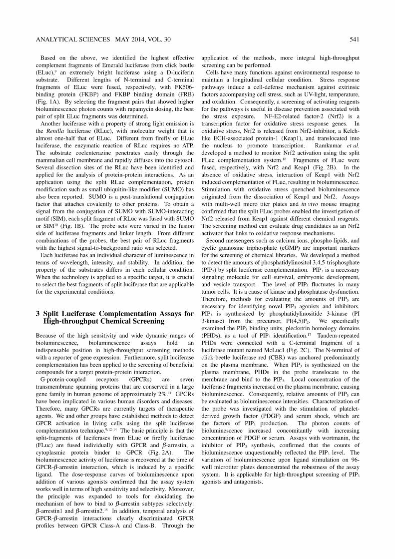
Fig. 1 Development of a pair of split luciferase probes. (A) Screening of a pair of split ELuc fragments by rapamycin-induced interaction of FKBP and FRB. The left figure portrays a schematic diagram of the probes. The N-terminal and C-terminal of ELuc (ELucN and ELucC) are attached to FKBP and FRB, respectively. The FKBP–FRB interaction brings fragments of ELuc into proximity. Then complementation occurs. The assays were performed with different combination of the variable lengths of ELuc fragments explained in the right panel. (B) Bioluminescence detection assays of SUMO–SIM interactions by split luciferase complementation. The left figure shows a model of probes. When SUMO binds to SIM, reconstituted RLuc is recovered in the bioluminescence activity. The right figure shows the combination of the probe candidates. A series of split RLuc probes with different fusion order and linker length was prepared. Their abilities of luciferase complementation were examined.
ANALYTICAL SCIENCES MAY 2014, VOL. 30 541
Based on the above, we identified the highest effective complement fragments of Emerald luciferase from click beetle (ELuc),9 an extremely bright luciferase using a D-luciferin substrate. Different lengths of N-terminal and C-terminal fragments of ELuc were fused, respectively, with FK506-binding protein (FKBP) and FKBP binding domain (FRB) (Fig. 1A). By selecting the fragment pairs that showed higher bioluminescence photon counts with rapamycin dosing, the best pair of split ELuc fragments was determined.
Another luciferase with a property of strong light emission is the Renilla luciferase (RLuc), with molecular weight that is almost one-half that of ELuc. Different from firefly or ELuc luciferase, the enzymatic reaction of RLuc requires no ATP. The substrate coelenterazine penetrates easily through the mammalian cell membrane and rapidly diffuses into the cytosol. Several dissection sites of the RLuc have been identified and applied for the analysis of protein–protein interactions. As an application using the split RLuc complementation, protein modification such as small ubiquitin-like modifier (SUMO) has also been reported. SUMO is a post-translational conjugation factor that attaches covalently to other proteins. To obtain a signal from the conjugation of SUMO with SUMO-interacting motif (SIM), each split fragment of RLuc was fused with SUMO or SIM10 (Fig. 1B). The probe sets were varied in the fusion side of luciferase fragments and linker length. From different combinations of the probes, the best pair of RLuc fragments with the highest signal-to-background ratio was selected.
Each luciferase has an individual character of luminescence in terms of wavelength, intensity, and stability. In addition, the property of the substrates differs in each cellular condition. When the technology is applied to a specific target, it is crucial to select the best fragments of split luciferase that are applicable for the experimental conditions.
3 Split Luciferase Complementation Assays for High-throughput Chemical Screening
Because of the high sensitivity and wide dynamic ranges of bioluminescence, bioluminescence assays hold an indispensable position in high-throughput screening methods with a reporter of gene expression. Furthermore, split luciferase complementation has been applied to the screening of beneficial compounds for a target protein–protein interaction.
G-protein-coupled receptors (GPCRs) are seven transmembrane spanning proteins that are conserved in a large gene family in human genome of approximately 2%.11 GPCRs have been implicated in various human disorders and diseases. Therefore, many GPCRs are currently targets of therapeutic agents. We and other groups have established methods to detect GPCR activation in living cells using the split luciferase complementation technique.9,12–14 The basic principle is that the split-fragments of luciferases from ELuc or firefly luciferase (FLuc) are fused individually with GPCR and β-arrestin, a cytoplasmic protein binder to GPCR (Fig. 2A). The bioluminescence activity of luciferase is recovered at the time of GPCR–β-arrestin interaction, which is induced by a specific ligand. The dose–response curves of bioluminescence upon addition of various agonists confirmed that the assay system works well in terms of high sensitivity and selectivity. Moreover, the principle was expanded to tools for elucidating the mechanism of how to bind to β-arrestin subtypes selectively: β-arrestin1 and β-arrestin2.15 In addition, temporal analysis of GPCR–β-arrestin interactions clearly discriminated GPCR profiles between GPCR Class-A and Class-B. Through the
application of the methods, more integral high-throughput screening can be performed.
Cells have many functions against environmental response to maintain a longitudinal cellular condition. Stress response pathways induce a cell-defense mechanism against extrinsic factors accompanying cell stress, such as UV-light, temperature, and oxidation. Consequently, a screening of activating reagents for the pathways is useful in disease prevention associated with the stress exposure. NF-E2-related factor-2 (Nrf2) is a transcription factor for oxidative stress response genes. In oxidative stress, Nrf2 is released from Nrf2-inhibitor, a Kelch-like ECH-associated protein-1 (Keap1), and translocated into the nucleus to promote transcription. Ramkumar et al. developed a method to monitor Nrf2 activation using the split FLuc complementation system.16 Fragments of FLuc were fused, respectively, with Nrf2 and Keap1 (Fig. 2B). In the absence of oxidative stress, interaction of Keap1 with Nrf2 induced complementation of FLuc, resulting in bioluminescence. Stimulation with oxidative stress quenched bioluminescence originated from the dissociation of Keap1 and Nrf2. Assays with multi-well micro titer plates and in vivo mouse imaging confirmed that the split FLuc probes enabled the investigation of Nrf2 released from Keap1 against different chemical reagents. The screening method can evaluate drug candidates as an Nrf2 activator that links to oxidative response mechanisms.
Second messengers such as calcium ions, phospho-lipids, and cyclic guanosine triphosphate (cGMP) are important markers for the screening of chemical libraries. We developed a method to detect the amounts of phosphatidylinositol 3,4,5-trisphosphate (PIP3) by split luciferase complementation. PIP3 is a necessary signaling molecule for cell survival, embryonic development, and vesicle transport. The level of PIP3 fluctuates in many tumor cells. It is a cause of kinase and phosphatase dysfunction. Therefore, methods for evaluating the amounts of PIP3 are necessary for identifying novel PIP3 agonists and inhibitors. PIP3 is synthesized by phosphatidylinositide 3-kinase (PI 3-kinase) from the precursor, PI(4,5)P2. We specifically examined the PIP3 binding units, pleckstrin homology domains (PHDs), as a tool of PIP3 identification.17 Tandem-repeated PHDs were connected with a C-terminal fragment of a luciferase mutant named McLuc1 (Fig. 2C). The N-terminal of click-beetle luciferase red (CBR) was anchored predominantly on the plasma membrane. When PIP3 is synthesized on the plasma membrane, PHDs in the probe translocate to the membrane and bind to the PIP3. Local concentration of the luciferase fragments increased on the plasma membrane, causing bioluminescence. Consequently, relative amounts of PIP3 can be evaluated as bioluminescence intensities. Characterization of the probe was investigated with the stimulation of platelet-derived growth factor (PDGF) and serum shock, which are the factors of PIP3 production. The photon counts of bioluminescence increased concomitantly with increasing concentration of PDGF or serum. Assays with wortmanin, the inhibitor of PIP3 synthesis, confirmed that the counts of bioluminescence unquestionably reflected the PIP3 level. The variation of bioluminescence upon ligand stimulation on 96-well microtiter plates demonstrated the robustness of the assay system. It is applicable for high-throughput screening of PIP3 agonists and antagonists.

542 ANALYTICAL SCIENCES MAY 2014, VOL. 30
4 Split Luciferase Complementation Assays for Monitoring Intracellular Environmental Conditions
Intracellular environmental factors such as temperature, pH, ions and reactive-oxygen species directly influence the activities of living cells. Environmental factors have been analyzed using traditional biochemical methods and recently developed imaging technologies using fluorescent probe molecules.18,19 Biochemical approaches do not accommodate evaluation of living cells. Even fluorescent probes need excitation light, which sometimes induces side effects in living cells. To overcome that shortcoming, bioluminescence-based techniques including split luciferase complementation are beneficial for measurements of different environmental conditions.
Calcium ion (Ca2+) is a major bioactive molecule that plays a
critical role as a messenger for numerous cell events. A useful intracellular Ca2+ indicator is a genetically encoded fluorescent indicator, “cameleon”.20 The core of the indicator is composed of calmodulin (CaM) fused with a CaM-binding domain (M13). The Ca2+ dependent conformational change of CaM interacts spontaneously with M13 in the probe. Cameleon shows the CaM–M13 interaction as a change in the fluorescence resonance energy transfer (FRET) ratio. The principle was applied to the bioluminescent Ca2+ indicator.21 Fluorescence proteins were replaced by split RLuc fragments, which reconstituted upon CaM–M13 interactions. The bioluminescent probe enabled monitoring of the Ca2+ level. Saito et al. improved the split-Luciferase-based Ca2+ indicator with the highest luminescence intensity.22 First, they developed a pair of highly efficient bioluminescence resonance energy transfer (BRET) using enhanced yellow fluorescence protein (named Venus) with mutation (VenusΔC10) and an enhanced RLuc (RLuc8), called
Fig. 2 Luciferase fragment complementation assays for high-throughput screening. (A) Detection of GPCR–β-arrestin interaction. The left figure presents a schematic diagram of the measurement of GPCR–β-arrestin interactions using luciferase fragment complementation. The N-terminal fragments of luciferase are fused with GPCR, whereas C-terminal fragments of luciferase are fused with β-arrestin. Ligand-induced interaction of GPCR with β-arrestin brings two luciferase fragments into proximity, followed by reconstitution. The right graph shows the dose–response luminescence curve of split luciferase probe with β2-adrenergic receptor (ADRB2). Dosing of the agonist, isoproterenol (Iso), induced an increase in photon counts. This diagram was modified from Ref. 14. (B) Monitoring the Nrf2 activation by quenching of bioluminescence. The figure shows the detection principle of repression of Nrf2 by Keap1. In oxidative stress, Nrf2 is released from Keap1 and is translocated to the nucleus. The release enables detection by the dissociation of FLuc fragments as a decrease of bioluminescence. (C) Schematic model for detection of membrane PIP3 production. C-terminal fragment of McLuc1 is connected with tandem PHDs. The N-terminal fragment of CBR is anchored predominantly on the plasma membrane. An increase in PIP3 induces migration of PHDs to the plasma membrane through binding to PIP3. Subsequently, the reconstitution of luciferase occurrs.

ANALYTICAL SCIENCES MAY 2014, VOL. 30 543
“Nano-lantern”. Next, VenusΔC10 and split RLuc8 fragments were integrated into the split-Luciferase-based Ca2+ indicator (Fig. 3A). When CaM–M13 undergoes a conformational change, reconstituted RLuc8 induces BRET to VenusΔC10. Comparing the probe to conventional split luciferase complementation assays, the Nano-lantern-based probe generates brighter luminescence that enables higher temporal resolution. In fact, the new Ca2+ indicator demonstrates the potential for imaging Ca2+ in living cells. Time-course changes in Ca2+ level upon histamine stimulation were monitored distinctly in a single cell at a video rate.
Acidification of living cells occurs in abnormal conditions. For example, oxidative stress during ischemic treatment produces reactive oxygen species and a low-pH condition in living tissues. Moreover, such acidic tissues are observed in cancer tumors and inflamed tissue regions. Therefore, monitoring of the acidification is important for the diagnosis of abnormal tissues. Because of the low penetration of light in living subjects, bioluminescence methods with longer wavelength are often beneficial for in vivo analysis. We established a bioluminescence in vivo pH monitoring method based on the combination of a photoreactive protein and split luciferase complementation.23 A light, oxygen, and voltage domain (LOV2) from a plant Phototropin1 protein comprises a
LOV core domain and Jα-helix, which mutually interact in the dark (Fig. 3B). The light irradiation induced the temporal dissociation of Jα-helix from the LOV core domain. When turning off the light, the structure moves to the dark state in which Jα-helix interacts with the LOV core domain. A bioluminescent pH indicator was developed using this intriguing property of LOV2. The indicator comprises LOV2 connected with the N-terminal fragment of FLuc and McLuc1. In the dark, the luciferase fragments reconstitute and generate bioluminescence. Light irradiation engenders dissociation of the luciferase fragments, resulting in a temporal quenching of luminescence and its gradual restoration thereafter. In summary, the new luciferase, designated as photo-inactivatable luciferase (PI-Luc), enables control of the inactivation of bioluminescence by light. A unique characteristic of the bioluminescence is its pH dependence. In living cells, the bioluminescence recovery time (RT) increased concomitantly with decreasing pH, particularly from pH 6 to 7. Accordingly, PI-Luc can detect intracellular pH by its RT counts. We established a novel pH imaging system using PI-Luc. Then we monitored the real-time pH variation in mouse tissues. PI-Luc-expressed mouse footpad showed light-dependent bioluminescence inactivation and its recovery, which was sufficient to calculate the RT counts. The RT-based imaging revealed time-lapse changes in the
Fig. 3 Luciferase fragment complementation assays for monitoring cellular environmental components. (A) Ca2+-indicator based split luciferase complementation and BRET. The N-terminal and C-terminal fragments of RLuc8 were fused, respectively, with each side of the Ca2+ sensor region (CaM and M13). Mutated Venus (VenusΔC10) was connected with the N-terminal fragments of RLuc8 (RLuc8ΔN3). When Ca2+ binds to CaM, the interaction of CaM to M13 induced recovery of RLuc8 activity. Upon generating bioluminescence, BRET to Venus occurred. (B) Schematic model of photo-inactivatable luciferase, PI-Luc. The bioluminescence is generated in the dark. Light irradiation dissociates the Jα-helix, resulting in the inactivation of bioluminescence temporally. The right graph shows the calibration of the bioluminescence recovery time (RT) for different pH in living cells. The RT variations were increased concomitantly with decreasing pH. The graph was modified from Ref. 23.

544 ANALYTICAL SCIENCES MAY 2014, VOL. 30
acidification level of mouse footpad after ischemia-reperfusion treatments. As such, we found that in vivo bioluminescence assay with split luciferase complementation technique enables dynamic analysis of the temporal variation of environmental conditions in living tissues.
5 Conclusions
Through continuous efforts to identify and improve luciferases, various luciferase-based probes have been established during the last decade. Considering different properties of luciferases, and their benefits and shortcomings, we can determine the most appropriate bioluminescence analysis to monitor cellular events of interest. Split luciferase complementation methods are showing growing prospects of practical applicability for wider fields of bioanalysis. Additional applications based on split luciferase complementation are now under development and are expected to be announced in the near future.
6 Acknowledgements
This work was supported by the Japan Society for the Promotion of Science (JSPS), Japan Science and Technology (JST), and MEXT, Japan.
7 References
1. B. Stynen, H. Tournu, J. Tavernier, and P. Van Dijck, Microbiol. Mol. Biol. Rev., 2012, 76, 331.
2. T. K. Kerppola, Nat. Methods, 2006, 3, 969. 3. A. K. M. Kafi, M. Hattori, and T. Ozawa, Nano LIFE,
2010, 01, 79. 4. N. Hida, M. Awais, M. Takeuchi, N. Ueno, M. Tashiro, C.
Takagi, T. Singh, M. Hayashi, Y. Ohmiya, and T. Ozawa, PloS One, 2009, 4, e5868.
5. S. B. Kim, M. Sato, and H. Tao, Anal. Chem., 2009, 81, 67.
6. S. B. Kim, T. Ozawa, S. Watanabe, and Y. Umezawa, Proc. Natl. Acad. Sci. U. S. A., 2004, 101, 11542.
7. S. B. Kim, Y. Umezawa, and H. Tao, Anal. Sci., 2009, 25, 1415.
8. S. B. Kim, M. Hattori, and T. Ozawa, Int. J. Mol. Sci., 2012, 13, 16986.
9. N. Misawa, A. K. M. Kafi, M. Hattori, K. Miura, and T. Ozawa, Anal. Chem., 2010, 82, 2552.
10. M. Hirohama, A. R. D. Voet, T. Ozawa, H. Saitoh, Y. Nakao, K. Y. J. Zhang, A. Ito, and M. Yoshida, Anal. Biochem., 2013, 448, 92.
11. J. A. Allen and B. L. Roth, Annu. Rev. Pharmacol. Toxicol., 2011, 51, 117.
12. A. K. M. Kafi, M. Hattori, N. Misawa, and T. Ozawa, Pharmaceuticals, 2011, 4, 457.
13. K. E. Luker, M. Gupta, and G. D. Luker, Anal. Chem., 2008, 80, 5565.
14. H. Takakura, M. Hattori, M. Takeuchi, and T. Ozawa, ACS Chem. Biol., 2012, 7, 901.
15. M. Hattori, M. Tanaka, H. Takakura, K. Aoki, K. Miura, T. Anzai, and T. Ozawa, Mol. BioSyst., 2013, 9, 957.
16. K. M. Ramkumar, T. V. Sekar, K. Foygel, B. Elango, and R. Paulmurugan, Anal. Chem., 2013, 85, 7542.
17. L. Yang, Y. Nasu, M. Hattori, H. Yoshimura, A. Kanno, and T. Ozawa, Anal. Chem., 2013, 85, 11352.
18. N. Soh, K. Makihara, T. Ariyoshi, D. Seto, T. Maki, H. Nakajima, K. Nakano, and T. Imato, Anal. Sci., 2008, 24, 293.
19. S. Takenaka and B. Juskowiak, Anal. Sci., 2011, 27, 1167. 20. A. Miyawaki, O. Griesbeck, R. Heim, and R.Y. Tsien, Proc.
Natl. Acad. Sci. U. S. A., 1999, 96, 2135. 21. A. Kaihara, Y. Umezawa, and T. Furukawa, Anal. Sci.,
2008, 24, 1405. 22. K. Saito, Y.-F. Chang, K. Horikawa, N. Hatsugai, Y. Higuch,
M. Hashida, Y. Yoshida, T. Matsuda, Y. Arai, and T. Nagai, Nat. Commun., 2012, 3, 1262.
23. M. Hattori, S. Haga, H. Takakura, M. Ozaki, and T. Ozawa, Proc. Natl. Acad. Sci. U. S. A., 2013, 110, 9332.